About this Research Topic
The fundamental challenge for using quantitative magnetic resonance imaging (qMRI) to infer microstructural information or to detect microscopic tissue alterations is the inherent averaging that occurs across the imaging volume elements, known as voxels (i.e., pixels with thickness). Voxel-averaged MR images can only provide macroscopic information with respect to the relatively large voxel size, which is typically 1 to 8 mm cube. In a mammalian brain, an individual voxel contains multiple chemical and physical microenvironments such as axons, neurons, glia, myelin, and cerebrospinal fluid. While microscopy and histopathology are the gold standard techniques to measure these quantities, this limits investigations to ex-vivo measures. To date, investigating these cell-level processes using macroscopic averages remains unattainable using existing qMRI methods. As a result, the inability to separate normal and pathological tissue within a voxel is the cause of the limited sensitivity and specificity of conventional qMRI in detecting and diagnosing pathologies.
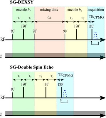
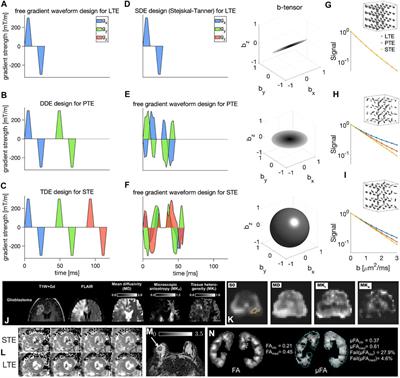
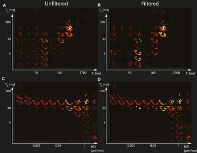
Water molecules, encapsulated within biological tissues, interact with their local chemical environment via nuclear relaxation processes and follow diffusion patterns that are governed by the local tissue density and geometry. Using a combination of magnetic field profiles to probe these mechanisms, MRI provides exquisite sensitivity to both the chemical composition, through relaxation parameters, and microstructure, through diffusion parameters, of biological tissues. By jointly encoding multiple MR dimensions, such as relaxation times (T1 and T2) and diffusion, multidimensional MRI overcomes the voxel-averaging limitation, yielding a multidimensional distribution of those MR parameters in each voxel; these distributions are specific fingerprints of various chemical and physical microenvironments within the voxel. Therefore, multidimensional MRI accomplishes two fundamental goals: (1) it provides unique intra-voxel distributions instead of an averaged value over the whole voxel; this allows identification of multiple components within a given voxel, while (2) the multiplicity of dimensions inherently facilitates their disentanglement; this allows higher accuracy and precision in derived quantitative values. Acquisition, computational, and pulse design technological breakthroughs have positioned multidimensional MRI as a powerful emerging imaging modality to studying biological media, from placenta to central nervous system, exhibiting extraordinary sensitivity and specificity in differentiating normal from abnormal cell-level processes.
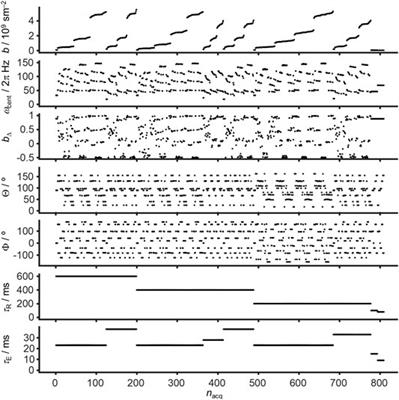
We are welcoming studies and manuscripts covering all aspects of multidimensional MRI: From theoretical studies focusing on the mathematical and physiological background of multidimensional MRI, to novel acquisition and sampling selection strategies to clinical studies covering the full width of the research and clinical landscape. Extensions beyond conventional diffusion encodings including mechanisms studying exchange, length and time scales, heterogeneity in isotropy and directions, as well as studies including further molecular tissue properties beyond T1 and T2 such as inversion efficiency and magnetization transfer effects are welcome. Studies bridging the gap from NMR mechanisms to their application in imaging, and studies at different field strength from ultra low to ultra high field are welcome. We are encouraging the submission of the following types of manuscripts: Original Research, Review, Mini Review, Perspective, Brief Research Report, and Technology and Code. Topics for submitted papers can be in one of the following general categories:
- development of processing methods, instrumentation, or experimental design for multidimensional MRI*.
- studies of normal or diseased media using multidimensional MRI.
- introduction of new theoretical or simulation models of multidimensional MRI.
*the term multidimensional includes relaxation-diffusion, T1-T2, diffusion tensor distribution, diffusion exchange, and relaxation exchange.
In-vitro, in-silico, as well as ex-vivo and in-vivo human or animal studies are all welcome.
Alexis Reymbaut is an employee of Random Walk Imaging AB, Lund, Sweden, which holds patents related to tensor-valued diffusion MRI acquisitions/analysis.
Keywords: multidimensional, MRI, diffusion, relaxation, exchange, multiexponential
Important Note: All contributions to this Research Topic must be within the scope of the section and journal to which they are submitted, as defined in their mission statements. Frontiers reserves the right to guide an out-of-scope manuscript to a more suitable section or journal at any stage of peer review.