About this Research Topic
In preclinical animal models, researchers can, within the same thin slice of tissue, probe activity within neurons, examine neurons’ projections (e.g., with tract or viral tracing), and determine the neurochemical phenotypes of those neurons (e.g., with immunohistochemistry). Great specificity and mechanistic understanding can be achieved using preclinical approaches like these. In humans, neuroimaging offers researchers the opportunity to noninvasively probe brain structure, function, and connectivity. However, like all techniques in science, neuroimaging has limitations. For example, the blood oxygen level-dependent (BOLD) signal in functional MRI (fMRI) is a proxy for neural activation, not neural activity itself; diffusion-weighted imaging and tractography provide inferences of white matter structure, not neural or synaptic targets.
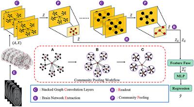
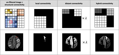
Despite these limitations, there is great potential for the integration of neuroimaging modalities and techniques to achieve a greater mechanistic understanding of many developmental, cognitive, social, and clinical issues. New tools and methods have been and are currently being developed to examine neuroimaging data in multimodal and integrative ways. Multimodal neuroimaging analyses can be applied to address myriad scientific questions that can result in both a broader picture of neural dynamics at play and a greater depth of understanding regarding neural mechanisms.
The ultimate goal of this research topic is to boost the development of multimodal neuroimaging methods and to remove the constraints of single modality neuroimaging, by leveraging unique information encoded in multiple sources and fusing them to obtain comprehensive inferences. This research topic will also serve as a place to exhibit neuroscientific discoveries that cannot be achieved by exploiting single modalities and thus demonstrate the power of multimodal analysis.
Original research manuscripts utilizing multimodal neuroimaging methods in human brain across the lifespan, as well as manuscripts that combine the use of post-mortem human brain tissue with neuroimaging methods, are highly welcome to contribute to this Research Topic. Particularly we invite authors to submit multimodal analyses:
• Using multimodal brain imaging data, including but not limited to structural MRI, functional MRI, diffusion-weighted imaging, MR-spectroscopy, PET, EEG, MEG, CT, etc.
• Using combinations of post-mortem human brain tissue (e.g., with histology or immunocytochemistry) and neuroimaging approaches
• Focused on any developmental stage (from infants to geriatric populations)
• Focused on applications toward a variety of developmental, cognitive, social, and clinical outcomes
• Focused on a variety of goals (e.g., neuroanatomical mapping of specific neural systems, prediction of brain or mental states)
Keywords: multimodal, neuroimaging, functional MRI, diffusion-weighted imaging, infant, adolescent, adult, geriatric, human brain mapping, state prediction
Important Note: All contributions to this Research Topic must be within the scope of the section and journal to which they are submitted, as defined in their mission statements. Frontiers reserves the right to guide an out-of-scope manuscript to a more suitable section or journal at any stage of peer review.