Craniopharyngiomas are benign brain tumors that derive from embryonic malformations and are commonly found at the sellar/parasellar, suprasellar region from sella turcica to the third ventricle along with the pituitary–hypothalamic axis. The clinical manifestations include pituitary/hypothalamic deficiencies, ...
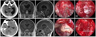
Craniopharyngiomas are benign brain tumors that derive from embryonic malformations and are commonly found at the sellar/parasellar, suprasellar region from sella turcica to the third ventricle along with the pituitary–hypothalamic axis. The clinical manifestations include pituitary/hypothalamic deficiencies, visual impairment, and intracranial pressure increase. These symptoms are caused by the tumor mass impacting the optic nerve/chiasma and hypothalamic–pituitary axis. Except for that the chemotherapy based on BRAF mutations in papillary types of craniopharyngiomas and its future significance was reported, there is no effective pharmaceutical treatment for craniopharyngiomas, while radiotherapy is proven as an adjuvant therapy to achieve excellent long-term local control after subtotal resection or small recurrent tumor. Therefore, surgical resection remains the first option of craniopharyngiomas treatment. Craniopharyngiomas tend to adhere to circumjacent neurovascular structures such as optic nerves and hypothalamus. Due to this nature, the surgical resection of craniopharyngiomas is very challenging. Quality of life and neuropsychological function are frequently impaired due to the close anatomical proximity to the optic chiasm, hypothalamus and pituitary stalk. The common complications following surgery are visual deterioration, neuroendocrine deficiencies and hypothalamic injury. Hypothalamic involvement and treatment-related hypothalamic lesions frequently result in hypothalamic obesity, physical fatigue and psychosocial deficits. Therefore, research on how to protect neurological structures and functions of the optic nerve, pituitary stalk and hypothalamus during the surgical resection of craniopharyngiomas is crucial.
This Research Topic is to focus on the protection of important structures during surgical resection of craniopharyngiomas, the mechanism of the development of craniopharyngioma and its related neurocognitive deficits.
The aim of this research topic is to integrate the high-quality and up-to-date advances in the above fields. Therefore, we welcome both original research articles and review articles that cover, but not limited to the following topics:
- Improving surgical techniques for the protection of optic nerve, pituitary stalk and hypothalamus during the resection of craniopharyngiomas.
- Developing Intraoperative electrophysiological monitoring technology for optic nerve protection during resection of craniopharyngiomas.
- Applying the Brain Imaging method to locate hypothalamus nuclei for selecting appropriate surgical approach of craniopharyngiomas.
- Investigating mechanisms of neurocognitive deficits and neuroendocrine deficiencies caused by craniopharyngiomas.
- Identifying risk factors of post- craniopharyngiomas-operative hypothalamic obesity.
- Studying molecular mechanisms of the growth and invasion of craniopharyngiomas.
- Exploring the novel radiotherapy and chemotherapy of craniopharyngiomas.
- Addressing significant advances in Next Generation Sequencing, animal models and other approaches for craniopharyngiomas
Keywords:
Craniopharyngioma, Optic Nerve, Pituitary Stalk, Hypothalamus, Surgical Technique, Brain imaging Method, Electrophysiological Monitoring Technology
Important Note:
All contributions to this Research Topic must be within the scope of the section and journal to which they are submitted, as defined in their mission statements. Frontiers reserves the right to guide an out-of-scope manuscript to a more suitable section or journal at any stage of peer review.