- Science news
- Engineering
- Surprising spider hair discovery may inspire stronger adhesives
Surprising spider hair discovery may inspire stronger adhesives
By K.E.D. Coan, science writer

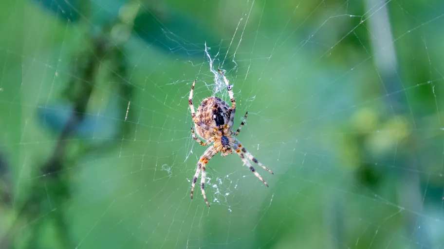
Cupiennius salei wandering spider. Image credit: Kevin Wells Photography / Shutterstock.com
A recent study by the open access publisher Frontiers shows the first evidence that the individual hair-like structures that form spiders’ adhesive feet are far more diverse than expected. By looking at a sample set of these hairs, researchers have found that they have varied shapes as well as attachment properties. Understanding how spiders climb a wide range of surfaces may help scientists design new and better adhesives.
Just how do spiders walk straight up -- and even upside-down across -- so many different types of surfaces? Answering this question could open up new opportunities for creating powerful, yet reversible, bioinspired adhesives. Scientists have been working to better understand spider feet for the past several decades. Now, a new study in Frontiers in Mechanical Engineering is the first to show that the characteristics of the hair-like structures that form the adhesive feet of one species -- the wandering spider Cupiennius salei -- are more variable than previously thought.
“When we started the experiments, we expected to find a specific angle of best adhesion and similar adhesive properties for all of the individual attachment hairs,” says the group leader of the study, Dr Clemens Schaber of the University of Kiel in Germany. “But surprisingly, the adhesion forces largely differed between the individual hairs, e.g. one hair adhered best at a low angle with the substrate while the other one performed best close to perpendicular.”
► Read original article► Download original article (pdf)
The feet of this species of spider are made up of close to 2,400 tiny hairs (one hundredth of one millimeter thick). Schaber, and his colleagues Bastian Poerschke and Stanislav Gorb, collected a sample of these hairs and then measured how well they stuck to a range of rough and smooth surfaces, including glass. They also looked at how well the hairs performed at various contact angles.
Unexpectedly, each hair showed unique adhesive properties. When the team looked at the hairs under a powerful microscope, they also found that each one showed clearly different -- and previously unrecognized -- structural arrangements. The team believes that this variety may be key to how spiders can climb so many surface types.This current work studied only a small number of the thousands of hairs on each foot, and it’s beyond the scope of existing resources to consider studying them all. But the team expects that not all of the hairs are unique, and that it might be possible to find clusters or repeating patterns instead.
“Although it is still very difficult to fabricate nanostructures like those of the spider -- and especially to achieve the stability and reliability of the natural materials -- our findings can further optimize existing models for reversible and residue-free artificial adhesives,” says Schaber. “The principle of different shapes and alignments of adhesive contacts as found in the spider attachment system can improve the attachment ability of bioinspired materials to a broad range of substrates with different properties.”

Scanning Electron Microscope (SEM) images of the microstructure of the adhesive hairs ('setae'). (A) Side view showing the up to 1.8 mm long hair shaft (not shown in full length) and the tip region covered with 'microtrichia' (minute hair-like structures on the hairs proper). (B) Top view of the 'scopula pad' (a dense tuft of hairs) on the lower side of the pretarsus. Covering the tip region of the hairs are spatula-shaped microtrichia, which stick to the substrate during walking. (C) Higher magnification of the spatula-shaped microtrichia. Credit: Poerschke, Gorb & Schaber 2021

SEM images of the bases of pretarsal (ie, on lowest part of leg) adhesive hairs. (A) On the left are the hair shafts of the adhesive setae closest to the exoskeleton. At their insertion, the hair shaft becomes thinner and a stopper-like structure on the exoskelecton meets the seta and attaches to it. (b) Further magnification of the same region: the asterisk marks the pivot point where setae can bend upwards. Distal vs. proximal here means away from vs. towards the claw on the tip of the leg. Credit: Poerschke, Gorb & Schaber 2021
REPUBLISHING GUIDELINES: Open access and sharing research is part of Frontiers’ mission. Unless otherwise noted, you can republish articles posted in the Frontiers news site — as long as you include a link back to the original research. Selling the articles is not allowed.


