- Science news
- Health
- CT scans of Egyptian mummy reveal new details about the death of a pivotal pharaoh
CT scans of Egyptian mummy reveal new details about the death of a pivotal pharaoh
New interpretations based on medical imaging suggest Seqenenre-Taa-II was executed by multiple attackers and embalmers had skillfully concealed some head wounds
By Peter Rejcek, science writer
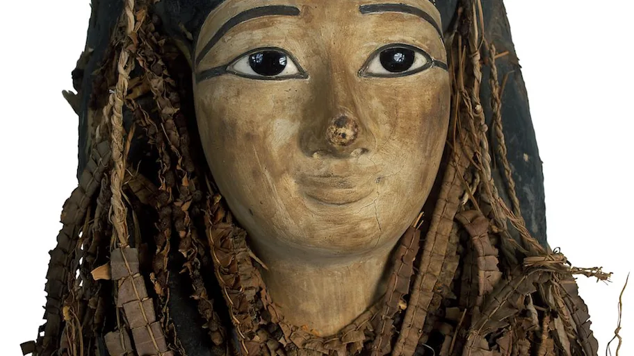
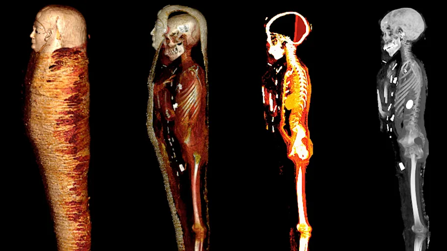
A CT scan study of the mummy of Pharaoh Seqenenre-Taa-II, an Egyptian ruler whose death eventually helped reunite the kingdom, revealed new details about how the king died. A recent paper suggests that the pharaoh died close to the battlefield and was ceremoniously executed by several people using Hyksos weapons. Additionally, the computer-processed x-rays revealed his embalmers had skillfully concealed some of the wounds, implying professional mummification of the body, despite its poor preservation.

Modern medical technology is helping scholars tell a more nuanced story about the fate of an ancient king whose violent death indirectly led to the reunification of Egypt in the 16th century BCE. The research was published in Frontiers in Medicine.
Pharaoh Seqenenre-Taa-II, ‘the Brave’, briefly ruled over southern Egypt during the country’s occupation by the Hyksos, a foreign dynasty that held power across the kingdom for approximately a century (1650-1550 BCE). In his attempt to oust the Hyksos, Seqenenre-Taa-II was killed. Scholars have debated the exact nature of the pharaoh’s death since his mummy was first discovered and studied in the 1880s.
These and subsequent examinations — including an x-ray study in the 1960s — noted the dead king had suffered several severe head injuries but no other wounds to his body. The prevailing theory, based on the evidence, was that the king had been captured in battle and then executed afterward, possibly by the Hyksos king himself. Others have suggested he was murdered in his sleep by a palace conspiracy.
Download original article (pdf)
In addition, the poor condition of the mummy suggested the embalming had been done hastily, away from the royal mummification workshop.
But computed tomography (CT) scans of the mummified remains of Seqenenre revealed new details about his head injuries, including previously undetected lesions that embalmers had skillfully concealed.
The authors of the new paper offer a novel interpretation of the events before and after the king’s death based on the computer-processed x-ray images: Seqenenre had indeed been captured on the battlefield, but his hands had been tied behind his back, preventing him from defending against the attack.
“This suggests that Seqenenre was really on the front line with his soldiers risking his life to liberate Egypt,” says lead author Dr Sahar Saleem, a professor of radiology at Cairo University who specializes in paleoradiology. This investigative technique employs medical imaging technologies to non-invasively study a cross section of archaeological remains, including bodies. It can help determine age at death, sex, and even how the person died.
For example, the CT scans, combined with other evidence, suggest the execution had been carried out by multiple attackers, which the scientists confirmed by studying five different Hyksos weapons that matched the king’s wounds.
“In a normal execution on a bound prisoner, it could be assumed that only one assailant strikes, possibly from different angles, but not with different weapons,” Saleem explains. “Seqenenre’s death was rather a ceremonial execution.”
The CT study also determined that Seqenenre was about 40 when he died, based on the detailed morphology revealed in the images, providing the most precise estimate to date.
Saleem and co-author Zahi Hawass, an archaeologist and former Egyptian minister of antiquities, have pioneered the use of CT scans to study the New Kingdom pharaohs and warriors, including well-known names such as Hatshepsut, Tutankhamun, Ramesses III, Thutmose III and Rameses II. Yet Seqenenre, based on the available evidence, appears to be the only one among this illustrious group to have been on the frontline of the battlefield.
In addition, the CT study revealed important details about the mummification of Seqenenre’s body. For instance, the embalmers used a sophisticated method to hide the king’s head wounds under a layer of embalming material that functioned similarly to the fillers used in modern plastic surgery. This would imply that mummification took place in a real mummification laboratory rather than in a poorly equipped place, as previously interpreted.
Saleem says the CT scan study provides important new details about a pivotal point in Egypt’s long history. “Seqenenre’s death motivated his successors to continue the fight to unify Egypt and start the New Kingdom,” she says.
Original article: Computed tomography study of the mummy of King Seqenenre Taa II: New insights into his violent death


REPUBLISHING GUIDELINES: Open access and sharing research is part of Frontiers’ mission. Unless otherwise noted, you can republish articles posted in the Frontiers news blog — as long as you include a link back to the original research. Selling the articles is not allowed.


