 Qi Xu
Qi Xu Minhui Luo2
 Jianghong Luo
95% of researchers rate our articles as excellent or good
Learn more about the work of our research integrity team to safeguard the quality of each article we publish.
Find out more
ORIGINAL RESEARCH article
Front. Nutr. , 18 November 2022
Sec. Nutrition and Food Science Technology
Volume 9 - 2022 | https://doi.org/10.3389/fnut.2022.1053315
This article is part of the Research Topic Dietary Phytochemicals: Identification, Bioactivities, and Delivery Strategy View all 28 articles
Introduction: Camellia oil (CO), a common edible oil in China, contains a variety of active ingredients. In this study, we explored the combining effect and optimal feeding time of CO and squalene on hyperlipemia-induced reproductive damage rats and probably provided supportive data for use of CO for health benefits.
Methods: We established the hyperlipidaemia-induced reproductive damage model, and then the successfully modeled rats were randomly classified into four groups including a model control (MC) group, a camellia oil (CO) group, a camellia oil + squalene (COS) group, and a sildenafil (SN) group, which were feeding with different subjects during days 30 and 60. The normal (NC) group was fed under the same conditions.
Results: Our results showed that compared with the MC group, the CO, COS, and SN groups could significantly decline the serum TG, TC and LDL-C levels, increase the serum testosterone levels, the sperm counts in epididymidis and organ coefficients of penises, and no pathological change in penis and testis at days 30 and 60. Compared with the pure CO, the mixture of CO and squalene could significantly enhance the effect of decreasing the concentrations of TG, TC, and LDL-C and increasing the serum testosterone level and sperm count of epididymal tail, and the results of day 30 were better than those of day 60.
Discussion: CO and squalene have a combining effect on lowering blood lipid, improving the level of testosterone and the number of epididymal tail sperm, and promoting the recovery of erectile and sexual function on hyperlipidemia-induced reproductive damage rats on day 30.
Hyperlipidemia, a common metabolic syndrome (1), the typical symptoms include the abnormal elevation of any or all lipids or lipoproteins in the blood (2). Hyperlipidemia is a critical damage-inducing factor for cardiovascular disease and frequently brings about many complications, such as cardiac damage (3), sexual dysfunction (4), cognitive impairment (5), inflammation, and insulin resistance (6). Strong associations are observed between hyperlipidemia and sexual dysfunction especially erectile dysfunction (ED). Experimental studies showed that the reduction in arterial blood flow induced by hyperlipidemia can directly affect the functions of cortical center, pituitary–testis axis, and corpus cavernosum penis (7) and inhibit the production of testosterone (8), which cause a decline in sexual function. Nowadays, many synthetic pharmaceuticals, like sildenafil (SN), are widely used for the management of ED. However, their long-term use always causes many serious side effects, including dizziness, headaches, heartburn, indigestion, stuffy nose, and vasodilatation (9). Given the potential risks and side effects associated with current treatments, it is important to explore more efficient and safe candidates.
Meanwhile, many promising sources can be used to regulate and manage ED (10). Some studies showed that black tea brew possessed marked aphrodisiac activity in terms of prolongation of latency of ejaculation shortening of mount- and intromission latencies and elevation of serum testosterone level (11). Moringa oleifera Lam. Leaf Tea could enhance sexual function and the male reproductive system (12). Camellia oil (CO), which obtain from the seeds of Camellia oleifera Abel, is a common edible oil in China and contains a variety of active ingredients (13, 14), such as Squalene, which can raise the activity of internal Superoxide Dismutase (SOD) and blood oxygen content (15), stimulate blood circulation (16), and improve sexual function (17). Vitamin E can promote the secretion of sex hormones and maintain the normal function of the Genital organs (18). Camellia Saponin can reduce serum cholesterol and prevent cardiovascular diseases (19, 20). The research group observed in a previous study is that CO can lower blood lipids, promote the secretion of the sex hormone testosterone, and improve sexual function (21). To further enhance the effectiveness of camellia oil, in this research, squalene, an active ingredient of CO, is proposed to be added to feed rats with hyperlipidemia-induced sexual dysfunction as a combining substance and observe the changes in blood lipid and testosterone levels, epididymal sperm count, and testis and corpus cavernosum tissue structures. This study aims to explore the combining effect and optimal feeding time of CO and squalene on hyperlipidemia-induced sexual dysfunction rats and provide supportive data for the health benefits of CO use.
High-purity CO purchased from Jiang Xi Qiyunshan Food Co., Ltd., (Ganzhou, China) contained 78.8/100 g monounsaturated fatty acid, 9.0/100 g linoleic acid, 2.3/100 g linolenic acid, 1.3/100 g stearic acid, 8.6/100 g palmitic acid, 0.0/100 g protein, 0.0/100 g cholesterol, and 0.0/100 g sugar.
Basal and high-fat feeds were provided by Guangdong Medical Laboratory Animal Center (Guangdong, China). High-fat feed (100 g) consisted of 78.8 g basal feed, 10.0 g lard, 10.0 g yolk powder, 1 g cholesterol, and 0.2 g cholate.
Squalene and SN were purchased from Beijing InnoChem Science and Technology Co., Ltd., (Beijing, China). Total cholesterol (TC), triglyceride (TG), and low-density lipoprotein cholesterol (LDL-C) test kits were obtained from Nanjing Jiancheng Bioengineering Institute (Nanjing, China). The testosterone ELISA kit was purchased from Cloud-Clone Corp., Wuhan (Wuhan, China). The hematoxylin–eosin (HE) dye was acquired from Beijing Zhongshan Jinqiao Biotechnology Co., Ltd., (Beijing, China). All other used reagents were analytical grade.
Specific pathogen-free male Sprague Dawley rats (weight = 150–180 g) (Certificate number SCXK [xiang] 2019-0004) were purchased from Hunan Slac Jingda Laboratory Animal Co., Ltd., (Hunan, China). Animals were housed in cages in a specific pathogen-free room with controlled temperature (18–26°C), relative humidity (40–60%), and 12/12 h of light–dark periods with ad libitum food and water prior to experiments. All procedures were approved by the Animal Experiment Ethics Committee, Gannan Medical University, China.
Rats were fed with a high-fat diet to establish a hyperlipidemia-induced reproductive damage model. Rats were randomly divided into the normal (NC, 12 rats) and model (MC, 48 rats) control groups and supplied with basal and high-fat feeds, respectively, for 4 weeks. Serum TC, TG, LDL-C, and testosterone levels of rats were measured using the corresponding kits in accordance with the manufacturer’s instructions. Compared with the NC group, the MC group showed significant increments in serum TC, TG, and LDL-C levels and prominent reduction in the serum testosterone content.
The successfully modeled rats were randomly classified into four groups (12 mice per group): Converted by recommended intake of oils and fats, squalene, SN of human and “equivalent dose ratio table of human and animal body surface area conversion.” The MC group received 3 mL/kg⋅bw⋅day of 0.9% normal saline. The CO group was treated with 3 mL/kg⋅bw⋅day of CO. The CO + squalene (COS) group was supplemented with 3 mL/kg⋅bw⋅day suspension solution (CO: squalene = 30:1) containing CO and squalene. The SN group was supplemented with 3 mL/kg⋅bw⋅day suspension solution (CO: sildenafil = 3:1) containing CO and sildenafil. Rats in the NC group were orally administered with 3 mL/kg⋅bw⋅day of 0.9% normal saline. During the experimental period, blood was collected at days 30 and 60. Half of the rats in each group were sacrificed at days 30 and 60. The testis, epididymis, and penis of rats were obtained.
The obtained testis, epididymis, and penis were rinsed with 0.9% normal saline, blotted dry using filter paper, and weighed. Organ indices were calculated using the equation: weight of organ/weight of rat.
The blood of rats was centrifuged at 3,000 rpm and 4°C for 10 min to collect the serum. TC, TG, LDL-C, and testosterone levels of serum were detected using assay kits as specified by the manufacturer.
The cauda of epididymidis was cut off, placed into 4 mL of 0.9% normal saline, and cut. The cauda of the epididymidis was incubated at 37°C for 20 min to allow running out of sperm. Afterward, the supernatant was obtained through filtration by using 100-mesh strainer. The supernatant (10 μL) was transferred into the sperm count board, and the sperm number was counted under electron microscopy.
Parts of testis and penis were fixed in 10% formalin for over 24 h and embedded in paraffin. Then, paraffin sections (5 μm thick) were stained with HE dye and histopathologically observed using electron microscopy.
All data were presented as mean ± standard deviation. Statistical analysis was carried out using the SPSS 23.0 software (SPSS Inc., Chicago, IL, United States). One-way analysis of variance was adopted to compare significant differences among all groups by Tukey’s analysis. Two-tailed p < 0.05 was considered statistically significant.
The elevation of serum TC, TG, and LDL-C levels is the typical symptom of hyperlipidemia (22). As shown in Figure 1A, the TC, TG, and LDL-C levels of rats in high-fat diet treatment groups, including MC, CO, COS, and SN groups, were significantly elevated compared with those in the NC group. Results indicated that the hyperlipidemia model was successfully established. The serum testosterone levels of rats were determined as illustrated in Figure 1B to evaluate whether hyperlipidemia induced reproductive damage. Compared with the NC group, the hyperlipidemia group had prominently decreased serum testosterone level, and this finding was consistent with those of previous reports (21, 23, 24). This result suggested that hyperlipidemia-induced reproductive damage occurred in the high-fat diet model.

Figure 1. Serum lipid (A) and testosterone (B) levels of rats at the 30th day in hyperlipidemia-induced reproductive damage modeling. TG, triglyceride; TC, total cholesterol; LDL-C, low-density lipoprotein cholesterol; NC, normal control; MC, model control; CO, Camellia oil; COS, Camellia oil + squalene; SN, sildenafil. Different letters represent significant differences (p < 0.05).
The sexual organ (i.e., testis, epididymis, and penis) indices of rats are shown in Table 1. No significant difference was observed in the testis and epididymis organ indices of rats among NC, MC, CO, COS, and SN groups. Compared with the MC group, the NC, CO, COS, and SN groups had significantly increased penis organ indices at days 30 and 60, suggesting that the CO, COS, and SN groups could improve the relaxation and atrophy of corpus cavernosum penis in rats.
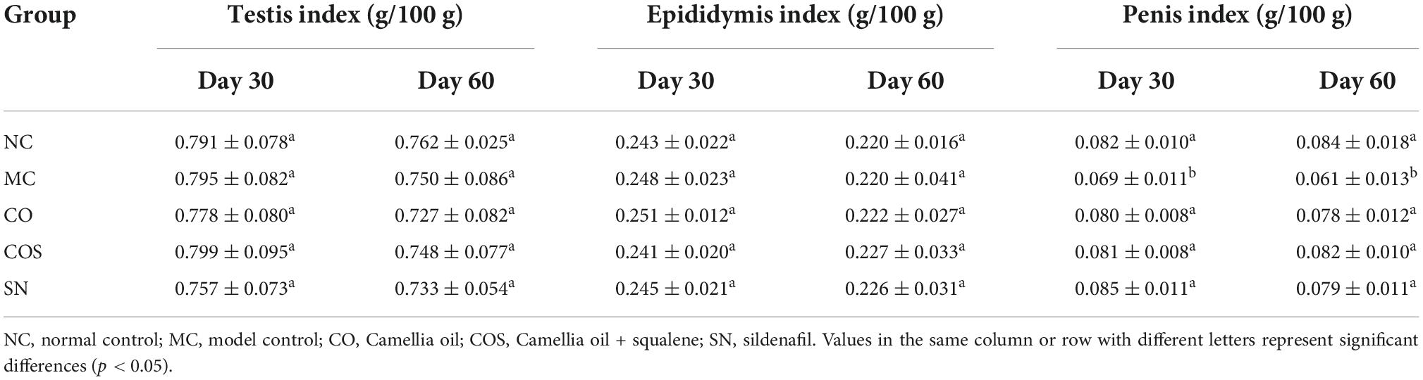
Table 1. Sexual organ indices of hyperlipidemia-induced reproductive damage rats.
The serum lipid (i.e., TG, TC, and LDL-C) levels of normal and hyperlipidemia rats are shown in Figure 2. Compared with the NC group, the serum lipid levels of rats were notably increased in the MC group at days 30 and 60. The treatments of test substances (i.e., CO and COS) could reverse this phenomenon. As illustrated in Figures 2A,B), at day 30, the serum TG and TC levels of rats in CO, COS, and SN groups declined compared with those in the MC group. Meanwhile, the serum TG and TC levels of rats in COS group declined compared with those in the CO group, indicating that CO and squalene could significantly enhance the effect of decreasing the serum TG and TC levels of hyperlipidemia rats. At day 60, the serum TG and TC levels of rats in the CO and COS groups notably decreased, while there was no significant difference between CO and COS groups. Figure 2C reveals that the LDL-C levels of rats supplemented with the above test substances (i.e., CO and COS) were markedly lower than those in the MC group at days 30 and 60.
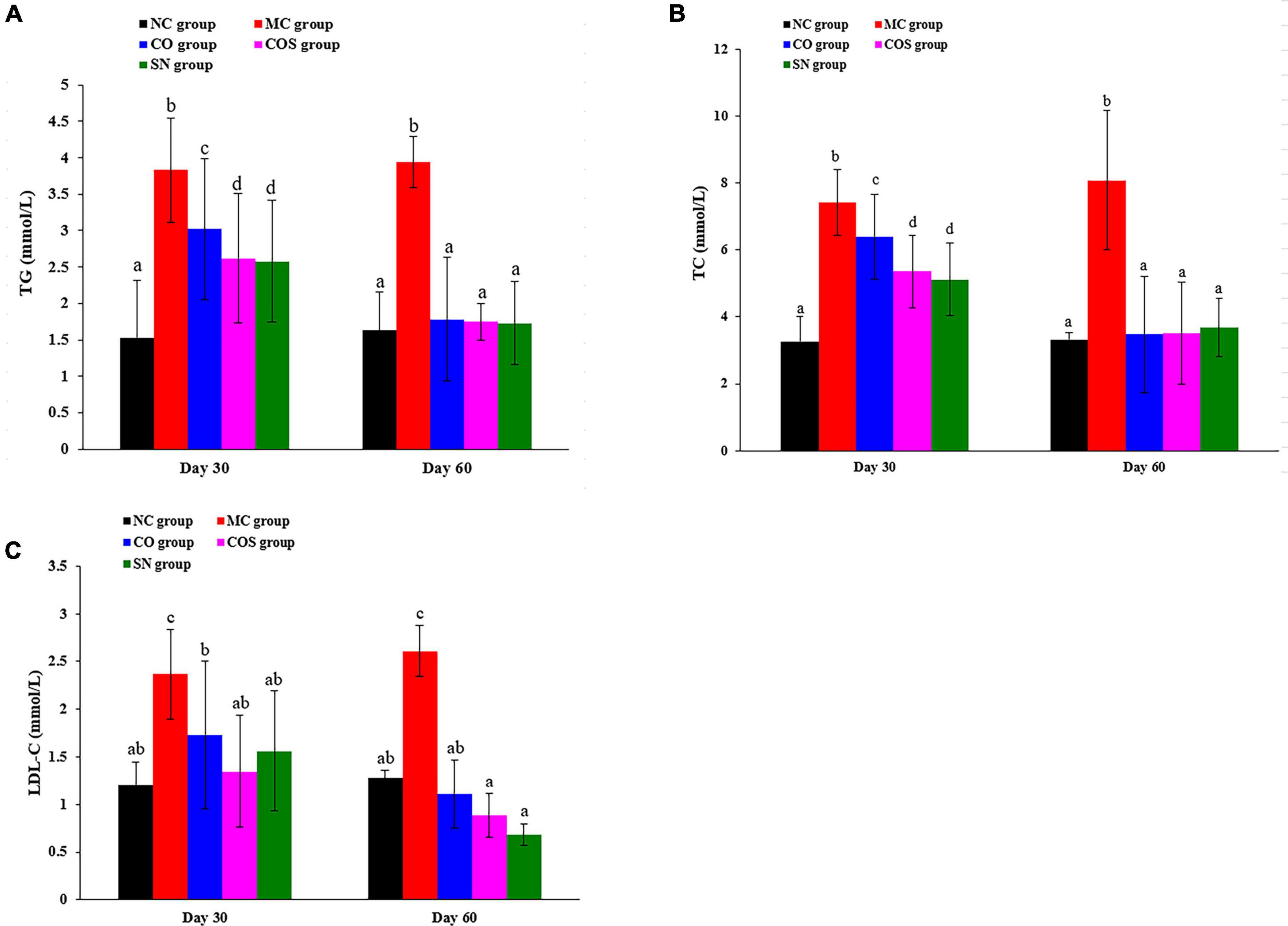
Figure 2. After the end of modeling, serum lipid levels of hyperlipidemia-induced reproductive damage rats at the another 30th and 60th day by feeding test substance: (A) TG, (B) TC, and (C) LDL-C levels. TG, triglyceride; TC, total cholesterol; LDL-C, low-density lipoprotein cholesterol; NC, normal control; MC, model control; CO, Camellia oil; COS, Camellia oil + squalene; SN, sildenafil. All values are expressed as mean ± SD. Different letters represent significant differences (p < 0.05).
The serum testosterone levels of rats were detected (Figure 3). Compared with those in the NC group, the serum testosterone levels of rats in the MC group significantly decreased at days 30 and 60. At day 30, the serum testosterone levels of rats in the COS and SN groups dramatically increased, whereas no outstanding increment was observed in the CO group. Results suggested that CO and squalene exerted combining effect against hyperlipidemia-induced reduction in the serum testosterone level of rats. At day 60, rats in the CO, COS, and SN groups showed significant elevation in serum testosterone levels in comparison with those in the MC group. At day 30, the serum testosterone level showed varying degrees of decrease in the COS and SN groups.
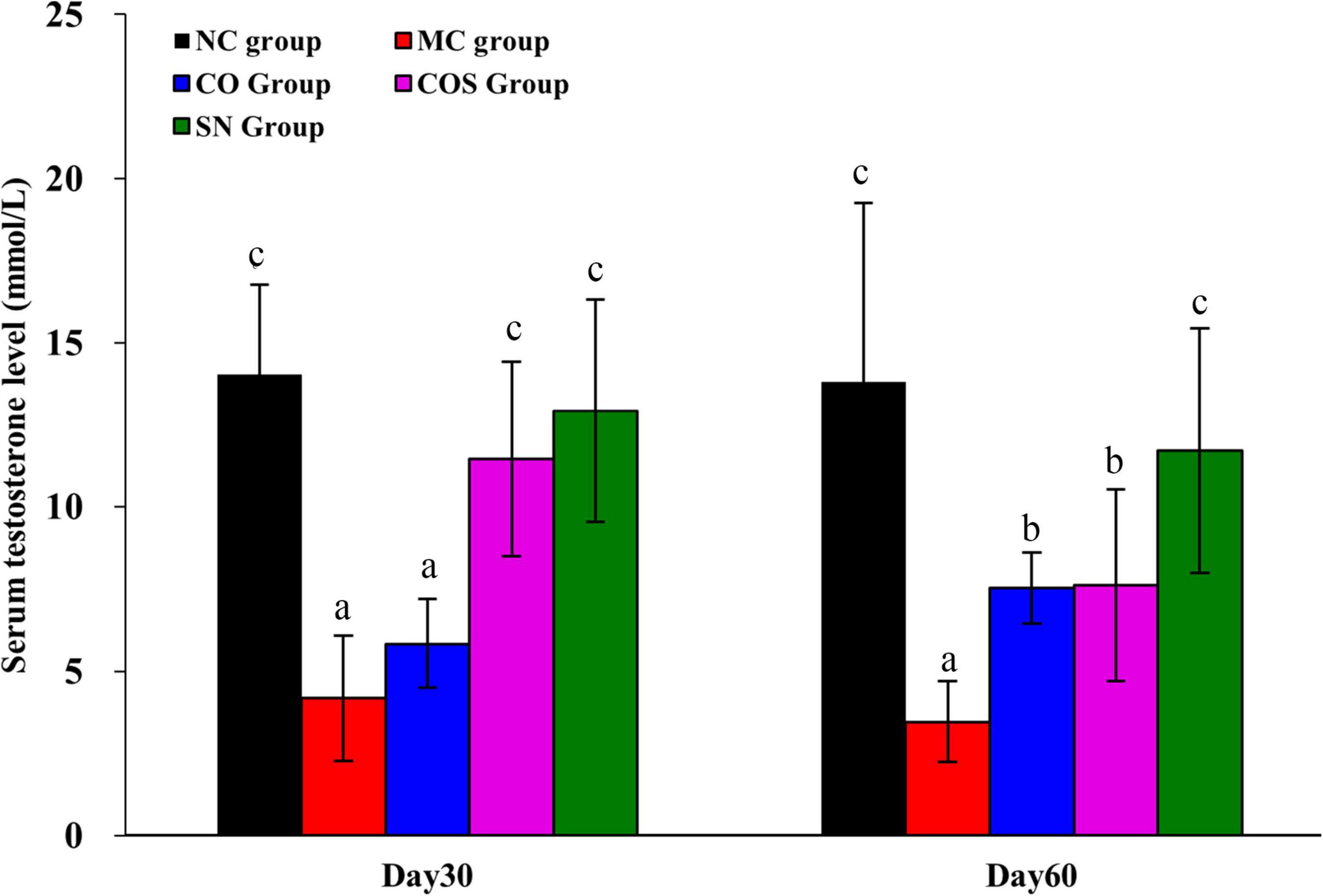
Figure 3. After the end of modeling, serum testosterone levels of hyperlipidemia-induced reproductive damage rats at the another 30th and 60th day by feeding test substance. NC, normal control; MC, model control; CO, Camellia oil; COS, Camellia oil + squalene; SN, sildenafil. All values are expressed as mean ± SD. Different letters represent significant differences (p < 0.05).
The sperm counts in the epididymidis of rats are shown in Figure 4. Compared with those in the NC group, the sperm counts in the epididymidis of rats in the MC, CO, COS, and SN groups were prominently reduced. The oral administration of CO, COS, and SN could significantly increase the sperm counts in the epididymidis of rats at days 30 and 60 compared with that of MC. The sperm counts in the epididymidis of rats in all groups at day 60 were not significantly different compared with those at day 30.
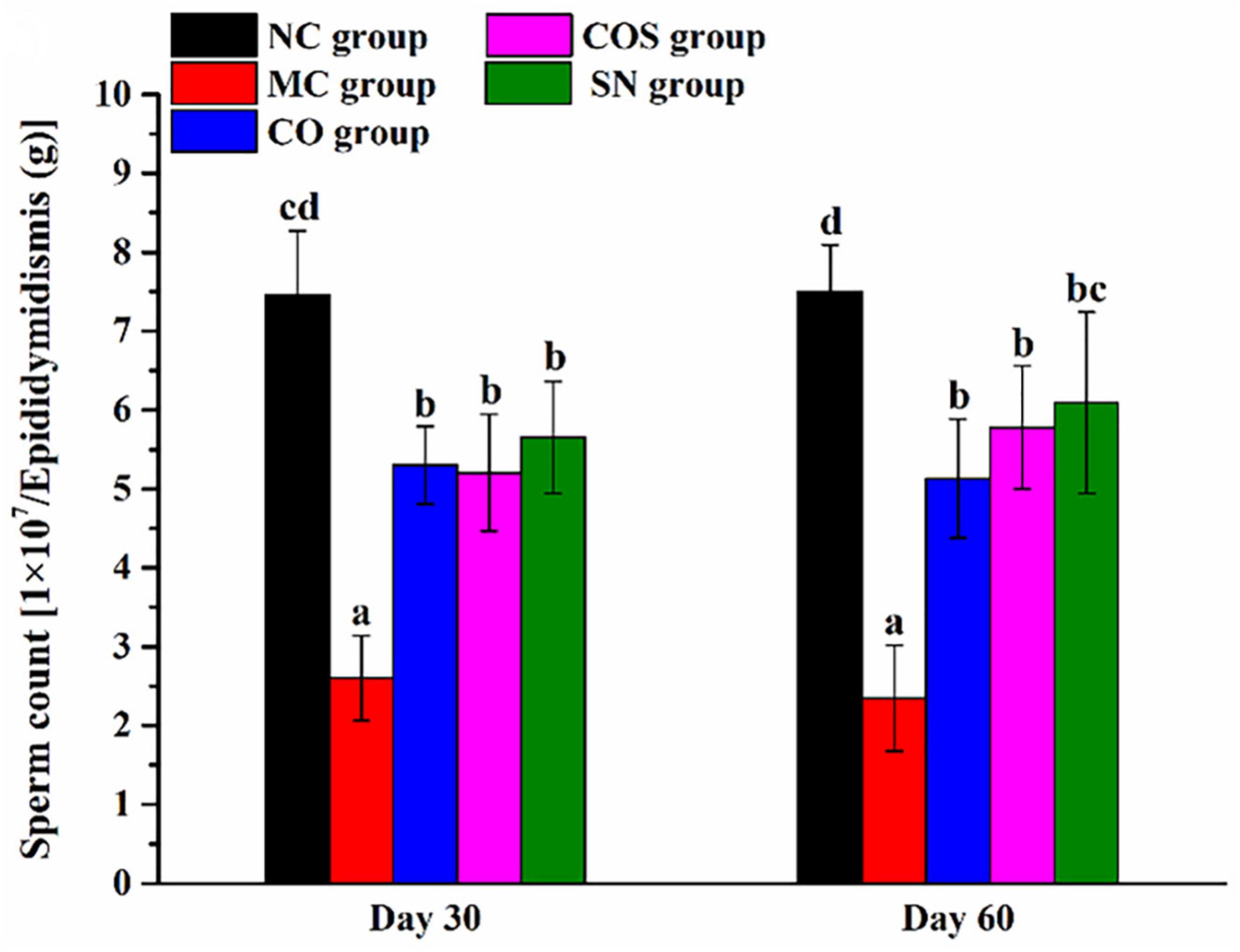
Figure 4. Sperm counts in the epididymidis of hyperlipidemia-induced reproductive damage rats. NC, normal control; MC, model control; CO, Camellia oil; COS, Camellia oil + squalene; SN, sildenafil. All values are expressed as mean ± SD. Different letters represent significant differences (p < 0.05).
The histomorphologies of testis and penis are shown in Figures 5, 6. Compared with the NC group, the CO, COS, and SN groups had no pathological change in the corpus cavernosum penis and testes. In the MC group, the number of smooth muscle cells and cavernous sinus decreased significantly in the corpus cavernosum penis, and the smooth muscle of the cavernous body were distributed unevenly and arranged disorderly and loosely. At the same time, all levels of spermatogenic cells in the testis were arranged disorderly, and some spermatogenic cells showed necrosis, apoptosis, and nuclear pyknosis at days 30 and 60.
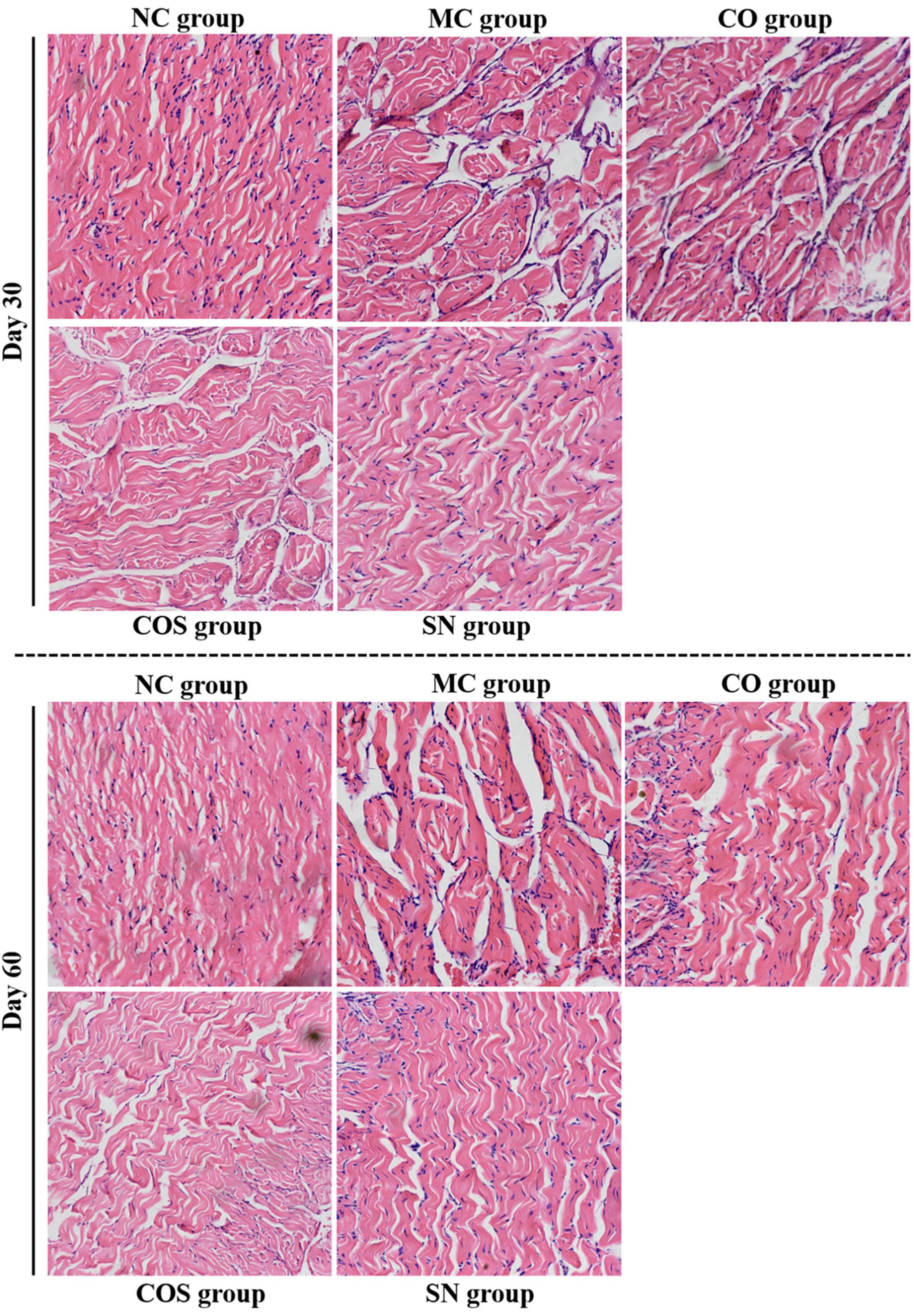
Figure 5. Penis histomorphologies of hyperlipidemia-induced reproductive damage rats. Photomicrographs of penis tissue sections stained with hematoxylin and eosin (H&E) (× 200). NC, normal control; MC, model control; CO, Camellia oil; COS, Camellia oil + squalene; SN, sildenafil.
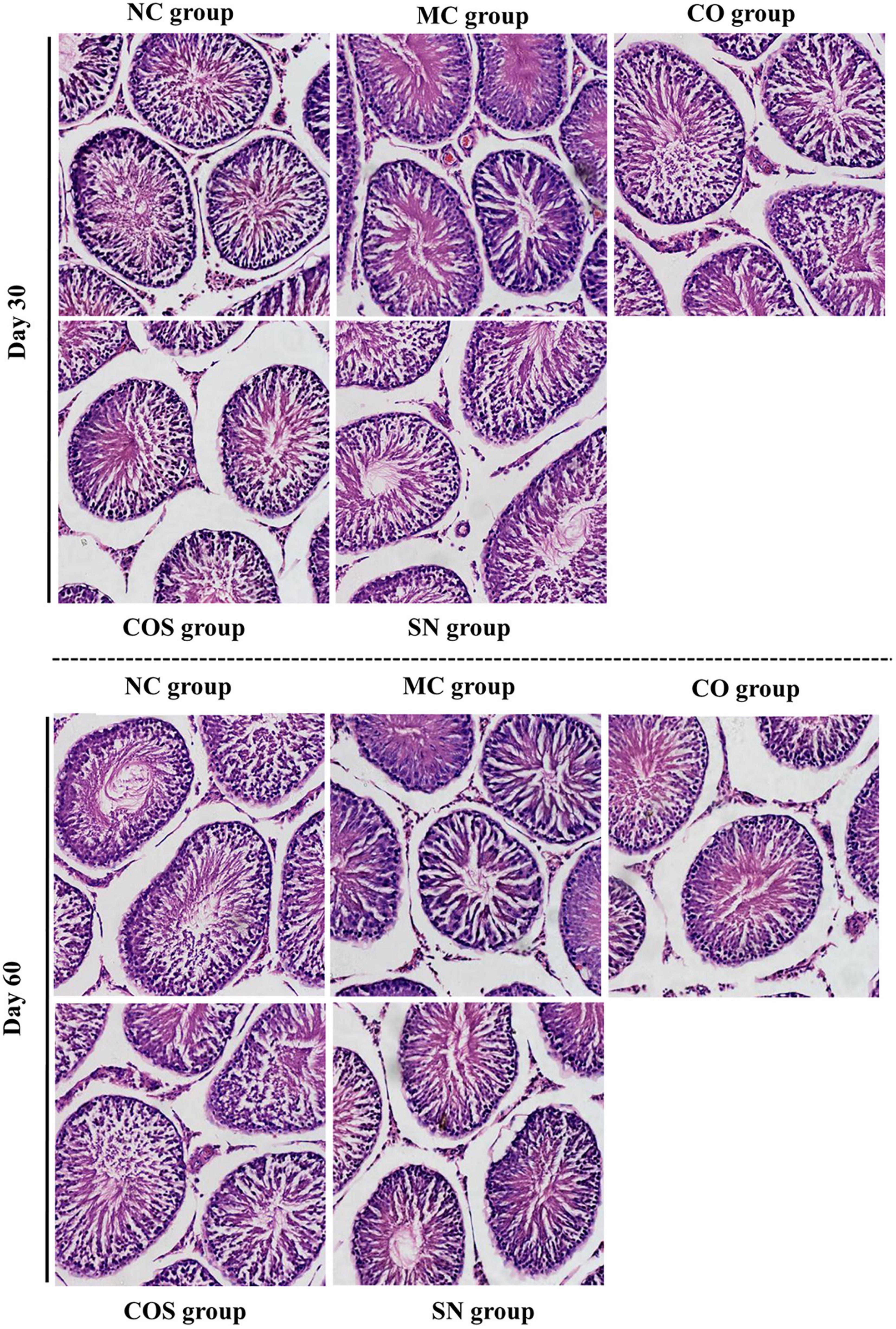
Figure 6. Testis histomorphologies of hyperlipidemia-induced reproductive damage rats. Photomicrographs of testis tissue sections stained with hematoxylin and eosin (H&E) (× 200). NC, normal control; MC, model control; CO, Camellia oil; COS, Camellia oil + squalene; SN, sildenafil.
Hyperlipemia is a prevalent, chronic condition that affects penile hemodynamics (25), Huang et al. found that hyperlipidemia could damage arterial endothelial cell function through cell adhesion molecule-1, cause inadequate blood supply to the penis and reduced blood flow to the Corpus cavernosum penis arteries, which could lead to erectile dysfunction and sexual dysfunction by causing of cavernosal fibrosis (26). At present, researches on tea mainly focus on the effect of tea polyphenols on hyperlipidemia (27) and reproductive system (19, 28, 29), Jiraporn et al. (12) reported that due to Moringa oleifera Lam. Leaf Tea containing rich total phenols, flavonoids, and antioxidants, it could enhance sexual function and the male reproductive system. However, the effect of Camellia oil and its active ingredient squalene on hyperlipidemia-induced sexual dysfunction rats has not been reported.
This study was designed to explore the combining effect and optimal feeding time of CO and squalene on hyperlipidemia-induced sexual dysfunction rats. As expected, the hyperlipidemia rat model was successfully induced by feeding with a high-fat diet for 4 weeks (30). The serum levels of TC, TG, and LDL-C of hyperlipidemic rats orally administered with CO and COS significantly decreased compared with those orally administered with MC at day 60. The results of our study indicated that CO can significantly reduce blood lipids that are rich in monounsaturated fatty, oleic, linoleic, and linolenic acids. This is in agreement with other studies (19, 30–32). Furthermore, we studied the effect of camellia oil on reducing blood lipids at different doses in our previous study (21), including very low (0.75 mL/kg⋅bw⋅day), low (1.5 mL/kg⋅bw⋅day), medium (3 mL/kg⋅bw⋅day) and high (4.5 mL/kg⋅bw⋅day) dose, and found that medium (3 mL/kg⋅bw⋅day) dose was optimal. Therefore, in this present study, we investigated this dose of camellia oil with its active ingredient squalene to observe their combining effect on reducing blood lipids. The results indicated that compared with CO group, COS group significantly reduced the level of blood lipids at day 30, suggesting that CO and squalene could play an enhancing role in combining the effect of reducing blood lipids, and this effect was better than that of pure CO. Studies showed that squalene probably plays a role in hindering the intestinal cholesterol absorption or restraining the activities of key enzymes, such as hepatic acyl-CoA oxidase, fatty acid synthase, and hydroxyl-3-methylglutarylcoenzyme A reductase in endogenous cholesterol biosynthesis (33, 34).
Testosterone, the main male hormone in the body, can promote the development of male reproductive organs and sperm and maintain sexual function (35). Wannasiri et al. (36) and Vigueras-Villaseñor et al. (37) induced hyperlipidemia in male Wistar or Sprague-Dawley rats by feeding with a high-fat diet, found that the testosterone level was decreased in the high-fat diet group. It is proposed that elevated estradiol levels derived from peripheral aromatization of androgen inhibits the hypothalamic-pituitary-gonadal axis, resulting in a reduction of testosterone level (38, 39). Our research indicated that the testosterone level and sperm count of epididymal tail were decreased in the MC group as well, which was probably due to hyperlipidemia. However, in the CO, COS, and SN groups were significantly higher than those in the MC group at day 60. We inferred that the three groups of test substances significantly increased the testosterone levels and sperm counts in the epididymal tail of male rats with hyperlipidemia. However, only COS and SN groups significantly increased testosterone levels at day 30, suggesting that CO mixed with squalene could play a role in combining the effect of promoting the level of testosterone and the number of epididymal tail sperm, and this effect was better than that of pure CO. This finding might be because squalene can participate in cholesterol biosynthesis and various biochemical reactions in the body; accelerate the synthesis of steroid hormones, such as testosterone (40, 41); increase the activity of SOD and blood oxygen content (15); promote blood circulation; and reduce blood lipid levels, which in turn improve sexual function. At day 60, the testosterone level and sperm count in the epididymal tail did not increase compared with the day 30, suggesting that the testosterone level and sperm count in the epididymis did not increase with feeding time. Therefore, we considered that the optimal feeding duration of CO and squalene on hyperlipidemia-induced sexual dysfunction rats was 30 days.
Current animal studies showed that hyperlipidemia can impair erectile function by changing the morphological structures of sexual organs, e.g., penile corpus cavernosum lesions. Li et al. (42) assessed erectile function by performing cavernous nerve electrostimulation followed by intracavernosal pressure/mean arterial pressure measurements, as well as plasma lipid profile assessment, and then pointed out that hyperlipidemia caused cavernous fibrosis by increasing plasma lipid levels, fibrosis and apoptosis, decreasing smooth muscle/collagen ratio and autophagy. The organ coefficient and pathological results of this study showed that the organ coefficient of penis decreased significantly and that the penis and testis showed pathological changes in different degrees in the MC group, such as the number of smooth muscle cells and cavernous sinus decreased significantly in the corpus cavernosum penis, and the smooth muscle of the cavernous body were distributed unevenly and arranged disorderly and loosely, all levels of spermatogenic cells in the testis were arranged disorderly, and some spermatogenic cells showed necrosis, apoptosis, and nuclear pyknosis. These findings were consistent with those of previous reports (42–44). Compared with the MC group, the CO, COS, and SN groups had significantly increased penile organ coefficient and no pathological change in penis and testis, suggesting that the three groups of test substances could improve the damage of penis and testis and promote the recovery of erectile function.
In conclusion, results demonstrated that the addition of squalene to CO as a combining substance to feed hyperlipidemia-induced reproductive damage rats for 30 days could play an important role in combining the effect of lowering blood lipid, promoting the level of testosterone and the number of epididymal tail sperm, improving the damage of penis and testis, and promoting the recovery of erectile and sexual function. However, further studies should be carried out to elucidate the molecular mechanisms of CO and squalene in lowering blood lipid and improving sexual function in vivo.
The original contributions presented in this study are included in the article/supplementary material, further inquiries can be directed to the corresponding author.
This animal study was reviewed and approved by Ethics Committee of Gannan Medical University.
QX: methodology, investigation, and writing—original draft preparation. ML: methodology, validation, investigation, formal analysis, and data curation. GC and QZ: investigation and resource. YG: validation, writing—review and editing, and software. JL: conceptualization, writing—review and editing, funding acquisition, and supervision. All authors contributed to the article and approved the submitted version.
This research was supported by the Open Project of Key Laboratory of Prevention and treatment of cardiovascular and cerebrovascular diseases, Ministry of Education (XN202001) and University-Level Scientific Research Projects of Gannan Medical University (ZD201811 and YB201935).
The authors extend sincere thanks to Zhi Wang from Ganzhou People’s Hospital, Ganzhou, China for providing pathological histology analysis service.
The authors declare that the research was conducted in the absence of any commercial or financial relationships that could be construed as a potential conflict of interest.
All claims expressed in this article are solely those of the authors and do not necessarily represent those of their affiliated organizations, or those of the publisher, the editors and the reviewers. Any product that may be evaluated in this article, or claim that may be made by its manufacturer, is not guaranteed or endorsed by the publisher.
1. Samson SL, Garber AJ. Metabolic syndrome. Endocrinol Metab Clin North Am. (2014) 43:1–23. doi: 10.1016/j.ecl.2013.09.009
2. Wang JS, Dai HH, Yan YB, Gong XH, Li X, Li HS. Research of stroke combined hyperlipidemia-induced erectile dysfunction in rat model. Aging Male. (2019) 22:278–86. doi: 10.1080/13685538.2018.1484443
3. Pei ZW, Guo Y, Zhu HL, Dong M, Zhang Q, Wang F. Thymoquinone protects against hyperlipidemia-induced cardiac damage in low-density lipoprotein receptor-deficient (LDL-R–/–) mice via its anti-inflammatory and antipyroptotic effects. Biomed Res Int. (2020) 2020:4878704. doi: 10.1155/2020/4878704
4. Schulster ML, Liang SE, Najari BB. Metabolic syndrome and sexual dysfunction. Curr Opin Urol. (2017) 27:435–40. doi: 10.1097/MOU.0000000000000426
5. Uppin V, Acharya P, Kempaiah BB, Talahalli RR. Zerumbone augments cognitive enhancement potentials of EPA+ DHA: insight from a hyperlipidaemic rat model. Br J Nutr. (2020) 124:1353–60. doi: 10.1017/S0007114520002445
6. Jung TW, Park J, Sun JL, Ahn SH, El-Aty AMA, Hacimuftuoglu A, et al. Administration of kynurenic acid reduces hyperlipidemia-induced inflammation and insulin resistance in skeletal muscle and adipocytes. Mol Cell Endocrinol. (2020) 518:110928. doi: 10.1016/j.mce.2020.110928
7. Jing J, Ding N, Ding XM, Peng LP, Hu XC, Zhao ZJ, et al. Effects of high-fat diet on male mice reproductive function and changes in oxidized low-density lipoprotein in testis. J Med Postgrad. (2016) 29:133–7. doi: 10.16571/j.cnki.1008-8199.2016.02.004
8. Dong YJ, Zheng C, Li YF, Zhang CM, Shen SL, Liang JH. Effects of serum total testosterone and sex hormone-binding globulin levels on erectile function in hyperlipidaemia rats. J Guangxi Med Univ. (2017) 34:1699–702. doi: 10.16190/j.cnki.45-1211/r.2017.12.005
9. Sperling H. Side effects of erectile dysfunction drug treatment. Urologe A. (2017) 56:451–5. doi: 10.1007/s00120-017-0341-4
10. Masuku NP, Unuofin JO, Lebelo SL. Promising role of medicinal plants in the regulation and management of male erectile dysfunction. Biomed Pharmacother. (2020) 130:110555. doi: 10.1016/j.biopha.2020.110555
11. Ratnasooriya WD, Fernando TS. Effect of black tea brew of camellia sinensis on sexual competence of male rats. J Ethnopharmacol. (2008) 118:373–7. doi: 10.1016/j.jep.2008.04.023
12. Laoung-On J, Saenphet K, Jaikang C, Sudwan P. Effect of Moringa oleifera lam. Leaf tea on sexual behavior and reproductive function in male rats. Plants. (2021) 10:2019. doi: 10.3390/plants10102019
13. Zhao ZL, Fan JS, Fu MH, Zhang M, Chen W, Li T, et al. A review of the main constituents and pharmacological studies of camellia oil. Tradit Chin Med. (2020) 9:368–73.
14. Wang M, Zhang YC, Wan Y, Zou Q, Shen LC, Fu GM, et al. Effect of pretreatments of camellia seeds on the quality, phenolic profile, and antioxidant capacity of camellia oil. Front Nutr. (2022) 9:1023711. doi: 10.3389/fnut.2022.1023711
15. Zhang F, Zhu F, Chen B, Su E, Chen Y, Cao F. Composition, bioactive substances, extraction technologies and the influences on characteristics of Camellia oleifera oil: a review. Food Res Int. (2022) 156:111159. doi: 10.1016/j.foodres.2022.111159
16. Ibrahim N’, Fairus S, Zulfarina MS, Mohamed NI. The efficacy of squalene in cardiovascular disease risk-a systematic review. Nutrients. (2020) 12:414. doi: 10.3390/nu12020414
17. Kim SK, Karadeniz F. Biological importance and applications of squalene and squalane. Adv Food Nutr Res. (2012) 65:223–33. doi: 10.1016/B978-0-12-416003-3.00014-7
18. Yin HP, Xu JP, Zhou XQ, Wang Y. Effects of vitamin E on reproductive hormones and testis structure in chronic dioxin-treated mice. Toxicol Ind Health. (2012) 28:152–61. doi: 10.1177/0748233711408381
19. Bumrungpert A, Pavadhgul P, Kalpravidh RW. Camellia oil-enriched diet attenuates oxidative stress and inflammatory markers in hypercholesterolemic subjects. J Med Food. (2016) 19:895–8. doi: 10.1089/jmf.2016.3659
20. Su MH, Shih MC, Lin KH. Chemical composition of seed oils in native taiwanese Camellia species. Food Chem. (2014) 156:369–73. doi: 10.1016/j.foodchem.2014.02.016
21. Xu Q, Luo MH, Tang Q, Liu K, Zhan ZY, Luo JH. Effect of Camellia oil on sexual dysfunction induced by hyperlipidemia in male rats. Acta Nutr Sin. (2018) 40:574–7. doi: 10.3969/j.issn.0512-7955.2018.06.010
22. Nie C, Zhang F, Ma X, Guo R, Zhou S, Zhao L, et al. Determination of quality markers of xuezhiling tablet for hyperlipidemia treatment. Phytomedicine. (2018) 44:231–8. doi: 10.1016/j.phymed.2018.03.004
23. Dupont C, Ralliard-Rousseau D, Tarrade A, Faure C, Dahirel M, Sion B, et al. Impact of maternal hyperlipidic hypercholesterolaemic diet on male reproductive organs and testosterone concentration in rabbits. J Dev Orig Health Dis. (2014) 5:183–8. doi: 10.1017/S2040174414000087
24. Migliaccio V, Sica R, Scudiero R, Simoniello P, Putti R, Lionetti L. Physiological adaptation to simultaneous chronic exposure to high-fat diet and dichlorodipheniletylhene (DDE) in Wistar rat testis. Cells. (2019) 8:443. doi: 10.3390/cells8050443
25. Huang YC, Wu CT, Chen MF, Kuo YH, Li JM, Shi CS. Intracavernous injection of autologous platelet-rich plasma ameliorates hyperlipidemia-associated erectile dysfunction in a rat model. Sex Med. (2021) 9:100317. doi: 10.1016/j.esxm.2020.100317
26. Huang YC, Ho DR, Lin JH, Huang KT, Chen CS, Shi CS. Dietary modification is associated with normalization of penile hemodynamics in rats fed a high-fat diet. J Sex Med. (2019) 16:791–802. doi: 10.1016/j.jsxm.2019.03.013
27. Wen JJ, Li MZ, Chen CH, Hong T, Yang JR, Huang XJ, et al. Tea polyphenol and epigallocatechin gallate ameliorate hyperlipidemia via regulating liver metabolites and remodeling gut microbiota. Food Chem. (2023) 404(Pt A.):134591. doi: 10.1016/j.foodchem.2022.134591
28. Adedara IA, Okpara ES, Busari EO, Omole O, Owumi SE, Farombi EO. Dietary protocatechuic acid abrogates male reproductive dysfunction in streptozotocin-induced diabetic rats via suppression of oxidative damage, inflammation and caspase-3 activity. Eur J Pharmacol. (2019) 849:30–42. doi: 10.1016/j.ejphar.2019.01.033
29. Gu Q, Wang X, Xie L, Yao X, Qian L, Yu Z, et al. Green tea catechin EGCG could prevent obesity-related precocious puberty through NKB/NK3R signaling pathway. J Nutr Biochem. (2022) 108:109085. doi: 10.1016/j.jnutbio.2022.109085
30. Shen TT, Wu SX. Effects of tea seed oil on hyperlipidemic rats induced by high-fat diet. Food Sci Technol. (2017) 13:101–9. doi: 10.3136/fstr.23.101
31. Chiang SS, Chen LS, Chu CY. Active food ingredients production from cold pressed processing residues of Camellia oleifera and Camellia sinensis seeds for regulation of blood pressure and vascular function. Chemosphere. (2021) 267:129267. doi: 10.1016/j.chemosphere.2020.129267
32. Xiao X, He L, Chen Y, Wu L, Wang L, Liu Z. Anti-inflammatory and antioxidative effects of Camellia oleifera abel components. Future Med Chem. (2017) 9:2069–79. doi: 10.4155/fmc-2017-0109
33. Woo MN, Bok SH, Choi MS. Hypolipidemic and body fat-lowering effects of Fatclean in rats fed a high-fat diet. Food Chem Toxicol. (2009) 47:2076–82. doi: 10.1016/j.fct.2009.05.041
34. Kwak YS, Kyung JS, Kim JS, Cho JY, Rhee MH. Anti-hyperlipidemic effects of red ginseng acidic polysaccharide from Korean red ginseng. Biol Pharm Bull. (2010) 33:468–72. doi: 10.1248/bpb.33.468
35. Corona G, Rastrelli G, Morgentaler A, Sforza A, Mannucci E, Maggi M. Meta-analysis of results of testosterone therapy on sexual function based on international index of erectile function scores. Eur Urol. (2017) 72:1000–11. doi: 10.1016/j.eururo.2017.03.032
36. Wannasiri S, Chansakaow S, Sireeratawong S. Effects of Solanum torvum fruit water extract on hyperlipidemia and sex hormones in high-fat fed male rats. Asian Pac J Trop Biomed. (2017) 7:401–5. doi: 10.1016/j.apjtb.2017.01.027
37. Vigueras-Villaseñor RM, Rojas-Castañeda JC, Chávez-Saldaña M, Gutiérrez-Pérez O, García-Cruz ME, Cuevas-Alpuche O, et al. Alterations in the spermatic function generated by obesity in rats. Acta Histochem. (2011) 113:214–20. doi: 10.1016/j.acthis.2009.10.004
38. Fui MN, Dupuis P, Grossmann M. Lowered testosterone in male obesity: mechanisms, morbidity and management. Asian J Androl. (2014) 16:223–31. doi: 10.4103/1008-682X.122365
39. Leisegang K, Almaghrawi W, Henkel R. The effect of Nigella sativa oil and metformin on male seminal parameters and testosterone in Wistar rats exposed to an obesogenic diet. Biomed Pharmacother. (2021) 133:111085. doi: 10.1016/j.biopha.2020.111085
40. Shi T, Wu G, Jin Q, Wang X. Camellia oil authentication: a comparative analysis and recent analytical techniques developed for its assessment. A review. Trends Food Sci Technol. (2020) 97:88–99. doi: 10.1016/j.tifs.2020.01.005
41. Li S, Liu Y, Wang CL. The health benefits and application of squalene. Food Res Dev. (2016) 14:206–9. doi: 10.3969/j.issn.1005-6521.2016.14.050
42. Li R, Cui K, Wang T, Wang S, Li X, Qiu J, et al. Hyperlipidemia impairs erectile function in rats by causing cavernosal fibrosis. Andrologia. (2017) 49:12693.
43. Hu JL, Chen HX, Chen HR, Wu Y, Sun XW, Li Z, et al. Novel noninvasive quantification of penile corpus cavernosum lesions in hyperlipidemia-induced erectile dysfunction in rabbits by two-dimensional shear-wave elastography. Asian J Androl. (2019) 21:143–9. doi: 10.4103/aja.aja_78_18
Keywords: camellia oil, squalene, hyperlipidemia model, improve erectile and sexual function, combining effect
Citation: Xu Q, Luo M, Cheng G, Zhong Q, Guo Y and Luo J (2022) Combining effect of camellia oil and squalene on hyperlipidemia-induced reproductive damage in male rats. Front. Nutr. 9:1053315. doi: 10.3389/fnut.2022.1053315
Received: 25 September 2022; Accepted: 07 November 2022;
Published: 18 November 2022.
Edited by:
Wei Liu, Nanchang University, ChinaReviewed by:
Hongyu Chen, Shanghai Academy of Agricultural Sciences, ChinaCopyright © 2022 Xu, Luo, Cheng, Zhong, Guo and Luo. This is an open-access article distributed under the terms of the Creative Commons Attribution License (CC BY). The use, distribution or reproduction in other forums is permitted, provided the original author(s) and the copyright owner(s) are credited and that the original publication in this journal is cited, in accordance with accepted academic practice. No use, distribution or reproduction is permitted which does not comply with these terms.
*Correspondence: Jianghong Luo, bHVvamgxMjEyQGdtdS5lZHUuY24=
Disclaimer: All claims expressed in this article are solely those of the authors and do not necessarily represent those of their affiliated organizations, or those of the publisher, the editors and the reviewers. Any product that may be evaluated in this article or claim that may be made by its manufacturer is not guaranteed or endorsed by the publisher.
Research integrity at Frontiers
Learn more about the work of our research integrity team to safeguard the quality of each article we publish.