 Jessica Lenka
 Enrique González-Tortuero3
 Shweta Kuba
 Natalie Ferry
- 1School of Science, Engineering and Environment, University of Salford, Salford, United Kingdom
- 2School of Life Sciences, Faculty of Natural Sciences, Keele University, Staffordshire, United Kingdom
- 3Department of Applied Sciences, Faculty of Health and Life Sciences, Northumbria University, Newcastle upon Tyne, United Kingdom
- 4School of Health and Life Sciences, Teesside University, Middlesborough, United Kingdom
The microbiota within the guts of insects plays beneficial roles for their hosts, such as facilitating digestion and extracting energy from their diet. The African palm weevil (APW) lives within and feeds on the high lignin-containing trunk of palm trees; therefore, their guts could harbour a large community of lignin-degrading microbes. In this study, we aimed to explore the bacterial community within the gut of the APW larvae, specifically with respect to the potential for lignin degradation in various gut segments as a first step to determining the viability of mining bacterial lignin-degrading enzymes for the bioconversion of lignocellulosic biomass to biofuels and biomaterials. Bacterial metagenomic DNA was extracted from the foregut, midgut, and hindgut of larvae of the APW, and the V3–V4 hypervariable region of the 16S rRNA gene was sequenced using the Illumina MiSeq platform. The generated data were analysed and taxonomically classified to identify the different bacterial phylotypes within the gut community cumulatively and per gut segment. We then determined the presence, diversity, and abundance of bacteria associated with lignin degradation within each larval gut compartment as a basis for suggesting the gut segment(s) where lignin degradation occurs the most. All sequences were classified and belonged to the bacterial kingdom. Firmicutes (54.3%) and Proteobacteria (42.5%) were the most dominant phyla within the gut, followed distantly by Bacteroidota (1.7%) and Actinobacteriota (1.4%). Enterococcus, Levilactobacillus, Lactococcus, Shimwellia, Megasphaera, Klebsiella, Pectinatus, Salmonella, Lelliotia, and Enterobacter constituted the most abundant genera found across all gut segments. The foregut and midgut had many similar genera, whilst the hindgut appeared unique. Overall, 29.5% of total gut bacteria comprising 21 genera were lignin degraders found predominantly in the Firmicutes and Proteobacteria phyla (56.8 and 39.5%, respectively), then moderately in Actinobacteriota (2.5%) and Bacteroidota (1.1%). The most abundant ligninolytic genera were Levilactobacillus (46.4%), Klebsiella (22.9%), Enterobacter (10.7%), Lactiplantibacillus (5.9%), Citrobacter (2.2%), Corynebacterium (1.8%), Paucilactobacillus (1.8%), Serratia (1.5%), Bacteroides (1.1%), and Leucobacter (1.0%) found in different amounts in different gut compartments. The foregut had the most diverse and highest abundance of lignin-degrading phylotypes, and we present reasons that point to the foregut as the main location for the depolymerization of lignin in the APW larval gut.
1 Introduction
Beneficial associations between insects and their gut microbial inhabitants, especially concerning the host’s nutrition, can be exploited for biotechnological applications (Harrison et al., 2021; Rajagopal, 2009; Chukwuma et al., 2021). Wood-feeding insects are known to be able to digest and utilise plant biomass by the synergistic association they enjoy with the microorganisms that inhabit their gut (Ali et al., 2019; Chew et al., 2018; Scully et al., 2013; Chauhan, 2020; Kougias et al., 2018). Recently, much attention has been given to understanding the composition of the inhabitant microbes and how they are naturally adapted to facilitate these bioconversion processes (Prasad et al., 2018; Ransom-Jones et al., 2017).
Molecular techniques such as PCR and high-throughput sequencing have facilitated the studies of microbial communities without depending on the ability to culture individual members of the community, as the optimum conditions for growing different species of microbes vary or are yet undetermined for most species (Lazarevic et al., 2016; Stewart, 2012). Structural survey methods of studying microbiomes aim to identify the taxonomic profiles of the study environments concerning the types of microorganisms present (diversity) and their amounts (abundance or richness), from which functional capability can be predicted if desired (Kunath et al., 2017; Knight et al., 2018).
There is a plethora of studies that have investigated insect gut bacterial compositions using the 16S rRNA amplicon sequencing technique and have primarily identified Proteobacteria, Firmicutes, Actinobacteria, and Bacteroidota as the predominant bacterial phyla in insect guts, amongst many other species and environment-specific findings (Prasad et al., 2018; Scully et al., 2013; Do et al., 2014; Ali et al., 2019; Bozorov et al., 2019). The V3–V4 hypervariable region of the 16S rRNA gene has been targeted in many published sequencing studies of phylogenetic and taxonomic classification of insect gut microbiomes (Ben Guerrero et al., 2016; Lazarevic et al., 2016; García-López et al., 2020; Lluch et al., 2015; Chew et al., 2018). Some of these studies have also pointed out the fact that the gut microbiome of insects is non-static and influenced by factors such as environment (Yun et al., 2014), host phylogeny (Franzini et al., 2016; Mohammed et al., 2018), developmental stage and season (Valzano et al., 2012; Jia et al., 2013), nutrition and diet (Montagna et al., 2015; Muhammad et al., 2017; Ben Guerrero et al., 2016), gut physiology and conditions for pH, temperature, and oxygen availability (Egert et al., 2003; Chew et al., 2018). Regardless, there are core members of the community that are only mildly influenced by such factors that may persist, thereby defining the most fundamental functions performed by the microbiome (Pal and Karmakar, 2018; Reich et al., 2018; Franzini et al., 2016; Ben Guerrero et al., 2016).
Research on bacteria capable of lignin degradation has only recently gained much attention, as most studies of microbial lignin degradation have centred on fungi (Bugg et al., 2011a). The ability to directly degrade and modify lignin has been shown in several bacterial phyla such as Proteobacteria, some Firmicutes, and Actinobacteria, the majority of which were obtained from the guts of ruminants, termites, and other wood-feeding insects (Bugg et al., 2011b; Huang et al., 2012; Arumugam et al., 2014; Bugg and Rahmanpour, 2015; Kassim et al., 2016; Janusz et al., 2017). Other bacteria identified and shown to possess lignin-degrading or modifying abilities from several other research outcomes have been compiled and are presented in Table 1.
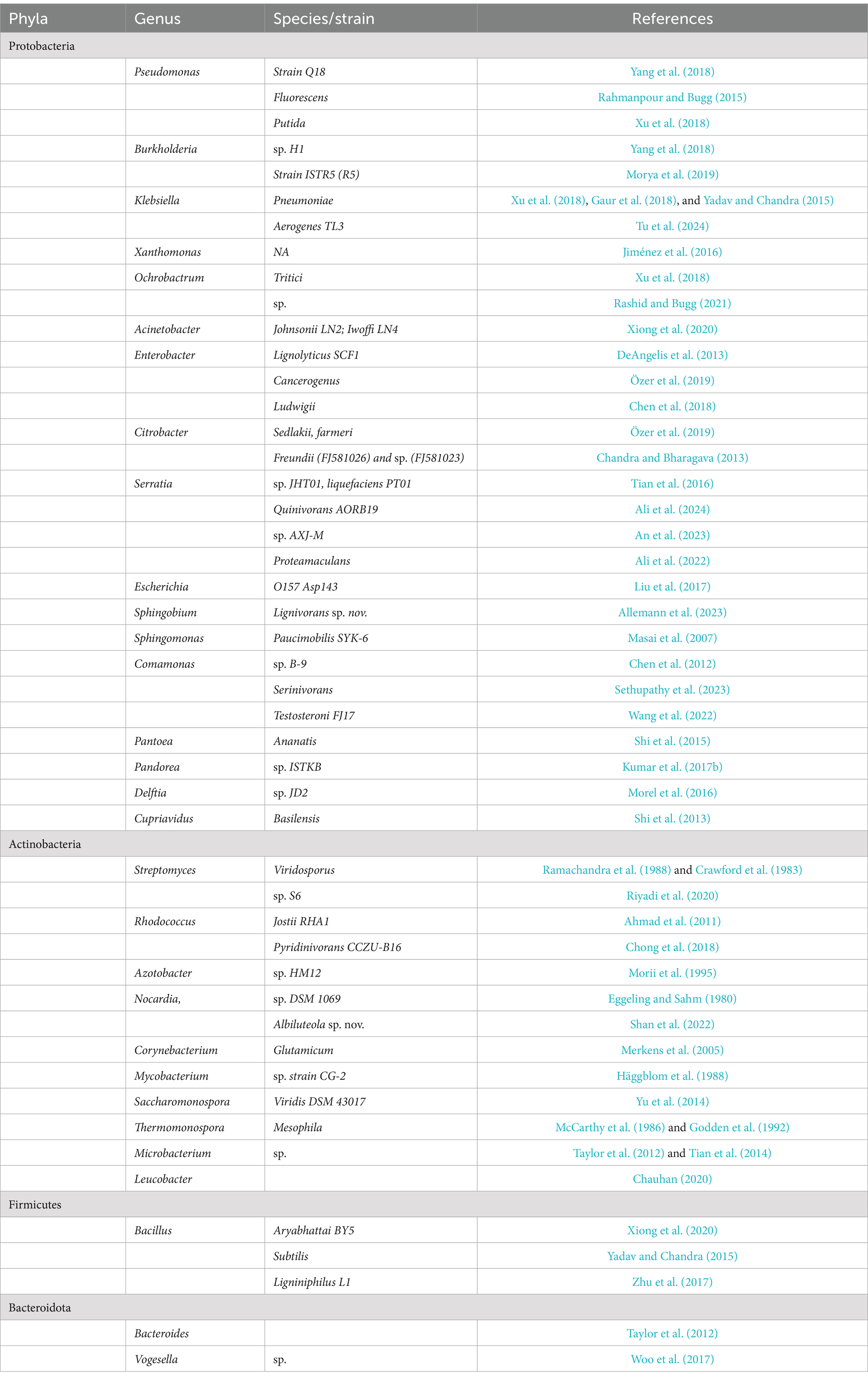
Table 1. Lignin-degrading bacterial genera identified from literature reports.
A list of different lignin-associated bacterial genera (grouped according to phyla) identified through a literature search of published articles.
Despite the increase in research on gut microbial communities, studies about how these communities are organised within each gut compartment using culture-independent methods are not readily available, as most gut bacterial diversity studies have been about the whole gut communities or are taxa-specific. This presents a need for broader and systematic identification of the diversity in each segment of the gut of these insects to provide a wider description of the microbial community and relate the contribution of members of the community in each gut segment to the overall host’s metabolism, adaptability, and survival (Engel and Moran, 2013; Poelchau et al., 2016).
Industrial-scale bioprocessing of lignocellulosic biomass as viable substitutes to fossil-based sources is plagued by a lack of efficient pre-treatment and lignin valorization strategies that align with the global outcry for green and sustainable processes to minimise environmental damage and their climate change consequences. In biorefineries, substituting currently used chemical and thermophysical methods of biomass pre-treatment with biological enzyme-based methods will go a long way in alleviating costs and slowing down climate change. Given this, researchers have prioritised exploring natural biomass-utilising systems such as the guts of wood-feeding insects to maximise the chances of isolating the most efficient candidate enzymes of microbial origin, which serve to facilitate the breakdown of the host’s lignocellulose-rich diet for potential application in industrial bioconversion of lignocellulose to biobased products as alternatives to chemical methods (Olsson, 2016; Brown and Chang, 2014). Detailed studies surrounding the enzymology of ligninolytic enzymes are being intensified, and several classes of enzymes potentially possessing ligninolytic activity have been identified from lignin-degrading fungi and bacteria (Fisher and Fong, 2014).
The African palm weevil (Rhynchophorus phoenicis) belongs to the Curculionidae family of beetles (Coleoptera). It is an important pest affecting mostly oil palm trees in Nigeria, Cameroon, and other subtropical African countries where it is found. Other host plants of this insect include sugar cane, coconut, raffia palm, and the sago palm (Omotoso and Adedire, 2007; Mba et al., 2017). The weevil lives their entire life cycle within the trunk of palm trees, feeding on the palm tissue, which has been reported to have high lignin content (Al-Zuhair et al., 2015; Fadele et al., 2017; Ameh et al., 2016; Nasser et al., 2016; Bensah et al., 2015), and the larval stage of development is the most destructive stage of this insect (Bamidele et al., 2013; Harris et al., 2015; Angzzas et al., 2016; Omotoso, 2013). Despite this lignin content, the APW overcomes the lignin barrier as it excavates and burrows deeply into the interior of the trunk of healthy trees, leading to their eventual destruction. In such a sense, the weevils most likely benefit from the synergistic relationship with their gut microbiota that enables them to degrade lignin (Geib et al., 2008), hence our interest in profiling the bacteria inhabiting the gut of the insect with respect to the potential for lignin degradation. Thus, these insect guts could be reservoirs for novel lignocellulose/lignin-degrading enzymes that could be explored for increased efficiency of industrial plant biomass bioconversion processes into energy and material products. To the best of our knowledge, there has not been any comprehensive investigation or exploration of the gut microbiota of Rhynchophorus phoenicis. Therefore, in this study, we present the first attempt at exploring the bacterial community within the gut of the APW larvae, specifically with respect to the potential for lignin degradation as a first step to determining the viability of mining bacterial lignin-degrading enzymes.
2 Methods
2.1 Field collection of APW larvae
Actively feeding larvae of the African palm weevil (R. phoenicis) were collected from different freshly felled palm tree trunks at the Ejekimomi forest reserves of Amukpe village in Sapele, Delta state, Nigeria (5°52′29.9″N 5°42′14.3″E) in 2019 (Figure 1).
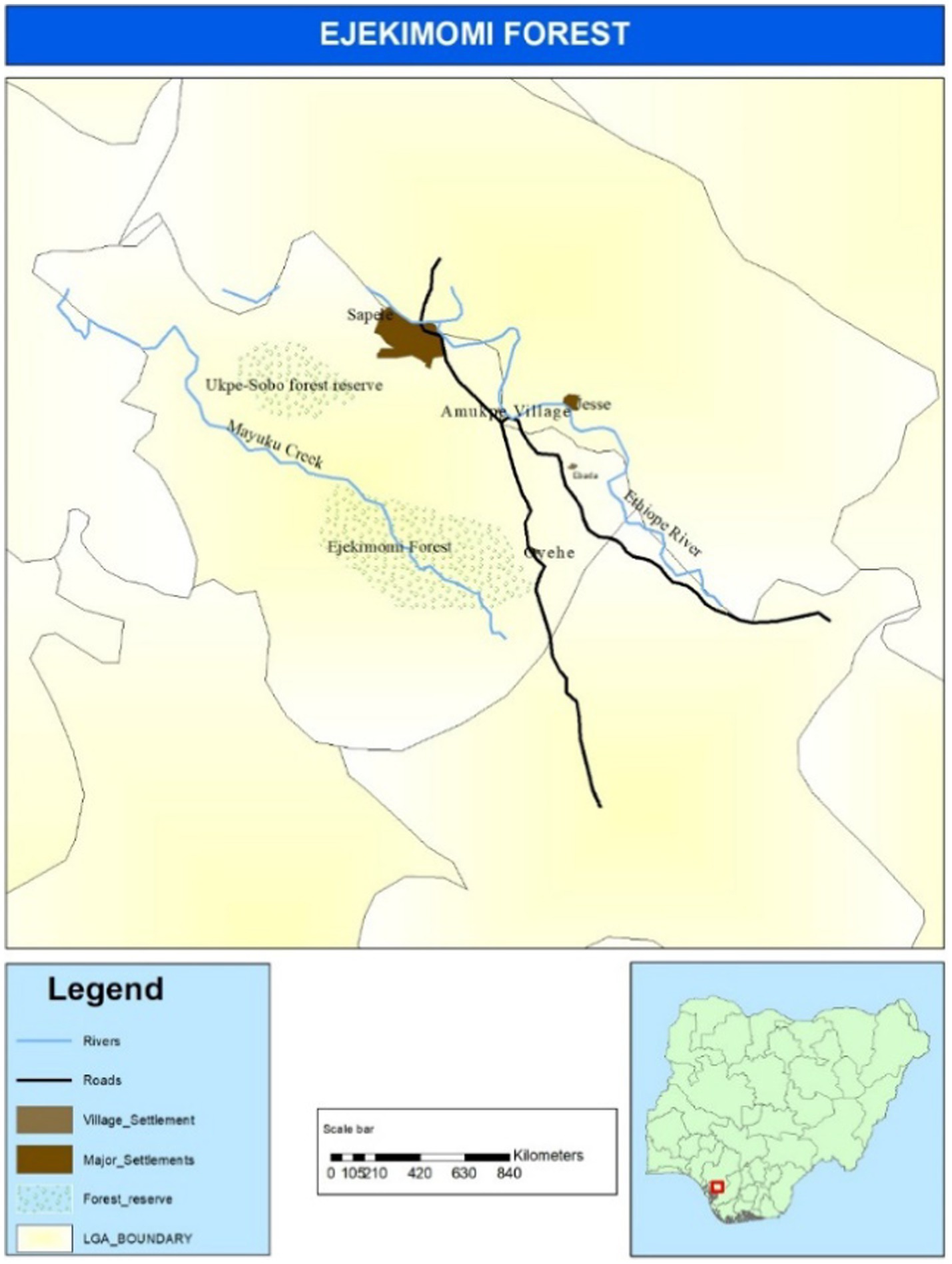
Figure 1. Map showing the location of Ejekimomi forest reserve area in Amukpe village of Sapele town, in Delta state Nigeria where African palm weevil larvae were collected.
Figure 1 is a map of the Ejekimomi forest reserve area in Amukpe village, Sapele town, in Delta State, Nigeria, where African palm weevil larvae were collected. R. phoenicis larvae were identified based on their morphological characteristics. The larvae were washed in sterile water to remove dirt and chopped tree particles; surface sterilisation was conducted using 70% ethanol and 10% bleach, and later, it was rinsed a second time in distilled water (Hammer et al., 2015; Mohammed et al., 2018). Larvae were packaged in sterile containers in the laboratory-prepared and sterilised NAP buffer (Camacho-Sanchez et al., 2013). The samples were stored at 4°C until dissection and DNA extraction (see Supplementary Data 2.0).
2.1.1 Ethics statement
Ethical clearance is unnecessary for work on insects (Franzini et al., 2016). Also, Rhynchophorus phoenicis has not been listed as protected or endangered in national or regional laws. However, ethical approval was obtained to undertake this research due to the Nagoya protocol which emphasises the need for agreement and benefit sharing when accessing genetic materials from a different country as enshrined in the provisions of the biodiversity convention (Ajai, 1997; Omotoso, 2013). The sample collection was done in open and unprotected forests with the agreement and support of the local community.
2.2 Dissection and bacterial DNA extraction from larval guts of APW
Stored larvae were removed from the NAP buffer and dried in a Petri dish. Ten randomly selected larvae were cut open from the mouth to the end of the abdomen using a sterile scalpel and forceps, and the whole guts were aseptically removed separately. Each whole gut was further sectioned into “Foregut,” “Midgut,” and “Hindgut” based on the description of the boundaries of each gut segment (Omotoso, 2013). Gut tissue pieces from the same gut segments were collected in one tube each. The tissues in each tube were cut into smaller pieces, homogenised, centrifuged, and collected supernatant. The supernatant collected was split into two, making two technical replicates per gut segment sample, and bacterial DNA was extracted from these using the QIAamp DNA microbiome kit from Qiagen, UK (Cat. No. 51704), according to the manufacturer’s instructions. A negative extraction control, which contained no DNA, was prepared and processed alongside the gut segment samples. Twenty-five microliters (25 μL) of bacterial DNA were eluted from each QIAamp mini column into 1.5 mL Eppendorf tubes. The concentration and purity of the eluted DNA samples were measured using a Nanodrop spectrophotometer (Thermo Fisher, United Kingdom).
2.3 Library preparation and sequencing
DNA samples from the different gut segments (foregut, midgut, and hindgut samples), extraction control (control sample), and ZymoBIOMICS microbial community standard (ZYMO research, United States), which contains a well-defined bacterial composition ideal for the validation of microbiomic workflows (standard sample), were sent to Macrogen, Inc. (NGS), Seoul, Republic of Korea, for library preparation and sequencing. The V3–V4 region of the 16S rRNA gene was amplified using the primers 337F/805R (F337: 5′-GACTCCTACGGGAGGCWGCAG-3′ and 805R: 5′-GACTACCAGGGTATCTAATCC-3′). Sequencing libraries were prepared using the Herculase II Fusion DNA Polymerase Nextera XT Index Kit V2 (Illumina) according to the protocols in the Illumina 16S metagenomic sequencing library preparation guide (Part #15044223 Rev. B). The libraries were purified, quality validated, diluted to 6 nm, and pooled. Paired-end sequencing (2 × 300 cycles) was carried out on an Illumina MiSeq device (Illumina Inc., San Diego, CA, United States) according to the manufacturer’s specifications.
2.4 Data processing and analysis
The data file containing forward and reverse reads for each sample was imported into R-studio software version 4.1.0 (R core team 2020) and was processed following guides from the DADA2 pipeline tutorial 1.16 (Callahan et al., 2016) with slight modifications to suit our reads and desired outcome. Data pre-processing involved quality profiling, trimming, and filtering raw data to eliminate read duplicates, low-quality reads, adapters, and barcode sequences. Paired reads that passed quality processing were denoised and merged, and amplicon sequence variants (ASVs) with corresponding frequencies for each sample were generated (Callahan et al., 2016). Taxonomy was assigned to each ASV using the AssignTaxonomy function to genus level using the Silva_nr99_v138 training set database (Glöckner et al., 2017) for bacterial 16S rRNA as the reference because taxonomic assignments at species level do not yield satisfactory resolution with amplicon sequencing in most cases (Callahan et al., 2016).
The taxonomy assigned ASVs were processed using the R package phyloseq version 1.36.0 (McMurdie and Holmes, 2013). Each sample was identified as a “True” sample (standard, foregut, midgut, and hindgut) or a “Negative” sample (control sample). To ensure no contamination by external sources was present, the automated prevalence-based strategy in the R package decontam version 1.12.0 (Davis et al., 2018) was used with the phyloseq object. ASVs corresponding to sequences identified as mitochondria and chloroplast sequences were removed, and all ASVs identified to belong to the same genus were merged. The different ASVs per gut segment were visualised in KronaTools version 2.8.1 (Ondov et al., 2011).
To assess the accuracy of the sequencing and taxonomic identification procedure, a separate phyloseq object was created containing only the mock bacterial community sample. This was analysed by examining ASV counts and comparing their observed relative proportions to the expected theoretical proportions of species declared in the ZymoBIOMICS microbial community DNA standard product literature (ZymoBIOMICS™ Microbial Community DNA Standard instruction manual, ver1.1.5). This information was represented as a bar chart using Microsoft Excel. A comparison between the observed and expected taxa was made using a chi-square test in R version 4.4.1.
Bacterial genera with <10 ASV counts of cumulative abundances (total abundance from all gut segments) were filtered out, and only those with >10 counts were used for further analysis. The most abundant bacterial phyla and genera identified in the APW gut were presented on a pie chart plotted in Microsoft Excel. Using Microsoft PowerPoint, a Venn diagram was created to show taxa shared between the different gut segments.
To evaluate potential differences in the microbial composition amongst the different gut segments, a non-metric multidimensional scaling (NMDS) plot using Bray–Curtis dissimilarity was constructed considering the number of counts. Then, permutational multivariate analyses of variance (PERMANOVA; Anderson, 2001) were performed to assess differences in the microbial composition amongst the gut segments. The maximum number of iterations was set to 1,000 in all analyses. Additionally, a Kruskal–Wallis test was performed to evaluate potential differences in the microbial diversity amongst the different gut segments when considering the Shannon and inverse Simpson indexes. Shannon index was calculated using the natural logarithm. In case there were significant differences between gut segments, to see which pairs of segments showed significant differences, Holm–Bonferroni-corrected Dunn tests were performed. All these statistical approaches were carried out at an alpha level of 0.05 and were performed in R version 4.4.1 using the vegan version 2.6-6.1 (Oksanen et al., 2024), FSA version 0.9.5 (Ogle et al., 2023), ggplot2 version 3.5.1 (Wickham, 2016), and gridExtra version 2.3 (Auguie, 2017).
All lignin-degrading bacterial genera identified within our samples were selected based on the current knowledge (Table 1), and their relative abundances by genus and gut segment were plotted using stacked column charts.
3 Results
3.1 Summary of raw amplicon sequence data statistics
A summary of the raw data generated following the sequencing of the 16S libraries on a 2 × 300 bp Illumina platform indicated a successful run with each sample having a high total number of paired end reads (except for the negative extraction control) and an average GC content of 54%. Additionally, 91% of the total reads sequenced had Phred quality scores higher than 20, whilst 82% had Phred quality scores of 30, suggesting that our dataset is very good quality data (Andrews, 2010; Table 2).

Table 2. Summary of the 16S rRNA sequencing data statistics.
3.2 Analysis of negative control sample (decontamination)
The duplicate negative control samples “Control” underwent all amplification, library preparation, sequencing, and bioinformatic analysis steps as the gut samples and were analysed for external contaminants using the R package decontam (Lazarevic et al., 2016). The output returned a “False” result with respect to the assumption that contaminating taxa are more likely to be present in the negative “control” sample compared to true samples. This observation, therefore, means that the “contaminant” taxa identified in the control samples are more present in the true samples than in the control. The negative control contained 42 ASVs, which were all present in the true samples and had a total abundance corresponding to just approximately 1.7% of the total taxa abundance found in the true samples. Only Enterococcus, Lactococcus, Acinetobacter, and Bacteroides were present at >0.1% each. All the other bacteria each had much lower values (<0.1%). Notwithstanding, these taxa were not removed from the true samples as contaminants as they are expected in the true samples and their abundances in the control sample are far lower than what was observed for each of these taxa in the true samples.
3.3 Analysis of mock microbial community standard
The mock microbial community DNA standard made up of eight bacterial strains with various theoretical compositions for each strain was sequenced and analysed alongside the other larval gut and negative extraction control samples. This sample served as a positive control for ascertaining the fidelity of the 16S rRNA amplicon sequencing process and the performance of the data analysis pipeline used. No significant differences existed between the bacterial composition in the “observed” standard sample and the “expected” theoretical values (chi-square test: p = 0.2303; Figure 2).
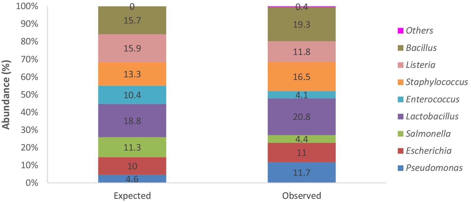
Figure 2. Stacked bar chart showing percentage abundances of bacterial strains expected within the mock microbial DNA community standard from theoretical data and actual observed bacterial genera identified following sequencing and analysis of the positive control “standard” sample.
3.4 Taxonomic profile of APW larval gut bacteria
3.4.1 Total bacterial diversity in the APW larval gut showing percentage abundances by phyla and genera
All taxa identified (100%) belonged to the kingdom Bacteria. In all, 165 genera spanning 7 phyla (Firmicutes, Proteobacteria, Actinobacteriota, Bacteroidota, Campylobacteria, Desulfobacterota, and Verrucomicrobiota) were identified. The dominant phyla with individual genera having a sequence abundance of 10 counts and above were Firmicutes (54.3% of total ASV abundance), Proteobacteria (42.5%), Bacteroidota (1.7%), and Actinobacteriota (1.4%) (Figure 3A). Enterococcus, Levilactobacillus, Lactococcus, Shimwellia, Megasphaera, Klebsiella, Pectinatus, Salmonella, Lelliotia, and Enterobacter were the most dominant genera, listed in decreasing order of abundance (Figure 3B), but approximately 16.4% of the ASVs were not resolved to the genus level. The ASV table (>10 average counts) generated can be seen in Supplementary Data 3.0.
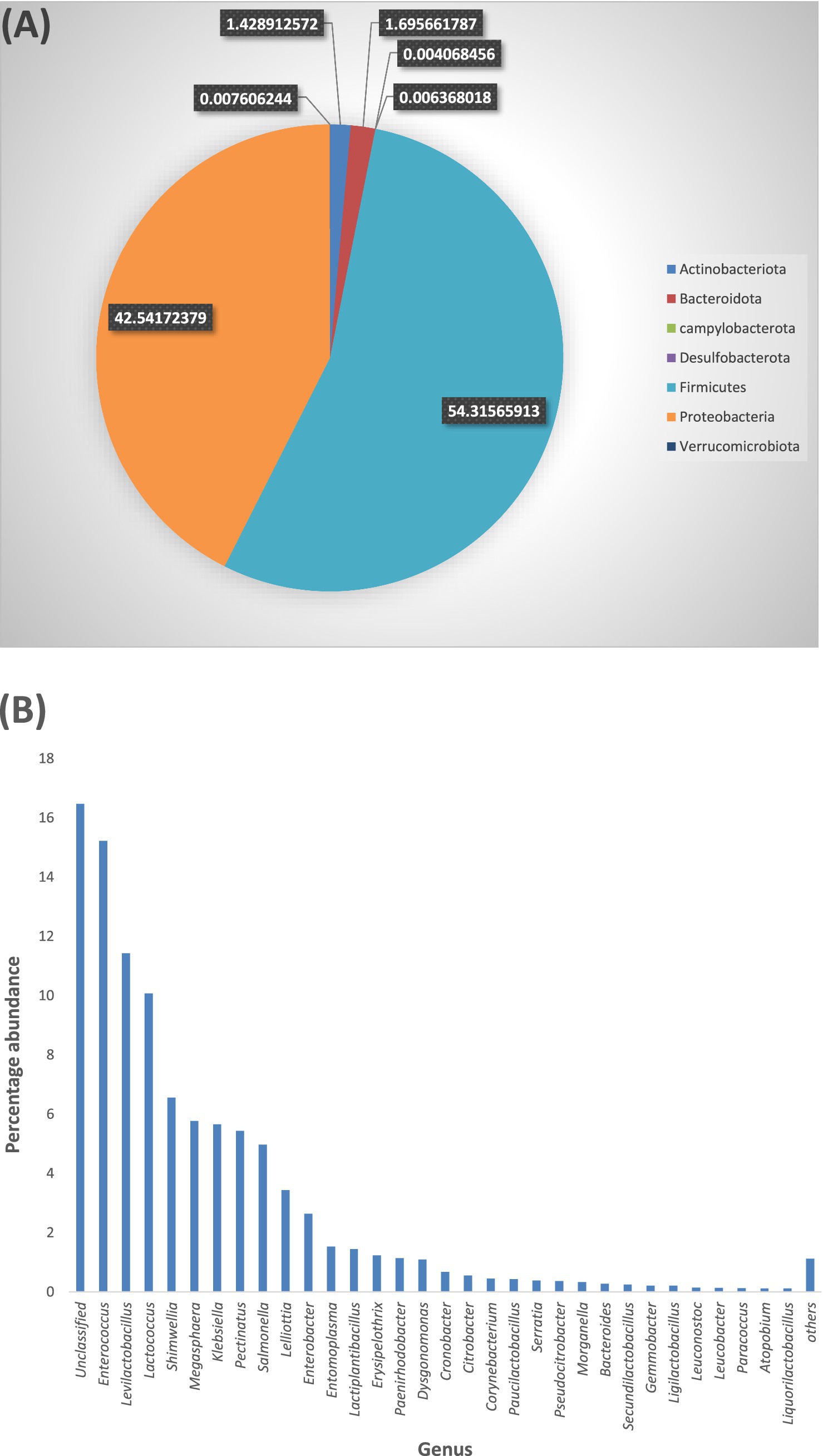
Figure 3. (A) Each section of the pie chart, having a unique colour, represents a bacterial phylum. The size of each section is indicative of the percentage abundance of the phylum. (B) All bacterial genera are represented in one colour (blue) on the bar chart with the length of the bars indicating the percentage abundance of each genus. To optimise the view, only genera with 0.1% abundances are shown individually on the bar chart, genera with <0.1% abundance are combined into “Others”. “Unclassified” represents the abundance of ASV counts that were not successfully resolved to genus level.
3.4.2 Genus-level bacterial diversity and percentage abundances in different gut segments of APW larva
3.4.2.1 Taxonomic profile per gut segment
The taxonomic profile and classification of bacteria identified in the foregut, midgut, and hindgut segments of the APW gut are shown in Figure 4. In each gut segment, a large percentage of the bacteria (foregut; 12%, midgut; 14%, and hindgut; 19%) were not resolved to specific bacterial genera. Enterococcus, Lactococcus, Shimwellia, Lelliotia, Klebsiella, Enterobacter, Bacteroides, Serratia, Salmonella, and Citrobacter were the genera found across all gut segments that were deemed to represent the core bacterial microbiota. The foregut and midgut shared lots of similar genera that were completely absent in the hindgut (Megasphaera, Pectinatus, Levilactobacillus, Paucilactobacillus, Secundilactobacillus, Leuconostoc, Salmonella, Pectinatus, Cronobacter, and Atopobium). The hindgut appeared to be unique, containing Erysipelothrix, Morganella, Gemmobacter, Paracoccus, Providencia, Leminorella, Yokenella, and Rhizobium exclusively, having only Leucobacter in common with the foregut and Ligilactobacillus with the midgut (Figure 5).
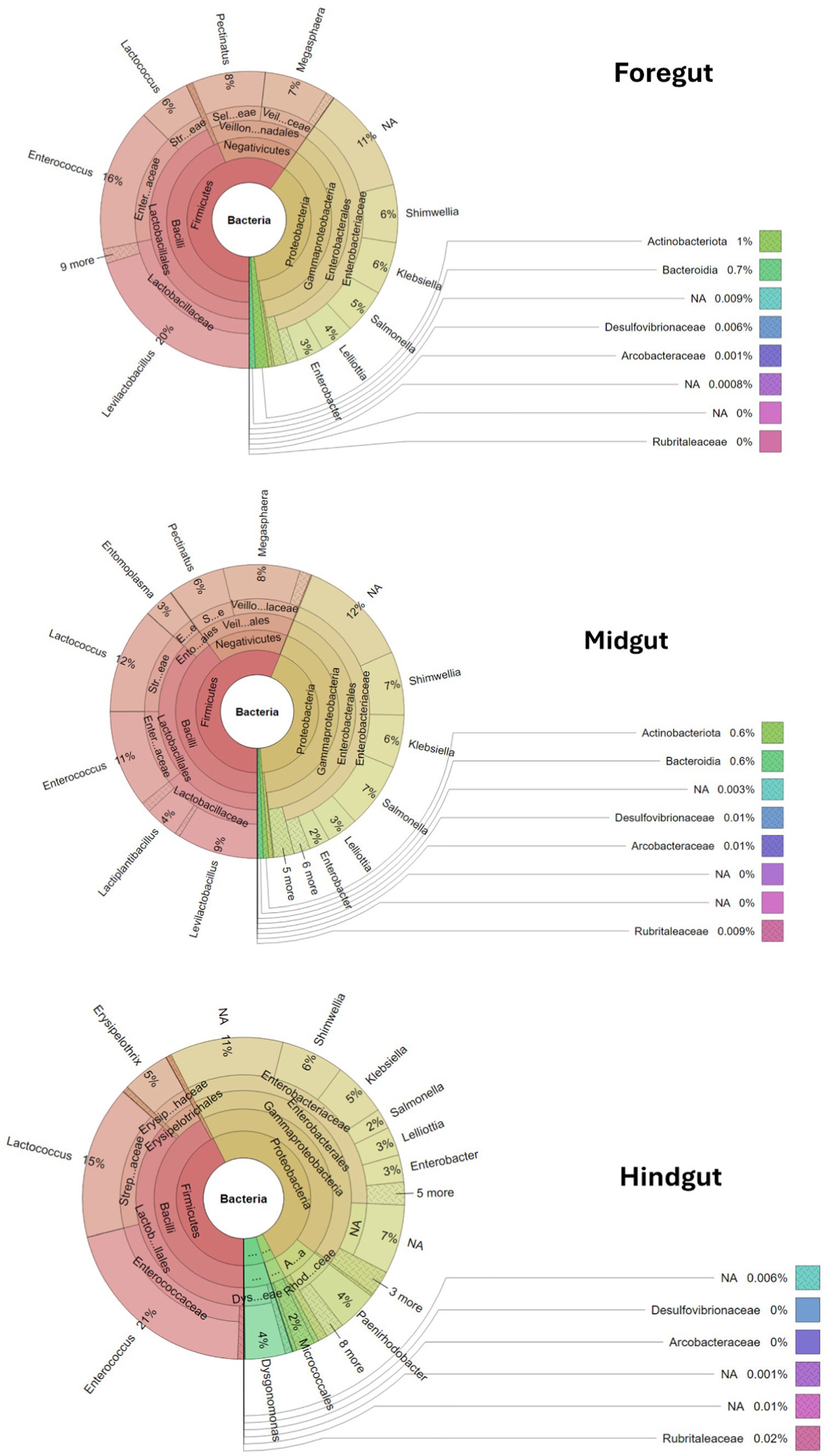
Figure 4. Krona plots showing the taxonomic classification of bacteria within the gut segments. Each circle represents a taxonomic level growing outwards from “Kingdom” to “genus”. Each phylum is represented by a specific colour and the different taxa at different levels within the phylum are represented by varying shades of the colour assigned to the phylum. The different genera and their percentage abundances identified can be seen in the outermost layer of the circles.
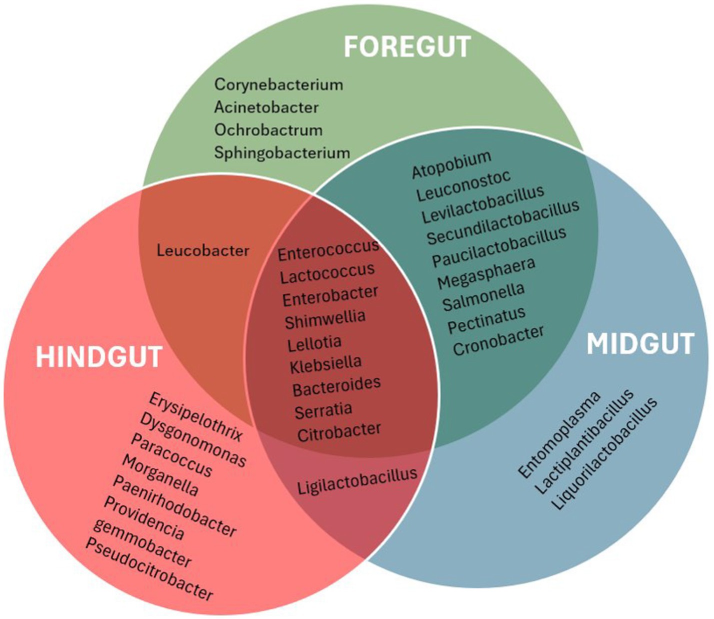
Figure 5. Venn diagram that presenting a visualization of the bacterial taxa found uniquely in different gut segments and those shared between the different gut segments. Only the most abundant genera with abundance ≥0.1% were used to produce the diagram for a more meaningful analysis.
3.4.2.2 Comparative analysis of bacterial composition across the APW gut segments
The dissimilarity between identified bacterial communities from each gut segment was calculated using the Bray–Curtis dissimilarity method, which is based on phylotype abundances and is shown in multidimensional space on a non-metric multidimensional scaling (NMDS) plot (Figure 6). Although the hindgut samples were more clearly separated from those of midgut and foregut and the points representing the same gut segment were closer to each other and separated from those representing other gut segments, there were no significant differences (PERMANOVA: p = 0.06667). This result might indicate that the midgut and foregut microbial communities are more similar. In contrast, the hindgut community is distinctively different, but due to the low variability amongst gut segments, there were no huge differences amongst them.

Figure 6. Each dot on the graph represents a particular sample and each gut segment is represented by a different colour and enclosed in a sphere to denote a specific community.
3.4.2.3 Evaluation of the microbial diversity within each APW gut segment
The species diversity was estimated using both the Shannon and inverse Simpson diversity indices (Figure 7). Whilst the Shannon index considers the richness component and rare cover of species present in the different gut segments, the inverse Simpson index emphasises the evenness component and, thus, the dominant cover species. Consequently, the Hindgut had the highest Shannon diversity index and the lowest inverse Simpson index. However, the foregut showed the lowest Shannon index, but the midgut showed the highest inverse Simpson index. Despite all these observations, there were no significant differences in the microbial diversity amongst the segments (Kruskal–Wallis test: p = 0.2765).
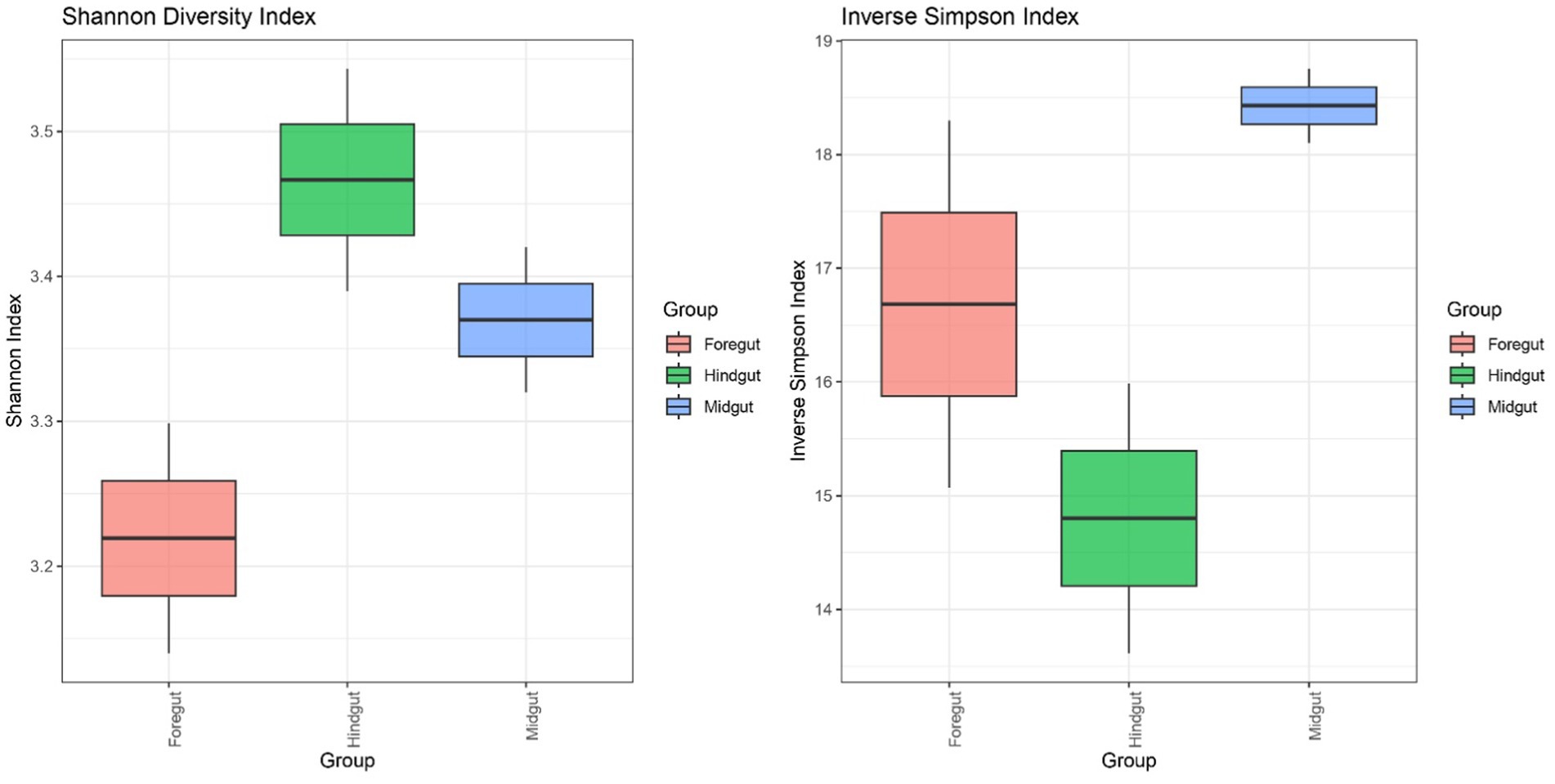
Figure 7. The boxes denote interquartile ranges (IQR) between the first and third quartiles (Q1-Q3) and the horizontal line inside the boxes defines the median. The whiskers which extend from Q1 and Q3 represent the lowest and highest points within 1.5-fold IQR respectively.
3.5 Diversity and relative abundance of all identified lignin-degrading bacteria in the different gut segments of APW larvae
A total of 21 bacterial genera reported to have lignin-degrading ability from several literature (Table 1) were identified across the different gut segments of the African palm weevil microbiome. They represented a total of 21.49% of all identified genera within the gut. Firmicutes constituted 56.79 and 39.56% of the lignin degraders were from the Proteobacteria phylum, whilst only 2.5 and 1.13% were from the Actinobacteriota and Bacteroidota phylum, respectively. Levilactobacillus (46.4%), Klebsiella (22.9%), Enterobacter (10.7%), Lactiplantibacillus (5.9%), Citrobacter (2.2%), Corynebacterium (1.8%), Paucilactobacillus (1.8%), Serratia (1.5%), Bacteroides (1.1%), and Leucobacter (1.0%) were the most dominant lignin-degrading genera in the gut cumulatively in the listed order (Figure 8).
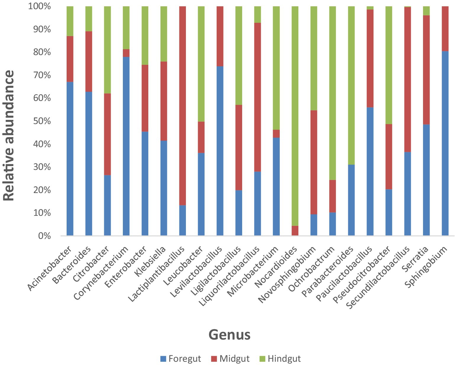
Figure 8. Stacked columns showing plots of relative abundances of individual lignin degrading bacteria identified in the Foregut, Midgut and Hindgut of the APW larvae as a percentage of the total gut bacterial abundance per gut segment. Each column represents a specific bacterial genus and is made up of colour coded regions (one colour for each of the gut segments), the size of which is proportional to the relative abundance of that genus in the different gut segments.
The foregut had 20 out of the 21 identified lignin-degrading genera (except Norcardiodes), adding up to a total abundance of 55.5%. The midgut followed closely, also having 20 of the identified genera (except Parabacteroides) and a total abundance of 32.9%. The hindgut had the least number of identified bacterial taxa (only 18 genera, with Lactiplantibacillus, Levilactobacillus, and Sphingobium absent) and the least total abundance of 11.5%. To facilitate comparison, we calculated the percentage abundance of each bacterial genera in each gut segment as a fraction of the total abundance of lignin-degrading taxa identified. The three gut segments shared 18 similar taxa in varying abundances, with the foregut having the highest abundance of each taxon in most cases. Overall, the foregut had the most diverse and abundant lignin-degrading genera, followed by the midgut, and the hindgut had the least.
4 Discussion
The quest to find enzymes capable of biological degradation of lignin as an alternative to chemical and physical methods of lignocellulose breakdown has resulted in research efforts geared towards bioprospecting these enzymes from environments where lignin degradation is known to occur naturally, such as in the guts of wood-feeding insects (Fisher and Fong, 2014; Ali et al., 2019). Recently, research on mining the microbiota of insects for genes that code for enzymes and bioactive compounds has greatly increased and is mainly being carried out via culture-independent methods (Steele et al., 2009; Hammer et al., 2015; Harnpicharnchai et al., 2007; Quince et al., 2017).
The DNA extracted from pooled gut tissues of preserved larvae had low concentration, and due to the COVID-19 lockdown, which could not allow us to return to the field (Nigeria) to collect more samples, we could only prepare duplicate samples from each gut segment pool. This posed a challenge to the attainment of at least 3 technical replicates to facilitate statistical analysis. However, being sequencing data that were subjected to quality control and filtering, the impact of using only two technical replicates is minimised, especially as the sequencing data statistics were of good quality (Table 2).
It is not just enough to have good quality data; it is also critical to assess that the sequencing did not introduce bias that will misrepresent the true composition of the microbial community after analysis, hence the need for controls and standards (Lazarevic et al., 2016; Reich et al., 2018). The negative control sample had only bacteria, which were also present in the true samples and are associated with insect guts (Egert et al., 2003; Janusz et al., 2017; Ceballos et al., 2017). This observation implies that there was no external or unexpected contamination by any foreign or exogenous bacteria. It is recommended that microbial taxa found in the control sample that correspond to genuine or biologically expected microbiota of interest should not be removed from valid samples except where they occur in higher relative abundances compared to the samples (Reich et al., 2018; Lazarevic et al., 2016).
Analysis of the mock microbial community DNA standard also found all the bacterial strains as expected, with only a few additional strains in meagre amounts of approximately 0.4% (Figure 2). Although the expected percentage abundances for the bacterial components in the community standard were slightly overrepresented as with Bacillus, Staphylococcus, Lactobacillus, Escherichia, and Pseudomonas or underrepresented as with Salmonella, Enterococcus, and Listeria. Overall, the discrepancies are minimal and validate our sequencing and bioinformatic analysis pipeline. The identified discrepancies could be because of primer and hypervariable region choice, PCR conditions, library preparation, sequencing, and data pre-processing, as well as several other variables known to introduce bias in 16S rRNA sequencing. The impact of variables cannot be eliminated but can only be minimised (Lluch et al., 2015; Jovel et al., 2016).
Microbiome studies of host-associated gut communities have identified four bacterial phyla (Proteobacteria, Actinobacteria, Firmicutes, and Bacteroidota) that predominantly colonise the guts of insects and most animals (Le, 2021; Batista-García et al., 2016; Colman et al., 2012; Engel and Moran, 2013; Franzini et al., 2016; Huang et al., 2012; Fisher and Fong, 2014). Several factors such as diet and nutrition, host taxonomy, developmental stage and habitat, seasons, gut morphology, and physicochemical conditions have been shown to affect the structure of the microbiota in most insect guts, and these findings have reported host phylogeny as being the most influential factor, with diet contributing significantly, especially in lignocellulose-feeding insects (Colman et al., 2012; Yun et al., 2014; Chew et al., 2018; Franzini et al., 2016; Huang et al., 2012; Jia et al., 2013; Tsegaye et al., 2019). Our results agree with preliminary findings of insect gut-associated bacterial communities, with the detection of the four mentioned phyla being predominant in the APW gut (Figure 3A) and similarity in taxa compared to other Rhynchophorus species (Valzano et al., 2012; Jia et al., 2013; Tagliavia et al., 2014; Kassim et al., 2016; Muhammad et al., 2017; Liao et al., 2020).
A large-scale cross-taxa analysis of insect-associated bacterial diversity and communities based on 137 insect specimens representing 39 species using 16S rRNA sequencing reported that, on average, most insect bacterial communities were not diverse, containing less than 8 phylotypes, and were dominated mainly by a single phylotype belonging to the phyla Proteobacteria or Firmicutes (Jones et al., 2013). However, they excluded phylotypes with less than 1% of the bacterial community in each sample, which must have eliminated many taxa with low abundance, thereby presenting a community with low diversity. Another large-scale deep sequencing effort, based on 305 individual insects belonging to 218 species, reported that the gut of insects harbours a diverse collection of bacteria (Yun et al., 2014). We identified a total of 165 bacterial genera out of which only 78, which had an abundance of 10 or more counts, were used for further analyses and gut microbiota description (Figure 3B). The situation explained above calls for caution when comparing findings across different studies, as subtle variations in methods and analysis parameters (e.g., the threshold for filtering low abundance data) if not carefully considered may lead to wrong conclusions (Knight et al., 2018; Thomas et al., 2012; Quince et al., 2017).
The most dominant genera were mostly aerobes and facultative anaerobes from the Firmicutes (Enterococcus, Levilactobacillus, Lactococcus, Megasphaera, and Pectinatus) and Proteobacteria phyla (Shimwellia, Klebsiella, Salmonella, Lelliotia, and Enterobacter). Investigations to detect the effect of different developmental stages on the gut microbiota of the red palm weevil (Rhynchophorus ferrugineus), a sister species to the APW (R. phoenicis), using non-culture-dependent 16S rRNA amplicon sequencing of the V4 hypervariable region also detected similar bacterial taxa, including Enterobacter, Citrobacter, Serratia, Klebsiella, Lactococcus, Entomoplasma, and Erysipelothrix, though in varying abundances (Muhammad et al., 2017). Similarly, Liao et al. (2020) identified Enterobacter, Lactococcus, and Erysepelothrix as dominant genera in the midgut of R. ferrugineus. These observations might indicate that the bacterial community has a strong phylogenetic signal, i.e., bacterial community structures are more similar amongst closely related insect species than in less related ones (Jones et al., 2013). The red palm weevil gut has also been reported to have a stable gut microbiota across all developmental stages, with differences owing more to nutrition than host taxonomy (Muhammad et al., 2017). The detection of similar abundant taxa or what could be called a “core microbiome” from our results studying the larval stage and those of studies in larval, pupal, and adult stages of Rhynchophorus species are in tandem with this report. There have been several other studies into the microbiota of the red palm weevil, being the most investigated species of the Rhynchophorus weevils, but the sequencing methods, experimental design, parameters used, and focus of these studies may not allow for an accurate comparison of total gut bacterial profile with our results (Le, 2021; Tagliavia et al., 2014; Montagna et al., 2015; Jia et al., 2013; Angzzas et al., 2016). To the best of our knowledge, this is the first attempt at profiling the microbiome of Rhynchophorus phoenicis, and there is no published record of gut microbiota studies of other Rhynchophorus relatives such as R. cruentatus, R. palmarum, and R. vulneratus.
All the gut segments of the APW larvae shared an appreciable number of core taxa, whilst the foregut and midgut particularly had more taxa in common, hence exhibiting greater similarity in community structure compared to the hindgut, which was more compositionally unique (Figures 4, 5). Alpha diversity estimation of the different gut segments using the Shannon diversity and inverse Simpson indices visualised by box plots (Figure 6) shows that the hindgut harboured more diverse bacterial taxa, followed by the midgut and then the foregut (the higher the Shannon index, the higher the diversity), corroborating the findings presented in Figure 5. However, taxa distribution within the foregut was more even than in the hindgut, with the most uneven distribution found in the midgut (the wider the size of the box, i.e., the interquartile range, the less even the distribution) (Liu et al., 2017; Li et al., 2018). Although the statistics do not present any significant differences, probably due to having only two technical replicates, the visual representation from Figure 5 and the alpha diversity analyses above support this assertion. These observations support the submission made by other researchers that the difference in morphology (shape, size) and physicochemical conditions (oxygen, temperature, pH, and mineral elements) within each gut compartment affects the structure of the microbial community that exists in it (Engel and Moran, 2013; Valzano et al., 2012; Egert et al., 2003; Chew et al., 2018).
In the absence of an existing database of lignin-degrading bacteria to our knowledge, we compiled information from many pieces of literature stemming from research where bacteria have been implicated or tentatively confirmed to be associated with the decomposition of any part of the lignin molecule (Table 1). We used that list as a reference document, and any bacteria on that list that is identified within the gut of the APW was considered as lignin-degrading bacteria. The lignin degraders constituted 29.5% of the total taxa identified within the larval gut and were drawn from the phyla Firmicutes predominantly (56.8%), Proteobacteria (39.5%), Actinobacteriota (2.5%), and Bacteroidota (1.1%). The dominance of lignin-degrading bacteria belonging to the Proteobacteria and Firmicutes phyla has been consistently reported in all previous research we have accessed on best-characterised lignin-degrading bacteria along with other phyla such as Bacteriodota and Actinobacteria (see a detailed list of sources in Table 1).
The distribution of the 21 lignin degradation-associated bacterial genera (Klebsiella, Enterobacter, Citrobacter, Corynebacterium, Serratia, Bacteroides, Leucobacter, Acinetobacter, Ochrobactrum, Microbacterium, Sphingobium, Novosphingobium, Thermomonas, Sphingomonas, Delftia, and Pseudomonas) across the different gut segments and their relative abundance per segment is presented in Figure 8.
The physical and chemical characteristics of the major components of lignocellulose and the physicochemical conditions such as pH and oxygen availability within the insect’s gut are the major determining factors responsible for the distribution pattern of the lignocellulose-degrading machinery in insects (Sun and Zhou, 2011; Yun et al., 2014). In a detailed morphological and histological description of the APW digestive tract, it was reported to have a foregut, midgut, and hindgut. The foregut is the largest gut segment made up of the buccal cavity, oesophagus, crop, and proventriculus, which are all adapted for intake, mechanical grinding, storage, and onward passage of food to the midgut. The midgut and hindgut are structurally and functionally adapted for the digestion of food, assimilation of nutrients, and excretion of wastes (Omotoso, 2013). The same alimentary tract structure was reported for R. ferrugineus by Harris et al. (2015) in a similar study of the morphology and histology of the RPW larval gut.
Different segments of the gut have unique characteristics, which make them susceptible to colonisation by different types of bacteria (Engel and Moran, 2013). The microbiome within a gut compartment is affected by morphology, which varies as insects metamorphose from one developmental stage to the next in most insect orders. The size and shape of the gut additionally influence the availability of oxygen due to the partial pressure of oxygen from the external environment, which in turn determines the metabolism of the inhabitant bacteria (Yun et al., 2014). For effective utilisation of lignocellulose by wood-feeding insects for energy, depolymerization of lignin must occur first to grant access to hydrolytic enzymes to release the stored-up energy in the carbohydrate polymers, cellulose and hemicellulose (Sun and Zhou, 2011; Kumar et al., 2017a; Silva et al., 2018). Lignin degradation is an aerobic oxidation process requiring oxidative enzymes such as peroxidases, oxidases, and laccases; hence, it is believed that these reactions are most likely to occur in the foregut, being the anterior part of the gut closest to the external environment where oxygen supply is highest (Chew et al., 2018; Sun and Zhou, 2011). In contrast, the midgut and hindgut have been reported as the sites for cellulose and hemicellulose degradation (Egert et al., 2003; Sun and Zhou, 2011; Chew et al., 2018; Yun et al., 2014). These fermentative processes occur by anaerobic hydrolysis; thus, it is reasonable to expect the degradation machinery to be domiciled in the interior, anaerobic compartments of the gut that are farther away from oxygen supply.
The presence almost exclusively of aerobic and facultatively anaerobic bacteria (except Bacteroides alone, which is anaerobic) within the gut of APW and the specific abundance distribution of lignin-degrading bacteria within each gut compartment demonstrates the adaptability of the APW larvae to digesting its diet and suggests where lignin degradation most likely occurs (Khiyami and Alyamani, 2008). Olsson (2016) has reported that the gut of mammals houses more obligate and facultative anaerobes, whilst insect guts have a prevalence of aerobes and facultative anaerobes and a large variety of lignin-associated enzymes.
From our results, the foregut of APW larvae possessed the most diverse and highest percentage abundance of lignin-degrading phylotypes compared to the midgut and hindgut. The presence of the proventriculus as part of the foregut of the APW’s digestive tract shows their adaptation to their food source (lignocellulosic palm tissues) and explains their ability to offer some sort of mechanical pre-treatment to the lignin in their diet as a first step towards extracting energy from the polysaccharides that occur in the later parts of the gut following a logical order. A similar investigation of bacterial community structure in the foregut, midgut, and hindgut of the wood-feeding termite bulbitermes sp. by Chew et al. (2018) suggested that lignin degradation was most probably held in the foregut due to the significantly higher relative abundance of the lignin-degrading bacteria, Actinomycetales, in the foregut compared to the other segments. They further justified their assertion following predictive functional profiling where they found energy and co-factor metabolism predominantly occurring in the hindgut, whereas oxidative xenobiotic degradation reactions (which are related to lignin degradation reactions) occurred mostly in the foregut. Overall, our results, supported by the studies of Chew et al. (2018), and the several other pieces of literature cited above, seem to rationalise the foregut of the APW larvae as being the site for lignin degradation prior to cellulose and hemicellulose degradation in the other gut compartments.
5 Conclusion
Our study, which represents the first known metaprofiling effort of the bacteria colonising the gut of the African palm weevil, R. phoenicis, to date, has revealed great similarity in bacterial community structure with those identified in most insects, specifically with the bacterial microbiota of the phylogenetically related red palm weevil, Rhynchophorus ferrugineus. An appreciable number of lignin-degrading bacteria within the larval gut suggests an immense potential for the discovery of lignin-degrading genes and enzymes. Furthermore, lignin degradation in the African palm weevil is believed to be domiciled in its foregut due to the presence of a proventriculus that serves to mechanically decrease the structural complexity of lignocellulose as a first step towards degradation and the greatest abundance of mostly aerobic and facultatively anaerobic bacteria capable of oxidatively decomposing lignin predominating the foregut. Our findings point towards the gut of the African palm weevil being a reservoir that harbours a consortium of bacteria capable of lignin degradation/modification from which lignin-degrading genes and enzymes can be harvested.
Data availability statement
The datasets presented in this study can be found in online repositories. The names of the repository/repositories and accession number(s) can be found at: https://www.ebi.ac.uk/biostudies/arrayexpress/studies/E-MTAB-13719?key=639be3f8-41bb-40e1-970c-44cc13632281, E-MTAB-13719.
Ethics statement
The animal study was approved by Research Innovation and Academic Engagement Ethical Approval panel, University of Salford. The study was conducted in accordance with the local legislation and institutional requirements.
Author contributions
JL: Conceptualization, Data curation, Investigation, Project administration, Writing – original draft, Writing – review & editing. SK: Data curation, Formal analysis, Writing – review & editing. NF: Conceptualization, Supervision, Writing – review & editing. EG-T: Data curation, Formal analysis, Writing – review & editing.
Funding
The author(s) declare that financial support was received for the research, authorship, and/or publication of this article. This study was funded by the Petroleum Technology Development Fund (PTDF) overseas PhD grant 2017/2018.
Acknowledgments
Special thanks to Prof. Ian Goodhead, Prof. Chloe James, Dr. Rachael Antwis, and Dr. Poppy Pescod (University of Salford) for their insight and support during the experimental and writing-up stages of the project. I also acknowledge financial support granted to me (JL) by the British Federation for Women Graduates (BFWG) during the writing-up phase of my thesis, from which this article has been produced. The authors want to acknowledge Dr. Manasseh Manyi (Federal University of Agriculture Makurdi, Benue State, Nigeria) for the taxonomic identification of the R. phoenicis larvae and the Biochemistry Laboratory of the University of Jos (Plateau State, Nigeria) for the maintenance of the APW weevils during the field collection of larvae.
Conflict of interest
The authors declare that the research was conducted in the absence of any commercial or financial relationships that could be construed as a potential conflict of interest.
Publisher’s note
All claims expressed in this article are solely those of the authors and do not necessarily represent those of their affiliated organizations, or those of the publisher, the editors and the reviewers. Any product that may be evaluated in this article, or claim that may be made by its manufacturer, is not guaranteed or endorsed by the publisher.
Supplementary material
The Supplementary material for this article can be found online at: https://www.frontiersin.org/articles/10.3389/fmicb.2024.1401965/full#supplementary-material
References
Ahmad, M., Roberts, J. N., Hardiman, E. M., Singh, R., Eltis, L. D., and Bugg, T. D. (2011). Identification of DypB from Rhodococcus jostii RHA1 as a lignin peroxidase. Biochemistry 50, 5096–5107. doi: 10.1021/bi101892z
Ajai, O. (1997). Access to genetic resources and biotechnology regulation in Nigeria. Rev. European Commun. Int. Environ. Law 6:42.
Ali, S. S., Al-Tohamy, R., Sun, J., Wu, J., and Huizi, L. (2019). Screening and construction of a novel microbial consortium SSA-6 enriched from the gut symbionts of wood-feeding termite, Coptotermes formosanus and its biomass-based biorefineries. Fuel 236, 1128–1145. doi: 10.1016/j.fuel.2018.08.117
Ali, N. S., Huang, F., Qin, W., and Yang, T. C. (2022). Identification and characterization of a new Serratia proteamaculans strain that naturally produces significant amount of extracellular laccase. Front. Microbiol. 13:878360. doi: 10.3389/fmicb.2022.878360
Ali, N. S., Thakur, S., Ye, M., Monteil-Rivera, F., Pan, Y., Qin, W., et al. (2024). Uncovering the lignin-degrading potential of Serratia quinivorans AORB19: insights from genomic analyses and alkaline lignin degradation. BMC Microbiol. 24:181. doi: 10.1186/s12866-024-03331-3
Allemann, M. N., Presley, G. N., Elkins, J. G., and Michener, J. K. (2023). Sphingobium lignivorans sp. nov., isolated from river sediment downstream of a paper mill. Int. J. Syst. Evol. Microbiol. 73:005704. doi: 10.1099/ijsem.0.005704
Al-Zuhair, S., Abualreesh, M., Ahmed, K., and Abdul Razak, A. (2015). Enzymatic delignification of biomass for enhanced fermentable sugars production. Energy Technol. 3, 121–127. doi: 10.1002/ente.201402138
Ameh, A. O., Ojo, A. A., and Gaiya, J. (2016). Preliminary investigation into the synthesis of furfural from sugarcane bagasse. FUW Trends Sci. Technol. J. 33, 582–586.
An, X., Cheng, Y., Zang, H., and Li, C. (2023). Biodegradation characteristics of lignin in pulping wastewater by the thermophilic Serratia sp. AXJ-M: performance, genetic background, metabolic pathway and toxicity assessment. Environ. Pollut. 322:121230. doi: 10.1016/j.envpol.2023.121230
Anderson, M. J. (2001). A new method for non-parametric multivariate analysis of variance. Austral Ecol. 26, 32–46. doi: 10.1111/j.1442-9993.2001.01070.pp.x
Andrews, S. (2010). FastQC: a quality control tool for high throughput sequence data. Available at: https://www.bioinformatics.babraham.ac.uk/projects/fastqc/ (Accessed October 2019).
Angzzas, S. M. K., Ashuvila, M. A., and Dayang, N. F. A. Z. (2016). Potential lignin degraders isolated from the gut of Rhynchophorus ferrugineus. International Conference on Mechanics, Materials and Structural Engineering (ICMMSE 2016). Atlantis Press.
Arumugam, N., Kalavathi, P., and Mahalingam, P. U. (2014). Lignin database for diversity of lignin degrading microbial enzymes (LD2L). Res. Biotechnol. 5, 13–18.
Auguie, B. (2017). gridExtra: miscellaneous functions for “grid” graphics. R package version 2.3. Available at: https://CRAN.R-project.org/package=gridExtra (Accessed November 2019).
Bamidele, O., Ajele, J., Kolawole, A., and Oluwafemi, A. (2013). Changes in the tissue antioxidant enzyme activities of palm weevil (Rynchophorous phoenicis) larva by the action of 2, 2-dichlorovinyl dimethyl phosphate. Afr. J. Biochem. Res. 7, 128–137.
Batista-García, R. A., del Rayo Sánchez-Carbente, M., Talia, P., Jackson, S. A., O’Leary, N. D., Dobson, A. D., et al. (2016). From lignocellulosic metagenomes to lignocellulolytic genes: trends, challenges, and future prospects. Biofuels Bioprod. Biorefin. 10, 864–882. doi: 10.1002/bbb.1709
Ben Guerrero, E., Soria, M., Salvador, R., Ceja-Navarro, J. A., Campos, E., Brodie, E. L., et al. (2016). Effect of different lignocellulosic diets on bacterial microbiota and hydrolytic enzyme activities in the gut of the cotton boll weevil (Anthonomus grandis). Front. Microbiol. 7:2093. doi: 10.3389/fmicb.2016.02093
Bensah, E. C., Kádár, Z., and Mensah, M. Y. (2015). Ethanol production from hydrothermally-treated biomass from West Africa. Bioresources 10, 6522–6537. doi: 10.15376/biores.10.4.6522-6537
Bozorov, T. A., Rasulov, B. A., and Zhang, D. (2019). Characterization of the gut microbiota of invasive Agrilus mali Matsumara (Coleoptera: Buprestidae) using high-throughput sequencing: uncovering plant cell-wall degrading bacteria. Sci. Rep. 9:4923. doi: 10.1038/s41598-019-41368-x
Brown, M. E., and Chang, M. C. (2014). Exploring bacterial lignin degradation. Curr. Opin. Chem. Biol. 19, 1–7. doi: 10.1016/j.cbpa.2013.11.015
Bugg, T. D., Ahmad, M., Hardiman, E. M., and Rahmanpour, R. (2011a). Pathways for degradation of lignin in bacteria and fungi. Nat. Prod. Rep. 28, 1883–1896. doi: 10.1039/c1np00042j
Bugg, T. D., Ahmad, M., Hardiman, E. M., and Singh, R. (2011b). The emerging role for bacteria in lignin degradation and bio-product formation. Curr. Opin. Biotechnol. 22, 394–400. doi: 10.1016/j.copbio.2010.10.009
Bugg, T. D., and Rahmanpour, R. (2015). Enzymatic conversion of lignin into renewable chemicals. Curr. Opin. Chem. Biol. 29, 10–17. doi: 10.1016/j.cbpa.2015.06.009
Callahan, B. J., Sankaran, K., Fukuyama, J. A., McMurdie, P. J., and Holmes, S. P. (2016). Bioconductor workflow for microbiome data analysis: from raw reads to community analyses. F1000Res. 5:1492. doi: 10.12688/f1000research.8986.2
Camacho-Sanchez, M., Burraco, P., Gomez-Mestre, I., and Leonard, J. A. (2013). Preservation of RNA and DNA from mammal samples under field conditions. Mol. Ecol. Resour. 13, 663–673. doi: 10.1111/1755-0998.12108
Ceballos, S. J., Yu, C., Claypool, J. T., Singer, S. W., Simmons, B. A., Thelen, M. P., et al. (2017). Development and characterization of a thermophilic, lignin degrading microbiota. Process Biochem. 63, 193–203. doi: 10.1016/j.procbio.2017.08.018
Chandra, R., and Bharagava, R. N. (2013). Bacterial degradation of synthetic and kraft lignin by axenic and mixed culture and their metabolic products. J. Environ. Biol. 34, 991–999
Chauhan, P. S. (2020). Role of various bacterial enzymes in complete depolymerization of lignin: a review. Biocatal. Agric. Biotechnol. 23:101498. doi: 10.1016/j.bcab.2020.101498
Chen, Y. H., Chai, L. Y., Zhu, Y. H., Yang, Z. H., Zheng, Y., and Zhang, H. (2012). Biodegradation of kraft lignin by a bacterial strain Comamonas sp. B-9 isolated from eroded bamboo slips. J. Appl. Microbiol. 112, 900–906. doi: 10.1111/j.1365-2672.2012.05275.x
Chen, B., Yu, T., Xie, S., Du, K., Liang, X., Lan, Y., et al. (2018). Comparative shotgun metagenomic data of the silkworm Bombyx mori gut microbiome. Sci. Data 5:180285. doi: 10.1038/sdata.2018.285
Chew, Y. M., Lye, S., Salleh, M. M., and Yahya, A. (2018). 16S rRNA metagenomic analysis of the symbiotic community structures of bacteria in foregut, midgut, and hindgut of the wood-feeding termite Bulbitermes sp. Symbiosis 76, 187–197. doi: 10.1007/s13199-018-0544-5
Chong, G. G., Huang, X. J., Di, J. H., Xu, D. Z., He, Y. C., Pei, Y. N., et al. (2018). Biodegradation of alkali lignin by a newly isolated Rhodococcus pyridinivorans CCZU-B16. Bioprocess Biosyst. Eng. 41, 501–510. doi: 10.1007/s00449-017-1884-x
Chukwuma, O. B., Rafatullah, M., Tajarudin, H. A., and Ismail, N. (2021). A review on bacterial contribution to lignocellulose breakdown into useful bio-products. Int. J. Environ. Res. Public Health 18:6001. doi: 10.3390/ijerph18116001
Colman, D. R., Toolson, E. C., and Takacs-Vesbach, C. D. (2012). Do diet and taxonomy influence insect gut bacterial communities? Mol. Ecol. 21, 5124–5137. doi: 10.1111/j.1365-294X.2012.05752.x
Crawford, D. L., Pometto, A. L. III, and Crawford, R. L. (1983). Lignin degradation by Streptomyces viridosporus: isolation and characterization of a new polymeric lignin degradation intermediate. Appl. Environ. Microbiol. 45, 898–904. doi: 10.1128/aem.45.3.898-904.1983
Davis, N. M., Proctor, D. M., Holmes, S. P., Relman, D. A., and Callahan, B. J. (2018). Simple statistical identification and removal of contaminant sequences in marker-gene and metagenomics data. Microbiome 6, 1–14. doi: 10.1186/s40168-018-0605-2
DeAngelis, K. M., Sharma, D., Varney, R., Simmons, B., Isern, N. G., Markilllie, L. M., et al. (2013). Evidence supporting dissimilatory and assimilatory lignin degradation in Enterobacter lignolyticus SCF1. Front. Microbiol. 4:280. doi: 10.3389/fmicb.2013.00280
Do, T. H., Nguyen, T. T., Nguyen, T. N., Le, Q. G., Nguyen, C., Kimura, K., et al. (2014). Mining biomass-degrading genes through Illumina-based de novo sequencing and metagenomic analysis of free-living bacteria in the gut of the lower termite Coptotermes gestroi harvested in Vietnam. J. Biosci. Bioeng. 118, 665–671. doi: 10.1016/j.jbiosc.2014.05.010
Egert, M., Wagner, B., Lemke, T., Brune, A., and Friedrich, M. W. (2003). Microbial community structure in midgut and hindgut of the humus-feeding larva of Pachnoda ephippiata (Coleoptera: Scarabaeidae). Appl. Environ. Microbiol. 69, 6659–6668. doi: 10.1128/AEM.69.11.6659-6668.2003
Eggeling, L., and Sahm, H. (1980). Degradation of coniferyl alcohol and other lignin-related aromatic compounds by Nocardia sp. DSM 1069. Arch. Microbiol. 126, 141–148. doi: 10.1007/BF00511219
Engel, P., and Moran, N. A. (2013). The gut microbiota of insects–diversity in structure and function. FEMS Microbiol. Rev. 37, 699–735. doi: 10.1111/1574-6976.12025
Fadele, O., Oguocha, I. N., Odeshi, A., and Soleimani, M. (2017). The effect of alkalization on properties of raffia palm fiber. Proceedings of the 26th CANCAM
Fisher, A. B., and Fong, S. S. (2014). Lignin biodegradation and industrial implications. AIMS Bioeng. 1, 92–112. doi: 10.3934/bioeng.2014.2.92
Franzini, P. Z., Ramond, J. B., Scholtz, C. H., Sole, C. L., Ronca, S., and Cowan, D. A. (2016). The gut microbiomes of two Pachysoma MacLeay desert dung beetle species (Coleoptera: Scarabaeidae: Scarabaeinae) feeding on different diets. PLoS One 11:e0161118. doi: 10.1371/journal.pone.0161118
García-López, R., Cornejo-Granados, F., Lopez-Zavala, A. A., Sánchez-López, F., Cota-Huízar, A., Sotelo-Mundo, R. R., et al. (2020). Doing more with less: a comparison of 16S hypervariable regions in search of defining the shrimp microbiota. Microorganisms 8:134. doi: 10.3390/microorganisms8010134
Gaur, N., Narasimhulu, K., and Pydi Setty, Y. (2018). Extraction of ligninolytic enzymes from novel Klebsiella pneumoniae strains and its application in wastewater treatment. Appl Water Sci 8:111. doi: 10.1007/s13201-018-0758-y
Geib, S. M., Filley, T. R., Hatcher, P. G., Hoover, K., Carlson, J. E., Del Mar Jimenez-Gasco, M., et al. (2008). Lignin degradation in wood-feeding insects. Proc. Natl. Acad. Sci. U.S.A. 105, 12932–12937. doi: 10.1073/pnas.0805257105
Glöckner, F. O., Yilmaz, P., Quast, C., Gerken, J., Beccati, A., Ciuprina, A., et al. (2017). 25 years of serving the community with ribosomal RNA gene reference databases and tools. J. Biotechnol. 261, 169–176. doi: 10.1016/j.jbiotec.2017.06.1198
Godden, B., Ball, A. S., Helvenstein, P., Mccarthy, A. J., and Penninckx, M. J. (1992). Towards elucidation of the lignin degradation pathway in actinomycetes. Microbiology 138, 2441–2448. doi: 10.1099/00221287-138-11-2441
Häggblom, M. M., Nohynek, L. J., and Salkinoja-Salonen, M. S. (1988). Degradation and O-methylation of chlorinated phenolic compounds by Rhodococcus and Mycobacterium strains. Appl. Environ. Microbiol. 54, 3043–3052. doi: 10.1128/aem.54.12.3043-3052.1988
Hammer, T. J., Dickerson, J. C., and Fierer, N. (2015). Evidence-based recommendations on storing and handling specimens for analyses of insect microbiota. PeerJ 3:e1190. doi: 10.7717/peerj.1190
Harnpicharnchai, P., Thongaram, T., Sriprang, R., Champreda, V., Tanapongpipat, S., and Eurwilaichitr, L. (2007). An efficient purification and fractionation of genomic DNA from soil by modified troughing method. Lett. Appl. Microbiol. 45, 387–391. doi: 10.1111/j.1472-765X.2007.02200.x
Harris, M. N., Norzainih, J. J., and Wahida, O. N. (2015). Morphology and histology of the digestive system of the red palm weevil larva, Rhynchophorus ferrugineus, Olivier (Coleoptera: Dryophthoridae). Interaction 22:24.
Harrison, X. A., McDevitt, A. D., Dunn, J. C., Griffiths, S. M., Benvenuto, C., Birtles, R., et al. (2021). Fungal microbiomes are determined by host phylogeny and exhibit widespread associations with the bacterial microbiome. Proc. R. Soc. B 288:20210552. doi: 10.1098/rspb.2021.0552
Huang, S., Sheng, P., and Zhang, H. (2012). Isolation and identification of cellulolytic bacteria from the gut of Holotrichia parallela larvae (Coleoptera: Scarabaeidae). Int. J. Mol. Sci. 13, 2563–2577. doi: 10.3390/ijms13032563
Janusz, G., Pawlik, A., Sulej, J., Świderska-Burek, U., Jarosz-Wilkołazka, A., and Paszczyński, A. (2017). Lignin degradation: microorganisms, enzymes involved, genomes analysis and evolution. FEMS Microbiol. Rev. 41, 941–962. doi: 10.1093/femsre/fux049
Jia, S., Zhang, X., Zhang, G., Yin, A., Zhang, S., Li, F., et al. (2013). Seasonally variable intestinal metagenomes of the red palm weevil (Rhynchophorus ferrugineus). Environ. Microbiol. 15, 3020–3029. doi: 10.1111/1462-2920.12262
Jiménez, D. J., de Lima Brossi, M. J., Schückel, J., Kračun, S. K., Willats, W. G. T., and Van Elsas, J. D. (2016). Characterization of three plant biomass-degrading microbial consortia by metagenomics-and metasecretomics-based approaches. Appl. Microbiol. Biotechnol. 100, 10463–10477. doi: 10.1007/s00253-016-7713-3
Jones, R. T., Sanchez, L. G., and Fierer, N. (2013). A cross-taxon analysis of insect-associated bacterial diversity. PLoS One 8:e61218. doi: 10.1371/journal.pone.0061218
Jovel, J., Patterson, J., Wang, W., Hotte, N., O'Keefe, S., Mitchel, T., et al. (2016). Characterization of the gut microbiome using 16S or shotgun metagenomics. Front. Microbiol. 7:459. doi: 10.3389/fmicb.2016.00459
Kassim, A. S. M., Ishak, N., Aripin, A. M., and Zaidel, D. N. F. A. (2016). Potential lignin degraders isolated from the gut of Rhynchophorus ferrugineus. International Conference on Mechanics, Materials and Structural Engineering (ICMMSE 2016)
Khiyami, M., and Alyamani, E. (2008). Aerobic and facultative anaerobic bacteria from gut of red palm weevil (Rhynchophorus ferrugineus). Afr. J. Biotechnol. 14, 1432–1437.
Knight, R., Vrbanac, A., Taylor, B. C., Aksenov, A., Callewaert, C., Debelius, J., et al. (2018). Best practices for analysing microbiomes. Nat. Rev. Microbiol. 16, 410–422. doi: 10.1038/s41579-018-0029-9
Kougias, P. G., Campanaro, S., Treu, L., Tsapekos, P., Armani, A., and Angelidaki, I. (2018). Spatial distribution and diverse metabolic functions of lignocellulose-degrading uncultured bacteria as revealed by genome-centric metagenomics. Appl. Environ. Microbiol. 84:e01244. doi: 10.1128/AEM.01244-18
Kumar, D., Singh, B., and Korstad, J. (2017a). Utilization of lignocellulosic biomass by oleaginous yeast and bacteria for production of biodiesel and renewable diesel. Renew. Sustain. Energy Rev. 73, 654–671. doi: 10.1016/j.rser.2017.01.022
Kumar, M., Singhal, A., Verma, P. K., and Thakur, I. S. (2017b). Production and characterization of polyhydroxyalkanoate from lignin derivatives by Pandoraea sp. ISTKB. ACS Omega, 9156–9163. doi: 10.1021/acsomega.7b01615
Kunath, B. J., Bremges, A., Weimann, A., McHardy, A. C., and Pope, P. B. (2017). “Metagenomics and CAZyme discovery” in Protein-carbohydrate interactions (New York, NY: Humana Press), 255–277.
Lazarevic, V., Gaïa, N., Girard, M., and Schrenzel, J. (2016). Decontamination of 16S rRNA gene amplicon sequence datasets based on bacterial load assessment by qPCR. BMC Microbiol. 16, 1–8. doi: 10.1186/s12866-016-0689-4
Le, N. G. (2021). “Isolation and characterization of novel enzymatic activities from gut metagenomes to support lignocellulose breakdown” in PhD Thesis (Amsterdam: Vrije Universiteit Amsterdam).
Li, Y., Hu, X., Yang, S., Zhou, J., Qi, L., Sun, X., et al. (2018). Comparison between the fecal bacterial microbiota of healthy and diarrheic captive musk deer. Front. Microbiol. 9:300. doi: 10.3389/fmicb.2018.00300
Liao, Q., Guo, Y., Zhou, J., Wan, Y., Carballar-Lejarazú, R., Sheng, L., et al. (2020). Characterization of bacterial communities associated with Rhynchophorus ferrugineus Olivier (Coleoptera: Curculionidae) and its host Phoenix sylvestris. Curr. Microbiol. 77, 3321–3329. doi: 10.1007/s00284-020-02196-9
Liu, X., Yuan, Z., Wang, J., Cui, Y., Liu, S., Ma, Y., et al. (2017). Crystal structure and biochemical features of dye-decolorizing peroxidase YfeX from Escherichia coli O157 Asp143 and Arg232 play divergent roles toward different substrates. Biochem. Biophys. Res. Commun. 484, 40–44. doi: 10.1016/j.bbrc.2017.01.081
Lluch, J., Servant, F., Païssé, S., Valle, C., Valière, S., Kuchly, C., et al. (2015). The characterization of novel tissue microbiota using an optimized 16S metagenomic sequencing pipeline. PLoS One 10:e0142334. doi: 10.1371/journal.pone.0142334
Masai, E., Katayama, Y., and Fukuda, M. (2007). Genetic and biochemical investigations on bacterial catabolic pathways for lignin-derived aromatic compounds. Biosci. Biotechnol. Biochem. 71, 1–15. doi: 10.1271/bbb.60437
Mba, A. R. F., Kansci, G., Viau, M., Hafnaoui, N., Meynier, A., Demmano, G., et al. (2017). Lipid and amino acid profiles support the potential of Rhynchophorus phoenicis larvae for human nutrition. J. Food Compos. Anal. 60, 64–73. doi: 10.1016/j.jfca.2017.03.016
McCarthy, A. J., Paterson, A., and Broda, P. (1986). Lignin solubilisation by Thermomonospora mesophila. Appl. Microbiol. Biotechnol. 24, 347–352. doi: 10.1007/BF00257062
McMurdie, P. J., and Holmes, S. (2013). Phyloseq: an R package for reproducible interactive analysis and graphics of microbiome census data. PLoS One 8:e61217. doi: 10.1371/journal.pone.0061217
Merkens, H., Beckers, G., Wirtz, A., and Burkovski, A. (2005). Vanillate metabolism in Corynebacterium glutamicum. Curr. Microbiol. 51, 59–65. doi: 10.1007/s00284-005-4531-8
Mohammed, W. S., Ziganshina, E. E., Shagimardanova, E. I., Gogoleva, N. E., and Ziganshin, A. M. (2018). Comparison of intestinal bacterial and fungal communities across various xylophagous beetle larvae (Coleoptera: Cerambycidae). Sci. Rep. 8:10073. doi: 10.1038/s41598-018-27342-z
Montagna, M., Chouaia, B., Mazza, G., Prosdocimi, E. M., Crotti, E., Mereghetti, V., et al. (2015). Effects of the diet on the microbiota of the red palm weevil (Coleoptera: Dryophthoridae). PLoS One 10:e0117439. doi: 10.1371/journal.pone.0117439
Morel, M. A., Iriarte, A., Jara, E., Musto, H., and Castro-Sowinski, S. (2016). Revealing the biotechnological potential of Delftia sp. JD2 by a genomic approach. AIMS Bioeng. 3, 156–175. doi: 10.3934/bioeng.2016.2.156
Morii, H., Nakamiya, K., and Kinoshita, S. (1995). Isolation of a lignin-decolorizing bacterium. J. Ferment. Bioeng. 80, 296–299. doi: 10.1016/0922-338X(95)90835-N
Morya, R., Kumar, M., Singh, S. S., and Thakur, I. S. (2019). Genomic analysis of Burkholderia sp. ISTR5 for biofunneling of lignin-derived compounds. Biotechnol. Biofuels 12, 1–14. doi: 10.1186/s13068-019-1606-5
Muhammad, A., Fang, Y., Hou, Y., and Shi, Z. (2017). The gut entomotype of red palm weevil Rhynchophorus ferrugineus Olivier (Coleoptera: Dryophthoridae) and their effect on host nutrition metabolism. Front. Microbiol. 8:2291. doi: 10.3389/fmicb.2017.02291
Nasser, R. A., Salem, M. Z., Hiziroglu, S., Al-Mefarrej, H. A., Mohareb, A. S., Alam, M., et al. (2016). Chemical analysis of different parts of date palm (Phoenix dactylifera L.) using ultimate, proximate and thermo-gravimetric techniques for energy production. Energies 9:374. doi: 10.3390/en9050374
Ogle, D. H., Doll, J. C., Wheeler, A. P., and Dinno, A. (2023). FSA: simple fisheries stock assessment methods. R package version 0.9.5. Available at: https://CRAN.R-project.org/package=FSA (Accessed August 2024).
Oksanen, J., Simpson, G., Blanchet, F., Kindt, R., Legendre, P., Minchin, P., et al. (2024). Vegan: community ecology package. R package version 2.6-6.1. Available at: https://CRAN.R-project.org/package=vegan (Accessed September 2024).
Olsson, N. (2016). “Lignin degradation and oxygen dependence” in Master’s thesis project in biology (Alnarp: SLU, Swedish University of Agricultural Sciences Faculty of Landscape Architecture, Horticulture and Crop Production Science Department of Plant Breeding).
Omotoso, O. T. (2013). Morphology and histology of the alimentary tract of adult palm weevil, Rhynchophorus phoenicis Fabricius (Coleoptera: Curculionidae). J. Dev. Biol. Tissue Eng. 5, 13–17.
Omotoso, O. T., and Adedire, C. O. (2007). Nutrient composition, mineral content and the solubility of the proteins of palm weevil, Rhynchophorus phoenicis f. (Coleoptera: Curculionidae). J. Zhejiang Univ. Sci. B 8, 318–322. doi: 10.1631/jzus.2007.B0318
Ondov, B. D., Bergman, N. H., and Phillippy, A. M. (2011). Interactive metagenomic visualization in a web browser. BMC Bioinformatics 12:385. doi: 10.1186/1471-2105-12-385
Özer, A., Rakıcı, E., Bektas, K. I., Çanakçı, S., and Belduz, A. O. (2019). Isolation of lignin-degrading Bacteria from different sources and testing of their Ligninolytic activities. J. Apither. Nat. 2, 30–45. doi: 10.35206/jan.554729
Pal, S., and Karmakar, P. (2018). Symbionts associated with insect digestive system and their role in insect nutrition. J. Entomol. Zool. Stud. 6, 421–425.
Poelchau, M. F., Coates, B. S., Childers, C. P., de Leon, A. A. P., Evans, J. D., Hackett, K., et al. (2016). Agricultural applications of insect ecological genomics. Curr. Opin. Insect Sci. 13, 61–69. doi: 10.1016/j.cois.2015.12.002
Prasad, R. K., Chatterjee, S., Sharma, S., Mazumder, P. B., Vairale, M. G., and Raju, P. S. (2018). “Insect gut bacteria and their potential application in degradation of lignocellulosic biomass: a review” in Bioremediation: applications for environmental protection and management. Energy, environment, and sustainability (Singapore: Springer), 277–299.
Quince, C., Walker, A. W., Simpson, J. T., Loman, N. J., and Segata, N. (2017). Shotgun metagenomics, from sampling to analysis. Nat. Biotechnol. 35, 833–844. doi: 10.1038/nbt.3935
Rahmanpour, R., and Bugg, T. D. (2015). Characterisation of Dyp-type peroxidases from Pseudomonas fluorescens Pf-5: oxidation of Mn(II) and polymeric lignin by Dyp1B. Arch. Biochem. Biophys. 574, 93–98. doi: 10.1016/j.abb.2014.12.022
Rajagopal, R. (2009). Beneficial interactions between insects and gut bacteria. Indian J. Microbiol. 49, 114–119. doi: 10.1007/s12088-009-0023-z
Ramachandra, M., Crawford, D. L., and Hertel, G. (1988). Characterization of an extracellular lignin peroxidase of the lignocellulolytic actinomycete Streptomyces viridosporus. Appl. Environ. Microbiol. 54, 3057–3063. doi: 10.1128/aem.54.12.3057-3063.1988
Ransom-Jones, E., McCarthy, A. J., Haldenby, S., Doonan, J., and McDonald, J. E. (2017). Lignocellulose-degrading microbial communities in landfill sites represent a repository of unexplored biomass-degrading diversity. mSphere 2, e00300–e00317. doi: 10.1128/mSphere.00300-17
Rashid, G. M., and Bugg, T. D. (2021). Enhanced biocatalytic degradation of lignin using combinations of lignin-degrading enzymes and accessory enzymes. Cat. Sci. Technol. 11, 3568–3577. doi: 10.1039/D1CY00431J
Reich, I., Ijaz, U. Z., Gormally, M., and Smith, C. J. (2018). 16S rRNA sequencing reveals likely beneficial core microbes within faecal samples of the EU protected slug Geomalacus maculosus. Sci. Rep. 8:10402. doi: 10.1038/s41598-018-28720-3
Riyadi, F. A., Tahir, A. A., Yusof, N., Sabri, N. S. A., Noor, M. J. M. M., Akhir, F. N. M., et al. (2020). Enzymatic and genetic characterization of lignin depolymerization by Streptomyces sp. S6 isolated from a tropical environment. Sci. Rep. 10:7813. doi: 10.1038/s41598-020-64817-4
Scully, E. D., Geib, S. M., Hoover, K., Tien, M., Tringe, S. G., Barry, K. W., et al. (2013). Metagenomic profiling reveals lignocellulose degrading system in a microbial community associated with a wood-feeding beetle. PLoS One 8:e73827. doi: 10.1371/journal.pone.0073827
Sethupathy, S., Xie, R., Liang, N., Shafreen, R. M. B., Ali, M. Y., Zhuang, Z., et al. (2023). Evaluation of a dye-decolorizing peroxidase from Comamonas serinivorans for lignin valorization potentials. Int. J. Biol. Macromol. 253:127117. doi: 10.1016/j.ijbiomac.2023.127117
Shan, Q., Han, C., Zhao, J., Yu, B., Jiang, M., Liu, T., et al. (2022). Nocardia albiluteola sp. nov., a novel lignin-degrading actinobacterium isolated from rhizosphere soil of pumpkin. Int. J. Syst. Evol. Microbiol. 72:005305. doi: 10.1099/ijsem.0.005305
Shi, Y., Chai, L., Tang, C., Yang, Z., Zhang, H., Chen, R., et al. (2013). Characterization and genomic analysis of kraft lignin biodegradation by the beta-proteobacterium Cupriavidus basilensis B-8. Biotechnol Biofuels. 6:1. doi: 10.1186/1754-6834-6-1
Shi, X., Liu, Q., Ma, J., Liao, H., Xiong, X., Zhang, K., et al. (2015). An acid-stable bacterial laccase identified from the endophyte Pantoea ananatis Sd-1 genome exhibiting lignin degradation and dye decolorization abilities. Biotechnol. Lett. 37, 2279–2288. doi: 10.1007/s10529-015-1914-1
Silva, C. O., Vaz, R. P., and Filho, E. X. (2018). Bringing plant cell wall‐degrading enzymes into the lignocellulosic biorefinery concept. Biofuels Bioprod. Biorefining 12, 277–289. doi: 10.1002/bbb.1832
Steele, H. L., Jaeger, K. E., Daniel, R., and Streit, W. R. (2009). Advances in recovery of novel biocatalysts from metagenomes. J. Mol. Microbiol. Biotechnol. 16, 25–37. doi: 10.1159/000142892
Stewart, E. J. (2012). Growing unculturable bacteria. J. Bacteriol. 194, 4151–4160. doi: 10.1128/JB.00345-12
Sun, J., and Zhou, X. J. (2011). “Utilization of lignocellulose-feeding insects for viable biofuels: an emerging and promising area of entomological science” in Recent advances in entomological research (Berlin: Springer), 434–500.
Tagliavia, M., Messina, E., Manachini, B., Cappello, S., and Quatrini, P. (2014). The gut microbiota of larvae of Rhynchophorus ferrugineus Oliver (Coleoptera: Curculionidae). BMC Microbiol. 14:136. doi: 10.1186/1471-2180-14-136
Taylor, C. R., Hardiman, E. M., Ahmad, M., Sainsbury, P. D., Norris, P. R., and Bugg, T. D. H. (2012). Isolation of bacterial strains able to metabolize lignin from screening of environmental samples. J. Appl. Microbiol. 113, 521–530. doi: 10.1111/j.1365-2672.2012.05352.x
Thomas, T., Gilbert, J., and Meyer, F. (2012). Metagenomics—a guide from sampling to data analysis. Microb. Informatics Exp. 2:3. doi: 10.1186/2042-5783-2-3
Tian, J. H., Pourcher, A. M., Bouchez, T., Gelhaye, E., and Peu, P. (2014). Occurrence of lignin degradation genotypes and phenotypes among prokaryotes. Appl. Microbiol. Biotechnol. 98, 9527–9544. doi: 10.1007/s00253-014-6142-4
Tian, J. H., Pourcher, A. M., and Peu, P. (2016). Isolation of bacterial strains able to metabolize lignin and lignin-related compounds. Lett. Appl. Microbiol. 63, 30–37. doi: 10.1111/lam.12581
Tsegaye, B., Balomajumder, C., and Roy, P. (2019). Microbial delignification and hydrolysis of lignocellulosic biomass to enhance biofuel production: an overview and future prospect. Bull. Natl. Res. Cent. 43, 1–16. doi: 10.1186/s42269-019-0094-x
Tu, Z., Geng, A., Xiang, Y., Zayas-Garriga, A., Guo, H., Zhu, D., et al. (2024). Lignin degradation by Klebsiella aerogenes TL3 under anaerobic conditions. Molecules 29:2177. doi: 10.3390/molecules29102177
Valzano, M., Achille, G., Burzacca, F., Damiani, C., Scuppa, P., Ricci, I., et al. (2012). Deciphering microbiota associated to Rhynchophorus ferrugineus in Italian samples: a preliminary study. J. Entomol. Acarol. Res. 44, 16–e16. doi: 10.4081/jear.2012.e16
Wang, L., Xue, C., Owens, G., and Chen, Z. (2022). Artificial intelligence modeling and molecular docking to analyze the laccase delignification process of rice straw by Comamonas testosteroni FJ17. Bioresour. Technol. 345:126565. doi: 10.1016/j.biortech.2021.126565
Woo, H. L., DeAngelis, K. M., Teshima, H., Davenport, K., Daligault, H., Erkkila, T., et al. (2017). High-quality draft genome sequences of four lignocellulose-degrading bacteria isolated from Puerto Rican forest soil: Gordonia sp., Paenibacillus sp., Variovorax sp., and Vogesella sp. Genome Announc. 5, 10–1128. doi: 10.1128/genomeA.00300-17
Xiong, Y. I., Zhao, Y., Ni, K., Shi, Y., and Xu, Q. (2020). Characterization of ligninolytic bacteria and analysis of alkali-lignin biodegradation products. Pol. J. Microbiol. 69, 339–347. doi: 10.33073/pjm-2020-037
Xu, Z., Qin, L., Cai, M., Hua, W., and Jin, M. (2018). Biodegradation of kraft lignin by newly isolated Klebsiella pneumoniae, Pseudomonas putida, and Ochrobactrum tritici strains. Environ. Sci. Pollut. Res. 25, 14171–14181. doi: 10.1007/s11356-018-1633-y
Yadav, S., and Chandra, R. (2015). Syntrophic co-culture of Bacillus subtilis and Klebsiella pneumonia for degradation of kraft lignin discharged from rayon grade pulp industry. J. Environ. Sci. 33, 229–238.
Yang, C., Yue, F., Cui, Y., Xu, Y., Shan, Y., Liu, B., et al. (2018). Biodegradation of lignin by Pseudomonas sp. Q18 and the characterization of a novel bacterial DyP-type peroxidase. J. Ind. Microbiol. Biotechnol. 45, 913–927. doi: 10.1007/s10295-018-2064-y
Yu, W., Liu, W., Huang, H., Zheng, F., Wang, X., Wu, Y., et al. (2014). Application of a novel alkali-tolerant thermostable DyP-type peroxidase from Saccharomonospora viridis DSM 43017 in biobleaching of eucalyptus kraft pulp. PLoS One 9:e110319. doi: 10.1371/journal.pone.0110319
Yun, J. H., Roh, S. W., Whon, T. W., Jung, M. J., Kim, M. S., Park, D. S., et al. (2014). Insect gut bacterial diversity determined by environmental habitat, diet, developmental stage, and phylogeny of host. Appl. Environ. Microbiol. 80, 5254–5264. doi: 10.1128/AEM.01226-14
Keywords: African palm weevil, lignin-degrading, 16S rRNA, bacterial community, lignocellulose, foregut, midgut, hindgut
Citation: Lenka J, González-Tortuero E, Kuba S and Ferry N (2025) Bacterial community profiling and identification of bacteria with lignin-degrading potential in different gut segments of African palm weevil larvae (Rhynchophorus phoenicis). Front. Microbiol. 15:1401965. doi: 10.3389/fmicb.2024.1401965
Edited by:
Aram Mikaelyan, North Carolina State University, United StatesReviewed by:
Fanchi Li, Soochow University, ChinaMadhavi Kakumanu, North Carolina State University, United States
Naima Bel Mokhtar, University of Patras, Greece
Copyright © 2025 Lenka, González-Tortuero, Kuba and Ferry. This is an open-access article distributed under the terms of the Creative Commons Attribution License (CC BY). The use, distribution or reproduction in other forums is permitted, provided the original author(s) and the copyright owner(s) are credited and that the original publication in this journal is cited, in accordance with accepted academic practice. No use, distribution or reproduction is permitted which does not comply with these terms.
*Correspondence: Natalie Ferry, Ti5mZXJyeUBzYWxmb3JkLmFjLnVr