 Zhuoshu Li1,2†
 Mingyi Zhao
94% of researchers rate our articles as excellent or good
Learn more about the work of our research integrity team to safeguard the quality of each article we publish.
Find out more
ORIGINAL RESEARCH article
Front. Educ., 19 July 2022
Sec. Digital Education
Volume 7 - 2022 | https://doi.org/10.3389/feduc.2022.874406
This article is part of the Research TopicMetaverse in Education: Opportunities and ChallengesView all 13 articles
Human anatomy is an important medical subject that includes abstract content and strong operability. The lack of specimens required for anatomical experimental teaching and unclear observation of fine structures of specimens lead to difficulties for students in learning. As a new technology in the field of computers, virtual reality (VR) has been widely used in the medical field and has great development potential and application value. Its use in the teaching of human anatomy has received increasing attention. This technology increases the sense of reality of medical students in learning and improves the learning effect, including initiative and enthusiasm of students. Publications were obtained from the Web of Science (WoS) Core Collection on April 30, 2022, with the following retrieval strategy: [(TS = VR) OR (TS = virtual reality)] AND (TS = anatomy) AND [(TS = education) OR (TS = train*) OR (TS = teach*) OR (TS = learn*) OR (TS = study*)] NOT TS = (surgery), and the time frame was from 1999 to 2022. Then, VOSviewer software, Excel and GraphPad Prism 9 were used to analyze the data. The keywords included cocitations, countries/territories, publication numbers, institutions, authors and journals of publications. A series of scientometric and visualized analyses were conducted, and a table for a detailed analysis of the application of VR in anatomy teaching was created. This paper mainly analyses the application status and progress of VR technology in anatomy teaching, which is shown to improve the anatomical learning effect of medical students. In conclusion, the application of VR technology in human anatomy has great potential.
Human anatomy basically studies the structure of the human body, and it’s a subject that bridges basic medical courses with clinical medicine courses. In addition to theoretical courses, human anatomy experimental teaching is required content of medical colleges and universities. Experimental teaching is essential teaching content, and usually occupies a large number of teaching hours. In order to deepen students’ understanding of the structure of the human body, as well as their mastery and memory of theoretical knowledge, anatomical experiment teaching is mostly carried out by autopsy, cadaver observation, and wall chart observation. However, this teaching method still has many defects. The current problems are the lack of medical specimens and the reduction of teaching hours (Moro et al., 2017). Despite the long history of cadavers in anatomy teaching, there are numerous financial, ethical and regulatory restrictions on their use. Currently, a greater emphasis on student autonomy has led to reductions in some basic science courses and a diminishing amount of time allocated to gross anatomy (Drake et al., 2002, 2009, 2014). As a result, many medical schools now use the teaching method of several students working together in groups to dissect specimens. In this case, a cadaver is usually shared by 10–12 students. This can lead to a lack of practical opportunities for each student. In addition, the availability of specimens, cost and time constraints greatly affect students’ learning outcomes, especially for small and complex structures, such as nerves, lymph nodes, and blood vessels (McLachlan and Patten, 2006). Substances used for fixation and preservation also pose a risk of contamination and toxicity (Akbar-Khanzadeh et al., 1994; Demiryürek et al., 2002). Another option is to use plastic models to study anatomy. It is not restricted by cadaver studies and has the advantage of presenting structures in three dimensions. However, these models are poor at showing fine structures and are expensive, and their decline in production has exacerbated the price problem in recent years (Nicholson et al., 2006). Therefore, VR technology provides a solution to these problems.
VR is the use of computer technology to create a realistic virtual environment with a variety of sensory functions, such as touch, hearing and sight. Users can learn human anatomy in an immersive virtual environment by interacting with entities through devices, such as a wearable perception helmet, tracking ball and perception gloves (Matthews, 2018). Compared with traditional human anatomy experiments, VR technology has many advantages. First, VR has the characteristics of digitalization, virtualization and automation, which creates a vivid environment for students to learn anatomy. In such a learning environment, students change from passive learning to active learning. Participants often find VR models interesting and engaging, with their enthusiasm for learning fully aroused (Weyhe et al., 2018; Erolin et al., 2019; Gloy et al., 2022). Second, the VR system provides a clear picture, which is convenient for students to observe. Seeing the anatomical structures from different angles and at different anatomical levels is very useful for learning complex structures, such as the heart, knee joint, and nervous system (Nicholson et al., 2006; Silén et al., 2008). In addition, VR constructs an immersive learning environment, making it easier for students to concentrate and improve their learning efficiency. Fun is recognized as an important factor in case learning and one of the key factors for success of problem-based learning in medical education (Neville, 2009; Telner et al., 2010). The interaction between the body and the three-dimensional model is crucial to understanding its physical structure, while autopsy has difficulty achieving interactivity. It is worth emphasizing that an important feature of VR that cannot be ignored is how much the users enjoy it, which is crucial to its application potential, as subjects often believe they will learn more while playing (Chittaro and Ranon, 2007b; Maresky et al., 2019). Moreover, the system allows students to repeat dissection exercises, which solves the problem of insufficient cadaver specimens and is conducive to the consolidation of students’ knowledge (Chittaro and Ranon, 2007a). Overall, the application of VR in anatomy teaching has great significance and broad prospects. Therefore, it is necessary to evaluate and analyze the current publications about VR in anatomy teaching. However, there is no precedent for a bibliometric analysis of this area.
Bibliometric analysis is a scientific method based on a large-scale literature database. It can be used to assess contributions to a field of research, including by countries, institutions, authors and journals. At present, it has gradually become a research hotspot in many fields (Ahmad and Slots, 2021; Bashir et al., 2021; Celik et al., 2021). In this study, VOSviewer (van Eck and Waltman, 2017), an important analytical tool, was used to perform a bibliometric analysis. The keywords, cocitations, countries/territories, publication numbers, institutions, authors, and journals of publications were analyzed. Finally, a series of scientometric and visualized analyses were done to provide a comprehensive knowledge map for the application of VR in anatomy teaching and to understand future research directions via bibliometric analysis.
The Core Collection database of Web of Science (WoS) was chosen to collect publications, since it is widely regarded as the most authoritative database of scientific publications on a wide range of research areas. The data was obtained from the Web of Science (WoS) Core Collection on April 30, 2022, with the following retrieval strategy: [(TS = VR) OR (TS = virtual reality)] AND (TS = anatomy) AND [(TS = education) OR (TS = train*) OR (TS = teach*) OR (TS = learn*) OR (TS = study*)] NOT TS = (surgery), and the time frame was from 1999 to 2022. Then, 646 publications were obtained. Afterward, 646 publications (Figure 1) were read and screened, and 287 of them were included in the final data analysis. The inclusion and exclusion criteria were as follows:
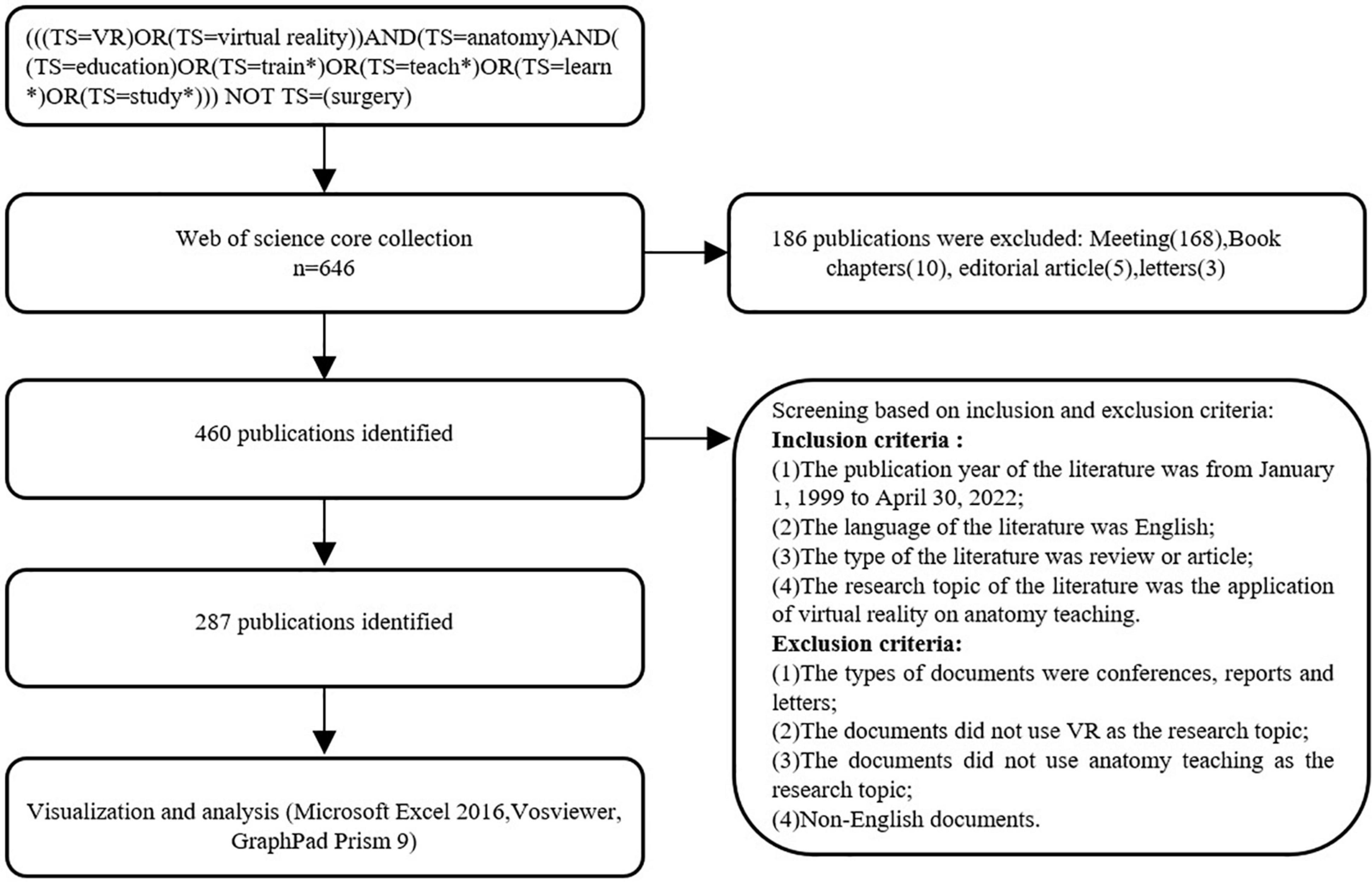
Figure 1. Flow chart for the analysis of virtual reality in anatomy teaching.
Inclusion Criteria:
(1) The publication year of the literature was from January 1, 1999, to April 30, 2022;
(2) The language of the literature was English;
(3) The type of literature was review or article;
(4) The research topic of the literature was the application of virtual reality to anatomy teaching.
Exclusion Criteria:
(1) The types of documents were conferences, reports and letters;
(2) The documents did not use VR as the research topic;
(3) The documents did not use anatomy teaching as the research topic;
(4) Non-English documents.
VOSviewer software (Leiden, Netherlands), Excel and GraphPad Prism 9 were used to analyze the data. A comprehensive description of various publishing characteristics was provided, including keywords, cocitations, countries/territories, publication numbers, institutions, authors, and journals.
As shown in Figure 2, VOSviewer was used to analyze keywords in 287 papers. Among all the keywords, reality, group, participant, application, and test, have higher frequencies. This shows that an increasing number of articles about the application of VR in anatomy teaching have been published. In addition, the emergence of the keyword “brain” was noted, which may indicate that the brain is a hot spot in VR applications due to its complex anatomical structure.
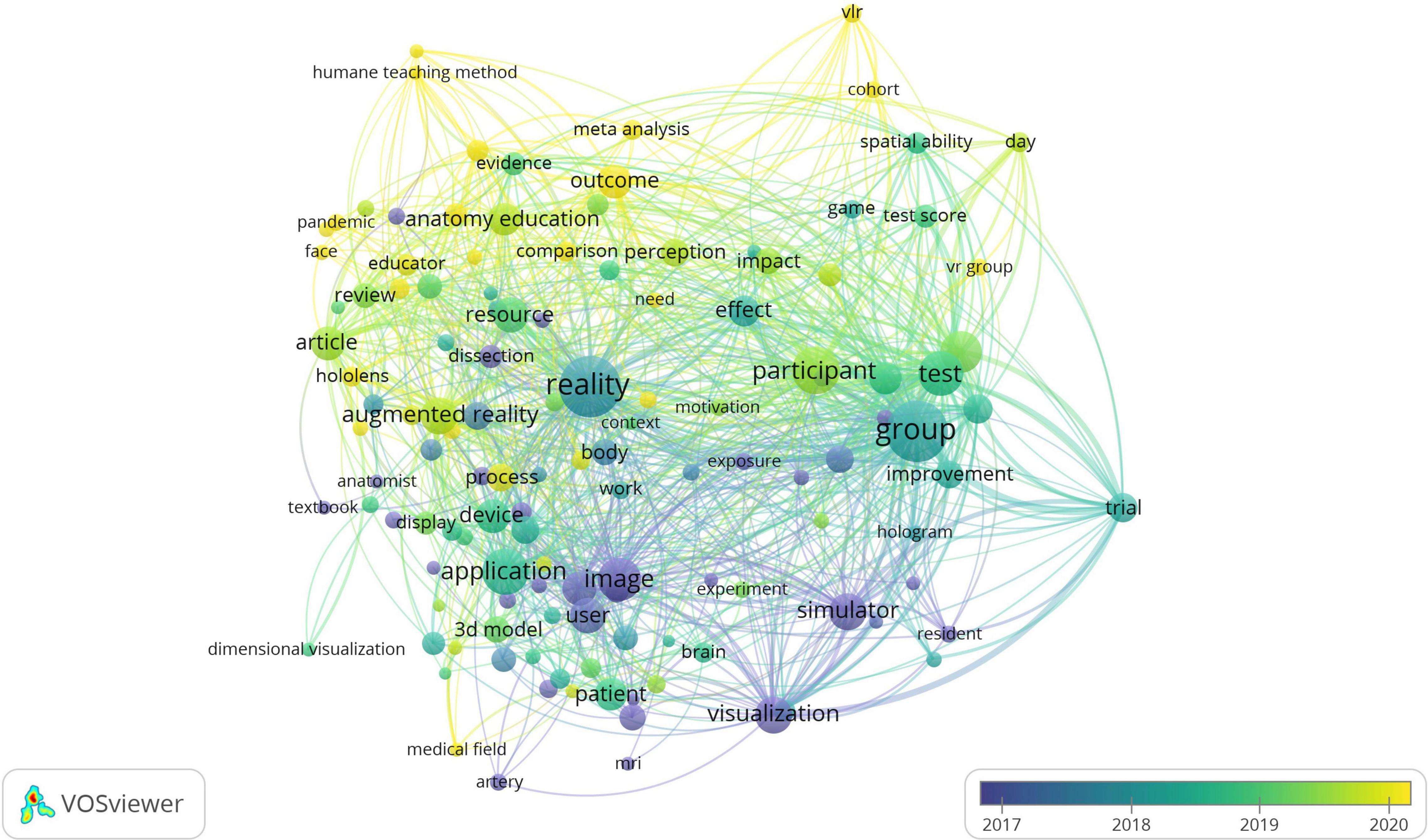
Figure 2. The co-occurrence network of keywords.
Then, an author cocitation analysis was conducted. The size of the nodes in the figure represents the author’s citation frequency, the thickness of the line represents the cocitation frequency, and different colors represent different clusters of clusters. Three distinct classes are seen, as shown in Figure 3. Among them, Moro, C, Yammine K. Nowinski, WL’s, articles were cited the most frequently. In addition, a cocitation analysis of the references was also carried out, as shown in Figure 4. There are four distinct classes. Among them, the paper published by Moro C (Moro et al., 2017) in 2006 is the most frequently cited, which is the classic literature in the application of VR in anatomy education.
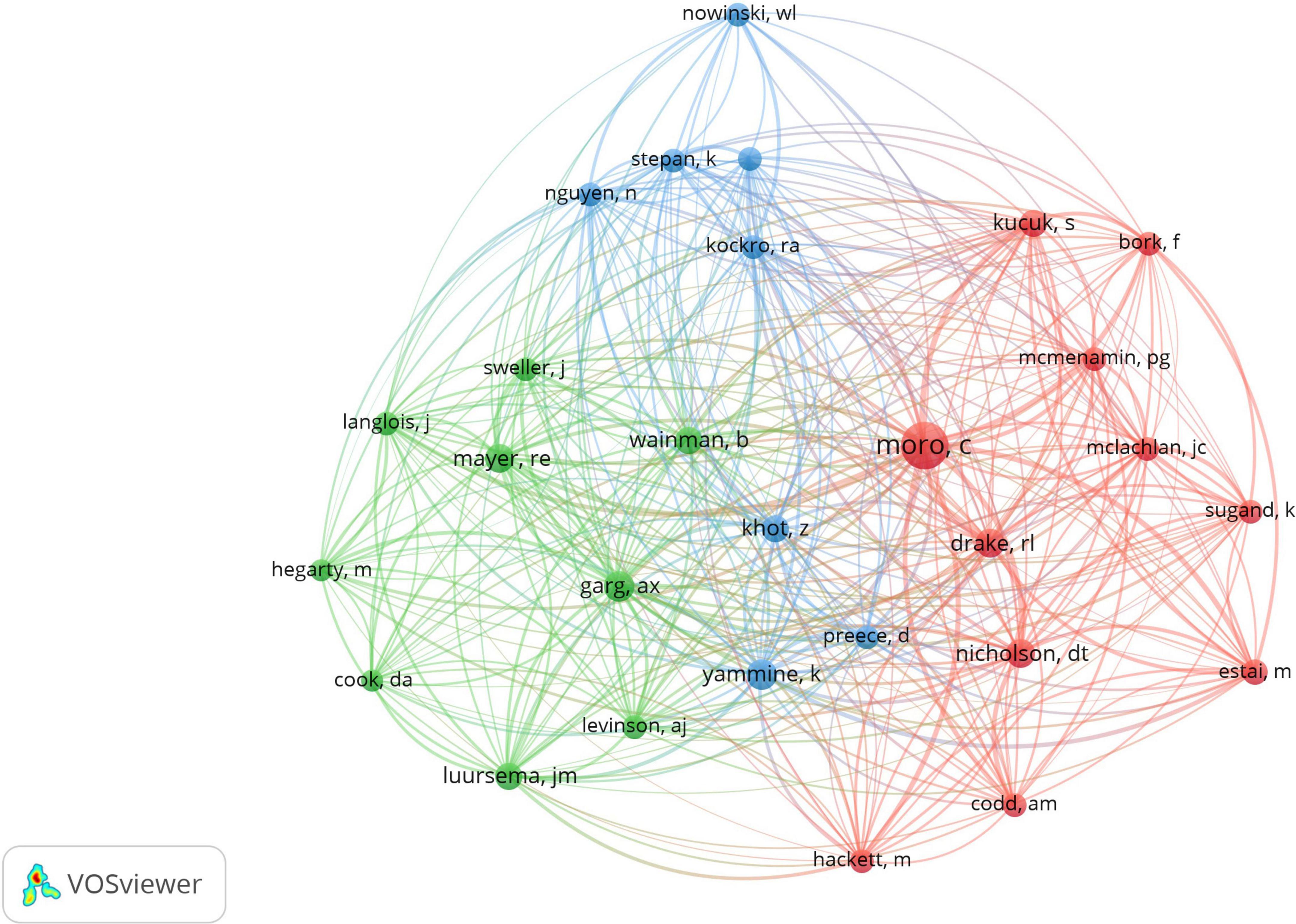
Figure 3. A visualization of author co-citation analysis.
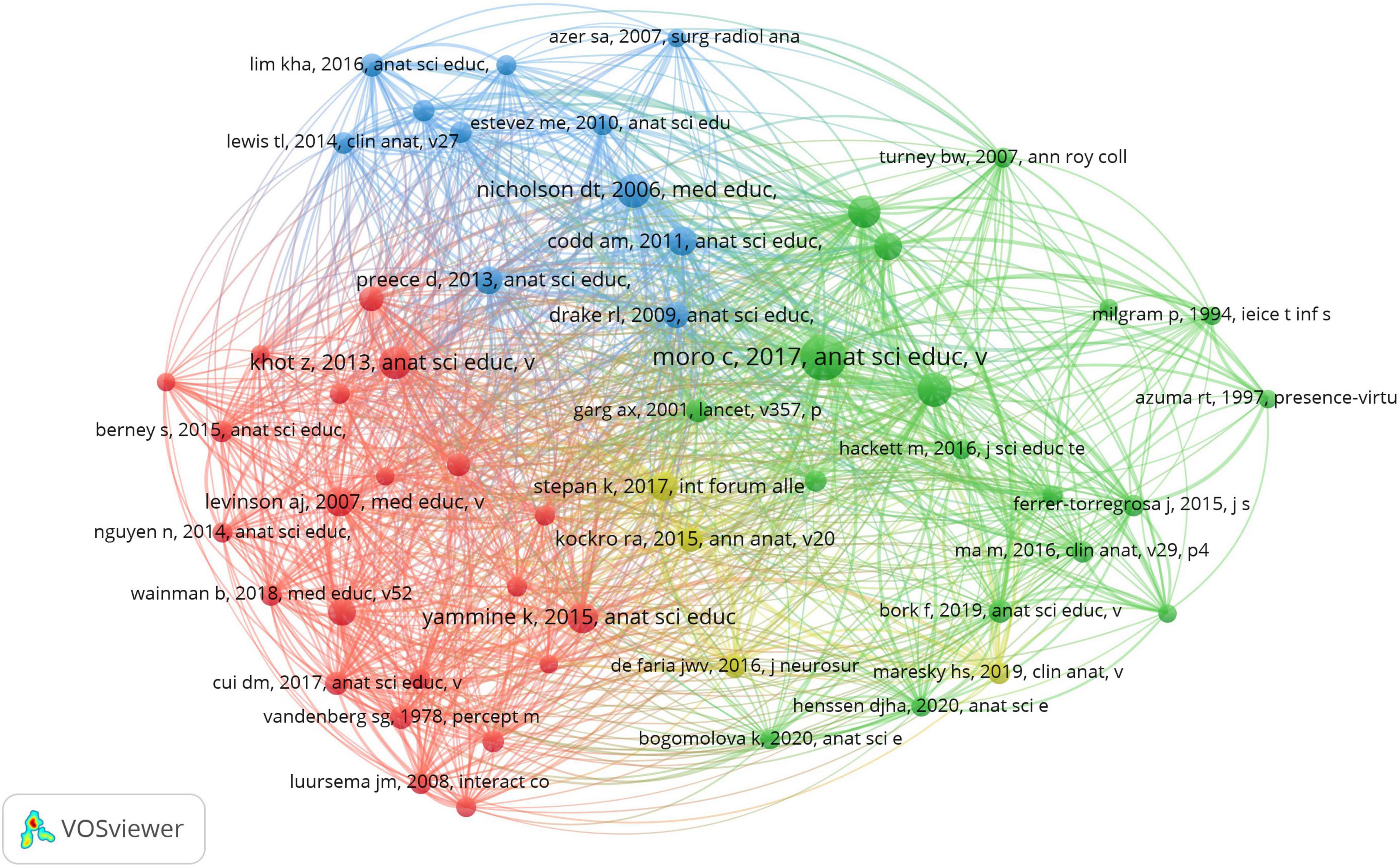
Figure 4. A visualization of co-citation analysis of reference.
As shown in Figure 5, the most published countries are the United States, Australia, and China. For further research, VOSviewer software was used to conduct visual analysis on cooperation relations between countries and territories, and the results are shown in Figure 6. Each country or region is represented by a circle, the size of which depends on the number of publications produced in that country. The curve connecting the two circles represents the cooperative relationship between the two connected countries. The thicker the curve is, the stronger the cooperation between the two countries. In general, there is less cooperation between countries. As seen from Figure 6, the cooperation between countries is not close. Among them, the United States has more cooperative relations with 10 countries or territories. Britain, China and Australia also have cooperation with a small number of countries. There is not much cooperation between other countries, so countries can strengthen cooperation in this field and jointly promote its development.
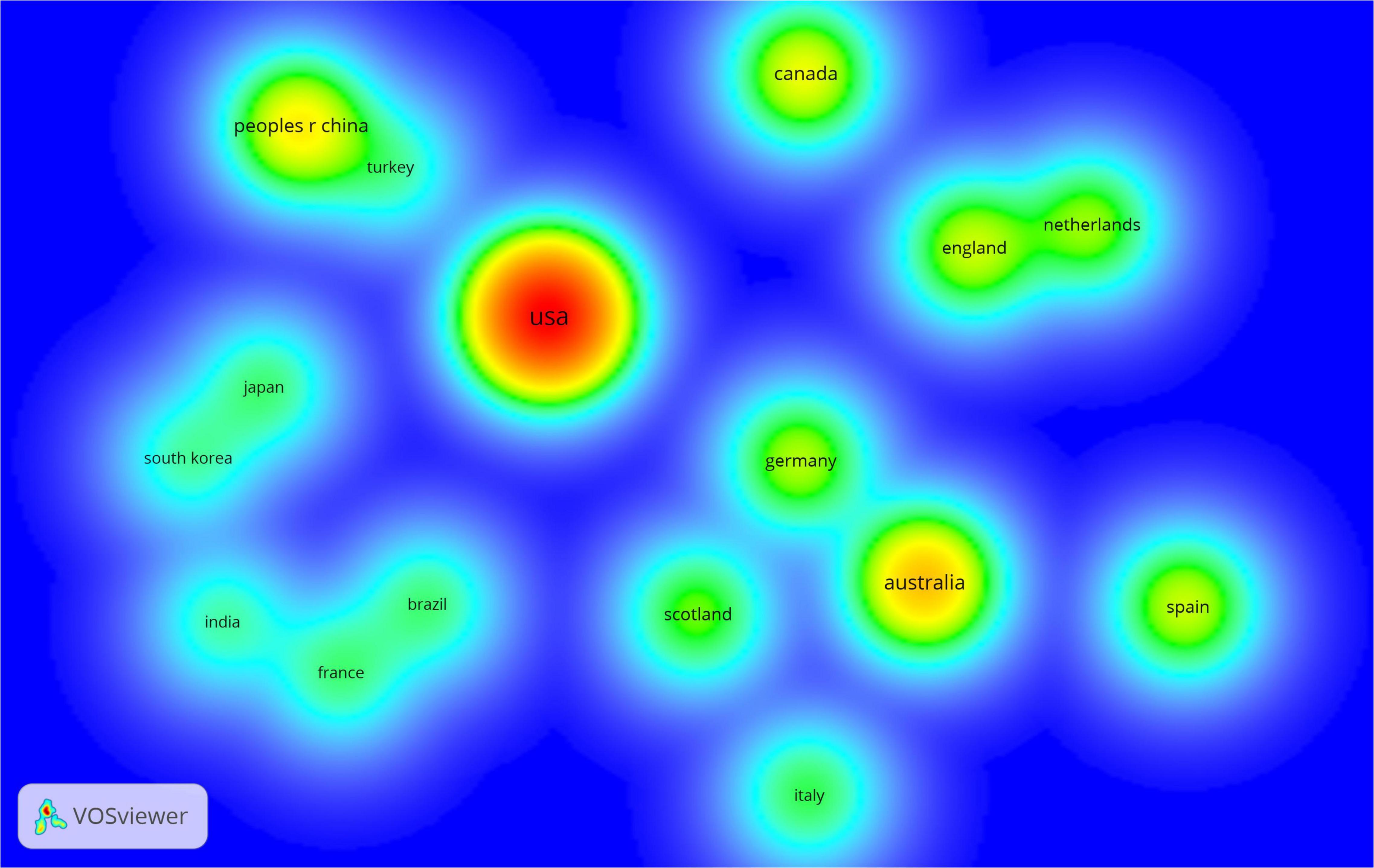
Figure 5. The co-author relationship of countries.
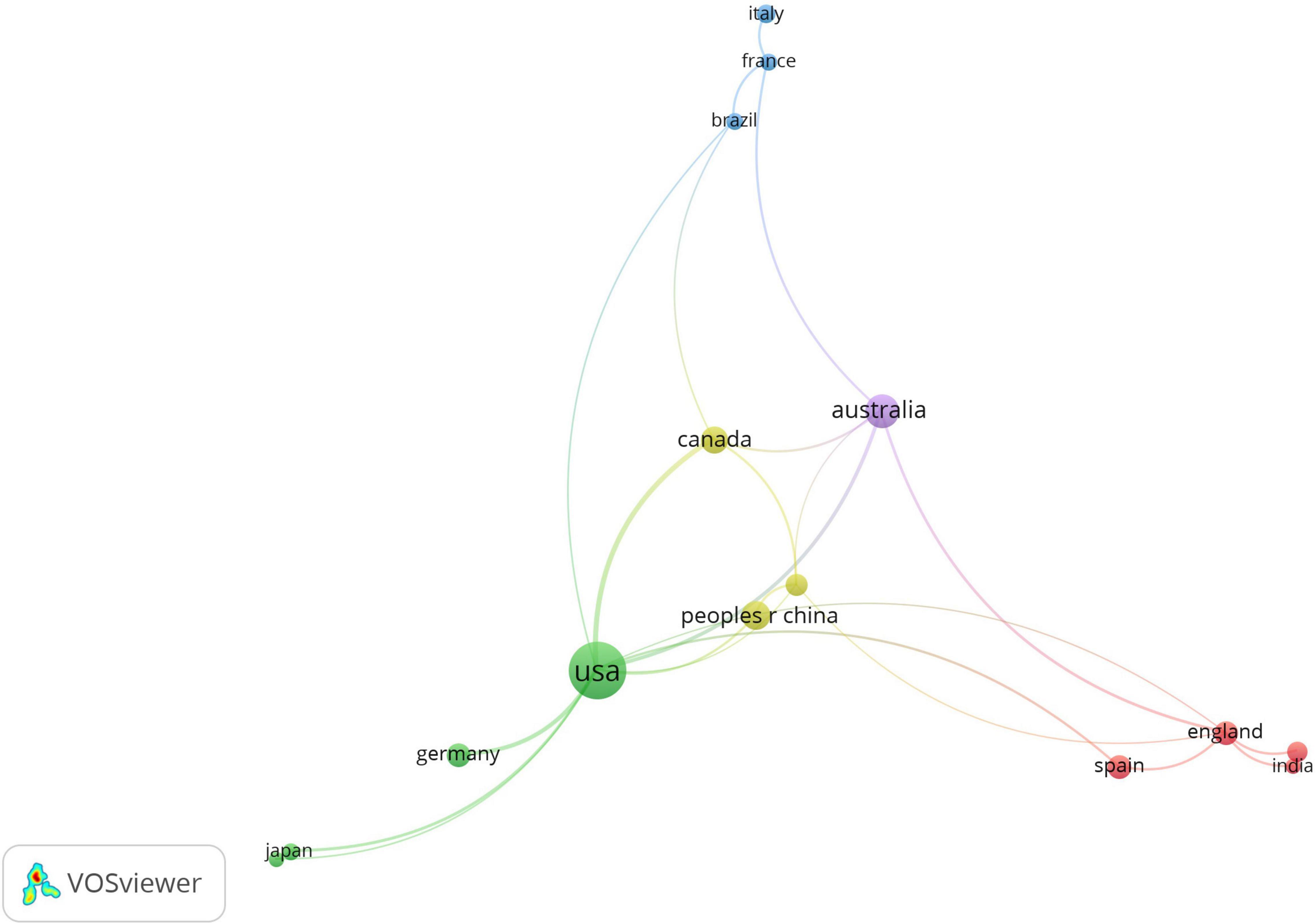
Figure 6. Country distribution for the articles.
The number of publications per year reflects the level and development of a field to some extent. We analyzed the number of published articles on the application of VR in anatomy teaching in the past decade (Figure 7). This indicates that the publications between 2011 and 2015 show an increasing trend year by year. After we fit the curve of the number of posts, we found that the number of posts in this field approaches an exponential relationship, which means that great breakthroughs of VR in anatomy education have been made in recent years.
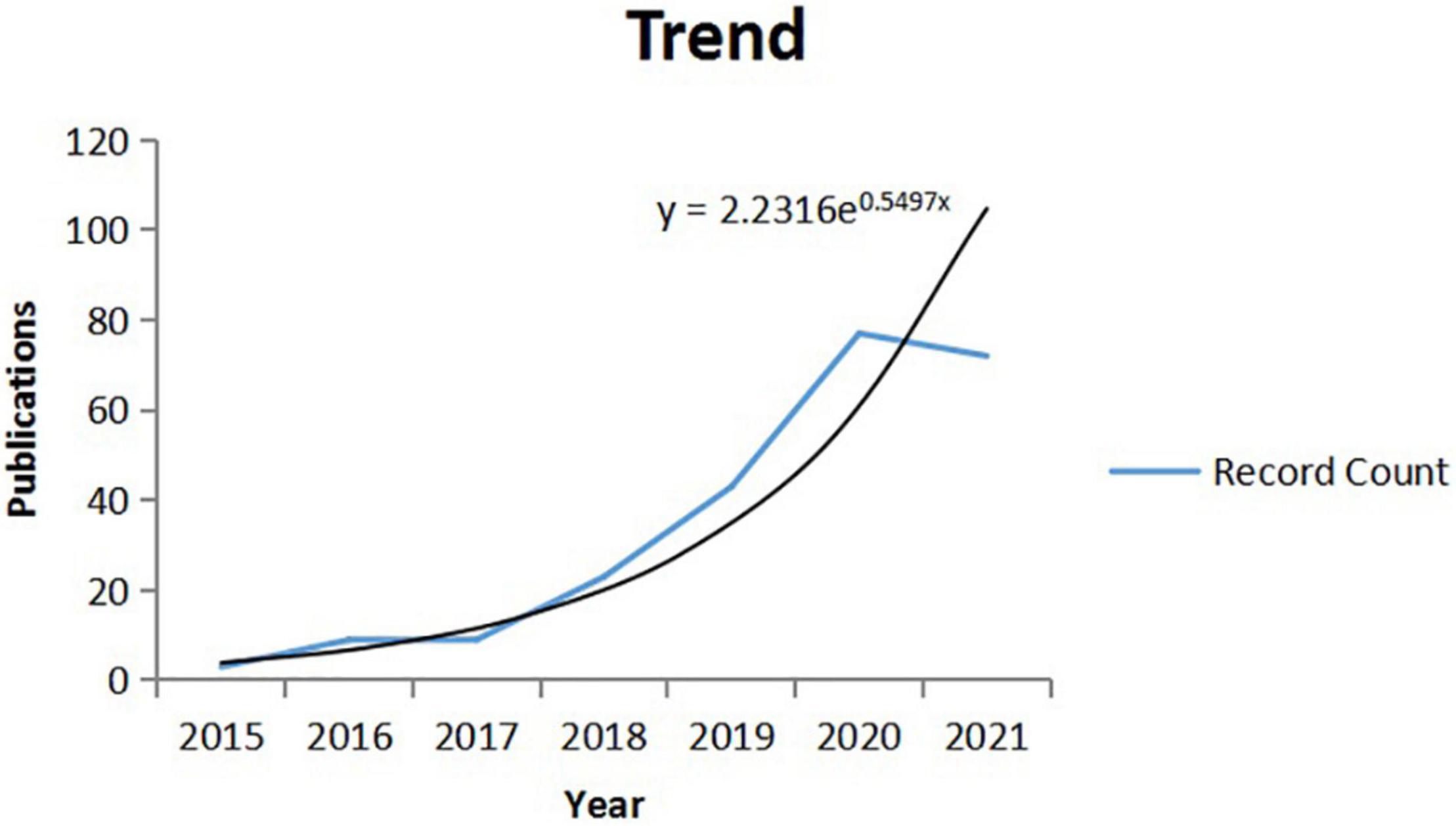
Figure 7. Year distribution of the articles between 2015 and 2021.
Figure 8 shows the top 10 organizations with the number of publications. Universities, such as McMaster University, Bond University, and Radboud University Nijmegen, have made outstanding contributions to the application of VR in anatomy. Similarly, Figure 9 shows the top 10 authors with the number of publications. Among them, Moro Christian, Stromberga Zane, and Wainman Bruce are the top three. In Figure 10, according to the average citation rate, the published journals in this field are ranked in descending order, and a total of 20 journals are included. This picture summarizes the journals with high academic profiles, the number of published journals from 2010 to 2022 in this field, and the impact factors of these journals. Among the 20 journals, the journal Brit J Anaesth has the highest impact factor (IF = 9.17), and the journal Anat Sci Educ has the highest number of publications in this field (Publication = 43).
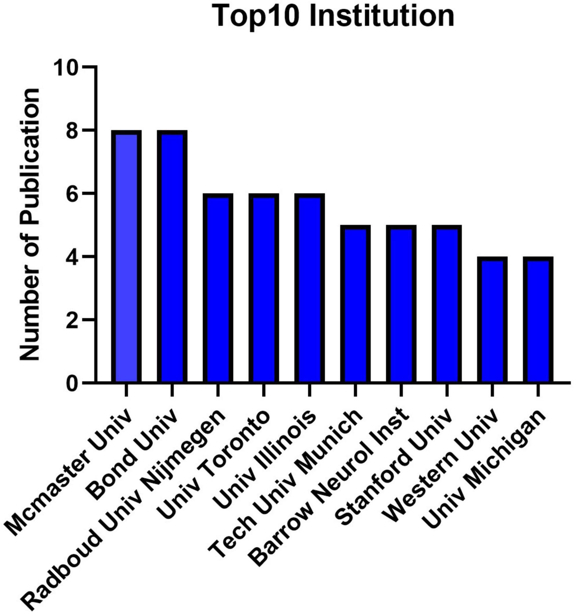
Figure 8. The contribution of the top 10 institutions.
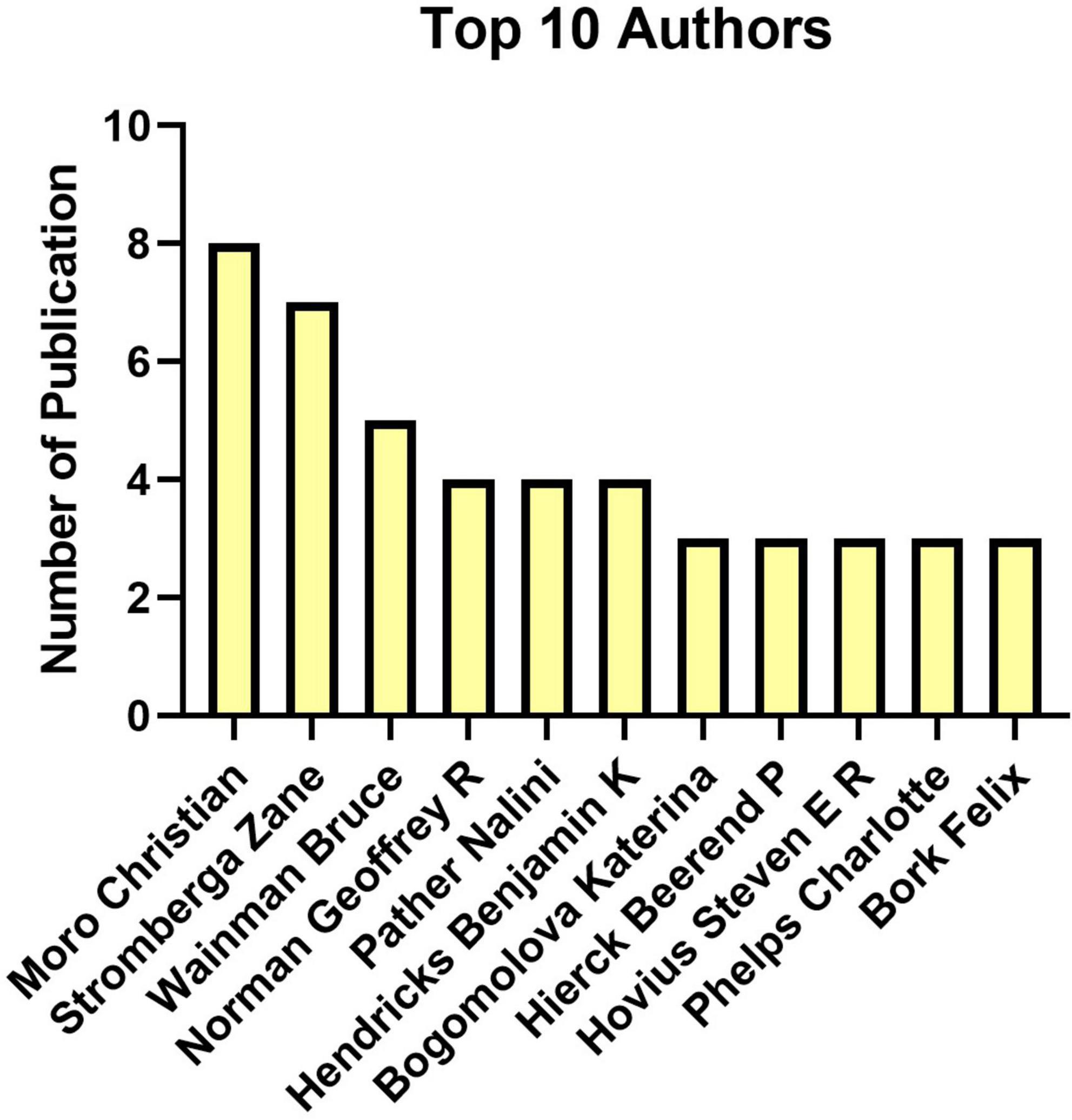
Figure 9. The publication of top 10 authors.
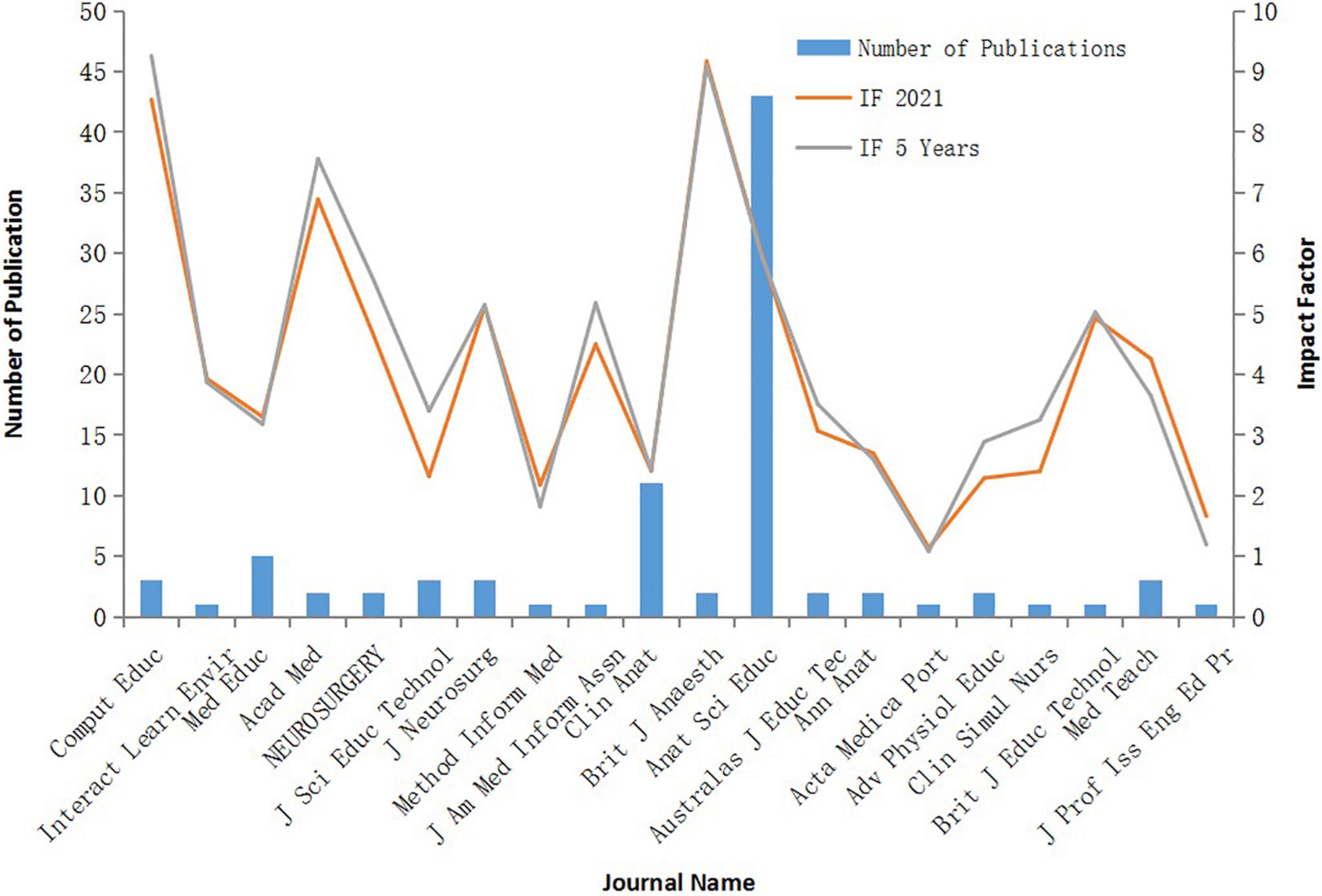
Figure 10. The impact factor and contribution of journal.
Human anatomy is the foundation for new medical students and an indispensable part of understanding other clinical disciplines, as it is closely related to other medical disciplines. Only on the basis of understanding the normal structure of the human body can one distinguish between pathological and physiological processes. Furthermore, the clinical operation and treatment of diseases are inseparable from human body structure and pathophysiological processes. However, due to the complexity of the course content and the limitation of traditional teaching methods, the teaching of anatomy is very difficult. Moreover, the lack of human specimens further increases challenges of anatomy teaching (George and De, 2010). Overall, there are many factors that hinder the application of traditional anatomy teaching, and new technologies are urgently needed to improve teaching methods in this area.
Fortunately, the application of VR has solved these problems. Virtual scenes of the real world significantly improve the intuition, accuracy, and real-time nature of the user’s sensory world (Shi et al., 2020). Therefore, the emergence of VR has an important influence on the development of medicine, especially the application of anatomy teaching. VR is a helpful tool for learning human anatomy and a useful adjunct to teaching (Fairen et al., 2020). To further understand what advantages VR has in anatomy teaching, the following 9 articles were analyzed (Table 1), which highlight the advantages of VR in anatomy teaching. The total citations of 287 articles were ranked and screened according to the following inclusion criteria: (1) the paper discusses the application of VR in anatomy teaching; (2) the paper compares the difference between VR teaching and anatomy teaching; (3) there are clear evaluation criteria for teaching effects; (4) participants have clear grouping criteria; and (5) the total citations were more than 50 times. Finally, the nine most cited articles were selected that met the criteria for analysis. Nicholson et al. (2006) conducted a study in 2006 to test the educational effectiveness of computer-generated three-dimensional models of the middle and inner ears. The subjects were divided into two groups, 29 in the control group and 28 in the experimental group. Students in the control group took the tutorial without exposure to the model, and students in the experimental group completed a Web-based tutorial that included the interactive model. However, there were some limitations to the study. For example, students in the experimental group took more time to complete the lessons and quizzes, which might indicate that the experimental group and the control group had different levels of effort, on average. A study on cardiac anatomy teaching conducted by Maresky et al. (2019) also suggested that VR could significantly improve learning effects and students’ interest in learning compared with traditional teaching methods. In 2015, Kockro et al. (2015) conducted an experiment on 169 second-year medical students. They were randomized into 2 groups. In the control group, they were taught by a standardized prerecorded audio lecture detailing the anatomy of the third ventricle, complete with a two-dimensional (2D) PowerPoint presentation. DextroBeam was used to visit the third ventricle. Immediately after class, students completed a 10-question multiple-choice exam based on what they learned and their subjective evaluation of the teaching methods. The results of this research showed that the students in the 2D group achieved a mean score of 5.19 (± 2.12) compared to 5.45 (± 2.16) in the 3D group, with the results in the 3D group being statistically non-inferior to those of the 2D group (p < 0.0001). The students rated the 3D method higher than 2D teaching in four domains. According to this research, stereoscopic enhanced 3D lectures are an effective way to learn anatomy, and students benefited greatly from the lectures. However, some studies suggested that VR teaching did not significantly improve students’ learning. Khot et al. (2013) showed that there was no significant difference in the teaching effects of VR, AR, and TB, and participants in VR teaching were more likely to suffer from headache, dizziness, blurred vision, and other adverse reactions.
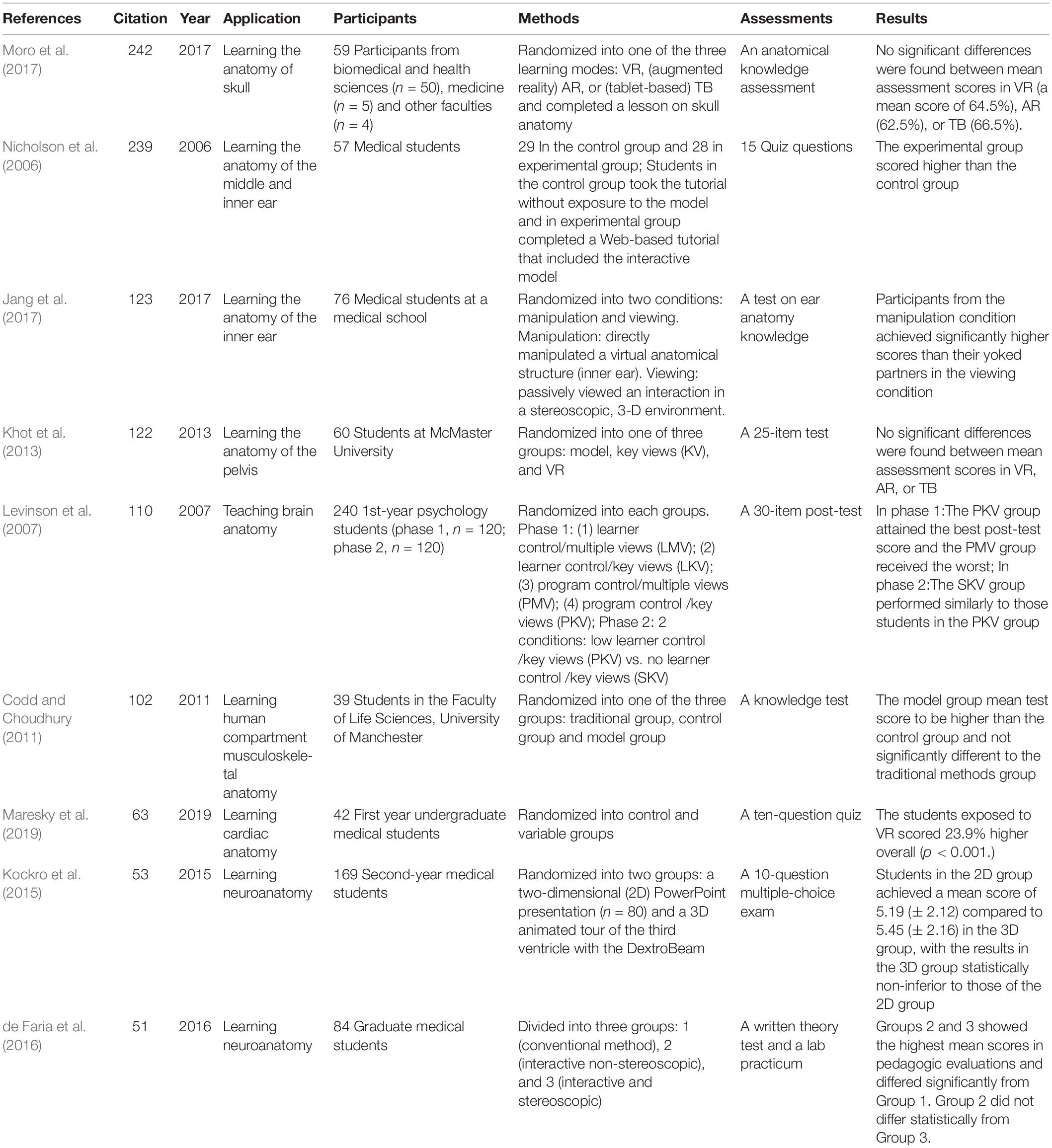
Table 1. Summary table of published studies applying virtual reality in anatomy education.
Through our analysis, VR plays a positive role in the learning process of anatomy, not only for students’ learning of normal anatomical structure, but also for students’ thinking and enthusiasm of anatomy learning. However, due to the small number of current studies, small sample size and variable control problems, the research results are also different, to some extent. Therefore, more research is needed to demonstrate the role of VR in anatomical learning. On the whole, there is great promise for the effective use of virtual as a means to supplement lesson content in anatomical education.
Through systematic analysis, one fully understands the advantages of VR in anatomy learning, especially during the epidemic. To avoid the risk of exposure, VR has become an excellent tool for students to learn anatomy, which is the basis of medical knowledge. VR enables medical students to understand the complex structures of the human body comprehensively and systematically. Generally, knowledge in textbooks and practical anatomy training is very practical for medical students, which is conducive to better understanding the structure of the human body, but VR enables them to understand knowledge from a new dimension. For example, in VR, students can take a muscle from the human body to understand the interaction and innervation of each muscle during exercise.
In addition, this research shows that VR has great potential in anatomy teaching in the future. In the past two decades, the overall number of publications has shown a significant upwards trend. Through literature research, it was found that VR has significant advantages in teaching results compared with traditional teaching modes. In recent years, VR has also been used in other fields, such as surgical teaching (Seymour et al., 2002; Sumdani et al., 2022), which reflects the broad development prospects of this technology.
However, little is known about how VR can be effectively used in medical education (Galvez et al., 2021). The reasons may include: First, VR equipment is expensive under the current conditions, which is a large expense for schools. Second, VR technology is not mature enough at present, and new theories are needed to break through the bottleneck to be more conducive to popularization. Third, VR-related technicians have not developed well in the medical field, which further hinders the popularization of VR in anatomy. Overall, the application of VR in anatomy still has considerable obstacles, and the efforts of relevant personnel are needed. According to Figure 4, which indicates that publications about VR in anatomy education show exponential growth, the field is at a rapid development stage, with new breakthroughs constantly. The future should focus more on establishing technological standards with high data quality and developing approved applications (Joda et al., 2019).
We have reason to believe that with the development of science and technology, VR will have a wider application in the field of anatomy and will also become a powerful modern teaching method in medical research institutions.
The original contributions presented in this study are included in the article/supplementary material, further inquiries can be directed to the corresponding author/s.
ZSL and ZXL collected the literature and drafted the initial manuscript, and drawn the figures. ZSL drew the table. CP, MZ, and QH were the lead investigators. All authors approved the final manuscript as submitted and are accountable for all aspects of the work.
The authors declare that the research was conducted in the absence of any commercial or financial relationships that could be construed as a potential conflict of interest.
All claims expressed in this article are solely those of the authors and do not necessarily represent those of their affiliated organizations, or those of the publisher, the editors and the reviewers. Any product that may be evaluated in this article, or claim that may be made by its manufacturer, is not guaranteed or endorsed by the publisher.
We thank everyone who contributed to the writing and all the publications and their authors involved in this study.
Ahmad, P., and Slots, J. (2021). A bibliometric analysis of periodontology. Periodontol 2000 85, 237–240.
Akbar-Khanzadeh, F., Vaquerano, M. U., Akbar-Khanzadeh, M., and Bisesi, M. S. (1994). Formaldehyde exposure, acute pulmonary response, and exposure control options in a gross anatomy laboratory. Am. J. Ind. Med. 26, 61–75. doi: 10.1002/ajim.4700260106
Bashir, M. F., Ma, B., Bilal, Komal, B., and Bashir, M. A. (2021). Analysis of environmental taxes publications: a bibliometric and systematic literature review. Environ. Sci. Pollut. Res. Int. 28, 20700–20716. doi: 10.1007/s11356-020-12123-x
Celik, E., Durmus, A., Adizel, O., and Nergiz Uyar, H. (2021). A bibliometric analysis: what do we know about metals(loids) accumulation in wild birds? Environ. Sci. Pollut. Res. Int. 28, 10302–10334. doi: 10.1007/s11356-021-12344-8
Chittaro, L., and Ranon, R. (2007a). Web3D technologies in learning, education and training – Foreword. Comput. Educ. 49, 1–2.
Chittaro, L., and Ranon, R. (2007b). Web3D technologies in learning, education and training: motivations, issues, opportunities. Comput. Educ. 49, 3–18.
Codd, A. M., and Choudhury, B. (2011). Virtual reality anatomy: is it comparable with traditional methods in the teaching of human forearm musculoskeletal anatomy? Anat. Sci. Educ. 4, 119–125. doi: 10.1002/ase.214
de Faria, J. W. V., Teixeira, M. J., Sousa, L. D., Otoch, J. P., and Figueiredo, E. G. (2016). Virtual and stereoscopic anatomy: when virtual reality meets medical education. J. Neurosurg. 125, 1105–1111.
Demiryürek, D., Bayramoğlu, A., and Ustaçelebi, S. (2002). Infective agents in fixed human cadavers: a brief review and suggested guidelines. Anat. Rec. 269, 194–197. doi: 10.1002/ar.10143
Drake, R. L., Lowrie, D. J. Jr., and Prewitt, C. M. (2002). Survey of gross anatomy, microscopic anatomy, neuroscience, and embryology courses in medical school curricula in the United States. Anat. Rec. 269, 118–122.
Drake, R. L., McBride, J. M., Lachman, N., and Pawlina, W. (2009). Medical education in the anatomical sciences: the winds of change continue to blow. Anat. Sci. Educ. 2, 253–259. doi: 10.1002/ase.117
Drake, R. L., McBride, J. M., and Pawlina, W. (2014). An update on the status of anatomical sciences education in United States medical schools. Anat. Sci. Educ. 7, 321–325. doi: 10.1002/ase.1468
Erolin, C., Reid, L., and McDougall, S. (2019). Using virtual reality to complement and enhance anatomy education. J. Vis. Commun. Med. 42, 93–101.
Fairen, M., Moyes, J., and Insa, E. (2020). VR4Health: personalized teaching and learning anatomy using VR. J. Med. Syst. 44:94. doi: 10.1007/s10916-020-01550-5
Galvez, R., Wallon, R. C., Shackelford, L., Amos, J. R., and Rowen, J. L. (2021). Use of Virtual Reality to Educate Undergraduate Medical Students on Cardiac Peripheral and Collateral Circulation. Med. Sci. Educ. 31, 19–22. doi: 10.1007/s40670-020-01104-x
George, A. P., and De, R. (2010). Review of temporal bone dissection teaching: how it was, is and will be. J. Laryngol. Otol. 124, 119–125. doi: 10.1017/S0022215109991617
Gloy, K., Weyhe, P., Nerenz, E., Kaluschke, M., Uslar, V., Zachmann, G., et al. (2022). Immersive Anatomy Atlas: Learning Factual Medical Knowledge in a Virtual Reality Environment. Anat. Sci. Educ. 15, 360–368. doi: 10.1002/ase.2095
Jang, S., Vitale, J. M., Jyung, R. W., and Black, J. B. (2017). Direct manipulation is better than passive viewing for learning anatomy in a three-dimensional virtual reality environment. Comput. Educ. 106, 150–165.
Joda, T., Gallucci, G. O., Wismeijer, D., and Zitzmann, N. U. (2019). Augmented and virtual reality in dental medicine: a systematic review. Comput. Biol. Med. 108, 93–100.
Khot, Z., Quinlan, K., Norman, G. R., and Wainman, B. (2013). The relative effectiveness of computer-based and traditional resources for education in anatomy. Anat. Sci. Educ. 6, 211–215. doi: 10.1002/ase.1355
Kockro, R. A., Amaxopoulou, C., Killeen, T., Wagner, W., Reisch, R., Schwandt, E., et al. (2015). Stereoscopic neuroanatomy lectures using a three-dimensional virtual reality environment. Ann. Anat. 201, 91–98. doi: 10.1016/j.aanat.2015.05.006
Levinson, A. J., Weaver, B., Garside, S., McGinn, H., and Norman, G. R. (2007). Virtual reality and brain anatomy: a randomised trial of e-learning instructional designs. Med. Educ. 41, 495–501. doi: 10.1111/j.1365-2929.2006.02694.x
Maresky, H. S., Oikonomou, A., Ali, I., Ditkofsky, N., Pakkal, M., and Ballyk, B. (2019). Virtual reality and cardiac anatomy: exploring immersive three-dimensional cardiac imaging, a pilot study in undergraduate medical anatomy education. Clin. Anat. 32, 238–243. doi: 10.1002/ca.23292
Matthews, D. (2018). Virtual-reality applications give science a new dimension. Nature 557, 127–128. doi: 10.1038/d41586-018-04997-2
McLachlan, J. C., and Patten, D. (2006). Anatomy teaching: ghosts of the past, present and future. Med. Educ. 40, 243–253.
Moro, C., Štromberga, Z., Raikos, A., and Stirling, A. (2017). The effectiveness of virtual and augmented reality in health sciences and medical anatomy. Anat. Sci. Educ. 10, 549–559.
Neville, A. J. (2009). Problem-based learning and medical education forty years on. A review of its effects on knowledge and clinical performance. Med. Princ. Pract. 18, 1–9. doi: 10.1159/000163038
Nicholson, D. T., Chalk, C., Funnell, W. R., and Daniel, S. J. (2006). Can virtual reality improve anatomy education? A randomised controlled study of a computer-generated three-dimensional anatomical ear model. Med. Educ. 40, 1081–1087. doi: 10.1111/j.1365-2929.2006.02611.x
Seymour, N. E., Gallagher, A. G., Roman, S. A., O’Brien, M. K., Bansal, V. K., Andersen, D. K., et al. (2002). Virtual reality training improves operating room performance – Results of a randomized, double-blinded study. Ann. Surg. 236, 458–464.
Shi, X. W., Yuan, H., Lu, M. X., Cai, J. H., and Zhang, X. Z. (2020). Current Status and Progress of Virtual Reality Technology in Medical Field. Laser Optoelectron. Prog. 57:10.
Silén, C., Wirell, S., Kvist, J., Nylander, E., and Smedby, O. (2008). Advanced 3D visualization in student-centred medical education. Med. Teach. 30, e115–24. doi: 10.1080/01421590801932228
Sumdani, H., Aguilar-Salinas, P., Avila, M. J., Barber, S. R., and Dumont, T. (2022). Utility of Augmented Reality and Virtual Reality in Spine Surgery: A Systematic Review of the Literature. World Neurosurg. 161, e8–e17. doi: 10.1016/j.wneu.2021.08.002
Telner, D., Bujas-Bobanovic, M., Chan, D., Chester, B., Marlow, B., Meuser, J., et al. (2010). Game-based versus traditional case-based learning: comparing effectiveness in stroke continuing medical education. Can. Fam. Physician. 56, e345–51.
van Eck, N. J., and Waltman, L. (2017). Citation-based clustering of publications using CitNetExplorer and VOSviewer. Scientometrics 111, 1053–1070. doi: 10.1007/s11192-017-2300-7
Keywords: model, neuroanatomy, dissection, technology, stereopsis
Citation: Li Z, Li Z, Peng C, Zhao M and He Q (2022) A Bibliometric Analysis of Virtual Reality in Anatomy Teaching Between 1999 and 2022. Front. Educ. 7:874406. doi: 10.3389/feduc.2022.874406
Received: 14 March 2022; Accepted: 22 June 2022;
Published: 19 July 2022.
Edited by:
Leman Figen Gul, Istanbul Technical University, TurkeyReviewed by:
Sonsoles López-Pernas, Universidad Politécnica de Madrid, SpainCopyright © 2022 Li, Li, Peng, Zhao and He. This is an open-access article distributed under the terms of the Creative Commons Attribution License (CC BY). The use, distribution or reproduction in other forums is permitted, provided the original author(s) and the copyright owner(s) are credited and that the original publication in this journal is cited, in accordance with accepted academic practice. No use, distribution or reproduction is permitted which does not comply with these terms.
*Correspondence: Cheng Peng, cGNoZW5nODNAc2luYS5jb20=; Mingyi Zhao, emhhb19taW5neWlAY3N1LmVkdS5jbg==; Qingnan He, aGVxbjI2MjlAY3N1LmVkdS5jbg==
†These authors have contributed equally to this work
Disclaimer: All claims expressed in this article are solely those of the authors and do not necessarily represent those of their affiliated organizations, or those of the publisher, the editors and the reviewers. Any product that may be evaluated in this article or claim that may be made by its manufacturer is not guaranteed or endorsed by the publisher.
Research integrity at Frontiers
Learn more about the work of our research integrity team to safeguard the quality of each article we publish.