
94% of researchers rate our articles as excellent or good
Learn more about the work of our research integrity team to safeguard the quality of each article we publish.
Find out more
REVIEW article
Front. Bioeng. Biotechnol., 14 January 2025
Sec. Biosensors and Biomolecular Electronics
Volume 12 - 2024 | https://doi.org/10.3389/fbioe.2024.1507460
 Qirui Zhao1,2,3
 Shan Li1,2,3Leonard Krall1,2,3Qianyu Li3Rongyuan Sun3Yuqi Yin3Jingyi Fu3Xu Zhang1,2,3Yonghua Wang1,2,3*Mei Yang1,2,3*
Single-cell protein analysis has emerged as a powerful tool for understanding cellular heterogeneity and deciphering the complex mechanisms governing cellular function and fate. This review provides a comprehensive examination of the latest methodologies, including sophisticated cell isolation techniques (Fluorescence-Activated Cell Sorting (FACS), Magnetic-Activated Cell Sorting (MACS), Laser Capture Microdissection (LCM), manual cell picking, and microfluidics) and advanced approaches for protein profiling and protein-protein interaction analysis. The unique strengths, limitations, and opportunities of each method are discussed, along with their contributions to unraveling gene regulatory networks, cellular states, and disease mechanisms. The importance of data analysis and computational methods in extracting meaningful biological insights from the complex data generated by these technologies is also highlighted. By discussing recent progress, technological innovations, and potential future directions, this review emphasizes the critical role of single-cell protein analysis in advancing life science research and its promising applications in precision medicine, biomarker discovery, and targeted therapeutics. Deciphering cellular complexity at the single-cell level holds immense potential for transforming our understanding of biological processes and ultimately improving human health.
Proteins, intracellular and membrane-bound, form the fundamental structural elements of cells and are organized into intricate molecular assemblies facilitating vital cellular processes like transcription, translation, metabolism, growth, adhesion, and signal transduction (Jelokhani-Niaraki, 2022). Consequently, quantifying protein expression levels and analyzing protein-protein interactions (PPIs) are essential for understanding cellular function and regulatory mechanisms. Deciphering these interactions can provide valuable insights into how cells respond to external stimuli, maintain homeostasis, and make fate decisions.
Conventional techniques like Western blotting and RT-PCR quantify collective responses of entire cell populations, assuming that the mean accurately reflects individual cell behavior. However, this assumption overlooks potentially significant variations within subpopulations, which can play pivotal roles in dictating overall population behavior (Hughes et al., 2014; Huang et al., 2023). Cellular processes exhibit inherent heterogeneity, evident in phenomena like stem cell differentiation, development, oncogenesis, immune responses, and neurodegenerative diseases (Mattiazzi Usaj et al., 2021; Liu et al., 2021; Budnik et al., 2018; Ryu et al., 2019a). As well, cells in complex organs, such as the liver, exhibit discrete metabolic and functional roles based on their specific localization (He et al., 2023). The tumor microenvironment (TME) also exemplifies this complexity, with intricate interactions between cancer cells and diverse non-malignant cells, each exhibiting unique behaviors under pathological conditions (Mayer et al., 2023).
To capture cellular diversity, researchers have emphasized developing methodologies for isolating and analyzing individual cells from complex biological samples (Bhagwat et al., 2018; Müller and Nebe-von-Caron, 2010; Pekle et al., 2019; Luan et al., 2020; Grigorev et al., 2023). Single-cell genomic, transcript, and proteomic analyses offer unprecedented insights into genomic variability, gene expression dynamics, and protein expression patterns (Reimegård et al., 2021; Shao et al., 2018). While single-cell RNA sequencing (scRNA-seq) revolutionized our understanding of cell population heterogeneity and gene expression (Reimegård et al., 2021), its limitations, including stochastic expression, mRNA half-life variability, amplification biases, and the inability to capture post-transcriptional modifications, necessitate integrating proteomics for comprehensive understanding (Larsson et al., 2019; Schwanhäusser et al., 2011; Ziegenhain et al., 2017). Protein-protein interactions (PPIs) are fundamental to cellular processes, their presence and strength can vary at the single-cell level. This variability is observed not only across different cell types and tissues but also within populations of genetically identical cells. Measuring PPIs at the single-cell level allows us to capture this heterogeneity and provides insights into how interactions may differ in response to factors like treatment or disease states. For instance, in a study on osimertinib-treated lung cancer models, significant cell-to-cell variation was found in PPIs such as Cyclin E and CDK2, FGFR1 and PIK3R1, and AKT1 and SRC. These interactions can be crucial for understanding how cancer cells respond to treatment and how resistance may develop, emphasizing the importance of single-cell resolution in identifying key interactomic events (Zhang et al., 2024). Single-cell protein analysis, on the other hand, can provide a more direct readout of cellular function, capturing post-translational modifications and protein-protein interactions that are crucial for regulating cellular behavior (Ryu et al., 2019a; Liu L. et al., 2020; Avin et al., 2017).
Recent studies have revealed only modest correlations between mRNA and protein levels (Edfors et al., 2016; Popovic et al., 2018; Liu et al., 2016), underscoring the idea that mRNA expression poorly predicts protein abundance. Proteins exhibit greater stability, and have higher concentrations than mRNAs which minimize random fluctuations. These traits allow proteins to play more immediate roles in sustaining cellular functions compared to transcripts (Liu L. et al., 2020; Liu et al., 2016; Frei et al., 2016). The generally longer half-lives and higher amounts of proteins also make them less susceptible to stochastic variations, allowing for more accurate quantification and analysis at the single-cell level (Popovic et al., 2018).
To comprehensively understand complex cell populations, researchers have developed analytical tools for quantitative and specific single-cell protein detection (Budnik et al., 2018; Ryu et al., 2019a; Bhagwat et al., 2018; Reimegård et al., 2021; Frei et al., 2016; Schulz et al., 2018; Stoeckius et al., 2017; Mimitou et al., 2021; Peterson et al., 2017; Bendall et al., 2012; Lun et al., 2019; Zhu et al., 2019). Operating with or without labels to minimize interference with cellular processes, these tools enable unraveling regulatory circuits, pathways, and mechanisms governing cellular behavior. Single-cell protein analysis has already yielded substantial contributions to our understanding of immune cell heterogeneity, stem cell differentiation, and tumor progression, paving the way for improved diagnostics, targeted therapies, and personalized medicine approaches (Mattiazzi Usaj et al., 2021; Liu et al., 2021; Satija and Shalek, 2014; Giesen et al., 2014).
The advancement of single-cell protein analysis technologies has been accompanied by the development of sophisticated data analysis and computational methods to extract meaningful biological insights from the vast amounts of complex data generated. These methods include data pre-processing, univariate and multivariate analysis, and advanced techniques such as machine learning, which have significantly improved the efficiency and accuracy of single-cell proteomics data analysis (Xie et al., 2020; Liu and Yang, 2021; Liu et al., 2019). As single-cell protein analysis technologies continue to evolve, the development of standardized data analysis pipelines and the integration of multi-omic data will be crucial for obtaining a comprehensive understanding of cellular heterogeneity and function, ultimately advancing our ability to decipher the complexity of biological systems.
This comprehensive review outlines advancements in single-cell separation techniques and single-cell protein analysis. The protein analysis section is further divided into two subtopics: (a) protein expression level analysis and (b) protein-protein interaction analysis. We summarize progress in isolation techniques, including filtration, fluorescence-activated cell sorting (FACS), magnetic-activated cell sorting (MACS), laser capture microdissection (LCM), manual picking, and microfluidics. Furthermore, we discuss breakthroughs in single-cell protein detection and PPI analysis, evaluating system performance in terms of multiplexity, analyte types, throughput, sensitivity, and specificity. We also highlight the importance of data analysis and computational methods in single-cell proteomics and discuss the future directions and potential applications of these technologies in deciphering cellular complexity. While significant progress has been made, challenges related to sensitivity, specificity, and throughput still remain. By comparing advantages and limitations, we provide insights into potential future directions, fostering advancements in this rapidly evolving field and deepening our comprehension of cellular biology and its biomedical applications. Addressing these challenges will be crucial for realizing the full potential of single-cell protein analysis and its widespread adoption in basic and translational research.
Isolating and accurately identifying target cells is a crucial prerequisite for single-cell analysis. The efficacy of single-cell isolation technologies is predominantly evaluated based on three critical parameters: efficiency, defined as the capacity to isolate a specific number of target cells within a given time frame; separation purity, referring to the proportion of target cells isolated relative to non-target cells post-separation; and recovery rate, indicating the quantity of target cells successfully retrieved post-separation compared to the initial count present in the sample (Hu et al., 2016; Gross et al., 2015). The selection of an appropriate isolation method is essential for reliable single-cell analyses, particularly when studying rare cell populations or investigating cellular heterogeneity, as distinct techniques exhibit varying effectiveness across these parameters.
Contemporary cell separation technologies can be broadly classified into two main categories based on their isolation properties. The first category encompasses techniques that rely on the physical attributes of cells, such as morphology, size, density, and deformability. This category includes methodologies like density gradient centrifugation, which separates cells based on their buoyant density; size-based filtration, which employs membranes with specific pore sizes to isolate cells of interest; manual cell picking, which involves the direct selection of individual cells using micropipettes; and a part of microfluidics-based capture, which utilizes the unique flow properties of cells in microchannels for isolation (Gross et al., 2015; Malter, 2016; Liu and Singh, 2013; Huang et al., 2008; Cha et al., 2022). A key advantage of these techniques is the ability to isolate cells in a label-free manner, without the need for internal or external markers, minimizing potential interference with cellular processes and preserving the native state of the cells (Gross et al., 2015).
The second category relies on the biological properties of cells, employing affinity-based methods. These methods, including fluorescence-activated cell sorting (FACS), magnetic-activated cell sorting (MACS), and laser capture microdissection (LCM), utilize the presence of specific surface proteins and their affinity for corresponding antibodies or probes (Potashnikova et al., 2018; Miltenyi et al., 1990; Vandewoestyne and Deforce, 2010). FACS utilizes fluorescently labeled antibodies to mark cells of interest, enabling their sorting according to fluorescence intensity. MACS uses antibody-coated magnetic beads to separate target cells from a heterogeneous population by use of an external magnetic field. While, LCM employs a laser to selectively isolate cells from tissue sections. These techniques offer high specificity and purity, enabling the isolation of rare cell populations based on their unique biological markers and specific morphology (Gross et al., 2015).
In this section, we briefly summarize the foundational principles, inherent advantages, limitations, and potential applications of the most prevalent cell separation methodologies (Table 1). Each technique’s unique attributes contribute to its suitability for specific research contexts, empathizing the importance of carefully evaluating the requirements of the study and accordingly selecting the most appropriate method. By understanding the strengths and weaknesses of each approach, researchers can optimize their experimental designs and ensure the reliable isolation of target cells for downstream single-cell analysis.
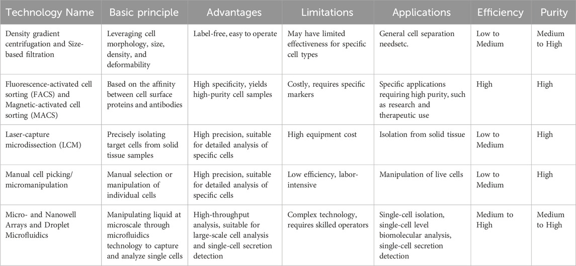
Table 1. Overview of single cell isolation techniques.
Fluorescence-Activated Cell Sorting (FACS), an advanced flow cytometry technique with sorting capabilities, and it is widely recognized as the most sophisticated and accessible method for identifying, categorizing, and isolating specific cell types from heterogeneous cell populations (Sharon et al., 2013; An and Chen, 2018; Herzenberg et al., 2002). This versatile technique differentiates cells based on a multitude of parameters, including size, granularity, and fluorescence intensity patterns arising from labeling with specific fluorescent markers. FACS enables concurrent quantitative and qualitative multi-parametric analysis of individual live or fixed cells at impressive throughput rates, rendering it an essential tool for single-cell isolation and characterization.
The FACS process begins with the preparation of a single-cell suspension, wherein target cells are labeled with fluorescent markers. Fluorophore-conjugated monoclonal antibodies, which recognize specific cell surface markers, are the primary choice of fluorescent probes utilized in this technique (Pan and Wan, 2020). As cells traverse through the cytometer’s fluidic system, they are hydrodynamically focused into a single-file stream and individually exposed to one or more laser beams. The interaction of the laser light with the cells generates fluorescence signals, which are collected by dedicated detectors (Adan et al., 2017). The forward scatter (FSC) and side scatter (SSC) signals provide information about the cell’s relative size and granularity, respectively, while the fluorescence detectors identify and characterize cells based on their predefined fluorescence signatures (Adan et al., 2017).
The sorting mechanism of FACS relies on an electrostatic deflection system. After laser excitation, the cell stream is fragmented into individual droplets, with each droplet encapsulating a single cell. Droplets can be formed by using high-frequency (cycles/second, Hz) vibration of the nozzle at an optimal amplitude. Based on the detected fluorescence and scatter signals, the droplets containing the desired single cells are assigned an electrical charge—either positive or negative by an electrical charging ring placed just at the point where the stream breaks into droplets. Next, as the droplets pass through an electric field, the charged droplets are deflected into designated collection tubes or well plates for downstream analysis or culture, while the uncharged droplets are directed into a waste container (Figure 1A) (Rambault et al., 2021). State-of-the-art FACS systems, such as the FACS-Aria™ III, boast an impressive throughput rate of up to 100,000 droplets per second, enabling the analysis of approximately 70,000 events per second (Gross et al., 2015). Moreover, FACS has proven adept at sorting individual cells from complex mixtures containing thousands of cells, utilizing up to 18 distinct surface markers for precise identification and separation (Hu et al., 2016). Additionally, an advanced FACS system developed by Dr. Li’s group has demonstrated over 90% recovery efficiency and more than 80% cell viability (Dong et al., 2017).

Figure 1. Overview of single-cell isolation technologies. (A) Schematic of fluorescence-activated cell sorting. As cells coated with fluorescent antibodies traverse through the cytometer, they are individually illuminated by a laser beam, enabling fluorescence detectors to identify and sort cells based on predefined characteristics. (B) Schematic of magnetic-activated cell sorting. When a heterogeneous cell mixture is exposed to an external magnetic field, desired cells labeled with magnetic beads become magnetically responsive. (C) Schematic of laser capture microdissection. The technique utilizes a laser to cut the tissue and let the cells adhere to the melted membrane. (D) Schematic of manual cell picking. The cells of interest are retrieved under a microscope by glass capillary transferred to microwells for further analysis. (E) Schematic of microfluidic used for single cell isolation. Upper: microwell Arrays for Single-Cell isolation; Lower: discrete aqueous droplets formation in droplet microfluidics.
One of the key advantages of FACS over other cell sorting techniques is its capability to analyze and sort cells simultaneously based on multiple parameters, encompassing both surface and intracellular markers (Bennett et al., 2016). This multi-parametric capability enables highly specific cell isolation and characterization, allowing researchers to identify and isolate rare cell populations with unique phenotypic profiles (Maes et al., 2020).
After single cell isolation, subsequent analysis is necessary. The high throughput and purity of FACS make it an invaluable tool in various applications, including immunophenotyping (An and Chen, 2018; Pan and Wan, 2020), cell cycle analysis (Potashnikova et al., 2018), subpopulation analysis (Antoniadi et al., 2022), cancer diagnosis (Sharon et al., 2013), and the isolation of rare cell populations for downstream single-cell analyses, such as single-cell RNA sequencing and single-cell proteomics (Zhu et al., 2019).
Despite its unparalleled versatility, FACS has certain limitations. The technique requires a relatively large initial cell number, typically in the range of 10 ^ 4 to 10 ^ 6 cells, which can hinder the isolation of individual cells from rare subpopulations (Gross et al., 2015; Shields et al., 2015). Moreover, the high-speed fluidic system and the potential for shear stress during sorting may compromise the viability and functional integrity of sorted cells, particularly for fragile cell types (Gross et al., 2015). To mitigate these issues, researchers have developed gentle sorting techniques, such as low-pressure sorting and the use of cell-friendly collection buffers, to improve cell viability and recovery (Teteris et al., 2012). Furthermore, recent advancements in microfluidics-based cell sorting technologies have enabled the isolation of single cells from rare populations with minimal sample requirements and reduced shear stress (Stavrakis et al., 2019).
Magnetic-Activated Cell Sorting (MACS) is another widely employed technique for the passive separation of cell populations, leveraging their cluster of specific extracellular markers to isolate specific cell types (Rambault et al., 2021; Shen et al., 2021). MACS has demonstrated the capability to achieve isolation of particular cell groups with a purity exceeding 90%, ideally even higher than 95% (Miltenyi et al., 1990; Desikan et al., 2022) and high recovery rate of more than 90% (Willasch et al., 2010). The principle underlying MACS involves the use of magnetic beads conjugated with targeting molecules such as antibodies, enzymes, lectins, or streptavidin. These conjugated beads are designed to selectively target surface molecules primarily on live cells, thereby enabling the efficient isolation of the desired cell population.
MACS offers two modes of separation: positive and negative. In positive separation, the heterogeneous cell mixture is incubated with magnetic beads coated with antibodies specific to the surface markers of the target cells (Pan and Wan, 2020). When exposed to an external magnetic field, the labeled cells become magnetically responsive and are retained within the field, while unlabeled cells can be easily removed through a washing process (Figure 1B). This approach is particularly useful when the target cell population is rare or when a high purity of the isolated cells is required (Nemescu et al., 2020). In contrast, negative separation is employed when targeting cell-specific substances are unavailable or when the aim is to deplete a specific cell population from the sample. In this mode, the unwanted cells are labeled with more than one antibody-conjugated magnetic beads and subsequently separated from the mixture, thereby isolating the unlabeled target cells in the solution (Pan and Wan, 2020).
Compared to FACS, which can separate cells based on the expression of multiple specific molecules simultaneously, MACS has a more limited capacity, sorting cells solely into positive and negative populations based on a single surface marker. Consequently, the purity level achievable with MACS is typically lower than that of FACS (Zeb et al., 2019). However, MACS offers several advantages, including its relative simplicity, cost-effectiveness, and the ability to process large sample volumes quickly. Additionally, MACS is generally gentler on the cells than FACS and results in lower cell loss, as it does not require high-pressure fluidic systems, making it more suitable for isolating fragile cell types (Sutermaster and Darling, 2019). To enhance the performance of MACS, an advanced method known as integrated Dielectrophoretic-Magnetic Activated Cell Sorter (iDMACS) was developed in 2009 (Kim and Soh, 2009). This innovative approach combines dielectrophoretic forces and magnetic particles. In iDMACS, cells are first labeled with magnetic beads and then subjected to a dielectrophoretic force, which helps to further separate the labeled and unlabeled cells based on their distinct dielectric properties. This additional separation step improves the purity of the isolated cell population (Kim and Soh, 2009). Recently, a novel purification method of human iPSC-derived cells at large scale by combining the miR-switch and MACS (miR-switch-MACS) was developed in 2022. This method achieved over 99% purity of chromogranin A-positive cells following puromycin selection to eliminate untransfected cells, surpassing the commonly used 95% purity threshold (Tsujisaka et al., 2022).
MACS enables efficient enrichment of target cell populations based on their unique surface markers, rendering it a valuable tool for a wide range of biological and medical research applications where high-purity cell samples are indispensable (Nemescu et al., 2020; Li et al., 2023). Its ability to process large sample volumes rapidly and its relative simplicity make it an attractive choice for many cell isolation tasks. Moreover, MACS can be used in conjunction with other cell separation techniques, such as FACS or density gradient centrifugation, to achieve even higher purity and specificity (Berteli et al., 2017). As the field of single-cell analysis advances, the innovation of new magnetic separation technologies and the fusion of MACS with microfluidic platforms show potential in applications of this robust cell isolation technique (Wang X. et al., 2019). Wang et al., combined Microfluidic chip with magnetic-activated cell sorting technology. Compared with EpCAM dependent traditional CTCs isolation system like CytoQuest™ CR system, their antigen-independent platform maintained a high detection sensitivity while achieving higher CTC counts consistent with clinical observations.
Laser Capture Microdissection (LCM) is an advanced technology for the precise isolation of pure cell populations or individual cells from solid tissue samples mounted on a microscope slide, as established by Emmert-Buck et al. (1996). This technique enables the targeted capture of cells of interest from heterogeneous tissue sections, allowing for subsequent molecular analyses such as PCR (Kandathil et al., 2013), microarrays (Wang et al., 2010), and proteomics (Dilillo et al., 2017).
The procedure for LCM begins with the visualization of target cells under a microscope. The tissue sample is typically prepared as a thin section and mounted on a special microscope slide. The operator then identifies the cells of interest and marks the section to be excised on the display by outlining it with a line. Subsequently, a laser follows this path to cut the tissue, isolating the desired cell or compartment (Figure 1C). The laser cutting procedure remains consistent across various LCM systems, but there are different methods for retrieving the dissected tissue, including contact-based extraction (Vandewoestyne et al., 2013), gravity-assisted microdissection (Gross et al., 2015), and laser pressure catapulting (Vogel et al., 2007). Contact-based extraction involves utilizing a thermoplastic film placed in contact with the tissue section. When activated by the laser, the film melts onto the targeted cells, enabling their separation from the surrounding tissue. In gravity-assisted microdissection, the laser cuts the tissue, and the dissected cells fall into a collection tube due to gravity. Conversely, laser pressure catapulting employs a focused laser pulse to propel the cut tissue into a collection device positioned above the sample. Typically, fixed cells—most commonly used in LCM—are subjected to a single downstream analysis. However, different downstream applications require distinct recovery protocols (such as variations in lysis solvents, extraction reagents, and treatment conditions) to obtain DNA, RNA, proteins, or metabolites (Guo et al., 2023).
Laser Capture Microdissection (LCM) offers several significant advantages. One of the most notable is its ability to maintain both precision and versatility when working with both fixed and live samples (Espina et al., 2007). LCM enables the accurate separation of even small numbers of cells, as well as single-cell isolation with high purity. With recent advancements in optical resolution, LCM can now also isolate cell organelles (Satori et al., 2012). This feature allows researchers to study cells in their native tissue environment, preserving the spatial relationships and potential interactions between different cell types. Additionally, LCM minimizes damage to adjacent cells following the initial dissection, ensuring that the collected cells are not contaminated by unwanted material. LSM, when combined with other techniques, has become more widely used in single-cell protein analysis. Gordon and Gousset demonstrated the integration of Laser Capture Microdissection (LCM) with mass spectrometry (MS) to identify low-abundance proteins in complex samples, achieving high protein extraction efficiency and excellent sample quality (Gordon and Gousset, 2021). However, LCM has some limitations, including a high operational barrier due to the need for specialized equipment and trained personnel, relatively low throughput compared to other cell isolation methods, and potential UV-induced damage to DNA or RNA during operation (Espina et al., 2007).
To overcome these limitations, recent advancements in LCM technology have focused on improving the speed, automation, and gentleness of the cell capture process. For example, the development of infrared (IR) LCM systems has enabled the use of longer wavelengths of light, reducing the risk of UV-induced damage to biomolecules (Vandewoestyne et al., 2013). Moreover, the integration of LCM with other technologies, such as microfluidics and single-cell sequencing, has expanded its applications and enhanced its throughput (Zhang et al., 2022).
Integration with immunohistological staining enhances LCM as a robust tool for analyzing solid samples at the single-cell level. By employing specific antibodies to mark cells of interest, researchers can visualize and isolate these specific cell types based on their protein expression profiles. This capability renders LCM an invaluable tool for diverse research efforts that necessitate the isolation of specific cells for subsequent molecular analysis, such as studies on tumor heterogeneity, neurodegenerative disorders, and developmental biology (Vandewoestyne and Deforce, 2010; Fink et al., 2006; Decarlo et al., 2011).
Manual cell picking, also known as micromanipulation, represents a straightforward, convenient, and effective approach for isolating single cells. Micromanipulation systems typically involve the use of an inverted microscope in conjunction with movable ultrathin glass capillaries controlled by motorized micromanipulation stages (Figure 1D). The cell sample is usually suspended in a dish or well-plate, allowing for direct visualization and precise isolation of individual live or fixed cells. The operator can observe and photograph the cells under the microscope, facilitating isolation based on morphological characteristics or fluorescent labeling.
A primary advantage of manual cell picking is its capability to isolate cells in a live state, preserving their viability and functionality for subsequent applications like single-cell sequencing, clonal expansion, or functional assays (Hu et al., 2016). This feature sets it apart from techniques like LCM, which primarily isolate single cells from sections of fixed tissue. Manual cell picking enables researchers to select and extract specific cells of interest based on their morphology, behavior, or response to stimuli, providing a powerful tool for studying cellular heterogeneity and elucidating the roles of individual cells within a population (Malter, 2016; Fröhlich and König, 2000; Li et al., 2011).
Microfluidics is a rapidly growing field of science and engineering that focuses on the manipulation and control of fluids at the microscale level. This technology enables the development of highly miniaturized platforms, often referred to as “lab-on-a-chip” devices, capable of handling biological samples in extremely small volumes, typically in the nanoliter range. The miniaturization offered by microfluidics provides several advantages in the study of biological systems, particularly in the realm of single-cell analysis and manipulation (Grigorev et al., 2023; Sims and Allbritton, 2007; Lecault et al., 2012; Liu D. et al., 2022).
Microfluidic systems offer a significant advantage in generating localized high protein concentrations. By confining cells and their secreted proteins within small volumes, requirement for amplification techniques to detect low-abundance proteins is not that necessary with microfluidic devices, enabling more precise quantification of biomolecules derived from single cells (Liu and Singh, 2013). This feature is particularly valuable in the study of rare cell types, such as hematopoietic stem cells and circulating tumor cells (CTCs), where sample availability is often limited (Yao et al., 2014). Furthermore, the integration of microfluidic cell isolation and detection strategies enables extensive parallelization, allowing for the quantitative analysis of hundreds to thousands of single cells simultaneously (Yu et al., 2014).
Microfluidic technologies encompass a diverse array of principles and techniques for single-cell isolation (Figure 1E). It is suitable for both live and fixed cells, with a recovery rate exceeding 90% and a purity greater than 90% for single-cell isolation. However, these values may vary depending on the experimental conditions (Yeo et al., 2016; Liu et al., 2018; Wang K. et al., 2019; Pritchard et al., 2019). Two of the most prominent approaches are microwell arrays and droplet microfluidics.
Microwell-shaped microfluidic devices are widely used for single-cell capture. These devices feature arrays of thousands of nanoliter-scale compartments that effectively trap individual cells, enabling high-throughput analysis and subsequent detection of cellular or secreted biomolecular signals within the confined microwells (Zhao et al., 2023). The detection of these signals can be achieved through surface-modified glass coverslips or nanobeads. The optimization of single-cell purity can be achieved by adjusting the microwell size according to the given sample. These systems can simultaneously process a large number of cells in parallel, making them suitable for applications such as studying antibody specificity, isotype, and affinity secreted from thousands of B cells (Story et al., 2008). While microwells offer the advantage of straightforward operation and fabrication, one potential limitation is the uneven distribution of reagents. To address this, several efforts have been made to achieve a more uniform flow field distribution (Chen et al., 2024; Hu et al., 2018).
Droplet microfluidics, introduced in the early 2000s, has emerged as a promising technique for the isolation of single cells (Ou et al., 2021). This approach utilizes oil-filled channels to generate and contain discrete aqueous droplets, enabling efficient cell isolation. In droplet generation, Gallium electrodes integrated within a microfluidic chip is commonly used to release a droplet as required with high specific spatial or temporal resolution (Berlanda et al., 2021). Femtoliter droplets of different viscous solutions could be produced by deforming an aqueous–oil interface inside a microchannel using a pulsed electric field (Shojaeian et al., 2019). Another approach employed two Laplace pressure barriers to generate droplets by first filling a reservoir before pinching off droplets into a main channel (Totlani et al., 2020). After droplet generation, fluorescence-activated droplet sorting is a well-established technique and can be found in many microfluidic devices. Besides, passive and active droplet sorting methods have also advanced in recent years. For example, Pan et al. showed that the interfacial tension changes with the pH, when specific surfactants are chosen, which allows for passive and label-free sorting based on pH (Pan et al., 2019). Passive high-throughput size-based sorting of hydrogel droplets was realized by inertial forces resulting in cross-streamline migration (Li et al., 2018).
The use of microfluidic droplets offers several notable advantages. First, it significantly reduces sample and reagent consumption, as the droplet volume typically ranges from femtoliter to nanoliters (Jiang et al., 2023). Secondly, the monodisperse nature and large spacing between microdroplets minimize cross-contamination, which is particularly important when working with cells. Thirdly, this technique allows for high throughput, enabling the isolation of several thousand single cells per second (Edd et al., 2008). Additionally, droplet microfluidics provides precise control and high reproducibility. These droplets also serve as convenient micro reaction compartments for subsequent analysis.
After single cell isolation, microfluidics has found wide application in cell analysis, such as small molecule detection (Sun et al., 2019), protease activity analysis (Stucki et al., 2021), gene function studies (van Tatenhove-Pel et al., 2020), and more (Table 2). For small molecule detection, Raman scattering (SERS)-microfluidic droplet platform is commonly used. Xu’s group applied this platform to enable the label-free simultaneous analysis of multiplexed metabolites, like pyruvate, adenosine triphosphate and lactate, at the single-cell level (Sun et al., 2019). Microfluidic droplet technology has broad applicational prospects in single-cell protein analysis, especially for secreted proteins, because of its relatively small size. Dynamic detection and analysis of specific proteins in single cells is still challenging by conventional flow cytometry. Yu et al. reported a microfluidic approach for the detection of MMP9 enzyme activity in individual tumor cell droplets using a flow-focusing capillary microfluidic device (Wu et al., 2021). Wimmers et al. presented a microfluidic single-cell droplet system for the immunofluorescence detection of type I interferon (IFN) production in human plasmacytoid dendritic cells (pDCs) (Wimmers et al., 2018). Its ability to handle large numbers of single cells in a highly controlled and reproducible manner has rendered it an indispensable technique for high-throughput single-cell manipulation and analysis in the field of biomedicine.
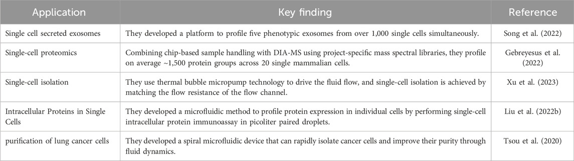
Table 2. Applications of microfluidic studies.
In summary, microfluidics provides a robust tool for the precise manipulation, analysis, and investigation of cellular heterogeneity at the individual cell level. The miniaturization, high throughput capacity, and precise control provided by microfluidic technologies empower the generation of single-cell data, facilitating the acquisition of valuable biological insights into diverse cellular systems (Satija and Shalek, 2014; Zhao et al., 2014). As the field of microfluidics progresses and merges with other single-cell analysis techniques, it is poised to play a crucial role in furthering our comprehension of cellular heterogeneity and its implications in diverse biological processes.
Endogenous gene tagging with fluorescent proteins or fluorophore-conjugated antibody labeling techniques have enabled highly sensitive quantification of endogenous protein levels. The fluorescence intensity obtained from these methods directly correlates with the quantity of protein molecules present, thereby enabling the assessment of relative protein abundance.
Fluorescence labeling of target proteins combined with FACS is considered the gold-standard approach for profiling proteins at the single-cell level (Hu et al., 2016). This method involves staining single cells with fluorescent-labeled antibodies or tagging them with a fluorescent protein, which are then detected as they pass through the flow chamber during FACS analysis. The fluorophores are excited by a laser, and the emitted fluorescence is subsequently measured. Through meticulous system calibration using protein-coated beads, the fluorescent intensities can be translated into single-cell protein expression levels (Adan et al., 2017).
FACS facilitates high-throughput detection (∼104 cells/s) and simultaneous measurement of ∼20 multiplexing membrane and intracellular protein parameters (Liu L. et al., 2020). However, this technique has some limitations. Dynamic cell monitoring over time poses a challenge, and the multiplexing capacity is constrained by spectral overlap, potentially impacting the accuracy of protein measurements. In the multi-colour detection system, each excitation laser, including green (532 nm), blue (488 nm) and violet (407 nm) ranges and red (633 nm), is configured with multiple detectors (for the green laser, fluorochromes PE, TRPE, Cy5PE, Cy5.5PE and Cy7PE could be applied and separated). If additional fluorochromes become available that have distinct emission spectra after excitation by the green laser, the detection capacity could be increased even further. Furthermore, physical stressors experienced by cells during FACS procedures could influence their status. A critical aspect is the requirement for a large number of cells (1 × 10^6/mL) in conventional flow cytometry sample preparation (Xie and Ding, 2022).
To address these challenges, flow cytometers incorporating microfluidics have been proposed as a solution (Liu et al., 2011; Wu et al., 2012; Su et al., 2011). Wu et al. introduced a microfluidic platform that integrates cell preparation and multi-color flow cytometry components. This on-chip system offers multiplexed and orthogonal data for profiling signaling pathways across various cell types, including primary cells (Wu et al., 2012). The integration of microfluidics with flow cytometry enables the analysis of smaller sample sizes, reduces the risk of sample contamination, and allows for the precise control of the cellular microenvironment during analysis. Besides, a recent study showed a workflow for generating double emulsions and performing multicolor cell sorting using a commercial FACS instrument. This workflow achieves a double emulsion detection rate exceeding 90%, enabling multicellular encapsulation and high-throughput immune cell activation sorting for the first time (Ding et al., 2024).
Despite these challenges, FACS remains a powerful tool for single-cell protein analysis, offering high throughput and multiplexing capabilities. Researchers continue to explore and develop new techniques that address these limitations, aiming to enable more comprehensive and accurate analysis of protein dynamics and interactions at the single-cell level.
Single-cell Western blotting (scWB) is another innovative technique that combines microfluidics and conventional Western blotting to analyze protein expression at a single-cell resolution (Hughes et al., 2014). It overcomes the issue of cross-reactivity by introducing a step of electrophoretic separation before antibody probing (Figure 2A). The scWestern analysis utilizes a microscope slide coated with a thin photoactive polyacrylamide gel. This gel is micropatterned with an array of thousands of microwells, allowing for the simultaneous analysis of 103–104 single cells/chip. Simply stated, the scWestern approach integrates all the essential steps of Western blotting into a dense array format. In this technique, single cells are placed into the microwells and lysed in situ. The proteins from the lysed cells are then separated by gel electrophoresis within each microwell. Following electrophoresis, the proteins are immobilized onto the PA gel through photoinitiated blotting. Finally, fluorescent-labeled antibodies are used for protein detection within each microwell.
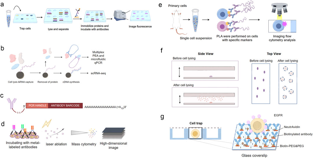
Figure 2. Overview of single-cell protein analysis technologies. (A) Schematic of Single Cell Western blotting. Single cells are placed onto the microwells and lysed in situ. The proteins from the lysed cells are then separated by gel electrophoresis within each microwell. Following electrophoresis, the proteins are immobilized onto the PA gel through photoinitiated blotting. Finally, fluorescent-labeled antibodies are used for protein detection within each microwell. (B) Schematic of Single-Cell Protein And RNA Co-profiling (SPARC). Single cells are isolated and lysed. Following oligo-dT mRNA hybridization, the protein-containing supernatant is removed for subsequent multiplex proximity extension analysis (PEA) and the mRNA is processed using a modified Smart-seq2 approach. (C) Illustration of the DNA-barcoded antibodies used in CITE-seq. (D) Schematic of Mass Cytometry. After incubation with metal-labeled oligonucleotides for RNA detection and antibodies for protein detection, tissues are subjected to laser ablation and mass-cytometric measurement of the metal abundances. The metal abundances per laser shot (in pixels) are then assembled into a high-dimension image. (E) Schematic of the Proximity Ligation Imaging Cytometry assay. (F) Schematics of in situ protein capture from single cells. Cells are sparsely immobilized on the coverslip surface. The cells are subsequently lysed, and the proteins released are captured by the antibody surface, where single-molecule imaging is performed. (G) Schematic of the single-cell capture trap.
scWB has emerged as a promising tool for investigating protein expression in individual cells, offering valuable insights into cellular heterogeneity and dynamics at a higher resolution (Rosàs-Canyelles et al., 2020; Kim et al., 2021; Grist et al., 2020). scWB enables detection of more than 10 proteins, including both membrane and intracellular proteins, in each cell during 4 h. The sensitivity of the scWB method is comparable to that of flow cytometry (FC). However, scWB encounters challenges in quantifying secreted proteins, low-abundance proteins, and small molecular weight proteins (Duncombe et al., 2016). To address these limitations, several new methods have been suggested and developed. For example, the integration of scWB with ultrasensitive detection methods, such as single-molecule fluorescence microscopy or mass spectrometry, has the potential to improve the sensitivity and dynamic range of protein measurements (Yen et al., 2014; Lomeli et al., 2021).
In addition to using fluorescent antibodies for protein expression measurement, employing DNA amplification and reporters offers improved accuracy and higher sensitivity. With protein signal outputs encoded by various DNA sequences, the multiplexing capacity is virtually unlimited when utilizing DNA reporters. Several approaches have emerged to enable simultaneous measurement of both mRNA and protein at the single-cell level (Reimegård et al., 2021; Stoeckius et al., 2017; Peterson et al., 2017; Mimitou et al., 2019).
One such approach is Single-Cell Protein and RNA Co-profiling (SPARC) (Reimegård et al., 2021), proposed by Dr. Gallant’s group in 2021. SPARC enables the simultaneous measurement of overall mRNA levels and targeted 89 intracellular proteins in individual cells (Figure 2B). Single cells were sorted on a BD FACS ARIAIII into a 96-well plate and lysed for protein analysis. Protein quantification is accomplished through multiplex, homogeneous protein extension assay (PEA), which is an affinity-based detection technique employing pairs of antibodies linked with oligonucleotides (Reimegård et al., 2021). In PEA, each antibody pair is designed to recognize a specific protein target, and when both antibodies bind to the same protein, the oligonucleotides are brought into close proximity, allowing for their extension and amplification. The resulting DNA amplicons are then quantified using next-generation sequencing or qPCR, providing a digital readout of protein abundance (Darmanis et al., 2016).
CITE-seq, pioneered by Stoeckius et al., is another well-established method in single-cell analysis (Stoeckius et al., 2017). This innovative technique utilizes a digital, sequencing-based platform to quantify single-cell protein expression levels. It involves conjugating antibodies to oligonucleotides, incorporating a unique barcode for antibody identification and facilitating PCR amplification (Figure 2C). The antibody-oligo complexes are then applied to single-cell suspensions. After incubation, a washing step removes unbound antibodies before proceeding to single-cell RNA sequencing (scRNA-seq). CITE-seq primarily allows for the analysis of cell surface proteins, providing valuable information about cell type, state, and function. Recently, Liu et al., extended co-indexing of transcriptomes and epitopes (CITE) to the spatial dimension and demonstrated high-plex protein and whole transcriptome co-mapping to measure 273 proteins and transcriptome in human tissues with 10–50 μm Spatial resolution (Liu et al., 2023). The simultaneous measurement of mRNA and surface proteins enables a more comprehensive characterization of cellular heterogeneity and can reveal novel cell subpopulations and their functional properties.
The integration of DNA reporters with single-cell protein analysis has greatly expanded the multiplexing capacity and sensitivity of these techniques. The ability to encode protein signals with unique DNA sequences allows for the simultaneous measurement of hundreds to thousands of proteins in individual cells, overcoming the limitations of spectral overlap encountered in fluorescence-based methods (Table 3). Furthermore, the amplification of DNA reporters through PCR or sequencing enables the detection of low-abundance proteins that may be missed by other techniques (Reimegård et al., 2021).
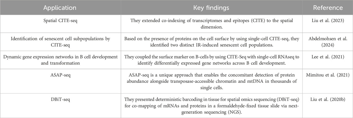
Table 3. Applications of dna reporter-based measurement of protein level insingle cells.
Chemiluminescence (CL) is known for its simple and cost-effective optical systems, as it does not require an external light source. This characteristic circumvents issues related to stray light and the instability of light sources, ultimately resulting in low backgrounds and high sensitivity.
The Enzyme-linked immunospot assay (ELISPOT) is a powerful technique developed in the 1980s for quantitatively detecting individual cells that secrete a specific protein of interest (Alexander et al., 2013). This method, based on an immuno-sandwich assay, is valuable for enumerating cell subsets with distinct secretory functions and for monitoring cellular responses to external stimuli or drugs (DiPiazza et al., 2016; Barabas et al., 2017). In ELISPOT, cells are cultured on a surface coated with antibodies that target the protein of interest. The secreted protein is then captured by the immobilized antibodies. A second, enzyme-conjugated antibody is then added, which binds to the captured protein. Upon addition of a substrate, the enzyme catalyzes a reaction that produces a visible spot on the membrane, with each spot representing an individual secreting cell.
The ELISPOT assay is well-known for its high sensitivity in detecting secreted proteins, with a detection limit of approximately six spots per 100,000 cells (Moodie et al., 2010). It enables high-throughput detection (∼106 cells/run) and simultaneous measurement of 1-3 secreted proteins. It has been particularly useful in studying immune responses, such as the detection of cytokine-secreting T cells or antibody-secreting B cells, and in assessing the impact of various interventions on cell function (Leehan and Koelsch, 2015). The ability to quantify the frequency of secreting cells and the relative amount of protein secreted by each cell makes ELISPOT a valuable tool for understanding the heterogeneity of cellular responses.
Recent advancements in ELISPOT technology have focused on improving its multiplexing capabilities and compatibility with other single-cell analysis techniques. For instance, FluoroSpot, a type of fluorescent ELISPOT, allows for the concurrent detection of multiple secreted proteins by utilizing fluorescent dyes instead of enzymes for signal generation (Axelsson, 2022). Additionally, the integration of ELISPOT with microfluidic devices has allowed for the isolation and analysis of individual secreting cells, providing a more comprehensive profile of their functional properties (Huang et al., 2012).
Recent advances in mass spectrometry (MS) have made single-cell MS one of the most powerful approaches to obtain the protein profile of a single cell (Slavov, 2021; Petelski et al., 2021). Mass spectrometry-based methods can generally be divided into two main categories: targeted and untargeted approaches.
Targeted MS approaches involve the specific detection and quantification of pre-selected proteins or peptides within a sample. This method is useful when researchers are interested in analyzing a particular set of proteins or biomarkers within a single cell.
Mass Cytometry, also known as CyTOF (Cytometry by Time-of-Flight), represents an innovative fusion of flow cytometry (FC) and mass spectrometry (MS), offering an alternative approach for the identification and quantification of target proteins (Table 4). Applying this technology to tissues or cells on slides, termed imaging mass cytometry (IMC), allows for visualization of normal and diseased tissues in situ (Tracey et al., 2021). This method facilitates high-dimensional, single-cell analysis of cell type and state (Giesen et al., 2014). In principle, mass cytometry closely resembles flow cytometry, where cells are labeled with antibodies conjugated with metal isotopes and subsequently analyzed via mass spectrometry. The use of specific isotopes circumvents issues related to spectral overlap, enabling simultaneous, multiplexed detection of target proteins (Zhang et al., 2020).
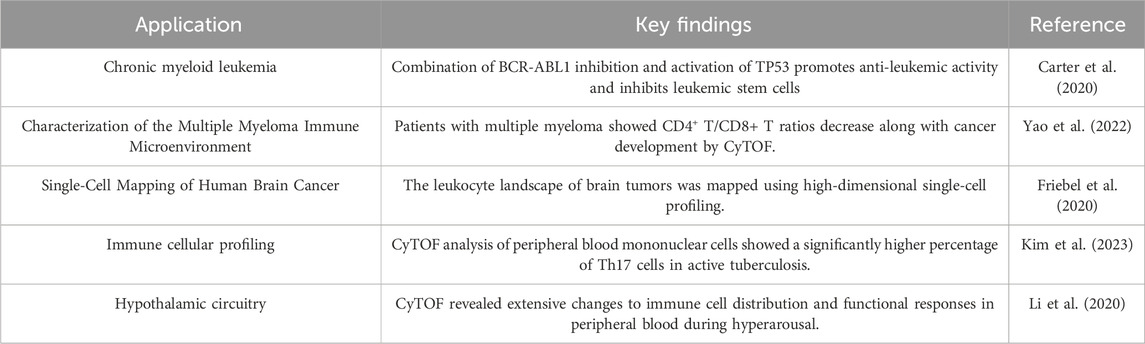
Table 4. Applications of cytof studies.
The workflow of mass cytometry includes the following steps: (1) cells are labeled with antibodies attached to distinct metal isotopes; (2) these labeled cells are introduced into the mass cytometer, where they are vaporized and ionized; (3) the metal ions are separated according to their mass-to-charge ratio; and (4) the quantity of each metal ion is measured, indicating the expression level of the associated protein (Olsen et al., 2019).
Mass cytometry has been broadly adopted and applied to many biological problems (Schulz et al., 2018; Lun et al., 2019; Krishnaswamy et al., 2014; Ajami et al., 2018). Friebel et al. successfully applied this technique, designed two CyTOF panels together, measuring 74 parameters at the single-cell level (Friebel et al., 2020) (Figure 2D). This innovative approach expands the capacity for comprehensive protein analysis within tissues, providing a valuable tool for studying complex biological systems (insert).
In terms of throughput, mass cytometry allows for the measurement of approximately ∼1,000 cells per second, which is inferior to the throughput achievable with fluorescent-labeled analysis approaches (Liu L. et al., 2020). Additionally, when compared to quantum-efficient fluorophores, mass reporters demonstrate lower sensitivities, posing challenges in the accurate measurement of low-abundance proteins in single cells. However, the high dimensionality and minimal spectral overlap of mass cytometry make it a powerful tool for dissecting the complexity of cellular systems and identifying rare cell subpopulations (Levine et al., 2021).
Recent advancements in liquid chromatography-mass spectrometry (LC-MS)-based proteomics methods have addressed significant challenges related to sensitivity and throughput, making single-cell mass spectrometry (scMS) more feasible. This approach has become increasingly utilized to gain valuable insights into cellular heterogeneity (Budnik et al., 2018; Zhu et al., 2019; Lombard-Banek et al., 2019; Cheung et al., 2021; Schoof et al., 2021). One of the main advantages of scMS is its ability to provide an unbiased, global view of the cellular proteome. Unlike targeted approaches that rely on antibodies, scMS can detect and quantify thousands of proteins without prior knowledge of their identity or function. This untargeted nature of scMS enables the discovery of novel proteins and pathways that may be missed by other methods (Lombard-Banek et al., 2019).
Current scMS approaches can quantify approximately 1,000 proteins per cell and analyze over a hundred cells per day of instrument time (Schoof et al., 2021). Despite having a lower throughput than some other single-cell protein analysis methods, the comprehensive coverage and capability to detect post-translational modifications make scMS a valuable tool for understanding cellular heterogeneity at the proteome level (Liu L. et al., 2020).
The Proximity Ligation Assay (PLA) represents a relatively novel method for studying protein-protein interactions (PPIs) with high specificity, applicable in both cells and tissue sections (Söderberg et al., 2006). In the assay, a pair of oligonucleotide-labeled antibodies is used to recognize two target proteins. When the two proteins are in close proximity (less than 30–40 nm apart) due to their interaction, the oligonucleotides are ligated, subsequently extended, and amplified to generate sequence products. Detection is accomplished by adding complementary fluorescently labeled oligonucleotides or by measuring the output of DNA sequencing using qPCR.
Dr. Abramson’s group has integrated the PLA assay with imaging flow cytometry (IFC) and developed Proximity Ligation Imaging Cytometry (FLIC) (Avin et al., 2017). This integration enables multiparametric fluorescent and localization analysis of cellular events, including the examination of PPIs and post-translational modifications (PTMs), as well as the assessment of subcellular signal distribution within cells (Figure 2E). FLIC combines the specificity of PLA with the high-throughput and multiplexing capabilities of IFC, allowing for the simultaneous analysis of multiple PPIs and cellular parameters in thousands of individual cells. Vistain et al. developed Prox-seq, a method that enables the detection of intracellular and membrane proteins, protein complexes, and mRNAs in thousands of single cells. Prox-seq combines single-cell RNA sequencing (scRNA-seq) with the proximity ligation assay (PLA). It utilizes pairs of DNA-conjugated antibodies that, when brought into close proximity, facilitate the ligation of DNA oligonucleotides (oligomers) on the antibodies. This ligation generates a PLA product that can be detected through next-generation sequencing. The method allows for the simultaneous measurement of up to 741 protein complexes across 8,700 single peripheral blood mononuclear cells (Vistain et al., 2022).
To investigate PPIs directly with high spatiotemporal resolution, genetically encoded biosensors based on Förster Resonance Energy Transfer (FRET) or Bioluminescence Resonance Energy Transfer (BRET) can be employed. These resonance energy transfer (RET) techniques offer the ability to record single-cell kinetics with millisecond resolution, facilitating the identification of protein complexes and the study of cell-to-cell heterogeneity (van Unen et al., 2016). Recently, Hoshino et al. designed γB2-FRET probes by fusing FRET donor and acceptor fluorescent proteins to a single γB2 molecule, successfully visualizing γB2 homophilic interactions in cultured hippocampal neurons (Hoshino et al., 2023). By utilizing these biosensors, researchers can visualize dynamic changes in PPIs in real-time, providing valuable insights into the molecular mechanisms underlying various cellular processes.
The Single-molecule pull-down (SiMPull) technique provides a method to directly capture native protein complexes from cell lysates, facilitating the identification of complex composition and PPIs at the single-molecule level (Jain et al., 2012). SiMPull involves immobilizing an antibody against a target protein on a glass surface, followed by the application of cell lysates containing the protein complexes. The captured complexes are then visualized using fluorescently labeled antibodies against the interacting partners, allowing for the direct observation of PPIs and the determination of their stoichiometry.
Expanding on this concept, various techniques have been suggested to investigate PPIs at the single-cell level (Ryu et al., 2019a; Zhao et al., 2023; Wang et al., 2018; Wedeking et al., 2015; Stolpner and Dickinson, 2022). Dr. Ha’s group developed an assay that involves lysing bacterial cells in situ and capturing the released proteins on an imaging surface coated with antibodies. This approach allows for the unambiguous assignment of captured proteins to their originating cells (Wang et al., 2018). The developed platform is compatible with high-throughput protein analysis and PPI analysis at the single-cell level through single-molecule imaging (Figure 2F). The ability to link protein interactions to specific cells enables the investigation of cell-to-cell variability in complex formation and function, providing insights into the mechanisms underlying cellular heterogeneity.
For mammalian cells, micro-patterned surfaces have been utilized to capture target cells and proteins. Yoon et al. implemented a method that involved in situ co-IP for individual cancer cells (Ryu et al., 2019a). This process included capturing single cells using a microfluidic device and inducing in situ lysis of the cells while immunoprecipitating EGFRs (the proteins of interest) to the surface. Subsequently, fluorescently labeled downstream interactors of EGFRs were directly added to stimulate protein-protein interactions, which were then visualized using a single-molecule fluorescence microscope. This innovative approach offers insights into the dynamics of protein interactions at the single-cell level, providing valuable information on signaling pathways and heterogeneity in cancer cells (Figure 2G).
The integration of co-IP with microfluidic devices and single-molecule imaging has greatly enhanced the sensitivity and resolution of PPI analysis at the single-cell level. By capturing protein complexes directly from individual cells, these techniques overcome the limitations of bulk co-IP experiments, which average out the heterogeneity in protein interactions across a population of cells. Moreover, the ability to visualize individual protein complexes allows for the quantitative analysis of their composition, stoichiometry, and dynamics, providing a more detailed understanding of their functional roles in cellular processes.
The advent of advanced single-cell protein analysis techniques, such as flow cytometry and mass spectrometry (MS), has revolutionized our ability to study cellular heterogeneity and protein dynamics at the individual cell level. These methods generate vast amounts of complex data, necessitating the development of sophisticated data analysis and computational approaches to extract meaningful biological insights.
In flow cytometry, data pre-processing typically involves gating, which entails selecting cells of interest by plotting the data in univariate histograms or bivariate (density) plots and defining an area of interest (Aghaeepour et al., 2013; Malek et al., 2015; Montante and Brinkman, 2019). While manual gating is convenient, it can be time-consuming, subjective, and prone to bias. To address these limitations, numerous semi-automated and automated gating methods have been developed, along with machine learning algorithms such as RchyOptimyx (Aghaeepour et al., 2012) and FloReMi (Van Gassen et al., 2016) that utilize these gating methods to identify cell types exhibiting significant differences between groups or for survival analysis. Following gating, data compensation and transformation are crucial steps to ensure accurate analysis (Tinnevelt et al., 2021). Modeling the distribution of single cells is typically performed using clustering (Aghaeepour et al., 2013), principal component analysis (PCA) (Ma et al., 2017), or t-distributed stochastic neighbor embedding (t-SNE) (Amir el et al., 2013).
Single-cell Mass Spectrometry (MS) stands as another potent technique for studying single-cell profiles due to its high sensitivity, broad detection range, and molecular identification capabilities. However, the raw data acquired from single-cell MS experiments are large and complex, necessitating systematic data analysis approaches (Liu and Yang, 2021). Unlike conventional MS proteomics, data analysis methods in current single-cell proteomics studies lack standardization. There is no widely agreed-upon, consistent analysis pipeline across different researchers and laboratories. The typical workflow for single-cell proteomics data analysis includes data pre-processing, univariate analysis, multivariate analysis, and advanced data analysis techniques (Liu and Yang, 2021). Data pre-processing involves noise and background signal removal, ion intensity normalization (Yin et al., 2018), and selection of proteins commonly detected in the majority of single cells (Pluskal et al., 2010). Univariate analysis, such as t-tests and analysis of variance (ANOVA), is used to reveal changes in cellular proteins corresponding to specific biological processes. Multivariate analysis, including unsupervised methods like PCA (Fang et al., 2020) and t-SNE (Wang R. et al., 2019), and supervised methods such as partial least squares discriminant analysis (PLS-DA) (Sun and Yang, 2019) and orthogonal projections to latent structures discriminant analysis (OPLS-DA) (Walker et al., 2013), are employed to reduce data dimensionality and identify patterns in protein expression.
Recent advancements in machine learning (ML) have introduced new data analysis methods for single-cell identification, proteomics, demonstrating high classification accuracy in distinguishing cell types (Xie et al., 2020), special omics, and enabling the extraction of trace-level signals from high-resolution mass spectra (Liu et al., 2019). Image-Based cell identification can bring significant insight to biomedical sciences. Deep learning algorithms could enable cell classification and isolation based on human-vision uninterpretable features within a complex cell population without labeling (Tang et al., 2023). Machine learning workflow developed by Xie et al., was trained to classify single cells according to their mass spectra based on cell groups of interest (GOI). The trained models achieved >80% classification accuracy (Xie et al., 2020). MEISTER, a mass spectrometry (MS) framework, integrates deep-learning-based reconstruction, three-dimensional (3D) molecular distributions and cell-specific mass spectra to 3D brain-wide single-cell biochemical mapping (Xie et al., 2024).
One of the key applications of single-cell protein analysis is the study of cellular heterogeneity in various contexts. For example, mass cytometry has been used to explore immune cell heterogeneity (Newell et al., 2012), while single-cell Western blotting (Sinkala et al., 2017), microfluidic single-cell imaging (Ryu et al., 2019b), and mass cytometry (Levine et al., 2015) have been employed to investigate cancer cell heterogeneity. These studies have revealed the presence of distinct cell subpopulations and provided insights into cell-cell interactions in complex biological samples, such as breast cancer tissues (Giesen et al., 2014).
In addition to studying cellular heterogeneity, single-cell proteomic technologies have proven to be valuable tools for investigating intracellular protein-protein interactions (PPIs) and signaling networks. By profiling a large number of proteins in individual cells, researchers can conduct pairwise protein expression correlation analysis to study protein activating and inhibitory interactions. Techniques such as microfluidic single-cell single-molecule imaging (Wedeking et al., 2015), proximity ligation assay (PLA) (Avin et al., 2017), and FRET assays (van Unen et al., 2016) have been successfully applied to elucidate PPIs and signaling pathways at the single-cell level.
Despite the remarkable progress made in single-cell protein analysis, there are still challenges to overcome in order to fully understand the complex and dynamic nature of the single-cell proteome. To address these challenges, the development of next-generation single-cell analytical techniques is crucial across multiple areas. One key area of focus is the enhancement of detection sensitivity, which will enable the identification and quantification of low-abundance and less stable proteins. Advancements in droplet microfluidic diagnostic techniques, such as the integration of the “picoliter single-cell reaction flask” principle, have shown promise in screening single cells for secreted molecules, including antibodies, cytokines, enzymes, and metabolites (Lan et al., 2023). Another important direction is the investigation of subcellular compartments and the precise analysis of proteins corresponding to specific organelles. Improved single-cell Western blotting methods, such as subcellular Western blotting techniques, have demonstrated the ability to separately assay proteins in the cytoplasm and nucleus of individual cells (Yamauchi and Herr, 2017).
To achieve a comprehensive understanding of single-cell states and functions, the development of complementary methods that integrate multiple types of data is essential. For example, combining high-throughput techniques for the simultaneous detection of mRNA and intracellular proteins can provide a more complete picture of the molecular regulatory networks within individual cells (Reimegård et al., 2021). Flow cytometry-based FISH (Flow-FISH) has been used to simultaneously measure transcript levels and protein expression in single cells, offering deeper insights into the regulation of gene transcription and translation (Van Hoof et al., 2014).
The application of machine learning (ML) techniques to single-cell protein analysis has the potential to revolutionize data analysis and interpretation. ML methods have significantly improved the efficiency of single-cell proteomics data analysis, enabling the identification of patterns and relationships that may be difficult to detect manually (Xie et al., 2020). As ML algorithms continue to evolve and become more sophisticated, their broader application in single-cell protein analysis is expected to facilitate the discovery of novel biomarkers, therapeutic targets, and disease mechanisms.
While the commercialization and clinical translation of single-cell protein analysis technologies have begun, there are still significant barriers to their widespread adoption in clinical and industrial settings. These technologies often require extensive expertise and hands-on time, and their repeatability may not yet meet the stringent requirements for industrial applications. To overcome these challenges, the development of more user-friendly and robust systems is essential. This can be achieved through device automation and further optimization, enabling researchers and practitioners to implement these methods across a wide range of applications.
The rapid development of single-cell protein analysis has revolutionized our ability to study the protein composition, expression levels, and functions of individual cells (Liu et al., 2021; Ryu et al., 2019a; Reimegård et al., 2021; Shao et al., 2018; Peterson et al., 2017; Maes et al., 2020; Sims and Allbritton, 2007; Xie and Ding, 2022; Pereira et al., 2022; Lohani et al., 2023; Yu et al., 2022; Hoover et al., 2023; Mund et al., 2022). By analyzing membrane proteins, cellular proteins, and secreted proteins, researchers can gain a more comprehensive understanding of cell states, functional changes, and cellular heterogeneity (Budnik et al., 2018; Giesen et al., 2014; Sharon et al., 2013; Rosàs-Canyelles et al., 2020; Crouch et al., 2024). This review provides a comprehensive summary of the advancements in single-cell protein analysis technologies over the past two decades, discussing various methods and techniques, and comparing their approaches for single-cell isolation based on factors such as purity, throughput, and efficiency.
Despite recent developments, current single-cell protein analysis techniques still have limitations that need to be addressed (Liu L. et al., 2020; Hu et al., 2016; Bennett et al., 2023). Improvements are needed in accuracy, reproducibility, and the ability to detect low-abundance proteins (Hughes et al., 2014; Hu et al., 2016; Gross et al., 2015; Lugli et al., 2017). The limited multiplexing capacity of existing methods hinders the comprehensive proteomic detection of over 10,000 proteins within a single cell. Additionally, most techniques cannot simultaneously detect membrane-bound, cytoplasmic, and secreted proteins, restricting our understanding of cell protein composition. Furthermore, current methods for analyzing PPIs are semi-quantitative and limited to known protein pairs, hindering the exploration of novel protein interactions.
The complexity and dynamic nature of proteins present a significant challenge in achieving high throughput, accuracy, multiplexing, and sensitivity simultaneously in single-cell proteomic analysis. Researchers must balance the need for detecting low abundance proteins with the desire to analyze a large number of cells efficiently. Future advancements in technology and methodology are expected to address these challenges, enhancing the capabilities of single-cell proteomic analysis and enabling more comprehensive insights into cellular heterogeneity and function. The integration of single-cell protein analysis with other omics technologies, such as single-cell transcriptomics and metabolomics, will provide a more holistic view of cellular processes and uncover novel regulatory mechanisms (Reimegård et al., 2021; Frei et al., 2016; Stoeckius et al., 2017; Peterson et al., 2017). The application of machine learning techniques to single-cell protein analysis will revolutionize data analysis and interpretation, facilitating the discovery of novel biomarkers, therapeutic targets, and disease mechanisms.
With ongoing technological advancements, single-cell protein analysis is expected to have significant applications in clinical diagnostics, cancer therapy, and drug development. Studying protein expression and PPIs in individual tumor cells can improve our understanding of tumor heterogeneity and drug resistance mechanisms, providing a basis for personalized therapy. Single-cell protein analysis can also aid in screening novel drug targets and assessing the impact of drugs on cell function, accelerating the drug development process (Barabas et al., 2017; Heath et al., 2016; Fitzgerald and Leonard, 2017). As technology advances, collaboration among researchers from different disciplines will be crucial to drive further progress in single-cell proteomics, ultimately leading to a deeper understanding of cellular heterogeneity and its implications for human health and disease.
QZ: Conceptualization, Investigation, Writing–original draft. SL: Writing–review and editing. LK: Writing–review and editing. QL: Writing–original draft. RS: Writing–original draft. YY: Writing–original draft. JF: Writing–original draft. XZ: Writing–review and editing. YW: Writing–review and editing. MY: Writing–review and editing.
The author(s) declare that financial support was received for the research, authorship, and/or publication of this article. This work was supported by General Program of National Natural Science Foundation of China (3207050372) and Basic Research Key Project of Yunnan (202101AS070002).
The authors declare that the research was conducted in the absence of any commercial or financial relationships that could be construed as a potential conflict of interest.
The author(s) declare that no Generative AI was used in the creation of this manuscript.
All claims expressed in this article are solely those of the authors and do not necessarily represent those of their affiliated organizations, or those of the publisher, the editors and the reviewers. Any product that may be evaluated in this article, or claim that may be made by its manufacturer, is not guaranteed or endorsed by the publisher.
Abdelmohsen, K., Mazan-Mamczarz, K., Munk, R., Tsitsipatis, D., Meng, Q., Rossi, M., et al. (2024). Identification of senescent cell subpopulations by CITE-seq analysis. Aging Cell 23 (11), e14297. doi:10.1111/acel.14297
Adan, A., Alizada, G., Kiraz, Y., Baran, Y., and Nalbant, A. (2017). Flow cytometry: basic principles and applications. Crit. Rev. Biotechnol. 37 (2), 163–176. doi:10.3109/07388551.2015.1128876
Aghaeepour, N., Finak, G., Hoos, H., Mosmann, T. R., Brinkman, R., Gottardo, R., et al. (2013). Critical assessment of automated flow cytometry data analysis techniques. Nat. Methods 10 (3), 228–238. doi:10.1038/nmeth.2365
Aghaeepour, N., Jalali, A., O'Neill, K., Chattopadhyay, P. K., Roederer, M., Hoos, H. H., et al. (2012). RchyOptimyx: cellular hierarchy optimization for flow cytometry. Cytom. A 81 (12), 1022–1030. doi:10.1002/cyto.a.22209
Ajami, B., Samusik, N., Wieghofer, P., Ho, P. P., Crotti, A., Bjornson, Z., et al. (2018). Single-cell mass cytometry reveals distinct populations of brain myeloid cells in mouse neuroinflammation and neurodegeneration models. Nat. Neurosci. 21 (4), 541–551. doi:10.1038/s41593-018-0100-x
Alexander, N., Fox, A., Lien, V. T. K., Dong, T., Lee, L. Y. H., Le Khanh Hang, N., et al. (2013). Defining ELISpot cut-offs from unreplicated test and control wells. J. Immunol. Methods 392 (1-2), 57–62. doi:10.1016/j.jim.2013.02.014
Amir el, A. D., Davis, K. L., Tadmor, M. D., Simonds, E. F., Levine, J. H., Bendall, S. C., et al. (2013). viSNE enables visualization of high dimensional single-cell data and reveals phenotypic heterogeneity of leukemia. Nat. Biotechnol. 31 (6), 545–552. doi:10.1038/nbt.2594
An, X., and Chen, L. (2018). Flow cytometry (FCM) analysis and fluorescence-activated cell sorting (FACS) of erythroid cells. Methods Mol. Biol. 1698, 153–174. doi:10.1007/978-1-4939-7428-3_9
Antoniadi, I., Skalický, V., Sun, G., Ma, W., Galbraith, D. W., Novák, O., et al. (2022). Fluorescence activated cell sorting-A selective tool for plant cell isolation and analysis. Cytom. A 101 (9), 725–736. doi:10.1002/cyto.a.24461
Avin, A., Levy, M., Porat, Z., and Abramson, J. (2017). Quantitative analysis of protein-protein interactions and post-translational modifications in rare immune populations. Nat. Commun. 8 (1), 1524. doi:10.1038/s41467-017-01808-6
Axelsson, B. (2022). Detection and enumeration of cytokine-secreting cells by FluoroSpot. Methods Mol. Biol. 2386, 81–99. doi:10.1007/978-1-0716-1771-7_6
Barabas, S., Spindler, T., Kiener, R., Tonar, C., Lugner, T., Batzilla, J., et al. (2017). An optimized IFN-γ ELISpot assay for the sensitive and standardized monitoring of CMV protein-reactive effector cells of cell-mediated immunity. BMC Immunol. 18 (1), 14. doi:10.1186/s12865-017-0195-y
Bendall, S. C., Nolan, G. P., Roederer, M., and Chattopadhyay, P. K. (2012). A deep profiler's guide to cytometry. Trends Immunol. 33 (7), 323–332. doi:10.1016/j.it.2012.02.010
Bennett, H. M., Stephenson, W., Rose, C. M., and Darmanis, S. (2023). Single-cell proteomics enabled by next-generation sequencing or mass spectrometry. Nat. Methods 20 (3), 363–374. doi:10.1038/s41592-023-01791-5
Bennett, M. L., Bennett, F. C., Liddelow, S. A., Ajami, B., Zamanian, J. L., Fernhoff, N. B., et al. (2016). New tools for studying microglia in the mouse and human CNS. Proc. Natl. Acad. Sci. U. S. A. 113 (12), E1738–E1746. doi:10.1073/pnas.1525528113
Berlanda, S. F., Breitfeld, M., Dietsche, C. L., and Dittrich, P. S. (2021). Recent advances in microfluidic technology for bioanalysis and diagnostics. Anal. Chem. 93 (1), 311–331. doi:10.1021/acs.analchem.0c04366
Berteli, T. S., Da Broi, M. G., Martins, W. P., Ferriani, R. A., and Navarro, P. A. (2017). Magnetic-activated cell sorting before density gradient centrifugation improves recovery of high-quality spermatozoa. Andrology 5 (4), 776–782. doi:10.1111/andr.12372
Bhagwat, N., Dulmage, K., Pletcher, C. H., Wang, L., DeMuth, W., Sen, M., et al. (2018). An integrated flow cytometry-based platform for isolation and molecular characterization of circulating tumor single cells and clusters. Sci. Rep. 8 (1), 5035. doi:10.1038/s41598-018-23217-5
Budnik, B., Levy, E., Harmange, G., and Slavov, N. (2018). SCoPE-MS: mass spectrometry of single mammalian cells quantifies proteome heterogeneity during cell differentiation. Genome Biol. 19 (1), 161. doi:10.1186/s13059-018-1547-5
Carter, B. Z., Mak, P. Y., Mu, H., Wang, X., Tao, W., Mak, D. H., et al. (2020). Combined inhibition of MDM2 and BCR-ABL1 tyrosine kinase targets chronic myeloid leukemia stem/progenitor cells in a murine model. Haematologica 105 (5), 1274–1284. doi:10.3324/haematol.2019.219261
Cha, H., Fallahi, H., Dai, Y., Yuan, D., An, H., Nguyen, N. T., et al. (2022). Multiphysics microfluidics for cell manipulation and separation: a review. Lab. Chip 22 (3), 423–444. doi:10.1039/d1lc00869b
Chen, W., Li, J., Wang, P., Ma, S., and Li, B. (2024). Study on the flow field distribution in microfluidic cells for surface plasmon resonance array detection. Materials 17, 2426. doi:10.3390/ma17102426
Cheung, T. K., Lee, C. Y., Bayer, F. P., McCoy, A., Kuster, B., and Rose, C. M. (2021). Defining the carrier proteome limit for single-cell proteomics. Nat. Methods 18 (1), 76–83. doi:10.1038/s41592-020-01002-5
Crouch, E. E., Diafos, L. N., Valenzuela, E. J., Wedderburn-Pugh, K., Birrueta, J. O., Caston, J., et al. (2024). Profiling human brain vascular cells using single-cell transcriptomics and organoids. Nat. Protoc. 19 (3), 603–628. doi:10.1038/s41596-023-00929-1
Darmanis, S., Gallant, C. J., Marinescu, V. D., Niklasson, M., Segerman, A., Flamourakis, G., et al. (2016). Simultaneous multiplexed measurement of RNA and proteins in single cells. Cell Rep. 14 (2), 380–389. doi:10.1016/j.celrep.2015.12.021
Decarlo, K., Emley, A., Dadzie, O. E., and Mahalingam, M. (2011). Laser capture microdissection: methods and applications. Methods Mol. Biol. 755, 1–15. doi:10.1007/978-1-61779-163-5_1
Desikan, S. A., Chavan, S., Ou, P., Roman, C. A., and Huan, C. (2022). A MACS protocol for purification of untouched germinal center B cells from unimmunized or germinal center-induced mice. Star. Protoc. 3 (2), 101388. doi:10.1016/j.xpro.2022.101388
Dilillo, M., Pellegrini, D., Ait-Belkacem, R., de Graaf, E. L., Caleo, M., and McDonnell, L. A. (2017). Mass spectrometry imaging, laser capture microdissection, and LC-MS/MS of the same tissue section. J. Proteome Res. 16 (8), 2993–3001. doi:10.1021/acs.jproteome.7b00284
Ding, Y., Zoppi, G., Antonini, G., Geiger, R., and deMello, A. J. (2024). Robust double emulsions for multicolor fluorescence-activated cell sorting. Anal. Chem. 96 (37), 14809–14818. doi:10.1021/acs.analchem.4c02363
DiPiazza, A., Richards, K., Batarse, F., Lockard, L., Zeng, H., García-Sastre, A., et al. (2016). Flow cytometric and cytokine ELISpot approaches to characterize the cell-mediated immune response in ferrets following influenza virus infection. J. Virol. 90 (17), 7991–8004. doi:10.1128/jvi.01001-16
Dong, Z., Ahrens, C. C., Yu, D., Ding, Z., Lim, H., and Li, W. (2017). Cell isolation and recovery using hollow glass microspheres coated with nanolayered films for applications in resource-limited settings. ACS Appl. Mater. and Interfaces 9 (18), 15265–15273. doi:10.1021/acsami.7b02197
Duncombe, T. A., Kang, C., Maity, S., Ward, T. M., Pegram, M. D., Murthy, N., et al. (2016). Hydrogel pore-size modulation for enhanced single-cell western blotting. Adv. Mater 28 (2), 327–334. doi:10.1002/adma.201503939
Edd, J. F., Di Carlo, D., Humphry, K. J., Köster, S., Irimia, D., Weitz, D. A., et al. (2008). Controlled encapsulation of single-cells into monodisperse picolitre drops. Lab. Chip 8 (8), 1262–1264. doi:10.1039/b805456h
Edfors, F., Danielsson, F., Hallström, B. M., Käll, L., Lundberg, E., Pontén, F., et al. (2016). Gene-specific correlation of RNA and protein levels in human cells and tissues. Mol. Syst. Biol. 12 (10), 883. doi:10.15252/msb.20167144
Emmert-Buck, M. R., Bonner, R. F., Smith, P. D., Chuaqui, R. F., Zhuang, Z., Goldstein, S. R., et al. (1996). Laser capture microdissection. Science 274 (5289), 998–1001. doi:10.1126/science.274.5289.998
Espina, V., Heiby, M., Pierobon, M., and Liotta, L. A. (2007). Laser capture microdissection technology. Expert Rev. Mol. Diagn 7 (5), 647–657. doi:10.1586/14737159.7.5.647
Fang, Z., Wang, R., Zhao, H., Yao, H., Ouyang, J., and Zhang, X. (2020). Mannose promotes metabolic discrimination of osteosarcoma cells at single-cell level by mass spectrometry. Anal. Chem. 92 (3), 2690–2696. doi:10.1021/acs.analchem.9b04773
Fink, L., Kwapiszewska, G., Wilhelm, J., and Bohle, R. M. (2006). Laser-microdissection for cell type- and compartment-specific analyses on genomic and proteomic level. Exp. Toxicol. Pathol. 57 (Suppl. 2), 25–29. doi:10.1016/j.etp.2006.02.010
Fitzgerald, V., and Leonard, P. (2017). Single cell screening approaches for antibody discovery. Methods 116, 34–42. doi:10.1016/j.ymeth.2016.11.006
Frei, A. P., Bava, F. A., Zunder, E. R., Hsieh, E. W. Y., Chen, S. Y., Nolan, G. P., et al. (2016). Highly multiplexed simultaneous detection of RNAs and proteins in single cells. Nat. Methods 13 (3), 269–275. doi:10.1038/nmeth.3742
Friebel, E., Kapolou, K., Unger, S., Núñez, N. G., Utz, S., Rushing, E. J., et al. (2020). Single-cell mapping of human brain cancer reveals tumor-specific instruction of tissue-invading leukocytes. Cell 181 (7), 1626–1642.e20. doi:10.1016/j.cell.2020.04.055
Fröhlich, J., and König, H. (2000). New techniques for isolation of single prokaryotic cells. FEMS Microbiol. Rev. 24 (5), 567–572. doi:10.1016/s0168-6445(00)00045-0
Gebreyesus, S. T., Siyal, A. A., Kitata, R. B., Chen, E. S. W., Enkhbayar, B., Angata, T., et al. (2022). Streamlined single-cell proteomics by an integrated microfluidic chip and data-independent acquisition mass spectrometry. Nat. Commun. 13 (1), 37. doi:10.1038/s41467-021-27778-4
Giesen, C., Wang, H. A. O., Schapiro, D., Zivanovic, N., Jacobs, A., Hattendorf, B., et al. (2014). Highly multiplexed imaging of tumor tissues with subcellular resolution by mass cytometry. Nat. Methods 11 (4), 417–422. doi:10.1038/nmeth.2869
Gordon, A., and Gousset, K. (2021). Utilization of laser capture microdissection coupled to mass spectrometry to uncover the proteome of cellular protrusions. Methods Mol. Biol. 2259, 25–45. doi:10.1007/978-1-0716-1178-4_3
Grigorev, G. V., Lebedev, A., Wang, X., Qian, X., Maksimov, G., and Lin, L. (2023). Advances in microfluidics for single red blood cell analysis. Biosens. (Basel) 13 (1), 117. doi:10.3390/bios13010117
Grist, S. M., Mourdoukoutas, A. P., and Herr, A. E. (2020). 3D projection electrophoresis for single-cell immunoblotting. Nat. Commun. 11 (1), 6237. doi:10.1038/s41467-020-19738-1
Gross, A., Schoendube, J., Zimmermann, S., Steeb, M., Zengerle, R., and Koltay, P. (2015). Technologies for single-cell isolation. Int. J. Mol. Sci. 16 (8), 16897–16919. doi:10.3390/ijms160816897
Guo, W., Hu, Y., Qian, J., Zhu, L., Cheng, J., Liao, J., et al. (2023). Laser capture microdissection for biomedical research: towards high-throughput, multi-omics, and single-cell resolution. J. Genet. Genomics 50 (9), 641–651. doi:10.1016/j.jgg.2023.07.011
He, J., Deng, C., Krall, L., and Shan, Z. (2023). ScRNA-seq and ST-seq in liver research. Cell Regen. 12 (1), 11. doi:10.1186/s13619-022-00152-5
Heath, J. R., Ribas, A., and Mischel, P. S. (2016). Single-cell analysis tools for drug discovery and development. Nat. Rev. Drug Discov. 15 (3), 204–216. doi:10.1038/nrd.2015.16
Herzenberg, L. A., Parks, D., Sahaf, B., Perez, O., and Roederer, M. (2002). The history and future of the fluorescence activated cell sorter and flow cytometry: a view from Stanford. Clin. Chem. 48 (10), 1819–1827. doi:10.1093/clinchem/48.10.1819
Hoover, M. Y., Ambrosi, T. H., Steininger, H. M., Koepke, L. S., Wang, Y., Zhao, L., et al. (2023). Purification and functional characterization of novel human skeletal stem cell lineages. Nat. Protoc. 18 (7), 2256–2282. doi:10.1038/s41596-023-00836-5
Hoshino, N., Kanadome, T., Takasugi, T., Itoh, M., Kaneko, R., Inoue, Y. U., et al. (2023). Visualization of trans homophilic interaction of clustered protocadherin in neurons. Proc. Natl. Acad. Sci. U. S. A. 120 (38), e2301003120. doi:10.1073/pnas.2301003120
Hu, J., Chen, R., Zhu, C., Ge, B., Zhu, X., Mi, L., et al. (2018). Label-free microarray-based binding affinity constant measurement with modified fluidic arrangement. BioChip J. 12 (1), 11–17. doi:10.1007/s13206-017-2102-2
Hu, P., Zhang, W., Xin, H., and Deng, G. (2016). Single cell isolation and analysis. Front. Cell Dev. Biol. 4, 116. doi:10.3389/fcell.2016.00116
Huang, D., Ma, N., Li, X., Gou, Y., Duan, Y., Liu, B., et al. (2023). Advances in single-cell RNA sequencing and its applications in cancer research. J. Hematol. Oncol. 16 (1), 98. doi:10.1186/s13045-023-01494-6
Huang, N. T., Chen, W., Oh, B. R., Cornell, T. T., Shanley, T. P., Fu, J., et al. (2012). An integrated microfluidic platform for in situ cellular cytokine secretion immunophenotyping. Lab. Chip 12 (20), 4093–4101. doi:10.1039/c2lc40619e
Huang, R., Barber, T. A., Schmidt, M. A., Tompkins, R. G., Toner, M., Bianchi, D. W., et al. (2008). A microfluidics approach for the isolation of nucleated red blood cells (NRBCs) from the peripheral blood of pregnant women. Prenat. Diagn 28 (10), 892–899. doi:10.1002/pd.2079
Hughes, A. J., Spelke, D. P., Xu, Z., Kang, C. C., Schaffer, D. V., and Herr, A. E. (2014). Single-cell western blotting. Nat. Methods 11 (7), 749–755. doi:10.1038/nmeth.2992
Jain, A., Liu, R., Xiang, Y. K., and Ha, T. (2012). Single-molecule pull-down for studying protein interactions. Nat. Protoc. 7 (3), 445–452. doi:10.1038/nprot.2011.452
Jelokhani-Niaraki, M. (2022). Membrane proteins: structure, function and motion. Int. J. Mol. Sci. 24 (1), 468. doi:10.3390/ijms24010468
Jiang, Z. Q., Shi, H., Tang, X., and Qin, J. (2023). Recent advances in droplet microfluidics for single-cell analysis. Trac-Trends Anal. Chem. 159, 116932. doi:10.1016/j.trac.2023.116932
Kandathil, A. J., Graw, F., Quinn, J., Hwang, H. S., Torbenson, M., Perelson, A. S., et al. (2013). Use of laser capture microdissection to map hepatitis C virus-positive hepatocytes in human liver. Gastroenterology 145 (6), 1404–1413.e10. doi:10.1053/j.gastro.2013.08.034
Kim, C. H., Kim, H. J., Park, J. E., Lee, Y. H., Choi, S. H., Seo, H., et al. (2023). CyTOF analysis for differential immune cellular profiling between latent tuberculosis infection and active tuberculosis. Tuberculosis 140, 102344. doi:10.1016/j.tube.2023.102344
Kim, J. J., Liang, W., Kang, C. C., Pegram, M. D., and Herr, A. E. (2021). Single-cell immunoblotting resolves estrogen receptor-α isoforms in breast cancer. PLoS One 16 (7), e0254783. doi:10.1371/journal.pone.0254783
Kim, U., and Soh, H. T. (2009). Simultaneous sorting of multiple bacterial targets using integrated dielectrophoretic-magnetic activated cell sorter. Lab. Chip 9 (16), 2313–2318. doi:10.1039/b903950c
Krishnaswamy, S., Spitzer, M. H., Mingueneau, M., Bendall, S. C., Litvin, O., Stone, E., et al. (2014). Conditional density-based analysis of T cell signaling in single-cell data. Science 346 (6213), 1250689. doi:10.1126/science.1250689
Lan, Y., Zhou, Y., Wu, M., Jia, C., and Zhao, J. (2023). Microfluidic based single cell or droplet manipulation: methods and applications. Talanta 265, 124776. doi:10.1016/j.talanta.2023.124776
Larsson, A. J. M., Johnsson, P., Hagemann-Jensen, M., Hartmanis, L., Faridani, O. R., Reinius, B., et al. (2019). Genomic encoding of transcriptional burst kinetics. Nature 565 (7738), 251–254. doi:10.1038/s41586-018-0836-1
Lecault, V., White, A. K., Singhal, A., and Hansen, C. L. (2012). Microfluidic single cell analysis: from promise to practice. Curr. Opin. Chem. Biol. 16 (3-4), 381–390. doi:10.1016/j.cbpa.2012.03.022
Lee, R. D., Munro, S. A., Knutson, T. P., LaRue, R. S., Heltemes-Harris, L. M., and Farrar, M. A. (2021). Single-cell analysis identifies dynamic gene expression networks that govern B cell development and transformation. Nat. Commun. 12 (1), 6843. doi:10.1038/s41467-021-27232-5
Leehan, K. M., and Koelsch, K. A. (2015). T cell ELISPOT: for the identification of specific cytokine-secreting T cells. Methods Mol. Biol. 1312, 427–434. doi:10.1007/978-1-4939-2694-7_43
Levine, J. H., Simonds, E., Bendall, S., Davis, K., Amir, E. a., Tadmor, M., et al. (2015). Data-driven phenotypic dissection of AML reveals progenitor-like cells that correlate with prognosis. Cell 162 (1), 184–197. doi:10.1016/j.cell.2015.05.047
Levine, L. S., Hiam-Galvez, K. J., Marquez, D. M., Tenvooren, I., Madden, M. Z., Contreras, D. C., et al. (2021). Single-cell analysis by mass cytometry reveals metabolic states of early-activated CD8(+) T cells during the primary immune response. Immunity 54 (4), 829–844.e5. doi:10.1016/j.immuni.2021.02.018
Li, C. X., Wang, G. q., Li, W. s., Huang, J. p., Ji, A. q., and Hu, L. (2011). New cell separation technique for the isolation and analysis of cells from biological mixtures in forensic caseworks. Croat. Med. J. 52 (3), 293–298. doi:10.3325/cmj.2011.52.293
Li, H., Gu, J., Sun, X., Zuo, Q., Li, B., and Gu, X. (2023). Isolation of swine bone marrow lin-/CD45-/cd133 + cells and cardio-protective effects of its exosomes. Stem Cell Rev. Rep. 19 (1), 213–229. doi:10.1007/s12015-022-10432-x
Li, M., van Zee, M., Goda, K., and Di Carlo, D. (2018). Size-based sorting of hydrogel droplets using inertial microfluidics. Lab. Chip 18 (17), 2575–2582. doi:10.1039/c8lc00568k
Li, S. B., Borniger, J. C., Yamaguchi, H., Hédou, J., Gaudilliere, B., and de Lecea, L. (2020). Hypothalamic circuitry underlying stress-induced insomnia and peripheral immunosuppression. Sci. Adv. 6 (37), eabc2590. doi:10.1126/sciadv.abc2590
Liu, C., Wu, D., Xia, M., Li, M., Sun, Z., Shen, B., et al. (2021). Characterization of cellular heterogeneity and an immune subpopulation of human megakaryocytes. Adv. Sci. (Weinh) 8 (15), e2100921. doi:10.1002/advs.202100921
Liu, D., Sun, M., Zhang, J., Hu, R., Fu, W., Xuanyuan, T., et al. (2022a). Single-cell droplet microfluidics for biomedical applications. Analyst 147 (11), 2294–2316. doi:10.1039/d1an02321g
Liu, L., Chen, D., Wang, J., and Chen, J. (2020a). Advances of single-cell protein analysis. Cells 9 (5), 1271. doi:10.3390/cells9051271
Liu, P., Meagher, R. J., Light, Y. K., Yilmaz, S., Chakraborty, R., Arkin, A. P., et al. (2011). Microfluidic fluorescence in situ hybridization and flow cytometry (μFlowFISH). Lab. Chip 11 (16), 2673–2679. doi:10.1039/c1lc20151d
Liu, R., and Yang, Z. (2021). Single cell metabolomics using mass spectrometry: techniques and data analysis. Anal. Chim. Acta 1143, 124–134. doi:10.1016/j.aca.2020.11.020
Liu, W., Zhang, R., Huang, S., Li, X., Liu, W., Zhou, J., et al. (2022b). Quantification of intracellular proteins in single cells based on engineered picoliter droplets. Langmuir 38 (26), 7929–7937. doi:10.1021/acs.langmuir.2c00341
Liu, Y., Beyer, A., and Aebersold, R. (2016). On the dependency of cellular protein levels on mRNA abundance. Cell 165 (3), 535–550. doi:10.1016/j.cell.2016.03.014
Liu, Y., DiStasio, M., Su, G., Asashima, H., Enninful, A., Qin, X., et al. (2023). High-plex protein and whole transcriptome co-mapping at cellular resolution with spatial CITE-seq. Nat. Biotechnol. 41 (10), 1405–1409. doi:10.1038/s41587-023-01676-0
Liu, Y., and Singh, A. K. (2013). Microfluidic platforms for single-cell protein analysis. J. Lab. Autom. 18 (6), 446–454. doi:10.1177/2211068213494389
Liu, Y., Li, T., Xu, M., Zhang, W., Xiong, Y., Nie, L., et al. (2018). A high-throughput liquid biopsy for rapid rare cell separation from large-volume samples. Lab. Chip 19 (1), 68–78. doi:10.1039/c8lc01048j
Liu, Y., Yang, M., Deng, Y., Su, G., Enninful, A., Guo, C. C., et al. (2020b). High-spatial-resolution multi-omics sequencing via deterministic barcoding in tissue. Cell 183 (6), 1665–1681.e18. doi:10.1016/j.cell.2020.10.026
Liu, Z., Portero, E. P., Jian, Y., Zhao, Y., Onjiko, R. M., Zeng, C., et al. (2019). Trace, machine learning of signal images for trace-sensitive mass spectrometry: a case study from single-cell metabolomics. Anal. Chem. 91 (9), 5768–5776. doi:10.1021/acs.analchem.8b05985
Lohani, V., A.r, A., Kundu, S., Akhter, M. Q., and Bag, S. (2023). Single-cell proteomics with spatial attributes: tools and techniques. ACS Omega 8 (20), 17499–17510. doi:10.1021/acsomega.3c00795
Lombard-Banek, C., Moody, S. A., Manzini, M. C., and Nemes, P. (2019). Microsampling capillary electrophoresis mass spectrometry enables single-cell proteomics in complex tissues: developing cell clones in live Xenopus laevis and zebrafish embryos. Anal. Chem. 91 (7), 4797–4805. doi:10.1021/acs.analchem.9b00345
Lomeli, G., Bosse, M., Bendall, S. C., Angelo, M., and Herr, A. E. (2021). Multiplexed ion beam imaging readout of single-cell immunoblotting. Anal. Chem. 93 (24), 8517–8525. doi:10.1021/acs.analchem.1c01050
Luan, Q., Macaraniag, C., Zhou, J., and Papautsky, I. (2020). Microfluidic systems for hydrodynamic trapping of cells and clusters. Biomicrofluidics 14 (3), 031502. doi:10.1063/5.0002866
Lugli, E., Zanon, V., Mavilio, D., and Roberto, A. (2017). FACS analysis of memory T lymphocytes. Methods Mol. Biol. 1514, 31–47. doi:10.1007/978-1-4939-6548-9_3
Lun, X. K., Szklarczyk, D., Gábor, A., Dobberstein, N., Zanotelli, V. R. T., Saez-Rodriguez, J., et al. (2019). Analysis of the human kinome and phosphatome by mass cytometry reveals overexpression-induced effects on cancer-related signaling. Mol. Cell 74 (5), 1086–1102.e5. doi:10.1016/j.molcel.2019.04.021
Ma, S., Dong, M., Zhang, F., Pan, Z., and Zhu, L. (2017). Cell data clustering method in flow cytometry based on kernel principal component analysis. Sheng Wu Yi Xue Gong Cheng Xue Za Zhi 34 (1), 115–122. doi:10.7507/1001-5515.201604088
Maes, E., Cools, N., Willems, H., and Baggerman, G. (2020). FACS-based proteomics enables profiling of proteins in rare cell populations. Int. J. Mol. Sci. 21 (18), 6557. doi:10.3390/ijms21186557
Malek, M., Taghiyar, M. J., Chong, L., Finak, G., Gottardo, R., and Brinkman, R. R. (2015). flowDensity: reproducing manual gating of flow cytometry data by automated density-based cell population identification. Bioinformatics 31 (4), 606–607. doi:10.1093/bioinformatics/btu677
Malter, H. E. (2016). Micromanipulation in assisted reproductive technology. Reprod. Biomed. Online 32 (4), 339–347. doi:10.1016/j.rbmo.2016.01.012
Mattiazzi Usaj, M., Yeung, C. H. L., Friesen, H., Boone, C., and Andrews, B. J. (2021). Single-cell image analysis to explore cell-to-cell heterogeneity in isogenic populations. Cell Syst. 12 (6), 608–621. doi:10.1016/j.cels.2021.05.010
Mayer, S., Milo, T., Isaacson, A., Halperin, C., Miyara, S., Stein, Y., et al. (2023). The tumor microenvironment shows a hierarchy of cell-cell interactions dominated by fibroblasts. Nat. Commun. 14 (1), 5810. doi:10.1038/s41467-023-41518-w
Miltenyi, S., Müller, W., Weichel, W., and Radbruch, A. (1990). High gradient magnetic cell separation with MACS. Cytometry 11 (2), 231–238. doi:10.1002/cyto.990110203
Mimitou, E. P., Cheng, A., Montalbano, A., Hao, S., Stoeckius, M., Legut, M., et al. (2019). Multiplexed detection of proteins, transcriptomes, clonotypes and CRISPR perturbations in single cells. Nat. Methods 16 (5), 409–412. doi:10.1038/s41592-019-0392-0
Mimitou, E. P., Lareau, C. A., Chen, K. Y., Zorzetto-Fernandes, A. L., Hao, Y., Takeshima, Y., et al. (2021). Scalable, multimodal profiling of chromatin accessibility, gene expression and protein levels in single cells. Nat. Biotechnol. 39 (10), 1246–1258. doi:10.1038/s41587-021-00927-2
Montante, S., and Brinkman, R. R. (2019). Flow cytometry data analysis: recent tools and algorithms. Int. J. Lab. Hematol. 41 (Suppl. 1), 56–62. doi:10.1111/ijlh.13016
Moodie, Z., Price, L., Gouttefangeas, C., Mander, A., Janetzki, S., Löwer, M., et al. (2010). Response definition criteria for ELISPOT assays revisited. Cancer Immunol. Immunother. 59 (10), 1489–1501. doi:10.1007/s00262-010-0875-4
Müller, S., and Nebe-von-Caron, G. (2010). Functional single-cell analyses: flow cytometry and cell sorting of microbial populations and communities. FEMS Microbiol. Rev. 34 (4), 554–587. doi:10.1111/j.1574-6976.2010.00214.x
Mund, A., Brunner, A. D., and Mann, M. (2022). Unbiased spatial proteomics with single-cell resolution in tissues. Mol. Cell 82 (12), 2335–2349. doi:10.1016/j.molcel.2022.05.022
Nemescu, D., Constantinescu, D., Gorduza, V., Carauleanu, A., Caba, L., and Navolan, D. B. (2020). Comparison between paramagnetic and CD71 magnetic activated cell sorting of fetal nucleated red blood cells from the maternal blood. J. Clin. Lab. Anal. 34 (9), e23420. doi:10.1002/jcla.23420
Newell, E. W., Sigal, N., Bendall, S., Nolan, G., and Davis, M. (2012). Cytometry by time-of-flight shows combinatorial cytokine expression and virus-specific cell niches within a continuum of CD8+ T cell phenotypes. Immunity 36 (1), 142–152. doi:10.1016/j.immuni.2012.01.002
Olsen, L. R., Leipold, M. D., Pedersen, C. B., and Maecker, H. T. (2019). The anatomy of single cell mass cytometry data. Cytom. A 95 (2), 156–172. doi:10.1002/cyto.a.23621
Ou, Y., Cao, S., Zhang, J., Dong, W., Yang, Z., and Yu, Z. (2021). Droplet microfluidics on analysis of pathogenic microbes for wastewater-based epidemiology. Trends Anal. Chem. 143, 116333. doi:10.1016/j.trac.2021.116333
Pan, C. W., Horvath, D. G., Braza, S., Moore, T., Lynch, A., Feit, C., et al. (2019). Sorting by interfacial tension (SIFT): label-free selection of live cells based on single-cell metabolism. Lab. Chip 19 (8), 1344–1351. doi:10.1039/c8lc01328d
Pan, J., and Wan, J. (2020). Methodological comparison of FACS and MACS isolation of enriched microglia and astrocytes from mouse brain. J. Immunol. Methods 486, 112834. doi:10.1016/j.jim.2020.112834
Pekle, E., Smith, A., Rosignoli, G., Sellick, C., Smales, C. M., and Pearce, C. (2019). Application of imaging flow cytometry for the characterization of intracellular attributes in Chinese hamster ovary cell lines at the single-cell level. Biotechnol. J. 14 (7), e1800675. doi:10.1002/biot.201800675
Pereira, A. C., Tenreiro, A., and Cunha, M. V. (2022). When FLOW-FISH met FACS: combining multiparametric, dynamic approaches for microbial single-cell research in the total environment. Sci. Total Environ. 806 (Pt 2), 150682. doi:10.1016/j.scitotenv.2021.150682
Petelski, A. A., Emmott, E., Leduc, A., Huffman, R. G., Specht, H., Perlman, D. H., et al. (2021). Multiplexed single-cell proteomics using SCoPE2. Nat. Protoc. 16 (12), 5398–5425. doi:10.1038/s41596-021-00616-z
Peterson, V. M., Zhang, K. X., Kumar, N., Wong, J., Li, L., Wilson, D. C., et al. (2017). Multiplexed quantification of proteins and transcripts in single cells. Nat. Biotechnol. 35 (10), 936–939. doi:10.1038/nbt.3973
Pluskal, T., Castillo, S., Villar-Briones, A., and Orešič, M. (2010). MZmine 2: modular framework for processing, visualizing, and analyzing mass spectrometry-based molecular profile data. BMC Bioinforma. 11, 395. doi:10.1186/1471-2105-11-395
Popovic, D., Koch, B., Kueblbeck, M., Ellenberg, J., and Pelkmans, L. (2018). Multivariate control of transcript to protein variability in single mammalian cells. Cell Syst. 7 (4), 398–411.e6. doi:10.1016/j.cels.2018.09.001
Potashnikova, D. M., Golyshev, S. A., Penin, A. A., Logacheva, M. D., Klepikova, A. V., Zharikova, A. A., et al. (2018). FACS isolation of viable cells in different cell cycle stages from asynchronous culture for RNA sequencing. Methods Mol. Biol. 1745, 315–335. doi:10.1007/978-1-4939-7680-5_18
Pritchard, R. H., Zhukov, A. A., Fullerton, J. N., Want, A. J., Hussain, F., la Cour, M. F., et al. (2019). Cell sorting actuated by a microfluidic inertial vortex. Lab. Chip 19 (14), 2456–2465. doi:10.1039/c9lc00120d
Rambault, M., Borkute, R., Doz-Deblauwe, E., Le-Vern, Y., Winter, N., Dorhoi, A., et al. (2021). Isolation of bovine neutrophils by fluorescence- and magnetic-activated cell sorting. Methods Mol. Biol. 2236, 203–217. doi:10.1007/978-1-0716-1060-2_16
Reimegård, J., Tarbier, M., Danielsson, M., Schuster, J., Baskaran, S., Panagiotou, S., et al. (2021). A combined approach for single-cell mRNA and intracellular protein expression analysis. Commun. Biol. 4 (1), 624. doi:10.1038/s42003-021-02142-w
Rosàs-Canyelles, E., Modzelewski, A. J., Geldert, A., He, L., and Herr, A. E. (2020). Assessing heterogeneity among single embryos and single blastomeres using open microfluidic design. Sci. Adv. 6 (17), eaay1751. doi:10.1126/sciadv.aay1751
Ryu, J. Y., Kim, J., Shon, M. J., Sun, J., Jiang, X., Lee, W., et al. (2019a). Profiling protein–protein interactions of single cancer cells within situlysis and co-immunoprecipitation. Lab a Chip 19 (11), 1922–1928. doi:10.1039/c9lc00139e
Ryu, J. Y., Kim, J., Shon, M. J., Sun, J., Jiang, X., Lee, W., et al. (2019b). Profiling protein-protein interactions of single cancer cells with in situ lysis and co-immunoprecipitation. Lab. Chip 19 (11), 1922–1928. doi:10.1039/c9lc00139e
Satija, R., and Shalek, A. K. (2014). Heterogeneity in immune responses: from populations to single cells. Trends Immunol. 35 (5), 219–229. doi:10.1016/j.it.2014.03.004
Satori, C. P., Kostal, V., and Arriaga, E. A. (2012). Review on recent advances in the analysis of isolated organelles. Anal. Chim. Acta 753, 8–18. doi:10.1016/j.aca.2012.09.041
Schoof, E. M., Furtwängler, B., Üresin, N., Rapin, N., Savickas, S., Gentil, C., et al. (2021). Quantitative single-cell proteomics as a tool to characterize cellular hierarchies. Nat. Commun. 12 (1), 3341. doi:10.1038/s41467-021-23667-y
Schulz, D., Zanotelli, V. R. T., Fischer, J. R., Schapiro, D., Engler, S., Lun, X. K., et al. (2018). Simultaneous multiplexed imaging of mRNA and proteins with subcellular resolution in breast cancer tissue samples by mass cytometry. Cell Syst. 6 (4), 25–36.e5. doi:10.1016/j.cels.2017.12.001
Schwanhäusser, B., Busse, D., Li, D., Dittmar, N., Schuchhardt, J., Wolf, J., et al. (2011). Global quantification of mammalian gene expression control. Nature 473 (7347), 337–342. doi:10.1038/nature10098
Shao, X., Wang, X., Guan, S., Lin, H., Yan, G., Gao, M., et al. (2018). Integrated proteome analysis device for fast single-cell protein profiling. Anal. Chem. 90 (23), 14003–14010. doi:10.1021/acs.analchem.8b03692
Sharon, Y., Alon, L., Glanz, S., Servais, C., and Erez, N. (2013). Isolation of normal and cancer-associated fibroblasts from fresh tissues by Fluorescence Activated Cell Sorting (FACS). J. Vis. Exp. (71), e4425. doi:10.3791/4425
Shen, M. J., Olsthoorn, R. C., Zeng, Y., Bakkum, T., Kros, A., and Boyle, A. L. (2021). Magnetic-activated cell sorting using coiled-coil peptides: an alternative strategy for isolating cells with high efficiency and specificity. ACS Appl. Mater Interfaces 13 (10), 11621–11630. doi:10.1021/acsami.0c22185
Shields, C. W. t., Reyes, C. D., and López, G. P. (2015). Microfluidic cell sorting: a review of the advances in the separation of cells from debulking to rare cell isolation. Lab. Chip 15 (5), 1230–1249. doi:10.1039/c4lc01246a
Shojaeian, M., Lehr, F. X., Göringer, H. U., and Hardt, S. (2019). On-Demand production of femtoliter drops in microchannels and their use as biological reaction compartments. Anal. Chem. 91 (5), 3484–3491. doi:10.1021/acs.analchem.8b05063
Sims, C. E., and Allbritton, N. L. (2007). Analysis of single mammalian cells on-chip. Lab. Chip 7 (4), 423–440. doi:10.1039/b615235j
Sinkala, E., Sollier-Christen, E., Renier, C., Rosàs-Canyelles, E., Che, J., Heirich, K., et al. (2017). Profiling protein expression in circulating tumour cells using microfluidic western blotting. Nat. Commun. 8, 14622. doi:10.1038/ncomms14622
Slavov, N. (2021). Single-cell protein analysis by mass spectrometry. Curr. Opin. Chem. Biol. 60, 1–9. doi:10.1016/j.cbpa.2020.04.018
Söderberg, O., Gullberg, M., Jarvius, M., Ridderstråle, K., Leuchowius, K. J., Jarvius, J., et al. (2006). Direct observation of individual endogenous protein complexes in situ by proximity ligation. Nat. Methods 3 (12), 995–1000. doi:10.1038/nmeth947
Song, F., Wang, C., Wang, C., Wang, J., Wu, Y., Wang, Y., et al. (2022). Multi-phenotypic exosome secretion profiling microfluidic platform for exploring single-cell heterogeneity. Small Methods 6 (9), e2200717. doi:10.1002/smtd.202200717
Stavrakis, S., Holzner, G., Choo, J., and deMello, A. (2019). High-throughput microfluidic imaging flow cytometry. Curr. Opin. Biotechnol. 55, 36–43. doi:10.1016/j.copbio.2018.08.002
Stoeckius, M., Hafemeister, C., Stephenson, W., Houck-Loomis, B., Chattopadhyay, P. K., Swerdlow, H., et al. (2017). Simultaneous epitope and transcriptome measurement in single cells. Nat. Methods 14 (9), 865–868. doi:10.1038/nmeth.4380
Stolpner, N., and Dickinson, D. J. (2022). Single-cell single-molecule pull-down (sc-SiMPull) for detection of protein complexes from embryonic lysates. Methods Mol. Biol. 2438, 59–81. doi:10.1007/978-1-0716-2035-9_4
Story, C. M., Papa, E., Hu, C. C. A., Ronan, J. L., Herlihy, K., Ploegh, H. L., et al. (2008). Profiling antibody responses by multiparametric analysis of primary B cells. Proc. Natl. Acad. Sci. U. S. A. 105 (46), 17902–17907. doi:10.1073/pnas.0805470105
Stucki, A., Vallapurackal, J., Ward, T. R., and Dittrich, P. S. (2021). Droplet microfluidics and directed evolution of enzymes: an intertwined journey. Angew. Chem. Int. Ed. Engl. 60 (46), 24368–24387. doi:10.1002/anie.202016154
Su, X., Kirkwood, S. E., Gupta, M., Marquez-Curtis, L., Qiu, Y., Janowska-Wieczorek, A., et al. (2011). Microscope-based label-free microfluidic cytometry. Opt. Express 19 (1), 387–398. doi:10.1364/oe.19.000387
Sun, D., Cao, F., Tian, Y., Li, A., Xu, W., Chen, Q., et al. (2019). Label-free detection of multiplexed metabolites at single-cell level via a SERS-microfluidic droplet platform. Anal. Chem. 91 (24), 15484–15490. doi:10.1021/acs.analchem.9b03294
Sun, M., and Yang, Z. (2019). Metabolomic studies of live single cancer stem cells using mass spectrometry. Anal. Chem. 91 (3), 2384–2391. doi:10.1021/acs.analchem.8b05166
Sutermaster, B. A., and Darling, E. M. (2019). Considerations for high-yield, high-throughput cell enrichment: fluorescence versus magnetic sorting. Sci. Rep. 9 (1), 227. doi:10.1038/s41598-018-36698-1
Tang, R., Xia, L., Gutierrez, B., Gagne, I., Munoz, A., Eribez, K., et al. (2023). Low-latency label-free image-activated cell sorting using fast deep learning and AI inferencing. Biosens. Bioelectron. 220, 114865. doi:10.1016/j.bios.2022.114865
Teteris, S. A., Hochheiser, K., and Kurts, C. (2012). Isolation of functional dendritic cells from murine kidneys for immunological characterization. Nephrol. Carlt. 17 (4), 364–371. doi:10.1111/j.1440-1797.2012.01581.x
Tinnevelt, G. H., Wouters, K., Postma, G. J., Folcarelli, R., and Jansen, J. J. (2021). High-throughput single cell data analysis - a tutorial. Anal. Chim. Acta 1185, 338872. doi:10.1016/j.aca.2021.338872
Totlani, K., Hurkmans, J. W., van Gulik, W. M., Kreutzer, M. T., and van Steijn, V. (2020). Scalable microfluidic droplet on-demand generator for non-steady operation of droplet-based assays. Lab. Chip 20 (8), 1398–1409. doi:10.1039/c9lc01103j
Tracey, L. J., An, Y., and Justice, M. J. (2021). CyTOF: an emerging technology for single-cell proteomics in the mouse. Curr. Protoc. 1 (4), e118. doi:10.1002/cpz1.118
Tsou, P. H., Chiang, P. H., Lin, Z. T., Yang, H. C., Song, H. L., and Li, B. R. (2020). Rapid purification of lung cancer cells in pleural effusion through spiral microfluidic channels for diagnosis improvement. Lab. Chip 20 (21), 4007–4015. doi:10.1039/d0lc00663g
Tsujisaka, Y., Hatani, T., Okubo, C., Ito, R., Kimura, A., Narita, M., et al. (2022). Purification of human iPSC-derived cells at large scale using microRNA switch and magnetic-activated cell sorting. Stem Cell Rep. 17 (7), 1772–1785. doi:10.1016/j.stemcr.2022.05.003
Vandewoestyne, M., and Deforce, D. (2010). Laser capture microdissection in forensic research: a review. Int. J. Leg. Med. 124 (6), 513–521. doi:10.1007/s00414-010-0499-4
Vandewoestyne, M., Goossens, K., Burvenich, C., Van Soom, A., Peelman, L., and Deforce, D. (2013). Laser capture microdissection: should an ultraviolet or infrared laser be used? Anal. Biochem. 439 (2), 88–98. doi:10.1016/j.ab.2013.04.023
Van Gassen, S., Vens, C., Dhaene, T., Lambrecht, B. N., and Saeys, Y. (2016). FloReMi: flow density survival regression using minimal feature redundancy. Cytom. A 89 (1), 22–29. doi:10.1002/cyto.a.22734
Van Hoof, D., Lomas, W., Hanley, M. B., and Park, E. (2014). Simultaneous flow cytometric analysis of IFN-γ and CD4 mRNA and protein expression kinetics in human peripheral blood mononuclear cells during activation. Cytom. A 85 (10), 894–900. doi:10.1002/cyto.a.22521
van Tatenhove-Pel, R. J., Hernandez-Valdes, J. A., Teusink, B., Kuipers, O. P., Fischlechner, M., and Bachmann, H. (2020). Microdroplet screening and selection for improved microbial production of extracellular compounds. Curr. Opin. Biotechnol. 61, 72–81. doi:10.1016/j.copbio.2019.10.007
van Unen, J., Stumpf, A. D., Schmid, B., Reinhard, N. R., Hordijk, P. L., Hoffmann, C., et al. (2016). A new generation of FRET sensors for robust measurement of Gαi1, Gαi2 and Gαi3 activation kinetics in single cells. PLoS One 11 (1), e0146789. doi:10.1371/journal.pone.0146789
Vistain, L., Van Phan, H., Keisham, B., Jordi, C., Chen, M., Reddy, S. T., et al. (2022). Quantification of extracellular proteins, protein complexes and mRNAs in single cells by proximity sequencing. Nat. Methods 19 (12), 1578–1589. doi:10.1038/s41592-022-01684-z
Vogel, A., Lorenz, K., Horneffer, V., Hüttmann, G., von Smolinski, D., and Gebert, A. (2007). Mechanisms of laser-induced dissection and transport of histologic specimens. Biophys. J. 93 (12), 4481–4500. doi:10.1529/biophysj.106.102277
Walker, B. N., Antonakos, C., Retterer, S. T., and Vertes, A. (2013). Metabolic differences in microbial cell populations revealed by nanophotonic ionization. Angew. Chem. Int. Ed. Engl. 52 (13), 3650–3653. doi:10.1002/anie.201207348
Wang, H., Gao, Y., Jin, X., and Xiao, J. (2010). Expression of contactin associated protein-like 2 in a subset of hepatic progenitor cell compartment identified by gene expression profiling in hepatitis B virus-positive cirrhosis. Liver Int. 30 (1), 126–138. doi:10.1111/j.1478-3231.2009.02151.x
Wang, K., Zhou, L., Zhao, S., Cheng, Z., Qiu, S., Lu, Y., et al. (2019b). A microfluidic platform for high-purity separating circulating tumor cells at the single-cell level. Talanta 200, 169–176. doi:10.1016/j.talanta.2019.03.035
Wang, R., Zhao, H., Zhang, X., Zhao, X., Song, Z., and Ouyang, J. (2019c). Metabolic discrimination of breast cancer subtypes at the single-cell level by multiple microextraction coupled with mass spectrometry. Anal. Chem. 91 (5), 3667–3674. doi:10.1021/acs.analchem.8b05739
Wang, X., Park, S., Zeng, L., Jain, A., and Ha, T. (2018). Toward single-cell single-molecule pull-down. Biophys. J. 115 (2), 283–288. doi:10.1016/j.bpj.2018.05.013
Wang, X., Sun, L., Zhang, H., Wei, L., Qu, W., Zeng, Z., et al. (2019a). Microfluidic chip combined with magnetic-activated cell sorting technology for tumor antigen-independent sorting of circulating hepatocellular carcinoma cells. PeerJ 7, e6681. doi:10.7717/peerj.6681
Wedeking, T., Löchte, S., Richter, C. P., Bhagawati, M., Piehler, J., and You, C. (2015). Single cell GFP-trap reveals stoichiometry and dynamics of cytosolic protein complexes. Nano Lett. 15 (5), 3610–3615. doi:10.1021/acs.nanolett.5b01153
Willasch, A., Eing, S., Weber, G., Kuçi, S., Schneider, G., Soerensen, J., et al. (2010). Enrichment of cell subpopulations applying automated MACS technique: purity, recovery and applicability for PCR-based chimerism analysis. Bone Marrow Transplant. 45 (1), 181–189. doi:10.1038/bmt.2009.89
Wimmers, F., Subedi, N., van Buuringen, N., Heister, D., Vivié, J., Beeren-Reinieren, I., et al. (2018). Single-cell analysis reveals that stochasticity and paracrine signaling control interferon-alpha production by plasmacytoid dendritic cells. Nat. Commun. 9 (1), 3317. doi:10.1038/s41467-018-05784-3
Wu, M., Perroud, T. D., Srivastava, N., Branda, C. S., Sale, K. L., Carson, B. D., et al. (2012). Microfluidically-unified cell culture, sample preparation, imaging and flow cytometry for measurement of cell signaling pathways with single cell resolution. Lab. Chip 12 (16), 2823–2831. doi:10.1039/c2lc40344g
Wu, W., Zhang, S., Zhang, T., and Mu, Y. (2021). Immobilized droplet arrays in thermosetting oil for dynamic proteolytic assays of single cells. ACS Appl. Mater Interfaces 13 (5), 6081–6090. doi:10.1021/acsami.0c21696
Xie, H., and Ding, X. (2022). The intriguing landscape of single-cell protein analysis. Adv. Sci. (Weinh) 9 (12), e2105932. doi:10.1002/advs.202105932
Xie, Y. R., Castro, D. C., Bell, S. E., Rubakhin, S. S., and Sweedler, J. V. (2020). Single-cell classification using mass spectrometry through interpretable machine learning. Anal. Chem. 92 (13), 9338–9347. doi:10.1021/acs.analchem.0c01660
Xie, Y. R., Castro, D. C., Rubakhin, S. S., Trinklein, T. J., Sweedler, J. V., and Lam, F. (2024). Multiscale biochemical mapping of the brain through deep-learning-enhanced high-throughput mass spectrometry. Nat. Methods 21 (3), 521–530. doi:10.1038/s41592-024-02171-3
Xu, C., Wang, K., Huang, P., Liu, D., and Guan, Y. (2023). Single-cell isolation microfluidic chip based on thermal bubble micropump technology. Sensors (Basel) 23 (7), 3623. doi:10.3390/s23073623
Yamauchi, K. A., and Herr, A. E. (2017). Subcellular western blotting of single cells. Microsyst. Nanoeng. 3, 16079. doi:10.1038/micronano.2016.79
Yao, L., Jayasinghe, R. G., Lee, B. H., Bhasin, S. S., Pilcher, W., Doxie, D. B., et al. (2022). Comprehensive characterization of the multiple myeloma immune microenvironment using integrated scRNA-seq, CyTOF, and CITE-seq analysis. Cancer Res. Commun. 2 (10), 1255–1265. doi:10.1158/2767-9764.crc-22-0022
Yao, X., Choudhury, A. D., Yamanaka, Y. J., Adalsteinsson, V. A., Gierahn, T. M., Williamson, C. A., et al. (2014). Functional analysis of single cells identifies a rare subset of circulating tumor cells with malignant traits. Integr. Biol. (Camb) 6 (4), 388–398. doi:10.1039/c3ib40264a
Yen, Y. K., Jiang, Y. W., Chang, S. C., and Wang, A. B. (2014). Western blotting by thin-film direct coating. Anal. Chem. 86 (10), 5164–5170. doi:10.1021/ac5010162
Yeo, T., Tan, S. J., Lim, C. L., Lau, D. P. X., Chua, Y. W., Krisna, S. S., et al. (2016). Microfluidic enrichment for the single cell analysis of circulating tumor cells. Sci. Rep. 6 (1), 22076. doi:10.1038/srep22076
Yin, R., Prabhakaran, V., and Laskin, J. (2018). Quantitative extraction and mass spectrometry analysis at a single-cell level. Anal. Chem. 90 (13), 7937–7945. doi:10.1021/acs.analchem.8b00551
Yu, H. Y., Lee, S., Ju, H., Kim, Y., Shin, J. H., Yun, H., et al. (2022). Intravital imaging and single cell transcriptomic analysis for engraftment of mesenchymal stem cells in an animal model of interstitial cystitis/bladder pain syndrome. Biomaterials 280, 121277. doi:10.1016/j.biomaterials.2021.121277
Yu, J., Zhou, J., Sutherland, A., Wei, W., Shin, Y. S., Xue, M., et al. (2014). Microfluidics-based single-cell functional proteomics for fundamental and applied biomedical applications. Annu. Rev. Anal. Chem. Palo Alto Calif. 7, 275–295. doi:10.1146/annurev-anchem-071213-020323
Zeb, Q., Wang, C., Shafiq, S., and Liu, L. (2019). “Chapter 6 - an overview of single-cell isolation techniques,” in Single-cell omics. Editors D. Barh,, and V. Azevedo (Academic Press), 101–135.
Zhang, N., Cai, S., Wang, M., Hu, T., Schneider, F., Sun, S. Y., et al. (2024). Graph-based spatial proximity of super-resolved protein–protein interactions predicts cancer drug responses in single cells. Cell. Mol. Bioeng. 17 (5), 467–490. doi:10.1007/s12195-024-00822-1
Zhang, T., Warden, A. R., Li, Y., and Ding, X. (2020). Progress and applications of mass cytometry in sketching immune landscapes. Clin. Transl. Med. 10 (6), e206. doi:10.1002/ctm2.206
Zhang, X., Wang, Z., Zhang, C., Li, Y., Lu, S., Steffens, S., et al. (2022). Laser capture microdissection-based mRNA expression microarrays and single-cell RNA sequencing in atherosclerosis research. Methods Mol. Biol. 2419, 715–726. doi:10.1007/978-1-0716-1924-7_43
Zhao, J. L., Ma, C., O’Connell, R., Mehta, A., DiLoreto, R., Heath, J., et al. (2014). Conversion of danger signals into cytokine signals by hematopoietic stem and progenitor cells for regulation of stress-induced hematopoiesis. Cell Stem Cell 14 (4), 445–459. doi:10.1016/j.stem.2014.01.007
Zhao, Q., Shen, Y., Li, X., Li, Y., Tian, F., Yu, X., et al. (2023). Nanobead-based single-molecule pulldown for single cells. Heliyon 9 (11), e22306. doi:10.1016/j.heliyon.2023.e22306
Zhu, Y., Scheibinger, M., Ellwanger, D. C., Krey, J. F., Choi, D., Kelly, R. T., et al. (2019). Single-cell proteomics reveals changes in expression during hair-cell development. Elife 8, e50777. doi:10.7554/elife.50777
Keywords: protein-protein interaction, proteomics, single-cell analysis, single-cell isolation, conventional approaches
Citation: Zhao Q, Li S, Krall L, Li Q, Sun R, Yin Y, Fu J, Zhang X, Wang Y and Yang M (2025) Deciphering cellular complexity: advances and future directions in single-cell protein analysis. Front. Bioeng. Biotechnol. 12:1507460. doi: 10.3389/fbioe.2024.1507460
Received: 07 October 2024; Accepted: 19 December 2024;
Published: 14 January 2025.
Edited by:
Yu Zhang, RMIT University, AustraliaReviewed by:
Donato Conteduca, Massachusetts General Hospital and Harvard Medical School, United StatesCopyright © 2025 Zhao, Li, Krall, Li, Sun, Yin, Fu, Zhang, Wang and Yang. This is an open-access article distributed under the terms of the Creative Commons Attribution License (CC BY). The use, distribution or reproduction in other forums is permitted, provided the original author(s) and the copyright owner(s) are credited and that the original publication in this journal is cited, in accordance with accepted academic practice. No use, distribution or reproduction is permitted which does not comply with these terms.
*Correspondence: Mei Yang, bWVpeWFuZ0B5bnUuZWR1LmNu; Yonghua Wang, eW9uZ2h1YXc4MDAyQHludS5lZHUuY24=
Disclaimer: All claims expressed in this article are solely those of the authors and do not necessarily represent those of their affiliated organizations, or those of the publisher, the editors and the reviewers. Any product that may be evaluated in this article or claim that may be made by its manufacturer is not guaranteed or endorsed by the publisher.
Research integrity at Frontiers
Learn more about the work of our research integrity team to safeguard the quality of each article we publish.