 Wei Chen1
 Zhe Yi1
 Lincoln Jian Rong Lim2,3Rebecca Qian Ru Lim4
 Aijie Zhang1Zhen Qian5Jiaxing Huang6,7Jia He6,7Bo Liu1,8*
- 1Department of Hand Surgery, Beijing Jishuitan Hospital, Capital Medical University, Beijing, China
- 2Department of Medical Imaging, Western Health, Footscray Hospital, Footscray, VIC, Australia
- 3Department of Surgery, The University of Melbourne, Melbourne, VIC, Australia
- 4Department of Hand & Reconstructive Microsurgery, Singapore General Hospital, Singapore, Singapore
- 5Institute of Intelligent Diagnostics, Beijing United-Imaging Research Institute of Intelligent Imaging, Beijing, China
- 6Institute of Automation, Chinese Academy of Sciences, Beijing, China
- 7School of Artificial Intelligence, University of Chinese Academy of Sciences, Beijing, China
- 8Beijing Research Institute of Traumatology and Orthopaedics, Beijing, China
In recent decades, there has been ongoing development in the application of computer vision (CV) in the medical field. As conventional contact-based physiological measurement techniques often restrict a patient’s mobility in the clinical environment, the ability to achieve continuous, comfortable and convenient monitoring is thus a topic of interest to researchers. One type of CV application is remote imaging photoplethysmography (rPPG), which can predict vital signs using a video or image. While contactless physiological measurement techniques have an excellent application prospect, the lack of uniformity or standardization of contactless vital monitoring methods limits their application in remote healthcare/telehealth settings. Several methods have been developed to improve this limitation and solve the heterogeneity of video signals caused by movement, lighting, and equipment. The fundamental algorithms include traditional algorithms with optimization and developing deep learning (DL) algorithms. This article aims to provide an in-depth review of current Artificial Intelligence (AI) methods using CV and DL in contactless physiological measurement and a comprehensive summary of the latest development of contactless measurement techniques for skin perfusion, respiratory rate, blood oxygen saturation, heart rate, heart rate variability, and blood pressure.
1 Introduction
1.1 Computer vision
Computer Vision (CV) is a branch of science that studies how to make machines “see.” CV aims to generate a high-level understanding of the input images or videos, enabling computers to have similar levels of human perception and task execution. CV trains machines to perform these functions, but they rely on cameras, data, and algorithms to do their work in less time, unlike humans, who are dependent on the retina, optic nerve, and visual cortex (Aloimonos and Rosenfeld, 1991). CV is widely used in many industries, such as Medicine, Energy, Public Utilities, Manufacturing, and Automotive industries. A key factor driving the growth of these applications is the steady flow of visual information from smartphones, security systems, cameras, and other visual inspection devices. The rapid progress of CV over the past decade is primarily due to three factors: 1) the maturity of deep learning (DL), 2) strides in Graphic Processing Unit (GPU), and 3) the open sourcing of large, labeled datasets with which are used to train these algorithms (Esteva et al., 2021).
1.2 Remote imaging photoplethysmography
Photoplethysmography (PPG) is used to measure blood flow and evaluate the physiological status of patients. Its principle is based on the optical intensity change of reflected or transmitted light from a light source that passes through a microvascular tissue bed with pulsatile blood flow (Tamura, 2019). The PPG waveform signal contains two key components: 1) the alternating current (AC) component, which fluctuates with the change of blood volume between systole and diastole in the cardiac cycle, and 2) the direct current (DC) component, which corresponds to the optical signal transmitted or reflected from the tissue and is dependent on the tissue structure and the average arterial and venous blood volumes (Tamura, 2019). Based on this principle, PPG can represent physiological signs related to blood flow, such as heart rate, pulse, blood pressure, blood oxygen saturation, and skin perfusion. While PPG sensors have several advantages over ECG sensors (easy to use, low cost, convenient, etc.), direct skin contact is needed to restrict a patient’s movement. It also has limited application in patients with significant skin conditions (burns/ulcers/wounds) and immature skin (infants).
As the application of CV in the field of healthcare, remote imaging photoplethysmography is a new technique based on the principle of PPG, which can sense the blood flow signal of outer skin layers (Marcinkevics et al., 2016). Compared to traditional contact PPG (cPPG), rPPG uses imaging devices (including industrial cameras, webcams, cell phone lenses, and other imaging devices) rather than a single sensor (e.g., photodiodes). This allows simultaneous assessment of multiple skin areas remotely. The skin’s absorption and reflection of light will change according to the patient’s hemodynamic status. Minor fluctuations of reflected light will carry specific physiological information, such as microcirculation perfusion, respiratory rate (RR), Oxygen saturation (SpO2), pulse rate (PR), heart rate (HR), and blood pressure (BP), etc., which can be read by traditional cameras (Jeong and Finkelstein, 2016; Gupta et al., 2020; Rasche et al., 2020; Lan et al., 2022; Boccignone et al., 2023). Figure 1 shows the schematic diagram of the rPPG principle. Presently, a research hotspot in the CV field is on achieving high-precision rPPG techniques in a low-cost and simplified way. The development and optimization of algorithms is one way to accomplish this goal. In search of the most robust algorithm for the extraction of the BVP signal from video recordings, numerous methods have been proposed: color-space-based [including red-green-blue (RGB), YCbCr, and hue-saturation-value (HSV)], blind-source-separation-based [BSS-based, including independent/principal component analysis (ICA and PCA), ensemble averaging (EA), empirical mode decomposition (EMD), and singular spectrum analysis (SSA)], model-based [including chrominance-based (CHROM), blood-volume-pulse-vector based (PBV), and plane-orthogonal-to-skin (POS)], and data-based [including spatial-subspace-rotation (2SR)] (Chaves-gonzalez et al., 2010; Poh et al., 2010; Sikdar et al., 2016; Wang et al., 2016; Wang et al., 2017a; Yu et al., 2021; Harford et al., 2022; Haugg et al., 2023). Table 1 summarizes these non-DL rPPG signal extraction methods.
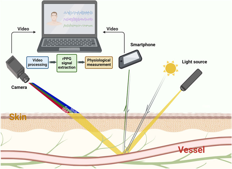
Figure 1. The schematic diagram of rPPG principle. The absorption and reflection of light by the skin varies according to the hemodynamic status under light sources, such as sunlight or lamps. Such changes will be recorded by imaging devices (including industrial cameras, webcams, cell phone lenses and other imaging devices) in the form of videos or pictures. Through the processing of computer and algorithm, rPPG waveforms that represent physiological information can be obtained from these videos.
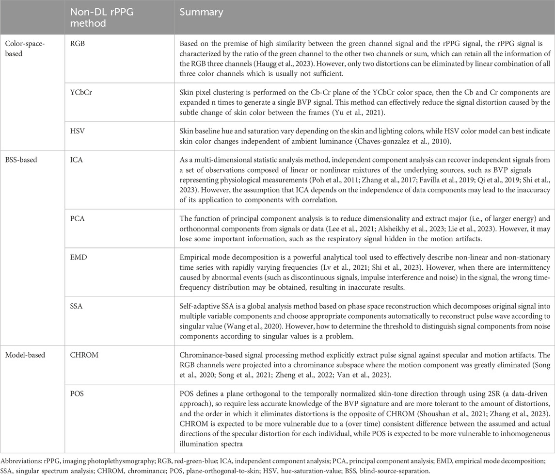
Table 1. Summary of non-DL rPPG signal extraction methods.
1.3 Deep learning
In recent years, the maturity and ongoing progress in the space of DL have injected new vitality into the CV field. DL-based CV techniques have been used in cardiology, pathology, dermatology, ophthalmology, and gastroenterology (Esteva et al., 2021). DL uses simple representations to extract abstract and higher-level features from data and uses artificial neurons as functional units to simulate human cognitive reasoning. The process of learning to perform tasks is called model training, and the ultimate goal of training is to minimize the error between the predicted results of the model and the ground truth. DL often involves three critical types of deep neural network (DNN): recurrent neural network (RNN), generative adversarial network (GAN), and convolutional neural network (CNN). At present, CNN is the most widely used CV. The structure of CNN is composed of three layers: 1) an input layer, 2) a hidden layer, and 3) an output layer. The process of CNN image classification usually includes dataset labeling, model learning, and performance evaluation (Helmy et al., 2023). This model can train a deeper network structure, extract more abstract image features, and reduce the number of neuron parameters to obtain better results with higher efficiency. DL has been successfully applied in contactless physiological and pathological measurements in recent years. Much has been achieved in the CV field, particularly in image registration, image retrieval, and image reconstruction and enhancement. With the support of the ever-increasing availability of datasets, DL will be pivotal in the rapid progress in medical image processing and analysis.
2 Peripheral blood perfusion
Changes in skin and flap color, temperature, or overall appearance (spots, swelling, etc.) often reflect a disease process. However, these changes are conventionally identified during clinical examination, which can be subjective and difficult to quantify. Digital cameras can provide an objective tool for real-time monitoring of skin changes, and this can be enhanced with rPPG signal analysis. Studies have shown that the amplitude of AC components in rPPG waveforms usually fluctuates with changes in central blood pressure or skin perfusion caused by local vasoconstriction (Tusman et al., 2019). The objective measurement of skin and flap blood perfusion can be achieved through the joint analysis of AC and DC components.
2.1 Skin perfusion
rPPG signals are affected by the wavelength of light, measurement site, motion artifacts, ambient light intensity, and ambient temperature (Tamura, 2019). Greenlight PPG signal can accurately reflect the change of skin blood flow caused by ambient temperature changes, while infrared light PPG signal does not reflect the change of skin blood flow under cold stimulation. Thus, skin blood perfusion information can be obtained using green light signals (Maeda et al., 2011). Furthermore, RGB color space is easily affected by luminance. By converting RGB pixel intensity values into the HSV color model, the interference of skin color changes related to ambient brightness can be eliminated (Chaves-gonzalez et al., 2010). Based on these, Harford et al. (2022) explored whether rPPG signals and color measurements could detect skin perfusion changes induced by drugs (phenylephrine and glyceryl trinitrate). They confirmed that skin perfusion changes induced by central (rather than local) administration could be detected from the rPPG waveforms of the skin. Similarly, rPPG signal intensity positively correlates with laser speckle imaging (LSI), used as a reference index for evaluating skin perfusion. This will enable practical evaluation of autonomic nervous system activity and skin perfusion (Rasche et al., 2020). In addition, using the rPPG positioning technique with a lock-in amplification algorithm and volumetric scan of the facial skin using a handheld swept-source optical coherence tomography (SS-OCT), the system can display the 3D structure of human skin microvasculature and obtain high-fidelity video of hemodynamic signals (He and Wang, 2022). The structural design of the exoscope combined with capillaroscopy and rPPG technique can reliably visualize the skin micro-vessels and study their local morphological characteristics. This can be used for the diagnosis and treatment of diseases related to blood microcirculation disorders (Machikhin et al., 2021). At the cellular level, vascular endothelial cells regulate vascular tension by releasing vasoactive substances such as nitric oxide and prostacyclin. As such, the changes of skin microcirculation perfusion caused by local heating detected by rPPG may also be extrapolated and used to evaluate endothelial function (Kamshilin et al., 2022a).
For different application scenarios, the imaging modalities and algorithms are different. Still, the fundamental purpose is to provide more auxiliary information for clinical diagnosis and treatment based on the detection of skin microcirculation. However, there are still some defects in the detection of skin microcirculation perfusion, such as local vascular disorders that will cause direct disturbance to the peripheral blood pulsation and contaminate the quantified measurements of microcirculation. In addition, the microcirculation situation may differ among individuals, and the algorithm’s applicability may need to be further optimized, such as the need for a large number of healthy datasets for correction or even considering additional imaging modalities to provide trans-regional calibration for microvascular measurements.
2.2 Flap perfusion
rPPG technique also performs well in post operative tissue perfusion and wound evaluation (Zaunseder et al., 2018; Mamontov et al., 2020; Kamshilin et al., 2022b; Lai et al., 2022). A systematic review published in 2022 evaluated the performance of near-infrared spectroscopy (NIRS) and hyperspectral imaging (HSI) in testing for flap failure following reconstructive surgery (Lindelauf et al., 2022). While both techniques allow for non-invasive skin flap blood supply monitoring, each modality has limitations. NIRS monitoring of tissue blood oxygen is achieved through a contact sensor (non-aseptic). While continuous monitoring can be achieved, it is unsuitable for all flap types and intraoperative monitoring. On the other hand, HSI is a contactless method that monitors flap perfusion. This can be applied to the intraoperative monitoring of all flap types (e.g., fascio-cutaneous, muscle, intestinal). However, the main limitation is its insufficient real-time monitoring ability. This makes postoperative monitoring tedious and labor-intensive. In recent times, Schraven et al. (2023) achieved continuous analysis of local flap perfusion based on the rPPG technique. This study utilized high-resolution and fully digital surgical microscopy for imaging. It put forward three parameters for evaluating perfusion quality robustly: perfusion index, correlation coefficient of the analyzed rPPG signal with a reference rPPG signal (a reference skin region), and magnitude of the flap. This identified flaps with perfect post operative reperfusion, specific incidents (e.g., vasospasm) during reperfusion, and complete failure. This allowed for early, immediate anastomotic revision to prevent flap failure. This promising result solves the defect of NIRS contacting the flap and overcomes the limitation that HSI can not be continuously monitored. However, this study only explored the practical results of rPPG monitoring during surgical procedures. Postoperative monitoring is also crucial and more complicated in clinical practice. Several parameters are used to distinguish arterial crisis and venous crisis, including flap color, flap temperature, capillary refill time, and swelling degree of the flap. However, these parameters are not absolute, and the experience of microsurgery practitioners is more important. Therefore, establishing a multi-parameter DL model to identify flap crises and achieve early warning is a promising way to solve this clinical problem. Figure 2 shows an ideal pipeline for contactless monitoring of flap blood supply.
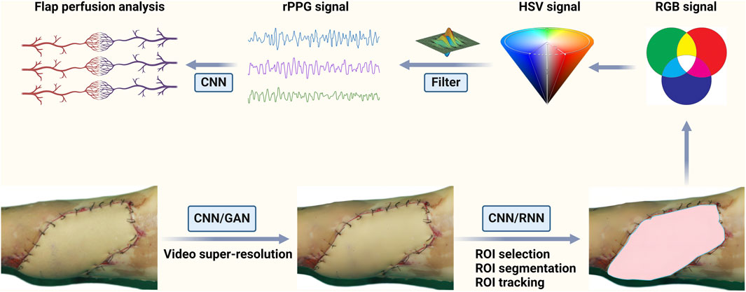
Figure 2. An ideal pipeline for contactless monitoring of flap blood supply. Firstly, the DL-based method is applied to original videos or images to realize super-resolution. Then the DL-based method is used to accomplish the preprocessing steps such as ROI selection, segmentation and tracking. Additionally, RGB signal can be converted into a HSV model with more detailed information. And the raw rPPG signal is obtained through a series of algorithms. Finally, the DL-based method is used to process the raw rPPG signal to obtain accurate physiological information.
A Taiwan study developed a smartphone application called “How’s the Flap” based on Apple’s CoreML framework for early flap crisis warning (Hsu et al., 2023). The datasets of this study contain internal training (230 cases of normal vs. 34 cases of congestion) and external validation (240 cases of normal vs. 16 cases of congestion), including 840 photographs of flaps with varying backgrounds, illumination intensity, flap sizes, and shapes. The accuracy of the model’s training and validation datasets reached 0.922 and 0.923, respectively. Finally, the Application was used to analyze 921 photographs to distinguish flap congestion, and the accuracy was 0.953. Although this study trains a satisfactory model, it is only suitable for venous crises that are easy to detect in clinical practice, and it may be more important to identify critical arterial crises. Therefore, the random forest ML model was proposed to identify arterial and venous insufficiency from images (Huang et al., 2023). The model was trained (80%) and validated (20%) using 805 flap photographs of 176 patients (555 cases of normal, 97 cases of arterial insufficiency, 153 cases of venous congestion), and Shapley Additive Explanations (SHAP) was used to explain the model. The results showed that the temperature and RGB values of flap color could predict the arterial and venous crises, respectively, and the model’s accuracy was 0.984. However, the photographs were segmented to enlarge datasets, which may lead to less generalizability and high homogeneity of the algorithm. In addition, the proposed model’s robustness is worth discussing because the flap photographs were taken in a standardized environment (the same background and illumination intensity).
3 Respiratory rate and oxygen saturation
3.1 Respiratory rate
RR is a vital sign that aids in detecting and evaluating respiratory dysfunction. Conventional electrocardiography (ECG) sensors and respiration belts are reliable methods for monitoring RR. The change of respiratory-induced rPPG waveform is usually related to the effect of respiration on cardiac activity, namely, respiratory-induced variation (RIV). The effect of respiration on the intensity of BVP, amplitude of cardiac output, and HR will enable rPPG waveforms to be used to measure RR (Buda et al., 1979).
3.1.1 Conventional methods for contactless RR estimation
Two main kinds of approaches have been proposed in the literature to achieve contactless RR estimation: 1) methods based on the direct extraction of morphological features attributable to breathing (that is, RIV) (Scully et al., 2012; Nam et al., 2014; Lázaro et al., 2015; Charlton et al., 2018) and 2) methods aimed at isolating the motion trend due to HR and RR (Wei et al., 2017; Schrumpf et al., 2019), implicitly related to RIV. For the first method, incremental merge segmenting (IMS) is the most utilized method. It uses several solutions to fuse the morphological features of respiration (Karlen et al., 2015). The second method is the most promising, single-channel BSS-based method to separate RR from HR and noise. The EMD and SSA are commonly used methods (Huang et al., 1998; Boccignone et al., 2023). Research shows that the morphological estimation of RIV is more reliable than those produced by a single-channel BSS-based method (Boccignone et al., 2023). However, a BSS-based method based on the selected dual region of interest (ROI) developed by Wei et al. (2017) obtained facial BVP and the respiratory signals corresponding to respiratory motion artifacts, thus achieving contactless synchronous measurement of RR and HR. Unlike other studies that rely on sophisticated video tracking and detection algorithms to attenuate motion artifacts, this algorithm takes advantage of motion artifacts and obtains hidden respiratory signals. Extension and improvement of this method may have the potential to detect multiple physiological indicators at the same time.
Unlike visible and near-infrared imaging systems, infrared thermography (IRT) does not require additional lighting and can work in a completely dark environment. For people who need to monitor asleep breathing (e.g., people with a substantial risk of sleep apnea) and critically ill patients who often wear oxygen masks, the rPPG signal provided by IRT may be a way of contactless monitoring of RR (Li et al., 2014a; Chan et al., 2019; Zhu et al., 2019). The skin of infants is fragile and sensitive to light stimulation. It is also challenging to use their small noses as an anatomical marker. In this instance, IRT based on a “black box” algorithm is a viable choice to evaluate RR (Pereira et al., 2019). However, the robustness of these algorithms’ development based on ward and family scenarios may not perform well. In complex public settings, the robust breath-tracking method based on the mobile thermal imaging system proposed by Cho et al. (2017) counteracts the confounding effects of ambient temperature changes and motion artifacts. This would enable accurate RR assessment in highly dynamic thermal scenes.
3.1.2 DL model for contactless RR estimation
Hardware improvements only provide limited gains in non-contact-based measurement accuracy. DL is a way to achieve high-precision rPPG technique in a simple and low-cost approach and is a current research hotspot in the CV field. The DL algorithm with CNN may achieve the purpose of extracting accurate rPPG signals from low-quality videos. BlazeFace and FaceMesh are face detection models based on MobileNetV1/V2 architecture, which can accurately locate the ROI (Bayar et al., 2022; Maity et al., 2022; Jewel et al., 2023; Kolosov et al., 2023). Accurate remote contactless RR estimation can be achieved with Eulerian Video Magnification (EVM) and rPPG techniques (Kolosov et al., 2023). However, the throughput, power consumption, efficiency, and value (throughput/cost) may differ when it runs on different commercial off-the-shelf hardware platforms. In addition, a multi-task temporal shift convolutional attention network (MTTS-CAN) also achieves contactless vital measurements and predicts both rPPG and respiratory signals (Liu et al., 2020). However, it will require complicated preprocessing. The Multi-task Siamese (MTS) model proposed by Lee et al. (2022a) combines the advantages of the Siamese neural network (based on 3D CNN) and multi-task architecture. This reduced the number of parameters by 16 times and accurately predicted heart and respiratory signals in a facial-based video. The MTS model outperformed the single-task model as well as the conventional multitask learning model for RR estimation, was computationally lightweight and may be helpful for applications in smartphones or portable devices. As mentioned, thermal imaging has many advantages and is one of the essential means to achieve contactless RR detection. However, due to the lack of information, selecting and tracking ROI in neonatal thermal images is challenging. One way around this is using the YOLO5Face (based on CSPNet) detection model to recognize the ROI in an RGB image and register it to thermal imaging. This can effectively solve the problem of extracting RR from neonatal thermal photos (Maurya et al., 2022). Whether based on the motion signal and rPPG signal in RGB video or the respiratory signal in thermal imaging video, the DL model can be trained through rich datasets to realize the dynamic estimation of RR. The only thing we need to do is to continuously simplify the algorithms and achieve robust RR estimation in the future.
3.2 Oxygen saturation
SpO2, the relative concentration of oxygenated hemoglobin relative to total hemoglobin, is one of the vital physiological indicators commonly used to monitor a patient’s respiratory function. The traditional finger-type photoelectric sensor is inconvenient for patients requiring long-term continuous essential monitoring. With rPPG techniques, remote pulse oximetry (RPO) can help with contactless vital monitoring. The principle that RPO can assist in SpO2 evaluation is based on the ratio of AC/DC ratios between two wavelengths of interest proposed by Beer-Lambert ‘s law. The limitation of this law is that it only considers the absorbance of chromophores in skin tissue and ignores the existence of light scatter (Kocsis et al., 2006). The robustness of RPO is also related to camera performance, light wavelength, motion artifacts, ambient light intensity, individual differences, posture, and temperature (Wieringa et al., 2005; Humphreys et al., 2007; Kong et al., 2013; Shao et al., 2016; Moço et al., 2019; Moço and Verkruysse, 2020). For visible light, different wavelengths penetrate the skin at different depths. The rPPG signals obtained by using blue and green wavelengths as light sources come from the arterioles of the upper dermal layers, while the signals received by using red wavelengths come from subdermal tissue (Verkruysse et al., 2017). This depth-gap may be more apparent when the skin properties or physiological conditions change (e.g., posture and temperature changes), so the robustness of visible light-based RPO in detecting SpO2 may be reduced (Moço et al., 2019; Moço and Verkruysse, 2020). However, these factors that affect the robustness of RPO are challenging to solve simultaneously, whether the improvement of equipment or algorithm is more aimed at a particular factor.
3.2.1 Multi-spectrum for enhancing RPO robustness
Applying a multi-wavelength light source or multi-spectral camera can effectively reduce the decline of RPO accuracy caused by the changes in ambient light. Wieringa et al. (2005) verified the feasibility of applying a three-wavelength light source to RPO measurement for the first time, but this method has not been well applied due to low SNR. Although the joint use of a dual-wavelength light-emitting diode array and semiconductor camera can estimate SpO2 measurements, its acquisition frame rate is low and is not highly accurate (Humphreys et al., 2007). The CMOS camera with trigger control function alternately records the lip rPPG signals of two specific wavelengths. This has the best SNR under orange and near-infrared wavelengths combined illumination. However, the accuracy of this method is still dependent on many surrounding environmental factors (Shao et al., 2016). While Kong et al. (2013) achieved accurate SpO2 measurement in ambient light using two cameras with narrowband filters to capture rPPG signals at two different wavelengths; the required equipment is complex and will not be readily applicable in the clinical setting.
Dynamic spectrum (DS) has the advantage of suppressing individual differences and measurement conditions. Li et al. (2014b) applied this theory to extract DS from the frequency domain of rPPG signals to calculate SpO2. The multispectral camera plays a significant role in material composition detection based on spectral imaging and can achieve fast and contactless material detection and recognition. Lan et al. (2022) used the multi-spectral camera to obtain the multi-wavelength rPPG signal from facial video, extract the DS values of multiple wavelengths, and obtain SpO2 measurements. This method simultaneously solves the influence of ambient illumination and individual differences on rPPG signals. It can potentially meet the needs of contactless SpO2 detection in a convenient and fast way. To further improve the robustness of RPO when detecting SpO2 under visible light sources, some calibration methods based on skin color, posture, and temperature changes have been proposed (Guazzi et al., 2015; Moço et al., 2019; Moço and Verkruysse, 2020).
3.2.2 Smartphone used for RPO
However, multispectral-based devices (light sources and cameras) are often inconvenient, expensive, and complex. The development of RPO based on smartphones has thus attracted more attention. Smartphones can record and analyze the varying color signals of a fingertip placed in contact with its optical sensor and can effectively evaluate HR, RR, and SpO2 (Scully et al., 2012; Nam et al., 2014; Karlen et al., 2015; Lázaro et al., 2015). Previous studies have successfully used rPPG signals from smartphones to estimate SpO2 (based on the traditional Beer-Lambert law) and introduced a multiple linear regression (MLR) algorithm to calibrate the RPO robustness decline caused by changes in physiological conditions (Sun et al., 2021). For special populations (e.g., children), this method has also been proven to monitor RR and intrathoracic pressure. It can also assist in diagnosing pneumonia and stratification of its severity (Lucy et al., 2021). Although the SpO2 detection based on Smartphones generally reflects peripheral tissue SpO2 and can not simulate the arterial SpO2 provided by the contact pulse oximetry, the mobile device with the built-in color camera as a remote sensor and flashlight as illumination is simple and readily available. With rapid advancements in smartphone technology, more opportunities for medical applications will arise. This will help to improve access to medical technology in undeveloped areas, as well as telehealth care and home health monitoring. Therefore, integrating accurate RR and RPO monitoring techniques into smartphones will accelerate the development of telehealth.
4 Heart rate/pulse rate
Cardiovascular pulse can be estimated and finally used in PR and HR estimation by analyzing the temporal signals of micro-motion or color variations across time. Studies have shown that for consumer cameras (e.g., a webcam or mobile camera), facial video is more reliable for evaluating HR than other body parts (such as wrists and calves) (Wang C. et al., 2018; Van der kooij and Naber, 2019). In the past decade, numerous studies have been conducted on HR detection through rPPG signals provided by facial video. There is currently a wide variety of model designs, parameter settings, algorithms, and equipment. Several methods have been developed for HR estimation using dimensionality reduction (e.g., BSS-based method), optical modeling (e.g., green channel), motion-based methods, and machine learning (ML). These methods are usually applied to face video processing, face BVP signal extraction, and HR computation phases to achieve HR detection (Wang C. et al., 2018).
Face video processing includes face detection and tracking, skin segmentation, and ROI selection. These processes aim to detect the face, improve the motion robustness, reduce the quantization error, and prepare the feature signal for further BVP signal extraction (Huang and Dung, 2016; Gudi et al., 2020; He et al., 2021; Woyczyk et al., 2021). However, some scholars have proposed a method of extracting HR from the whole video by ignoring the ROI selection and tracking process. However, this method is only suitable for instances with a stable video background environment over time (for example, sleep monitoring) (Wang W. et al., 2018). BVP signal extraction includes several postprocessing methods, such as bandpass filtering, detrending, and wavelet transform. This improves the accuracy of HR estimation by cleaning, filtering, or denoise rPPG signal (Huang and Dung, 2016; He et al., 2021). HR computation methods are divided into time domain analysis (peak detection methods) and frequency domain analysis (Malik et al., 1996; Sun et al., 2012). Some studies have tried to put forward unsupervised clustering-based methods to replace traditional peak detection, but they are still not as accurate as the improved BVP signal extraction method (Lee et al., 2019). A system review published in 2018 concluded that a facial skin area extraction, ICA, and peak detection pipeline achieved state-of-the-art accuracy (Wang C. et al., 2018). With the development of CV, these methods are being optimized and, at times, used to complement each other. As subtle facial color changes caused by cardiovascular activity are affected by noise such as ambient light, facial expressions, breathing, camera parameters, out-of-plane movements, and unconscious head shaking, researchers in the field of CV are mainly interested in how to reduce the interference of external factors and how to extract BVP signals quickly and accurately. Figure 3 shows the contactless HR estimation pipeline based on videos (including three phases).
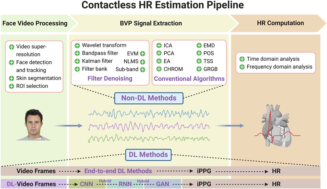
Figure 3. Contactless HR estimation pipeline based on videos. The contactless HR estimation pipeline is composed of face video processing, face BVP signal extraction and HR computation. Face video processing includes video super-resolution, face detection and tracking, skin segmentation, and ROI selection. BVP signal extraction includes the filter denoising methods for motion artifacts filtering and skin color normalization and the conventional algorithms for raw BVP signals construction. HR computation methods are divided into time domain analysis and frequency domain analysis. DL algorithms can be divided into end-to-end type and hybrid type. The former directly establish the mapping from video frames to the target HR values or BVP signals, while the latter use DL model in conjunction with traditional ML methods or different DL models to deal with different stages of HR estimation. The face image in the schematic diagram comes from Chicago Face Database (Ma et al., 2015).
4.1 Denoising for face video signal processing
4.1.1 Motion artifacts filtering
Motion artifacts are the most common interference factor in video recordings. A considerable number of methods have been developed to reduce or eliminate the error caused by motion artifacts, including Sub-band rPPG, continuous wavelet transform, bounded Kalman filter technique, and motion index (MI) indicator (Wang et al., 2017b; Lin et al., 2017; Finžgar and Podržaj, 2018; Prakash and Tucker, 2018; Abdulrahaman, 2023). The extent of eliminating the noise signals from the pulse signal in rPPG depends on the dimensionality of the acquired video signal. The Sub-band rPPG method proposed by Wang et al. (2017b) not only processes the given RGB signal in high dimension but also suppresses the distortion signals of each component, which effectively improves the robustness of multi-wavelength rPPG. Furthermore, the continuous wavelet transform-based Sub-Band rPPG method (SB-CWT) increases the degrees of freedom of distortion elimination by exerting wavelet transform decomposition on RGB video signals (Finžgar and Podržaj, 2018). This method has a good SNR and can estimate PR from RGB video signals without significant motion scenes. In addition, combined with a blur identification and denoising algorithm for each frame and a bounded Kalman filter technique for motion estimation and feature tracking, motion artifacts such as blur and noise caused by head motion can be minimized, but its application in complex and widely moving scenes needs further research (Prakash and Tucker, 2018). Lin et al. (2017) designed a motion index (MI) indicator to filter motion artifacts and used complexion tracking to detect the moving state of the target. At the same time, the near-infrared camera could achieve a better dark mode measurement of PR but ignore the diversity of complexion between individuals. The wavelet transform involves a two-stage denoising method proposed by Abdulrahman (2023), effectively removes motion artifacts, can significantly enhance the reconstructed signal, and can be applied to HR video monitoring of natural motion (not quick or large motions) scenes at different times of the day. Therefore, for different motion scenes, the demand for the algorithm to filter motion artifacts may be different. Additionally, the potential effects of varying skin colors caused by complexion or light source must be considered.
4.1.2 Skin color normalization or enhancement
In addition to motion artifacts, skin color is a crucial factor affecting the robustness of the rPPG signal. The skin color is affected by the change in light source and complexion, which brings much noise to the acquisition of the rPPG signal. The anti-interference performance of the normalized least mean square (NLMS) adaptive filter can rectify the illuminance variation. Still, it needs the desired signal established by a smooth rectifier in the background as the input, which is difficult to realize (Li et al., 2014c). A Distance-PPG method based on filter banks can weigh the average skin color changes in different tracking regions of the face and has an excellent anti-noise performance. Still, the algorithm implemented by this method is complex and time-consuming, and the pulse wave extracted by this method can not see apparent dicrotic waves (Kumar et al., 2015). Based on these limitations, Wang et al. (2020) first removed baseline offset and high-frequency random noise. Then, they used a self-adaptive SSA algorithm to extract details-preserving pulse waves from facial video in real situations.
Color enhancement can magnify subtle skin color changes. Unlike the traditional video based on RGB color space, the video based on YCbCr color space can obtain more subtle skin color changes, thus realizing the accurate extraction of BVP signals (Yu et al., 2021). Microsoft Kinect (a multi-mode camera) can provide additional information for RGB data, namely, depth, infrared, and skeleton frames, and processes the RGB images through the EVM color augmentation method to magnify the skin color changes caused by blood flow, so it is developed as a contactless HR estimation technique (Gambi et al., 2017). By integrating denoising techniques such as amplitude selective filter (MASF), wavelet decomposition, and robust PCA on RealSense (an RGB-NIR dual-modality camera), depth information can be obtained from short videos and HR information can be obtained accurately (Lie et al., 2023). Furthermore, Martinez-Delgado et al. (2022) combined a face detection algorithm based on OpenCV with the EVM algorithm to achieve a more accurate HR estimation. In addition, the EVM video amplification technique is usually used in combination with the DL model or PCA algorithm in HR estimation (Kolosov et al., 2023; Lin et al., 2023).
These video signal enhancement methods for filtering motion artifacts and dealing with skin color changes are the prerequisites for accurate rPPG signal extraction. However, rPPG signals often need further processing to obtain the components of BVP signals for accurate HR measurement. This step usually involves many more advanced algorithms, such as ICA and CHROM.
4.2 Conventional algorithms for contactless PR/HR estimation
4.2.1 Single ICA
As a commonly used method for BVP signal extraction, ICA begins with a random initialization of unmixing matrix with just a single prerequisite of unmixing matrix dimension, depending on the number of independent components, which is comparatively trivial than the wavelet transform method. ICA algorithm regards BVP extraction as a BSS problem, that is, extracting the desired signal with no or limited information from the mixed signal. Algorithms such as joint diagonalization approximation of matrices (JADE) and FastICA, which show motion tolerance to some extent, are based on the transformation or improvement of ICA (Poh et al., 2011; Shi et al., 2023). In addition, the multi-channel ICA algorithm is based on second-order blind identification (SOBI), which was proposed by Zhang et al. (2017) realizes the possibility of evaluating HR under low illumination. Similarly, integrating multiple simultaneously acquired BVP signals extracted by the ICA algorithm can also measure HR reliably (Favilla et al., 2019). The “Project_ICA” algorithm uses the skin reflection model to extract the BVP signal from the facial rPPG signal (Qi et al., 2019). This method combines advanced techniques such as feature point detection tracking and skin pixel detection, overcomes the decrease in robustness caused by motion artifacts and weak light and dark skin, and performs better than several classical ICA, CHROM, 2SR, and POS algorithms. However, it still has significant limitations in the application of black skin. Different algorithms have different advantages; for example, the ICA algorithm can recover independent signals from mixed signals, the CHROM algorithm explicitly extracts pulse signals against specular and motion artifacts, and the EMD is a powerful analytical tool used to effectively describe non-linear and non-stationary time series with rapidly varying frequencies. The high complexity of algorithms usually requires a longer running time, and how to combine the advantages of different algorithms to achieve fast and accurate HR estimation is a topic that scholars are committed to discussing.
4.2.2 Hybrid ICA
As one of the most commonly used and practical conventional algorithms, the ICA algorithm is often used with other algorithms to predict HR. Song et al. (2020) combined the advantages of ICA in independence and CHROM (a model-based method) in dealing with chromaticity, proposed a Semi-BSS-based rPPG method to realize the best performance of HR estimation. Still, this method requires super-high resolution (2.7 k) video. Combined with the remote ballistocardiography (rBCG) technique, rPPG signals can realize the combination of color and motion of BSS-based (EA, PCA, and ICA), thus effectively reducing the impact of illumination changes and motion artifacts on HR evaluation (Lee et al., 2021). In 2021, Lv et al. (2021) proposed an improved ensemble EMD (EEMD) algorithm, namely, complete EEMD with adaptive noise (CEEMDAN), and combined it with FastICA to realize remote HR measurement. However, there is still residual white noise in CEEMDAN, which leads to decomposition errors. To ensure the elimination of noise, the number of iterations of the algorithm will increase, which will lead to an increase in time cost. To solve the problem of decomposition errors and slow running speed caused by this residual noise, Shi et al. (2023) improved both EEMD and FastICA algorithms. By adding zero-mean random white noise generated according to the input signal to the sampled data, the Huber derivative approximation function is used instead of the nonlinear function in the FastICA algorithm to improve further accuracy, robustness, timeliness, and anti-interference performance. In addition, an under-complete ICA algorithm was proposed to restrict motion and illumination variation artifacts (Gupta et al., 2022). By using a non-linear cumulative density function (CDF) optimized by customized Levenberg-Marquardt algorithm (LMA) to estimate the unmixing matrix, this method can retain all the information of RGB three channels and has an excellent performance in constrained motion and illumination variations scenarios.
4.2.3 Other algorithms
Color subspace transformation methods such as CHROM and POS use orthonormal vector transformations to construct raw signals for BVP extraction (Wang et al., 2017a). Compared with the conventional ICA algorithm, it does not lose the critical information in the red and blue channels. Still, its main disadvantage is that improper weights assigned to color channels may reduce the BVP information (Gupta et al., 2022). POS algorithm can not only extract high-precision PR from videos captured by high-speed cameras but also process BVP signals in multiple respiratory modes (spontaneous, metronome, and forced) and video (smartphone and webcam) under different types of body movements, but it is challenging to achieve synchronization or desynchronization between HR and RR cycles (Shoushan et al., 2021; Zhang et al., 2023). A self-adaptive SSA algorithm can obtain cyclical components, remove aperiodic irregular noise, and extract the pulse wave that keeps the details from the facial video in real situations (Wang et al., 2020). The T-SNE-based signal separation (TSS) method can decompose the observed color traces into pulse-related vectors and noise vectors and then select the vector with the most significant spectral peak as the BVP signal for HR measurement (Wang et al., 2022). This proposed method is suitable for RGB and HSV color spaces and significantly suppresses the noise caused by head movement. Still, it is not robust to complex light interference and violent sports interference scenes. However, without relying on complex mathematical models or ML algorithms, combining RGB channels alone may also be a way to obtain robust BVP signals. Research shows that the sum of the green-to-red channel and green-to-blue channel ratios (GRGB) not only has lower computational complexity but also has the same effect as the POS algorithm, especially suitable for videos with a lot of movements and indoor lighting (e.g., gym and rotation) (Haugg et al., 2023). Table 2 summarizes these conventional rPPG signal extraction algorithms for HR estimation. Although there are many mature methods of using CV techniques based on traditional algorithms to extract rPPG signals used to estimate HR, the decline in the robustness of HR evaluation caused by subject motion and ambient lighting variations can still be optimized. Due to the success of DL in many CV and medical image processing applications, DL methods have been considered for rPPG to deal with its challenges.
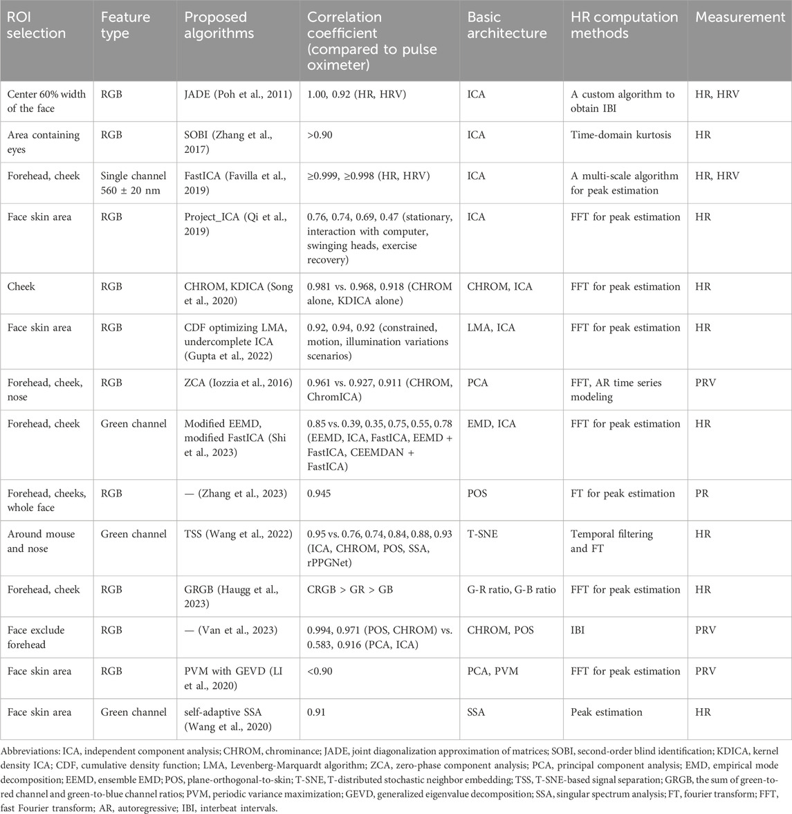
Table 2. Several conventional iPPG signal extraction algorithms for HR measurement.
4.3 DL for contactless PR/HR estimation
Before the advent of DL, several ML methods were used to remotely estimate HR, including linear regression, k-nearest neighbor (kNN) classifier, support-vector regression, adaptive hidden Markov models, and a general-to-specific transfer learning strategy named SynRhythm (Hsu et al., 2014; Monkaresi et al., 2014; Fan et al., 2015; Niu et al., 2018). As with many CV and signal processing applications, DL methods have shown promise in mapping complex physiological processes for contactless HR measurement. The number of research papers utilizing DL methods for remote HR measurement has increased yearly and is expected to grow continuously. The rPPG approaches for HR estimation based on DL can be generally divided into two types: 1) the end-to-end type and 2) the hybrid type. The former provides spatial-temporal (ST) visualization of physiological signals via the attention mechanism and directly establishes the mapping from video frames to the target HR values or BVP signals. At the same time, the latter uses the DL model in conjunction with traditional ML methods or DL models to deal with different stages of HR estimation (Figure 3).
4.3.1 End-to-end DL model
A method is classified as end-to-end if it takes in a series of video frames as input and directly outputs the rPPG signal or HR without any intermediate steps (Figure 3). End-to-end DL methods are indisputably great tools due to their straightforward model optimization process.
4.3.1.1 Single-stage CNN model
The Single-stage CNN model utilizes only one CNN architecture to extract HR or rPPG signals directly from facial video, even if there is no need for the preprocessing stage of face detection and tracking (Bousefsaf et al., 2019). The robustness of HR measurement under different skin types, facial expressions, and movements can be improved by integrating different attention mechanisms in CNN structure, such as motion, appearance, and ST attention model (Hu et al., 2022; Mcduff et al., 2022; Ouzar et al., 2023). An end-to-end ST network, X-iPPGNet, based on modified Xception integrated with a depthwise separable convolution, can realize instantaneous PR estimation directly from facial video recordings (Ouzar et al., 2023). Unlike most existing systems, X-iPPGNet has advantages with high and sharply fluctuating PR, ensuring robust PR prediction under various conditions (including head motions, facial expressions, and skin tone). This is because it learns the rPPG concept from scratch without incorporating prior knowledge or going through the extraction of BVP signals.
4.3.1.2 Multi-stage CNN model
The Multi-stage CNN model utilizes two or more linear CNN architectures to achieve more than one phase of HR estimation. A two-stage CNN named HR-CNN composed of the Extractor and HR estimator is trained end-to-end through alternating optimization and is robust to illumination changes and subject motion (Spetlik et al., 2018). Unlike the commonly used COHFACE and MAHNOB databases, the datasets used for training in this study are a new open-source ECG-Fitness database whose videos are not compressed. Similarly, another two-stage 3D CNN method comprised of ST video enhancement network (STVEN) and rPPGNet (composed of an ST convolutional network, a skin-based attention module, and a partition constraint module) generalizes well on novel data with only compressed videos available, which implies the promising potential for real-world applications (Yu et al., 2019a). In addition, the end-to-end model proposed by Perepelkina et al. (2020) uses CNN architectures in the three stages of the HR estimation pipeline. After using RetinaNet (based on MobileNet) to process facial ROI, HeartTrack (based on a 3D ST attention CNN) obtained the time series. Finally, 1D CNN was used to calculate HR. Furthermore, an utterly self-supervised training method based on pre-trained ResNet18 and 3D PhysNet CNN was designed to get rid of expensive ground truth physiological training data (Gideon and Stent, 2021).
4.3.1.3 Multi-scale network
We define a multi-scale network as a phase of HR estimation that uses more than one DL architecture; that is, the three-phase linear structure of the HR estimation pipeline is extended by multi-scale DL architecture. The Siamese-rPPG network proposed by Tsou et al. (2020) contains two 3D CNN architectures, which can not only extract the rPPG signal from the two face ROIs (without preprocessing) simultaneously but also effectively retain the ST characteristics of the rPPG signal. Furthermore, multi-task Siamese (MTS) combines the advantages of Siamese neural network and multi-task architecture to accurately predict cardiac signals while significantly reducing parameters (Lee et al., 2022a). Li et al. (2022) proposed a short-time end-to-end HR estimation framework based on facial features and temporal relationships of video frames. In the proposed method, a deep 3D multi-scale network with cross-layer residual structure is designed to construct an autoencoder and extract robust rPPG features by transferring the lost information in scale transformation. Then, an ST fusion mechanism is proposed to help the network focus on features related to rPPG signals. Yin et al. (2022) proposed an end-to-end multi-task learning model named PulseNet, combining the advantages of signal-based methods and DL methods, which can achieve accurate HR estimation in scenes that include changes in lighting and head movement. PulseNet uses (2 + 1)D convolution to decouple ST information and a skin-based attention mechanism to suppress background noise.
The central difference convolution (CDC) operator has potential advantages for rPPG feature extraction due to its ability to enrich temporal context. The 3D CDC network can achieve accurate HR measurement by combining the attention mechanism of ST, motion, and appearance, for example, the proposed CDCA-rPPGNet and AutoHR (Yu et al., 2020; Zhao et al., 2021; Liu et al., 2022). AutoHR proposed by Yu et al. (2020) is composed of neural architecture search (NAS) and the 3D temporal difference convolution (TDC). By combining a hybrid loss function considering constraints from both time and frequency domains and ST data augmentation strategies, AutoHR realizes accurate HR measurement. More complicatedly and accurately, a 3D ST convolutional network with multi-hierarchical fusion, including low-level face feature generation (LFFG), 3D ST stack convolution (STSC), multi-hierarchical feature fusion (MHFF), and signal predictor (SP), can reconstruct the rPPG signal representing HR from facial RGB video (Li et al., 2023).
4.3.1.4 Transformer
Transformer, a recently developed DL model, differs from the convolution structure of CNN based on local connection and weight sharing. It is based on self-attention mechanisms (Vaswani et al., 2017). Although the structure of the Transformer model is complex and requires many parameters, it can handle data noise and deformation better than the CNN structure. Yu et al. (2022) first proposed an end-to-end video transformer architecture, PhysFormer, for remote physiological measurement. On the one hand, the cascaded temporal difference Transformer blocks in PhysFormer benefit the rPPG feature enhancement via global ST attention based on the fine-grained temporal skin color differences. On the other hand, to alleviate the interference-induced overfitting issue and complement the weak temporal supervision signals, elaborate supervision in the frequency domain is designed, which helps PhysFormer learn more intrinsic rPPG-aware features. To better exploit the temporal contextual and periodic rPPG clues, the PhysFormer was extended to the two-pathway SlowFast-based PhysFormer++ with temporal difference periodic and cross-attention Transformers (Yu et al., 2023). However, the application of the Transformer to the physiological measurement of rPPG is still in its infancy, and future research should focus on designing a more efficient architecture while exploring a more accurate and efficient ST self-attention mechanism, particularly for long-sequence rPPG monitoring. Table 3 summarizes the application of end-to-end DL methods in contactless HR estimation.
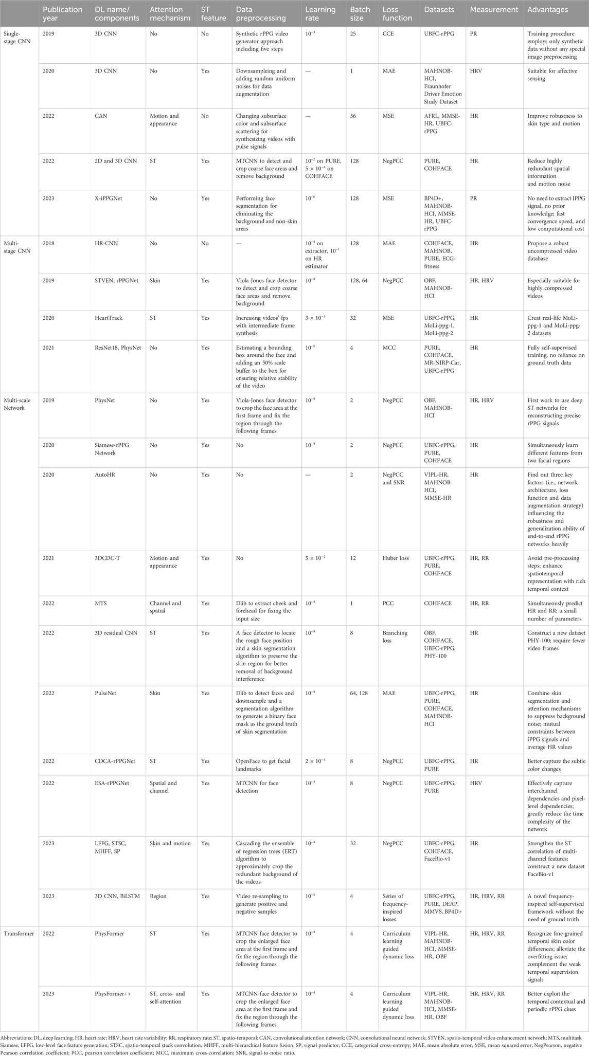
Table 3. Application of end-to-end DL methods in contactless HR estimation.
Although the end-to-end DL model shows great potential in HR estimation, it often results in a mysterious black box model that is difficult to understand. Therefore, optimizing the algorithm based on various factors that affect the robustness of rPPG is necessary. In addition, multiple DL models applied at different stages of HR measurement may increase the interpretability of the process.
4.3.2 Hybrid DL model
HR estimation is classified into three phases: face video processing, face BVP signal extraction, and HR computation. Using DL model(s) in one phase or different DL models in various phases is defined as hybrid DL, while the other phases still use the non-DL algorithms (Figure 3).
4.3.2.1 DL for face video processing
BlazeFace is a face detection model based on MobileNetV1/V2 architecture developed by Google, while FaceMesh integrates a face landmark model based on BlazeFace. These two models can eliminate any facial redundant areas that have no impact on HR or RR estimation to accurately locate an ROI (Bayar et al., 2022; Maity et al., 2022; Pagano et al., 2022; Jewel et al., 2023; Kolosov et al., 2023; Odinaev et al., 2023). The proposed cascade residual CNN-FPNR technique used for preprocessing and SNR enhancement facilitates segmentation in low-light ambient videos and provides high frame quality for HR estimation (Gupta et al., 2023). The AND-rPPG method based on a 2D temporal convolution network (TCN) architecture enables denoise temporal signals and action units from facial videos (Lokendra and Puneet, 2022). Then, the denoised temporal signals from all the facial regions are consolidated to compute the rPPG signal and estimate the HR. As a component of a two-stage DL model, rPPGRNet based on recurrent back projection network (RBPN) can form super-resolution images and then be used for HR estimation of subsequent THRNet (based on 3D ResNet-10) (Yue et al., 2021). The proposed DeepMag based on CNN architecture enables automated magnification of subtle color and motion signals from a specific source, even in the presence of large motions of various velocities (Chen and Mcduff, 2020). The magnified videos produced by DeepMag have fewer artifacts and blurring than the traditional EVM method.
4.3.2.2 CNN for face BVP signal extraction/feature decoder
A depth-wise separable convolution based on 3D MobileNet enables an estimate of HR from the feature images formed by spatial decomposition and temporal filtering of EVM (Qiu et al., 2019). Similarly, the proposed cross-verified feature disentangling strategy (CVD, based on CNN) enables disentangling the physiological features with non-physiological representations existing in a multi-scale ST map, which realizes robust multi-task physiological measurements (Niu et al., 2020a). In addition, a DL model based on ResNet-18 architecture is used to judge the quality of the ST feature image extracted by the conventional CHROM algorithm and to determine whether it is used in the fast Fourier transform (FFT) of subsequent HR estimation (Zheng et al., 2022). Similarly, for the ST images or time-frequency representation extracted by traditional algorithms, the CNN model can achieve robust HR estimation in continuous motion scenes (Hsu et al., 2017; Jaiswal and Meenpal, 2022; Chen and Li, 2023). Chen and Li (2023) applied CNN model based on ResNet101 architecture to HR reality monitoring in aerobics training with high accuracy. Jaiswal and Meenpal (2022) proposed a video-based noise-less cardiopulmonary measurement, which converts the 3D videos into 2D ST Images by wavelet decomposition, suppressing the noise while preserving temporal information of the rPPG signal. ST images are provided as input to CNN, which enables mapping the corresponding HR values under heterogeneous lighting conditions and continuous motion. Similarly, short-time Fourier transform (STFT) can transform the 1D color signal and frequency signal extracted from RGB videos to 2D time-frequency representation, subsequently used to train a VGG15 DL network to estimate the pulse (Hsu et al., 2017).
Temporal and spatial features are the key to accurately extracting rPPG signals from facial video. In addition to processing the ST signals obtained by traditional methods, CNN itself can also integrate the ST modular to improve the anti-noise ability, which is often realized by added attention mechanism or convolution modular, for example, the proposed DeeprPPG and ETA-rPPGNet networks (Niu et al., 2019; Liu and Yuen, 2020; Hu et al., 2021). Niu et al. (2019) input the ST map extracted from the video into the ResNet-18 CNN architecture integrated with channel attention and ST attention mechanism, thus outputting robust HR estimation. DeeprPPG, as a lightweight rPPG estimation network without preprocessing, is based on ST ConvNets (full 3D convolution/spatial 2D convolution + temporal 1D convolution), allows flexible ROI selection with different locations and sizes, and obtains the robust rPPG signal from multiple input skin regions (Liu and Yuen, 2020). The ETA-rPPGNet proposed by Hu et al. (2021) is comprised of a time-domain segment subnet and backbone net. The feature maps of the video generated by the time-domain segment subnet can effectively reduce redundant information. At the same time, the integrated time-domain attention mechanism in the backbone net can significantly improve the model’s anti-noise (insufficient light conditions and head movement) ability. ETA-rPPGNet shows superior performance in compressed datasets (compared with DeepPhys). Still, its short-term estimation performance is not as good as that of EVM-CNN because it needs to deal with redundant information. Furthermore, a novel global-local interaction and supervision network (GLISNet) utilizes the local path to learn the representations in the original scale and the global path to learn the representations in the other scale, thus capturing multi-scale information (Zhao et al., 2023). GLISNet can extract and fuse pulse signals from multi-scale ROIs without heavy computational load and preserve the rich temporal features of rPPG video to achieve accurate HR estimation.
4.3.2.3 RNN (+CNN) for face BVP signal extraction/feature decoder
Long short-term memory (LSTM), a typical RNN architecture, enables filter rPPG signal obtained by conventional methods (POS, PCA, CHROM, CWT, etc.), can more accurately identify the changes of HR and further evaluate the mental state or physical function of the population (Slapnicar et al., 2019). A two-layer LSTM was designed for regression from raw signals after normalization to estimate pulse wave signals and generate a large scale of synthetic HR signals which is used to pre-train the LSTM network to prevent over-fitting (Bian et al., 2019). This algorithm can effectively alleviate the problem of insufficient HR public database and achieve better performance than the baseline method (GREEN, ICA, CHROM, and POS). Maity et al. (2022) proposed a bi-directional LSTM (Bi-LSTM) network to filter the motion distortions in the rPPG signals, which shows better-filtering capability over the discriminative signature-based filtering during HR estimation.
Combined with CNN architecture, LSTM may realize more advanced performance. The proposed HR evaluation method named Meta-rPPG was comprised of ResNet (2D CNN) for feature extraction and an LSTM network for rPPG estimation, whose performance of HR estimation was better than that of EVM in different datasets (Lee et al., 2020; Pagano et al., 2022). As the most common CNN architectures, U-Net or ResNet combined with LSTM outperform the widely used prior-knowledge rPPG methodology in PR estimation, for example, a combination of POS and CWT (Niu et al., 2020b; Lampier et al., 2022). Furthermore, the combination of AlexNet, ResNet50V2, and LSTM can extract HR information from the rPPG signal obtained by the PCA algorithm (Alsheikhy et al., 2023).
4.3.2.4 GAN for face BVP signal extraction/feature decoder
GAN-based pulse feature disentanglement network (PFDNet) can extract the common robust features of rPPG and PPG pulse signals, and further recognize atrial fibrillation from facial videos with typical facial motions (Liu et al., 2023). The cbPPGGAN framework based on CycleGAN was used to enhance raw pulse signals extracted using traditional approaches while estimating more accurate HR under illumination variation (Yang et al., 2023). Furthermore, the proposed Dual-GAN model uses two GAN models to learn the mapping from the ST map to BVP and simulate noise distribution, respectively (Lu et al., 2021). The Dual-GAN structure allowed for indirect supervision for noise distribution and achieved better feature disentanglement for the BVP signal. This resulted in better prediction performance for HR, HRV, and RR. Table 4 summarizes the application of hybrid DL methods in contactless HR estimation.
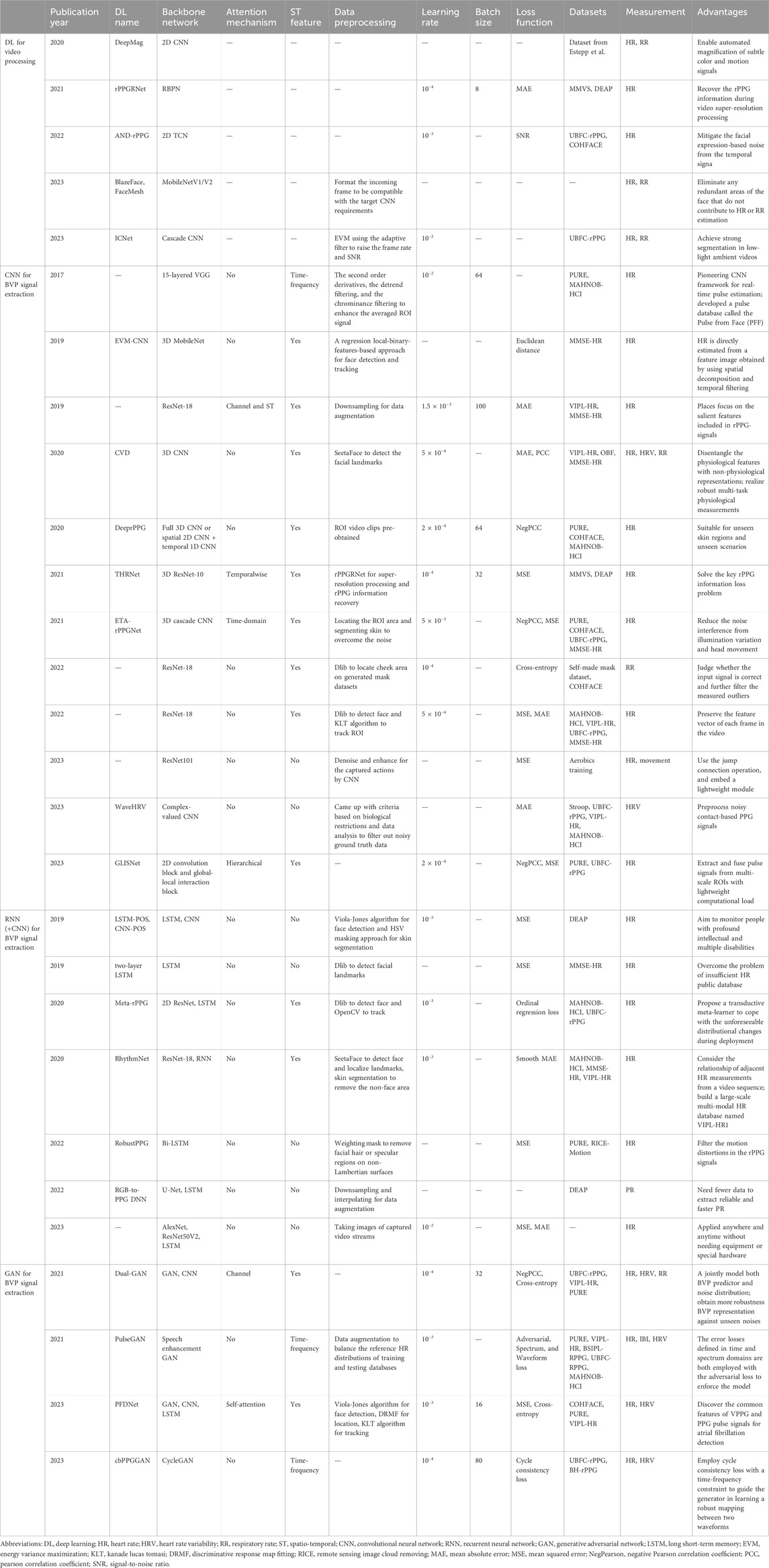
Table 4. Application of hybrid DL methods in contactless HR estimation.
The interest in contactless or remote HR measurement has steadily grown in healthcare and sports applications. Contactless methods involve the utilization of a video camera and image processing algorithms. Due to rapid development in ML, DL methods have shown significant promise in improving the performance of conventional algorithms for contactless HR estimation. As large labeled open-source datasets are used to train these algorithms, high-quality and diverse datasets are crucial for proper benchmarking and analysis of different methods and the future development of more complex DL models and architectures. In the longer term, the continuous update and iteration of smartphones and the popularity of robots in public places will provide a stronger foundation for HR contactless monitoring (Siddiqui et al., 2016; Poh and Poh, 2017; Lee et al., 2022b).
5 Heart/pulse rate variability
Heart rate variability (HRV) refers to the change of interval time between continuous heartbeats, while pulse rate variability (PRV) refers to the change of pulse interval time in relation to the BVP signal, indicating the change of instantaneous PR/HR. Both HRV and PRV reflect the ability of the autonomic nervous system to maintain the balance of the internal environment. The difference between the two methods is that HRV is usually calculated by ECG, while PRV is obtained by PPG signal. The analysis of HRV and PRV is a useful tool for a comprehensive description of autonomic dynamics and can provide useful information about changes in vagus nerve activity (which can be used to monitor stress and mood changes) (Rajendra acharya et al., 2006).
5.1 Relevance between HRV and PRV
The HRV standard defines the HRV evaluation of long-term (LT; 24 h) and short-term (ST; 5 min) through time-domain, frequency-domain, and non-linear metrics (Malik et al., 1996). In recent years, to achieve the lowest possible power consumption and computing load, the HRV evaluation index of ultra-short term (UST; less than 5 min) has been proposed. By combining UST with wearable technology or smartphone applications, one can assess a person’s wellbeing (mood, stress, health) while being user-friendly (speed and comfort) (Nussinovitch et al., 2011; Munoz et al., 2015; Castaldo et al., 2019; Finžgar and Podržaj, 2020). Studies have shown that PRV can be used as an effective and accurate index for estimating HRV in healthy subjects at rest as this helps simplify the recording of the signals used in HRV assessment. However, under physical or mental stress, motion artifacts would lead to a decrease in the level of consistency between HRV and PRV, amongst which UST-HRV and ST-HRV may be more affected (Schäfer and Vagedes, 2013; Iozzia et al., 2016). It has been shown that it is possible to use rPPG signals to generate HRV information in subjects with autonomic nerve excitation. Moreover, the rPPG signal extracted by POS and CHROM methods is the most accurate in predicting autonomic dynamics (Van et al., 2023). In addition, the multiple simultaneously acquired BVP signals extracted by the ICA algorithm seem to be able to evaluate HRV reliably (Favilla et al., 2019). A PhysioCam system developed by Davila et al. (2017) extends the application scenario of PRV characterization of HRV based on the rPPG signal. Its performance is similar to that of standard signals (ECG and PPG) in three physiological conditions (rest, single deep breath, and continuous fast and shallow breathing). However, the balance of achieving user-friendly and accurate PRV assessment (consequently HRV) in patients with multiple comorbidities is still a difficult one to strike at this point.
5.2 Conventional methods for contactless HRV/PRV estimation
Unlike the evaluation of HR or PR, the measurement of HRV or PRV requires accurate peak detection of BVP signals and continuous extraction of PR and BVP signals. This usually has a higher noise level and lower temporal resolution than cPPG thus rendering contactless remote measurement of PRV to be more complex. To achieve accurate measurement of PRV, some CV researchers have tried to improve the performance of the camera. This often makes the process more complex, expensive, and not applicable in daily life (Sun et al., 2012; Mcduff et al., 2014; Mcduff et al., 2018). It seems to be a potential method to improve the algorithms, such as improving BVP peak recognition, improving time-domain resolution, magnifying the subtle changes of respiration and skin color, and combining face detection and tracking (Sun et al., 2012; Melchor Rodríguez and Ramos-Castro, 2018; Li et al., 2020; Pai et al., 2021; Yu et al., 2021).
Using the periodic variance maximization (PVM) method to extract the BVP signal on rPPG, and using the event-related two-window algorithm to improve BVP peak recognition, contactless and accurate PRV detection based on rPPG can be realized (Li et al., 2020). Interpolating can compensate for the negative effects of a low initial sample rate and improve time-domain resolution and PRV measurements, thus providing further strong support for the low-cost webcam-based rPPG technique (Sun et al., 2012). A method based on YCbCr chromatic aberration developed by Yu et al. (2021) magnifies the subtle changes of skin color to make it easier to identify, and realizes the continuous extraction of BVP signals, which breaks away from the limitation that conventional rPPG techniques only measure a single PR instead of the whole signal. Furthermore, Melchor Rodríguez and Ramos-Castro (2018) utilized the Viola-Jones face detection algorithm and Kanade-Lucas-Tomasi (KLT) tracking algorithm to process the video obtained by webcam, and achieve robust rPPG PRV analysis under small-range motion conditions. Still, this method does not take into account more extensive and complex motion types. Pai et al. (2021) have developed an HRVCam algorithm based on a frequency demodulation framework (a combination of a new automated adaptive bandpass filter and the discrete energy separation algorithm (DESA)) for subjects with large changes in respiration and skin color, which was used to estimate the instantaneous frequency of the rPPG signal, thus improving the accuracy of estimated time-domain HRV metrics. These improved algorithms have achieved good results on the datasets based on traditional low-cost cameras and may be suitable for the promotion of rPPG monitoring physiological signs. Table 2 summarizes these conventional rPPG signal extraction algorithms in contactless HRV/PRV estimation.
5.3 DL model for contactless PRV estimation
5.3.1 End-to-end DL model
The measurement of HRV/PRV is based on the accurate detection of HR/PR, and the DL model involved can be roughly divided into end-to-end and hybrid DL. PhysNet, an end-to-end ST network constructed by 3D CNN or 2D CNN + RNN, can accurately evaluate the measurement metrics characterizing HRV however is highly complex and time-consuming (Yu et al., 2019b). In addition, a 3D CNN architecture without skin segmentation or other preprocessing was developed to realize HRV measurement (Luguev et al., 2020). More recently, an efficient ST attention network (ESA-rPPGNet) was developed, which is composed of ESA (based on MobileNet v3), 3D shuffle attention, and gated recurrent unit (GRU) (Kuang et al., 2022). ESA-rPPGNet can recover high-quality rPPG signals to accurately locate the peak of each heartbeat, thus improving the accuracy of HRV analysis and reducing the time complexity of the network. However, these methods are trained in a supervised manner, where PPG signals are recorded synchronously with facial videos for supervision. A novel frequency-inspired self-supervised framework for facial video-based remote physiological measurement was proposed, which learns to optimize rPPG estimation from multiple augmented videos of different signal frequencies and across temporally neighboring videos of similar signal frequencies, while there is no demand for PPG signal originating from ground truth (Yue et al., 2023). It has three main stages: data augmentation (involving a 3D Convolution layer, 3D Res-blocks, and Bi-LSTM), signal extraction (based on 3D ResNet-10), and network optimization. Its performance was better than most advanced self-supervised methods and equivalent to the most advanced supervised methods in HR, HRV, and RR estimation. Figure 4 shows the difference between supervised and self-supervised learning in rPPG signal prediction. Table 3 summarizes the application of end-to-end DL methods in contactless HRV estimation.
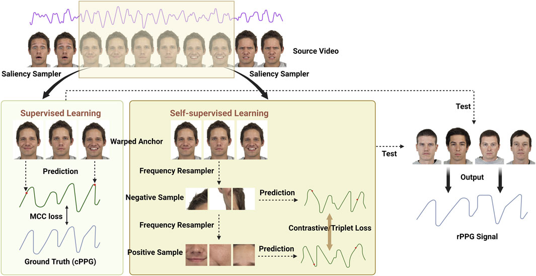
Figure 4. Supervised and self-supervised learning in rPPG signal prediction. A video clip is sampled from the source video first, then passed through the saliency sampler to generate the warped anchor. The anchor is passed through a PPG Estimator to get rPPG signal. If supervised training is employed, we employ a maximum cross-correlation (MCC) loss between the ground truth (cPPG) and predicted rPPG signal. If contrastive training is used, a random frequency ratio is sampled from a prior distribution. The warped clip is passed through the frequency resample to produce the negative sample, showing a subject with an artificially higher heart rate. This sample is passed through to produce the negative example PPG. The negative sample is again resampled with the inverse of random frequency ratio to produce a positive example PPG. Finally, the contrastive loss, multi-view triplet loss, is applied to the PPG samples, using a PSE MSE distance metric. The face images in the schematic diagram come from the Chicago Face Database (Ma et al., 2015).
5.3.2 Hybrid DL model
Wavelet scattering transform, a complex-valued CNN model, can denoise an extracted rPPG signal (Odinaev et al., 2023). Combined with adaptive bandpass filtering and inter-beat-interval (IBI) analysis, the contactless detection of HRV can be achieved. This transformation has been verified on different public datasets with satisfactory results. The proposed PulseGAN framework employs a combination of waveform, spectrum, and adversarial losses to enable extraction of high-quality rPPG pulse waveforms from rough input signals obtained by conventional methods (e.g., CHROM) to infer reliable cardiac features (e.g., HRV) (Song et al., 2021). In addition, the cbPPGGAN predicts a more realistic pulse waveform and a more accurate HRV estimation (Yang et al., 2023).
Cardiovascular disease is one of the most common diseases, and HRV may be a valuable indicator for predicting sudden cardiac death and arrhythmias. With increasing societal pressures, the youth will increasingly experience mental health and emotional stressors. As a physiological index reflecting stress and emotional changes, HRV monitoring helps evaluate the mental health of adolescents and prompts early intervention from psychiatrists. Real-time monitoring of HRV in various scenarios helps detect the occurrence of cardiovascular diseases and mental diseases, thus providing an early detection mechanism for a variety of global health problems.
6 Blood pressure
In the field of remote healthcare, non-invasive continuous BP measurement has become a growing topic. Classic non-invasive BP measurement techniques can obtain spontaneous systolic blood pressure (SBP) and diastolic blood pressure (DBP) at a point in time while invasive BP measurement techniques provide continuous BP monitoring. These techniques are however not suitable for long-term monitoring due to discomfort and are generally used in intensive care units. With the development of telemedicine, the demand for non-invasive continuous BP monitoring will continue to increase.
6.1 rPPG for contactless BP estimation
The research shows that the pulse transit time (PTT) determined by BP can be expressed not only by the time lag between the R wave of ECG and a subsequent pulse wave but also by the time lag between two PPG’s measured at different body locations. The principle of measuring BP by rPPG technology is based on its recognition of PTT (Geddes et al., 1981; Nitzan et al., 2002; Mukkamala et al., 2015; Sugita et al., 2015; Jeong and Finkelstein, 2016; Secerbegovic et al., 2016; Zhou et al., 2019; Fan et al., 2020). In addition to PTT-based methods, cuffless BP measurements are implemented by pulse arrival time (PAT, which requires an ECG sensor and a PPG sensor), pulse wave velocity (PWV, which requires two PPG sensors), and pulse wave analysis (PWA, which requires a PPG sensor) (Mccombie et al., 2006; Kim et al., 2015; Liu et al., 2017; El-Hajj and Kyriacou, 2020). On devices, it seems feasible for near-infrared cameras and smartphones to obtain rPPG signals that can characterize PTT. The accuracy is however affected by noise and motion artifacts (Krejcar et al., 2009; Chandrasekaran et al., 2013; Visvanathan et al., 2013). The development and optimization of algorithms is an effective means to achieve accurate contactless BP measurement. The development of AI represented by DL has brought revolutionary changes to contactless BP measurement.
6.2 DL model for contactless BP estimation
Research shows that artificial neural networks (ANN) can extract BP signals from face and finger videos (Lamonaca et al., 2013; Gonzalez et al., 2018; Luo et al., 2019). BP estimation algorithm based on DNN is one of the main research directions of continuous non-invasive BP monitoring by feeding features or waveforms to a neural network. Compared with the conventional ML-based measurement methods, DL models have a stronger ability to learn high-dimensional features and a better fit for complex nonlinear relationships.
6.2.1 Single CNN model
Using only one CNN model in one phase to realize BP estimation is defined as a single CNN model. The abilities of various DL algorithms (RhythmNet, GoogleNet, CNN with network regularization and attention module, ResNet50, ResNet18, VGG16 with BN layer, Small-rPGGNET, lightweight VGG16) to deal with RGB green channel 1D signal are compared (Xing et al., 2023). Among them, the simplified lightweight VGG16 network has the advantages of fewer network layers and rapid training convergence. It can achieve its best performance of BP estimation from facial videos. The DL algorithm based on the U-Net structure developed by Bousefsaf et al. (2021) can convert the rPPG signal acquired by wavelet transform into the cPPG signal, and successfully estimate BP from the cPPG signal. However, the videos involved in this study were captured by a fast camera, whose signals do not completely reflect those constituted from frames delivered by conventional cameras or webcams. Lin et al. (2023) proposed a method based on video magnification and DL which reduces the influence of interferences from human skin characteristics, breathing, and the external environment by extracting dual-path time series from facial video. This resulted in a highly precise estimation of vital signs. In this model, although the learning-based video motion magnification (VMM) algorithm can achieve the best accuracy, EVAM can better balance the running time and accuracy, while the small two-stage CNN algorithm can predict BP by extracting features from stable time series rather than the whole image, thus maintaining the effectiveness of training under limited samples.
6.2.2 Hybrid CNN model
Using different CNN models in one phase or different phases is defined as a hybrid CNN model. Iuchi et al. (2022) proposed a CNN architecture based on ResNet and CBAM, which established the relationship model between spatial information of facial pulse waves and BP, while the pattern of pulse contour-wise contribution pattern reflects the relationship between percussion wave and dicrotic wave. It was able to achieve its purpose of extracting continuous BP from RGB video. Wu et al. (2022) proposed three customized CNNs (Feature- Based Networks, Signal-Based Networks, and Feature-Signal-Combined Networks) based on residual blocks from ResNet, which use physiological indicators (including HR, HRV, BMI, and PTT) and multi-channel rPPG signals as model inputs. These calibration-free characteristics greatly improve the convenience, expand the application scope, and are widely verified in a large number of datasets of real patients who require BP monitoring. However, a single training dataset, long BP measurement time, and video resolution are vital factors that limit the generalization ability of the model. Joung et al. (2023) developed the PPG2BP-Net [(comprises a comparative paired 1D CNNs, one multi-layer perceptron (MLP), and one fully connected layer (FCL)] based on the large sample database with highly varying intrasubject BP which enabled the measurement of varying BP accurately in new daily users as the proposed subject-independent approach is regenerative for a new subject.
6.2.3 Hybrid DL model
Using CNN and DNN models at the same time to realize BP estimation is defined as a hybrid DL model. Hybrid DL models, including CNN, LSTM, and FCL, developed by Hamoud et al. (2023), can predict BP from images of ROI cropped from each frame of the video with just a smartphone. While this hybrid model establishes a link between BP and RR, there is a lack of datasets including populations with skin color changes and hypertension for verification. Cheng et al. (2023) proposed a multi-stage DL model based on rPPG signal, which combines CNN and bidirectional GRU (a variant network of LSTM) neural networks to automatically extract different morphological features of SBP and DBP waveforms. The proposed bidirectional GRU can establish the feature association between future information and past information, which solves the time series data features that are forgotten, thus reducing workload and improving the accuracy of BP measurement. Wu et al. (2023) proposed a multi-model structure, including face rPPG signal extraction (using multi-task cascade CNN), time difference feature extraction, the DL model architecture, model selection with subject information (considering the influence of BMI and age on BP), and synthetic data generation with InfoGAN (generates specified data by learning mutual information between latent noise and observations) to eliminate overfitting by the DL model and compensate for the lack of data. It was able to achieve good BP estimation on multiple rPPG datasets. Table 5 summarizes the application of the DL model in contactless BP estimation.
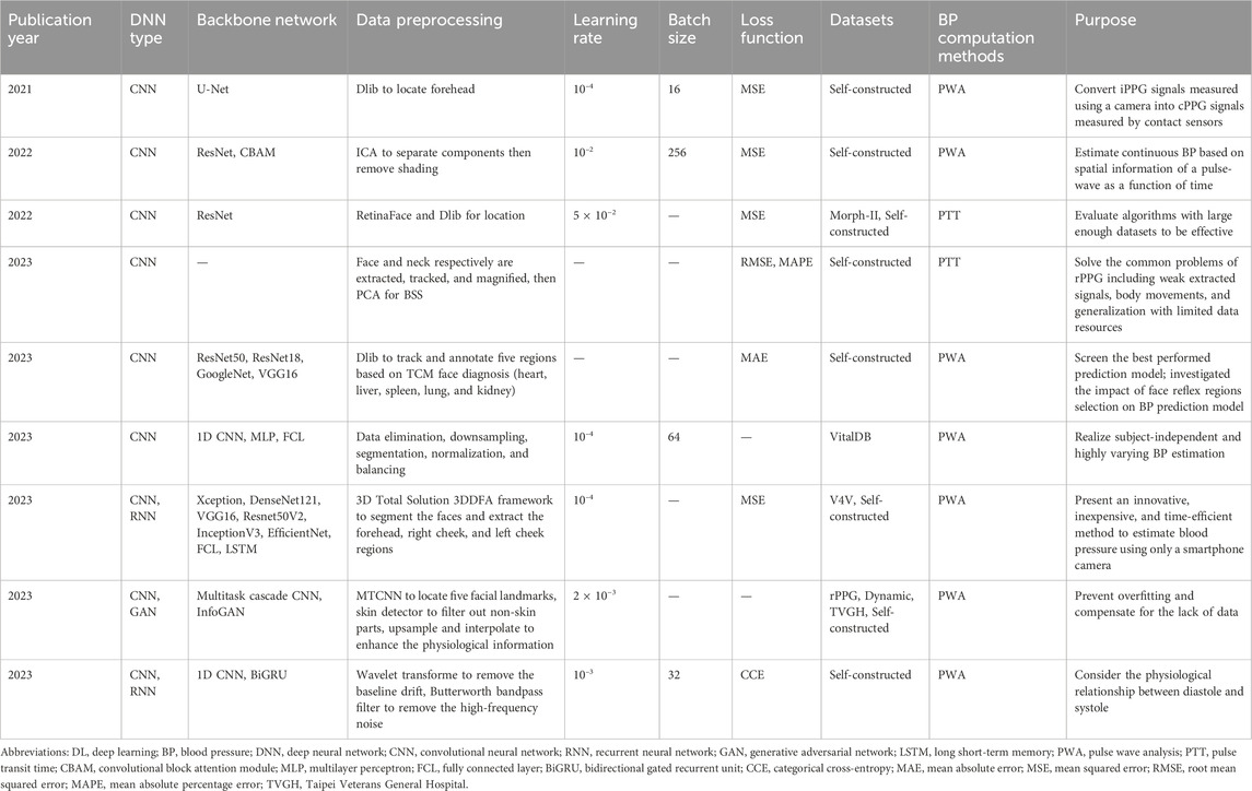
Table 5. Application of DL model in contactless BP estimation.
Hypertension is the leading cause of death worldwide and a key risk factor for many serious diseases, including cardiovascular diseases such as stroke and heart failure. BP is a major vital sign and must be monitored regularly for early detection, prevention, and treatment of cardiovascular disease. Conventional BP measurement techniques (invasive or cuff-based) are impractical, intermittent, and uncomfortable for patients. The method based on rPPG can realize the contactless monitoring of BP with improved patient comfort and mobility. CV-based methods can fully combine the advantages of computer algorithms and can extract key information characterizing BP from simple images or videos. With the development of DL, an exciting new field for contactless and continuous BP monitoring based on rPPG has been opened up. This will have a significant and transformative impact on monitoring the vital signs of patients, particularly those with high cardiovascular risk factors or diseases. It is encouraging to see a great amount of interest from both researchers and industry alike. While there are still challenges ahead, the continuous and relentless momentum of research provides hope for future PPG-based non-invasive, cuff-less, and continuous BP monitoring devices in the near future.
7 Limitations, prospects, and conclusion
There is already a tremendous amount of real-world applications for CV, and the technology is still young. Besides Healthcare, Autonomous vehicles, Google Translate app, Facial recognition, Real-time sports tracking, Agriculture, and Manufacturing are inseparable from the popularization of CV. As humans and machines continue to partner, the human workforce will be freed up to focus on higher-value tasks because the machines will automate processes that rely on image recognition. However, the popularity of AI will bring some problems. Data privacy issues are particularly common and prominent in the field of CV. With the open-source of a large number of datasets such as COHFACE, MAHNOB, and PURE, extensive videos and photographs containing face or identity information are disclosed. In the context of big data, in addition to adopting technical measures including anonymization, differential privacy, local differential privacy, and homomorphic encryption, strengthening data management is a another vital means to balance medical data sharing and privacy security. However, the application of these data privacy protection technologies needs to consider their efficiency and impact on data availability. The security management of medical and health data involves many departments, including medical institutions, AI suppliers, medical information management departments, etc. They are responsible for data collection, mining, storage, application and transmission. Therefore, the relevant departments are supposed to establish a security management system, series standard operating procedures, and a credible network security environment, strengthen supervision, reasonably utilize medical and health data in accordance with regulations, strictly standardize data use rights and data access control to protect data privacy and data security. Besides the concern about data privacy, another factor that influences remote contactless physiological monitoring must be considered, that is, the poor generalization of current task-specific algorithms, which causes weak accessibility for underserved populations. Generalist medical AI (GMAI), as a new concept proposed in recent years, can perform a variety of tasks using minimal or no task-specific labeled data (Moor et al., 2023). However, the development of GMAI usually founds on massive datasets, which brings about privacy issues. Therefore, when applying GMAI to the field of CV, we must consider the ethical issues and security risks involved, so that it can develop in a direction beneficial to accessible remote physiological monitoring for human health.
This paper aims to provide an in-depth and comprehensive literature review of the existing and proposed Artificial Intelligence methods with a focus on computer vision and deep learning in contactless physiological monitoring. Contactless physiological monitoring techniques based on images or video represented by rPPG have been applied in the evaluation of microcirculation perfusion, respiratory rate, oxygen saturation, heart rate, heart rate variability, and blood pressure while overcoming the limitations of conventional contact physiological measurements. The development of deep learning has injected new vitality into this field. Alongside continuous optimization of traditional algorithms, the gradual maturity of deep learning algorithms, and the miniaturization of imaging equipment, there is hope that these advancements will contribute greatly to comfortable, portable, and cost-effective remote healthcare services in the near future.
Author contributions
WC: Conceptualization, Investigation, Writing–original draft. ZY: Investigation, Writing–review and editing. LL: Writing–review and editing. RL: Resources, Writing–review and editing. AZ: Investigation, Writing–review and editing. ZQ: Writing–review and editing. JHA: Writing–review and editing. JHE: Writing–review and editing. BL: Conceptualization, Funding acquisition, Project administration, Resources, Supervision, Writing–review and editing.
Funding
The author(s) declare that financial support was received for the research, authorship, and/or publication of this article. This work was supported by Beijing Hospitals Authority Clinical medicine Development of special funding support (code: YGLX202314), National Natural Science Foundation of China (code: 82272581), Yunnan Provincial Science and Technology Talents and Platform Project (code: 202105AF150050), Beijing Hospitals Authority’s Ascent Plan (code: DFL20240402), and Beijing Municipal Health Commission (BJRITO-RDP-2024).
Conflict of interest
The authors declare that the research was conducted in the absence of any commercial or financial relationships that could be construed as a potential conflict of interest.
Publisher’s note
All claims expressed in this article are solely those of the authors and do not necessarily represent those of their affiliated organizations, or those of the publisher, the editors and the reviewers. Any product that may be evaluated in this article, or claim that may be made by its manufacturer, is not guaranteed or endorsed by the publisher.
References
Abdulrahaman, L. Q. (2023). Two-stage motion artifact reduction algorithm for rPPG signals obtained from facial video recordings. Arabian J. Sci. Eng. 49, 2925–2933. doi:10.1007/s13369-023-07845-2
Aloimonos, Y., and Rosenfeld, A. (1991). Computer vision. Science 253 (5025), 1249–1254. doi:10.1126/science.1891713
Alsheikhy, A., Said, Y. F., Shawly, T., and Lahza, H. (2023). A model to predict heartbeat rate using deep learning algorithms. Healthcare 11 (3), 330. doi:10.3390/healthcare11030330
Bayar, N., Guzel, K., and Kumlu, D. (2022). “A novel BlazeFace based pre-processing for MobileFaceNet in face verification,” in proceedings of the 2022 45th International Conference on Telecommunications and Signal Processing, TSP 2022, USA, 13-15 July 2022.
Bian, M., Peng, B., Wang, W., and Dong, J. (2019). An accurate LSTM based video heart rate estimation method. Pattern Recognit. Comput. Vis., 409–417. doi:10.1007/978-3-030-31726-3_35
Boccignone, G., D’Amelio, A., Ghezzi, O., Grossi, G., and Lanzarotti, R. (2023). An evaluation of non-contact photoplethysmography-based methods for remote respiratory rate estimation. Sensors 23 (7), 3387. doi:10.3390/s23073387
Bousefsaf, F., Djeldjli, D., Ouzar, Y., Maaoui, C., and Pruski, A. (2021). iPPG 2 cPPG: reconstructing contact from imaging photoplethysmographic signals using U-Net architectures. Comput. Biol. Med. 138, 104860. doi:10.1016/j.compbiomed.2021.104860
Bousefsaf, F., Pruski, A., and Maaoui, C. (2019). 3D convolutional neural networks for remote pulse rate measurement and mapping from facial video. Appl. Sci. 9 (20), 4364. doi:10.3390/app9204364
Buda, A. J., Pinsky, M. R., Ingels, N. B., Daughters, G. T., Stinson, E. B., and Alderman, E. L. (1979). Effect of intrathoracic pressure on left ventricular performance. N. Engl. J. Med. 301 (9), 453–459. doi:10.1056/nejm197908303010901
Castaldo, R., Montesinos, L., Melillo, P., James, C., and Pecchia, L. (2019). Ultra-short term HRV features as surrogates of short term HRV: a case study on mental stress detection in real life. BMC Med. Inf. Decis. Mak. 19 (1), 12. doi:10.1186/s12911-019-0742-y
Chandrasekaran, V., Dantu, R., Jonnada, S., Thiyagaraja, S., and Subbu, K. P. (2013). Cuffless differential blood pressure estimation using smart phones. IEEE Trans. Biomed. Eng. 60 (4), 1080–1089. doi:10.1109/tbme.2012.2211078
Chan, P., Wong, G., Dinh Nguyen, T., McNeil, J., and Hopper, I. (2019). Estimation of respiratory rate using infrared video in an inpatient population: an observational study. J. Clin. Monit. Comput. 34 (6), 1275–1284. doi:10.1007/s10877-019-00437-2
Charlton, P. H., Birrenkott, D. A., Bonnici, T., Pimentel, M. A. F., Johnson, A. E. W., Alastruey, J., et al. (2018). Breathing rate estimation from the electrocardiogram and photoplethysmogram: a review. IEEE Rev. Biomed. Eng. 11, 2–20. doi:10.1109/rbme.2017.2763681
Chaves-Gonzalez, J. M., Vega-Rodriguez, M. A., Gomez-Pulido, J. A., and Sánchez-Pérez, J. M. (2010). Detecting skin in face recognition systems: a colour spaces study. Digit. Signal Process. 20 (3), 806–823. doi:10.1016/j.dsp.2009.10.008
Cheng, H., Xiong, J., Chen, Z., et al. (2023). Deep learning-based non-contact IPPG signal blood pressure measurement research. Sensors 23 (12), 5528. doi:10.3390/s23125528
Chen, W., and Li, M. (2023). Standardized motion detection and real time heart rate monitoring of aerobics training based on convolution neural network. Prev. Med. 174, 107642. doi:10.1016/j.ypmed.2023.107642
Chen, W., and Mcduff, D. (2020). DeepMag: source-specific change magnification using gradient ascent. ACM Trans. Graph. 40 (1), 1–14. doi:10.1145/3408865
Cho, Y., Julier, S. J., Marquardt, N., and Bianchi-Berthouze, N. (2017). Robust tracking of respiratory rate in high-dynamic range scenes using mobile thermal imaging. Biomed. Opt. Express 8 (10), 4480. doi:10.1364/boe.8.004480
Davila, M. I., Lewis, G. F., and Porges, S. W. (2017). The PhysioCam: a Novel non-Contact sensor to measure heart rate variability in clinical and field applications. Front. Public Health 5, 300. doi:10.3389/fpubh.2017.00300
el-Hajj, C., and Kyriacou, P. A. (2020). A review of machine learning techniques in photoplethysmography for the non-invasive cuff-less measurement of blood pressure. Biomed. Signal Process. Control 58, 101870. doi:10.1016/j.bspc.2020.101870
Esteva, A., Chou, K., Yeung, S., Naik, N., Madani, A., Mottaghi, A., et al. (2021). Deep learning-enabled medical computer vision. npj Digit. Med. 4 (1), 5. doi:10.1038/s41746-020-00376-2
Fan, X., Wang, J., and Bayes, H. (2015). Proceedings of the 20th international conference on intelligent user interfaces, 405–416. doi:10.1145/2678025.2701364
Fan, X., Ye, Q., Yang, X., and Choudhury, S. D. (2020). Robust blood pressure estimation using an RGB camera. J. Ambient Intell. Humaniz. Comput. 11 (11), 4329–4336. doi:10.1007/s12652-018-1026-6
Favilla, R., Zuccala, V. C., and Coppini, G. (2019). Heart rate and heart rate variability from single-channel video and ICA integration of multiple signals. IEEE J. Biomed. Health Inf. 23 (6), 2398–2408. doi:10.1109/jbhi.2018.2880097
Finžgar, M., and Podržaj, P. (2018). A wavelet-based decomposition method for a robust extraction of pulse rate from video recordings. PeerJ 6, e5859. doi:10.7717/peerj.5859
Finžgar, M., and Podržaj, P. (2020). Feasibility of assessing ultra-short-term pulse rate variability from video recordings. PeerJ 8, e8342. doi:10.7717/peerj.8342
Gambi, E., Agostinelli, A., Belli, A., Burattini, L., Cippitelli, E., Fioretti, S., et al. (2017). Heart rate detection using microsoft kinect: validation and comparison to wearable devices. Sensors 17 (8), 1776. doi:10.3390/s17081776
Geddes, L. A., Voelz, M. H., Babbs, C. F., Bourland, J. D., and Tacker, W. A. (1981). Pulse transit time as an indicator of arterial blood pressure. Psychophysiology 18 (1), 71–74. doi:10.1111/j.1469-8986.1981.tb01545.x
Gideon, J., and Stent, S. (2021). “The way to my heart is through contrastive learning: remote photoplethysmography from unlabelled video,” in proceedings of the Proceedings of the IEEE International Conference on Computer Vision, USA, 20-23 June 1995.
Gonzalez, VIEJO C., Fuentes, S., Torrico, D., and Dunshea, F. R. (2018). Non-contact heart rate and blood pressure estimations from video analysis and machine learning modelling applied to food sensory responses: a case study for chocolate. Sensors 18 (6), 1802. doi:10.3390/s18061802
Guazzi, A. R., Villarroel, M., Jorge, J., Daly, J., Frise, M. C., Robbins, P. A., et al. (2015). Non-contact measurement of oxygen saturation with an RGB camera. Biomed. Opt. Express 6 (9), 3320–3338. doi:10.1364/boe.6.003320
Gudi, A., Bittner, M., and van Gemert, J. (2020). Real-time webcam heart-rate and variability estimation with clean ground truth for evaluation. Appl. Sci. 10 (23), 8630. doi:10.3390/app10238630
Gupta, A., Ravelo-Garcia, A. G., and Dias, F. M. (2022). A motion and illumination resistant non-contact method using undercomplete independent component analysis and levenberg-marquardt algorithm. IEEE J. Biomed. Health Inf. 26 (10), 4837–4848. doi:10.1109/jbhi.2022.3144677
Gupta, K., Sinhal, R., and Badhiye, S. S. (2023). Remote photoplethysmography-based human vital sign prediction using cyclical algorithm. J. Biophot. 17, e202300286. doi:10.1002/jbio.202300286
Gupta, P., Bhowmick, B., and Pal, A. (2020). MOMBAT: heart rate monitoring from face video using pulse modeling and Bayesian tracking. Comput. Biol. Med. 121, 103813. doi:10.1016/j.compbiomed.2020.103813
Hamoud, B., Kashevnik, A., Othman, W., and Shilov, N. (2023). Neural network model combination for video-based blood pressure estimation: new approach and evaluation. Sensors 23 (4), 1753. doi:10.3390/s23041753
Harford, M., Villarroel, M., Jorge, J., Redfern, O., Finnegan, E., Davidson, S., et al. (2022). Contactless skin perfusion monitoring with video cameras: tracking pharmacological vasoconstriction and vasodilation using photoplethysmographic changes. Physiol. Meas. 43 (11), 115001. doi:10.1088/1361-6579/ac9c82
Haugg, F., Elgendi, M., and Menon, C. (2023). GRGB rPPG: an efficient low-complexity remote photoplethysmography-based algorithm for heart rate estimation. Bioengineering 10 (2), 243. doi:10.3390/bioengineering10020243
He, L., Alam, K. S., Ma, J., Burkholder, E., Chung Chu, W. C., Iqbal, A., et al. (2021). “Remote photoplethysmography heart rate variability detection using signal to noise ratio bandpass filtering,” in 2021 IEEE International Conference on Digital Health (ICDH), China, 5-10 Sept. 2021, 133–141. doi:10.1109/icdh52753.2021.00025
Helmy, M., Truong, T. T., Jul, E., and Ferreira, P. (2023). Deep learning and computer vision techniques for microcirculation analysis: a review. Patterns 4 (1), 100641. doi:10.1016/j.patter.2022.100641
He, Q., and Wang, R. K. (2022). Imaging-photoplethysmography-guided optical microangiography. Opt. Lett. 47 (9), 2302. doi:10.1364/ol.452326
Hsu, G. S., Ambikapathi, A., and Chen, M. S. (2017). “Deep learning with time-frequency representation for pulse estimation from facial videos,” in proceedings of the IEEE International Joint Conference on Biometrics, IJCB, USA, 15 – 18 September, 2024.
Hsu, S.-Y., Chen, L.-W., Huang, R.-W., Tsai, T. Y., Hung, S. Y., Cheong, D. C. F., et al. (2023). Quantization of extraoral free flap monitoring for venous congestion with deep learning integrated iOS applications on smartphones: a diagnostic study. Int. J. Surg. Lond. Engl. 109 (6), 1584–1593. doi:10.1097/js9.0000000000000391
Hsu, Y., Lin, Y. L., and Hsu, W. (2014). “Learning-based heart rate detection from remote photoplethysmography features,” in proceedings of the ICASSP, IEEE International Conference on Acoustics, Speech and Signal Processing - Proceedings, USA, 14-19 April 2014.
Huang, N. E., Shen, Z., Long, S. R., Wu, M. C., Shih, H. H., Zheng, Q., et al. The empirical mode decomposition and the Hilbert spectrum for nonlinear and non-stationary time series analysis. Proc. R. Soc. a-Mathematical Phys. Eng. Sci., 1998, 454: 903–995. doi:10.1098/rspa.1998.0193
Huang, R.-W., Tsai, T.-Y., Hsieh, Y.-H., Hsu, C. C., Chen, S. H., Lee, C. H., et al. (2023). Reliability of postoperative free flap monitoring with a novel prediction model based on supervised machine learning. Plastic Reconstr. Surg. 152 (5), 943e–52e. doi:10.1097/prs.0000000000010307
Huang, R.-Y., and Dung, L.-R. (2016). Measurement of heart rate variability using off-the-shelf smart phones. Biomed. Eng. Online 15, 11. doi:10.1186/s12938-016-0127-8
Hu, M., Qian, F., Guo, D., Wang, X., He, L., and Ren, F. (2021). ETA-rPPGNet: effective time-domain attention network for remote heart rate measurement. IEEE Trans. Instrum. Meas. 70, 1–12. doi:10.1109/tim.2021.3058983
Hu, M., Qian, F., Wang, X., He, L., Guo, D., and Ren, F. (2022). Robust heart rate estimation with spatial–temporal attention network from facial videos. IEEE Trans. Cognitive Dev. Syst. 14 (2), 639–647. doi:10.1109/tcds.2021.3062370
Humphreys, K., Ward, T., and Markham, C. (2007). Noncontact simultaneous dual wavelength photoplethysmography: a further step toward noncontact pulse oximetry. Rev. Sci. Instrum. 78 (4), 044304. doi:10.1063/1.2724789
Iozzia, L., Cerina, L., and Mainardi, L. (2016). Relationships between heart-rate variability and pulse-rate variability obtained from video-PPG signal using ZCA. Physiol. Meas. 37 (11), 1934–1944. doi:10.1088/0967-3334/37/11/1934
Iuchi, K., Miyazaki, R., Cardoso, G. C., Ogawa-Ochiai, K., and Tsumura, N. (2022). Blood pressure estimation by spatial pulse-wave dynamics in a facial video. Biomed. Opt. Express 13 (11), 6035. doi:10.1364/boe.473166
Jaiswal, K. B., and Meenpal, T. (2022). Heart rate estimation network from facial videos using spatiotemporal feature image. Comput. Biol. Med. 151, 106307. doi:10.1016/j.compbiomed.2022.106307
Jeong, I. C., and Finkelstein, J. (2016). Introducing contactless blood pressure assessment using a high speed video camera. J. Med. Syst. 40 (4), 77. doi:10.1007/s10916-016-0439-z
Jewel, J. I., Hossain, M. M., and Haque, M. D. (2023). Design and implementation of a drowsiness detection system up to extended head angle using FaceMesh machine learning solution; proceedings of the lecture notes of the institute for computer sciences. Social-Informatics Telecommun. Eng. LNICST, F. doi:10.1007/978-3-031-34622-4_7
Joung, J., Jung, C.-W., Lee, H.-C., Chae, M. J., Kim, H. S., Park, J., et al. (2023). Continuous cuffless blood pressure monitoring using photoplethysmography-based PPG2BP-net for high intrasubject blood pressure variations. Sci. Rep. 13 (1), 8605. doi:10.1038/s41598-023-35492-y
Kamshilin, A. A., Zaytsev, V. V., Belaventseva, A. V., Podolyan, N. P., Volynsky, M. A., Sakovskaia, A. V., et al. (2022a). Novel method to assess endothelial function via monitoring of perfusion response to local heating by imaging photoplethysmography. Sensors 22 (15), 5727. doi:10.3390/s22155727
Kamshilin, A. A., Zaytsev, V. V., Lodygin, A. V., and Kashchenko, V. A. (2022b). Imaging photoplethysmography as an easy-to-use tool for monitoring changes in tissue blood perfusion during abdominal surgery. Sci. Rep. 12 (1), 1143. doi:10.1038/s41598-022-05080-7
Karlen, W., Garde, A., Myers, D., Scheffer, C., Ansermino, J. M., and Dumont, G. A. (2015). Estimation of respiratory rate from photoplethysmographic imaging videos compared to pulse oximetry. IEEE J. Biomed. Health Inf. 19 (4), 1331–1338. doi:10.1109/jbhi.2015.2429746
Kim, C. S., Carek, A. M., Mukkamala, R., Inan, O. T., and Hahn, J. O. (2015). Ballistocardiogram as proximal timing reference for pulse transit time measurement: potential for cuffless blood pressure monitoring. IEEE Trans. Biomed. Eng. 62 (11), 2657–2664. doi:10.1109/tbme.2015.2440291
Kocsis, L., Herman, P., and Eke, A. (2006). The modified Beer-Lambert law revisited. Phys. Med. Biol. 51 (5), N91–N98. doi:10.1088/0031-9155/51/5/n02
Kolosov, D., Kelefouras, V., Kourtessis, P., and Mporas, I. (2023). Contactless camera-based heart rate and respiratory rate monitoring using AI on hardware. Sensors 23 (9), 4550. doi:10.3390/s23094550
Kong, L., Zhao, Y., Dong, L., Jian, Y., Jin, X., et al. (2013). Non-contact detection of oxygen saturation based on visible light imaging device using ambient light. Opt. Express 21 (15), 17464. doi:10.1364/oe.21.017464
Krejcar, O., Slanina, Z., Stambachr, J., et al. (2009). “Noninvasive continuous blood pressure measurement and GPS position monitoring of patients,” in proceedings of the IEEE Vehicular Technology Conference, China, 8-10 June 1994.
Kuang, H., Lv, F., Ma, X., and Liu, X. (2022). Efficient spatiotemporal attention network for remote heart rate variability analysis. Sensors 22 (3), 1010. doi:10.3390/s22031010
Kumar, M., Veeraraghavan, A., and Sabharwal, A. (2015). DistancePPG: robust non-contact vital signs monitoring using a camera. Biomed. Opt. Express 6 (5), 1565–1588. doi:10.1364/boe.6.001565
Lai, M., van der Stel, S. D., Groen, H. C., van Gastel, M., Kuhlmann, K. F. D., Ruers, T. J. M., et al. (2022). Imaging PPG for in vivo human tissue perfusion assessment during surgery. J. Imaging 8 (4), 94. doi:10.3390/jimaging8040094
Lamonaca, F., Barbe, K., Kurylyak, Y., et al. (2013). Application of the artificial neural network for blood pressure evaluation with smartphones; proceedings of the proceedings of the 2013 IEEE 7th international conference on intelligent data acquisition and advanced computing systems. IDAACS 2013, F.
Lampier, L. C., ValadãO, C. T., Silva, L. A., Delisle-Rodríguez, D., Caldeira, E. M. d. O., and Bastos-Filho, T. F. (2022). A deep learning approach to estimate pulse rate by remote photoplethysmography. Physiol. Meas. 43 (7), 075012. doi:10.1088/1361-6579/ac7b0b
Lan, T., Li, G., and Lin, L. (2022). A non-contact oxygen saturation detection method based on dynamic spectrum. Infrared Phys. Technol. 127, 104421. doi:10.1016/j.infrared.2022.104421
LáZARO, J., Nam, Y., Gil, E., Laguna, P., and Chon, K. H. (2015). Respiratory rate derived from smartphone-camera-acquired pulse photoplethysmographic signals. Physiol. Meas. 36 (11), 2317–2333. doi:10.1088/0967-3334/36/11/2317
Lee, C. H. O. L. E. E., Cho, A., Lee, S., and Whang, M. (2019). Vision-based measurement of heart rate from ballistocardiographic head movements using unsupervised clustering. Sensors 19 (15), 3263. doi:10.3390/s19153263
Lee, E., Chen, E., and Lee, C. Y. (2020). Meta-rPPG: remote heart rate estimation using a transductive meta-learner; proceedings of the lecture notes in computer science including subseries lecture notes in artificial intelligence and lecture notes. Bioinformatics. doi:10.1007/978-3-030-58583-9_24
Lee, H., Cho, A., and Whang, M. (2021). Fusion method to estimate heart rate from facial videos based on RPPG and RBCG. Sensors 21 (20), 6764. doi:10.3390/s21206764
Lee, H., Ko, H., Chung, H., Nam, Y., Hong, S., and Lee, J. (2022b). Real-time realizable mobile imaging photoplethysmography. Sci. Rep. 12 (1), 7141. doi:10.1038/s41598-022-11265-x
Lee, H., Lee, J., Kwon, Y., Park, S., Sohn, R., et al. (2022a). Multitask siamese network for remote photoplethysmography and respiration estimation. Sensors 22 (14), 5101. doi:10.3390/s22145101
Li, M. H., Yadollahi, A., and Taati, B. (2014a). A non-contact vision-based system for respiratory rate estimation. Annu. Int. Conf. IEEE Eng. Med. Biol. Soc. 2014, 2119–2122. doi:10.1109/EMBC.2014.6944035
Li, B., Jiang, W., Peng, J., and Li, X. (2022). Deep learning-based remote-photoplethysmography measurement from short-time facial video. Physiol. Meas. 43 (11), 115003. doi:10.1088/1361-6579/ac98f1
Li, B., Zhang, P., Peng, J., and Fu, H. (2023). Non-contact PPG signal and heart rate estimation with multi-hierarchical convolutional network. Pattern Recognit. 139, 109421. doi:10.1016/j.patcog.2023.109421
Lie, W.-N., le, D.-Q., Lai, C.-Y., and Fang, Y. S. (2023). Heart rate estimation from facial image sequences of a dual-modality RGB-NIR camera. Sensors 23 (13), 6079. doi:10.3390/s23136079
Lin, Y.-C., Chou, N.-K., Lin, G.-Y., and Li, M. H. (2017). A real-time contactless pulse rate and motion status monitoring system based on complexion tracking. Sensors 17 (7), 1490. doi:10.3390/s17071490
Lin, B., Tao, J., Xu, J., He, L., Liu, N., and Zhang, X. (2023). Estimation of vital signs from facial videos via video magnification and deep learning. iScience 26 (10), 107845. doi:10.1016/j.isci.2023.107845
Lindelauf, A. A. M. A., Saelmans, A. G., van Kuijk, S. M. J., van der Hulst, R. R. W. J., and Schols, R. M. (2022). Near-infrared spectroscopy (NIRS) versus hyperspectral imaging (HSI) to detect flap failure in reconstructive surgery: a systematic review. Life 12 (1), 65. doi:10.3390/life12010065
Li, P., Benezeth, Y., Macwan, R., Nakamura, K., Gomez, R., Li, C., et al. (2020). Video-based pulse rate variability measurement using periodic variance maximization and adaptive two-window peak detection. Sensors 20 (10), 2752. doi:10.3390/s20102752
Liu, S.-Q., and Yuen, P. C. (2020). “A general remote photoplethysmography estimator with spatiotemporal convolutional network,” in 2020 15th IEEE International Conference on Automatic Face and Gesture Recognition (FG 2020), USA, 16-20 Nov. 2020, 481–488. doi:10.1109/fg47880.2020.00109
Liu, M., Po, L.-M., and Fu, H. (2017). Cuffless blood pressure estimation based on photoplethysmography signal and its second derivative. Int. J. Comput. Theory Eng. 9 (3), 202–206. doi:10.7763/ijcte.2017.v9.1138
Liu, X., Fromm, J., Patel, S., et al. (2020). Multi-task temporal shift attention networks for on-device contactless vitals measurement. Proc. Adv. Neural Inf. Process. Syst.
Liu, X., Wei, W., Kuang, H., and Ma, X. (2022). Heart rate measurement based on 3D central difference convolution with attention mechanism. Sensors 22 (2), 688. doi:10.3390/s22020688
Liu, X., Yang, X., Song, R., Wang, D., and Li, L. (2023). PFDNet: a pulse feature disentanglement network for atrial fibrillation screening from facial videos. IEEE J. Biomed. Health Inf. 27 (2), 1060–1071. doi:10.1109/jbhi.2022.3220656
Li, W., Lin, L., and Li, G. (2014b). Wavelength selection method based on test analysis of variance: application to oximetry. Anal. Methods 6 (4), 1082–1089. doi:10.1039/c3ay41601a
Li, X., Chen, J., Zhao, G., et al. (2014c). “Remote heart rate measurement from face videos under realistic situations,” in proceedings of the Proceedings of the IEEE Computer Society Conference on Computer Vision and Pattern Recognition, USA, 17-19 June 1997.
Lokendra, B., and Puneet, G. (2022). AND-rPPG: a novel denoising-rPPG network for improving remote heart rate estimation. Comput. Biol. Med., 141. doi:10.1016/j.compbiomed.2021.105146
Lucy, F. K., Suha, K. T., Dipty, S. T., Wadud, M. S. I., and Kadir, M. A. (2021). Video based non-contact monitoring of respiratory rate and chest indrawing in children with pneumonia. Physiol. Meas. 42 (10), 105017. doi:10.1088/1361-6579/ac34eb
Luguev, T., Seus, D., and Garbas, J. U. (2020). “Deep learning based affective sensing with remote photoplethysmography,” in proceedings of the 2020 54th Annual Conference on Information Sciences and Systems, China, 18-20 March 2020.
Lu, H., Han, H., and Zhou, S. K. (2021). DuAl-GaN: joint BVP and noise modeling for remote physiological measurement. Proc. Proc. IEEE Comput. Soc. Conf. Comput. Vis. Pattern Recognit. doi:10.1109/CVPR46437.2021.01222
Luo, H., Yang, D., Barszczyk, A., Vempala, N., Wei, J., Wu, S. J., et al. (2019). Smartphone-based blood pressure measurement using transdermal optical imaging technology. Circ. Cardiovasc Imaging 12 (8), e008857. doi:10.1161/circimaging.119.008857
Lv, W., Zhao, Y., Zhang, W., Liu, W., Hu, A., and Miao, J. (2021). Remote measurement of short-term heart rate with narrow beam millimeter wave radar. IEEE Access 9, 165049–165058. doi:10.1109/access.2021.3134280
Ma, D. S., Correll, J., and Wittenbrink, B. (2015). The Chicago face database: a free stimulus set of faces and norming data. Behav. Res. Methods 47 (4), 1122–1135. doi:10.3758/s13428-014-0532-5
Machikhin, A. S., Volkov, M. V., Khokhlov, D. D., Lovchikova, E. D., Potemkin, A. V., Danilycheva, I. V., et al. (2021). Exoscope-based videocapillaroscopy system for in vivo skin microcirculation imaging of various body areas. Biomed. Opt. Express 12 (8), 4627. doi:10.1364/boe.420786
Maeda, Y., Sekine, M., and Tamura, T. (2011). The advantages of wearable green reflected photoplethysmography. J. Med. Syst. 35 (5), 829–834. doi:10.1007/s10916-010-9506-z
Maity, A. K., Wang, J., Sabharwal, A., and Nayar, S. K. (2022). RobustPPG: camera-based robust heart rate estimation using motion cancellation. Biomed. Opt. Express 13 (10), 5447. doi:10.1364/boe.465143
Malik, M., Bigger, J. T., Camm, A. J., Kleiger, R. E., Malliani, A., Moss, A. J., et al. (1996). Heart rate variability: standards of measurement, physiological interpretation, and clinical use. Eur. Heart J. 17 (3), 354–381. doi:10.1093/oxfordjournals.eurheartj.a014868
Mamontov, O. V., Shcherbinin, A. V., Romashko, R. V., and Kamshilin, A. A. (2020). Intraoperative imaging of cortical blood flow by camera-based photoplethysmography at green light. Appl. Sci. 10 (18), 6192. doi:10.3390/app10186192
Marcinkevics, Z., Rubins, U., Zaharans, J., Miscuks, A., Urtane, E., and Ozolina-Moll, L. (2016). Imaging photoplethysmography for clinical assessment of cutaneous microcirculation at two different depths. J. Biomed. Opt. 21 (3), 035005. doi:10.1117/1.jbo.21.3.035005
Martinez-Delgado, G. H., Correa-Balan, A. J., May-Chan, J. A., Parra-Elizondo, C. E., Guzman-Rangel, L. A., and Martinez-Torteya, A. (2022). Measuring heart rate variability using facial video. Sensors 22 (13), 4690. doi:10.3390/s22134690
Maurya, L., Zwiggelaar, R., Chawla, D., and Mahapatra, P. (2022). Non-contact respiratory rate monitoring using thermal and visible imaging: a pilot study on neonates. J. Clin. Monit. Comput. 37 (3), 815–828. doi:10.1007/s10877-022-00945-8
Mccombie, D. B., Reisner, A. T., and Asada, H. H. (2006). “Adaptive blood pressure estimation from wearable PPG sensors using peripheral artery pulse wave velocity measurements and multi-channel blind identification of local arterial dynamics,” in proceedings of the Annual International Conference of the IEEE Engineering in Medicine and Biology - Proceedings, USA, 3-6 Nov. 1994, 3521–3524. doi:10.1109/IEMBS.2006.260590
Mcduff, D. J., Blackford, E. B., and Estepp, J. R. (2018). Fusing partial camera signals for noncontact pulse rate variability measurement. IEEE Trans. Biomed. Eng. 65 (8), 1725–1739. doi:10.1109/tbme.2017.2771518
Mcduff, D., Gontarek, S., and Picard, R. W. (2014). Improvements in remote cardiopulmonary measurement using a five band digital camera. IEEE Trans. Biomed. Eng. 61 (10), 2593–2601. doi:10.1109/tbme.2014.2323695
Mcduff, D., Hernandez, J., Liu, X., Wood, E., and Baltrusaitis, T. (2022). Using high-fidelity avatars to advance camera-based cardiac pulse measurement. IEEE Trans. Biomed. Eng. 69 (8), 2646–2656. doi:10.1109/tbme.2022.3152070
Melchor RodríGUEZ, A., and Ramos-Castro, J. (2018). Video pulse rate variability analysis in stationary and motion conditions. Biomed. Eng. OnLine 17 (1), 11. doi:10.1186/s12938-018-0437-0
MoçO, A., Stuijk, S., and de Haan, G. (2019). Posture effects on the calibratability of remote pulse oximetry in visible light. Physiol. Meas. 40 (3), 035005. doi:10.1088/1361-6579/ab051a
MoçO, A., and Verkruysse, W. (2020). Pulse oximetry based on photoplethysmography imaging with red and green light. J. Clin. Monit. Comput. 35 (1), 123–133. doi:10.1007/s10877-019-00449-y
Monkaresi, H., Calvo, R. A., and Hong, Y. (2014). A machine learning approach to improve contactless heart rate monitoring using a webcam. IEEE J. Biomed. Health Inf. 18 (4), 1153–1160. doi:10.1109/jbhi.2013.2291900
Moor, M., Banerjee, O., Abad, Z. S. H., Krumholz, H. M., Leskovec, J., Topol, E. J., et al. (2023). Foundation models for generalist medical artificial intelligence. Nature 616 (7956), 259–265. doi:10.1038/s41586-023-05881-4
Mukkamala, R., Hahn, J. O., Inan, O. T., Mestha, L. K., Kim, C. S., Toreyin, H., et al. (2015). Toward ubiquitous blood pressure monitoring via pulse transit time: theory and practice. IEEE Trans. Biomed. Eng. 62 (8), 1879–1901. doi:10.1109/tbme.2015.2441951
Munoz, M. L., van Roon, A., Riese, H., Thio, C., Oostenbroek, E., Westrik, I., et al. (2015). Validity of (Ultra-)Short recordings for heart rate variability measurements. PloS One 10 (9), e0138921. doi:10.1371/journal.pone.0138921
Nam, Y., Lee, J., and Chon, K. H. (2014). Respiratory rate estimation from the built-in cameras of smartphones and tablets. Ann. Biomed. Eng. 42 (4), 885–898. doi:10.1007/s10439-013-0944-x
Nitzan, M., Khanokh, B., and Slovik, Y. (2002). The difference in pulse transit time to the toe and finger measured by photoplethysmography. Physiol. Meas. 23 (1), 85–93. doi:10.1088/0967-3334/23/1/308
Niu, X., Han, H., Shan, S., et al. (2018). SynRhythm: learning a deep heart rate estimator from general to specific. Proc. Proc. - Int. Conf. Pattern Recognit. F. doi:10.1109/ICPR.2018.8546321
Niu, X., Shan, S., Han, H., and Chen, X. (2020b). RhythmNet: end-to-end heart rate estimation from face via spatial-temporal representation. IEEE Trans. Image Process. 29, 2409–2423. doi:10.1109/tip.2019.2947204
Niu, X., Yu, Z., Han, H., Li, X., Shan, S., and Zhao, G. (2020a). Video-based remote physiological measurement via cross-verified feature disentangling. Proc. Lect. Notes Comput. Sci. Incl. Subser. Lect. Notes Artif. Intell. Lect. Notes Bioinforma. F, 295–310. doi:10.1007/978-3-030-58536-5_18
Niu, X., Zhao, X., Han, H., et al. (2019). “Robust remote heart rate estimation from face utilizing spatial-temporal attention,” in proceedings of the Proceedings - 14th IEEE International Conference on Automatic Face and Gesture Recognition, FG 2019, China, 14-18 May 2019.
Nussinovitch, U., Elishkevitz, K. P., Katz, K., Nussinovitch, M., Segev, S., Volovitz, B., et al. (2011). Reliability of ultra-short ECG indices for heart rate variability. Ann. Noninvasive Electrocardiol. 16 (2), 117–122. doi:10.1111/j.1542-474x.2011.00417.x
Odinaev, I., Wong, K. L., Chin, J. W., Goyal, R., Chan, T. T., and So, R. H. Y. (2023). Robust heart rate variability measurement from facial videos. Bioengineering 10 (7), 851. doi:10.3390/bioengineering10070851
Ouzar, Y., Djeldjli, D., Bousefsaf, F., and Maaoui, C. (2023). X-iPPGNet: a novel one stage deep learning architecture based on depthwise separable convolutions for video-based pulse rate estimation. Comput. Biol. Med. 154, 106592. doi:10.1016/j.compbiomed.2023.106592
Pagano, T. P., Dos Santos, L. L., Santos, V. R., Sá, P. H. M., Bonfim, Y. d. S., Paranhos, J. V. D., et al. (2022). Remote heart rate prediction in virtual reality head-mounted displays using machine learning techniques. Sensors 22 (23), 9486. doi:10.3390/s22239486
Pai, A., Veeraraghavan, A., and Sabharwal, A. (2021). HRVCam: robust camera-based measurement of heart rate variability. J. Biomed. Opt. 26 (02), 022707. doi:10.1117/1.jbo.26.2.022707
Pereira, C. B., Yu, X., Goos, T., Reiss, I., Orlikowsky, T., Heimann, K., et al. (2019). Noncontact monitoring of respiratory rate in newborn infants using thermal imaging. IEEE Trans. Biomed. Eng. 66 (4), 1105–1114. doi:10.1109/tbme.2018.2866878
Perepelkina, O., Artemyev, M., Churikova, M., et al. (2020). “HeartTrack: convolutional neural network for remote video-based heart rate monitoring,” in proceedings of the IEEE Computer Society Conference on Computer Vision and Pattern Recognition Workshops, F, USA, Oct 2010.
Poh, M.-Z., Mcduff, D. J., and Picard, R. W. (2010). Non-contact, automated cardiac pulse measurements using video imaging and blind source separation. Opt. Express 18 (10), 10762–10774. doi:10.1364/oe.18.010762
Poh, M.-Z., Mcduff, D. J., and Picard, R. W. (2011). Advancements in noncontact, multiparameter physiological measurements using a webcam. IEEE Trans. Biomed. Eng. 58 (1), 7–11. doi:10.1109/tbme.2010.2086456
Poh, M.-Z., and Poh, Y. C. (2017). Validation of a standalone smartphone application for measuring heart rate using imaging photoplethysmography. Telemedicine e-Health 23 (8), 678–683. doi:10.1089/tmj.2016.0230
Prakash, S. K. A., and Tucker, C. S. (2018). Bounded Kalman filter method for motion-robust, non-contact heart rate estimation. Biomed. Opt. Express 9 (2), 873. doi:10.1364/boe.9.000873
Qi, L., Yu, H., Xu, L., Mpanda, R. S., and Greenwald, S. E. (2019). Robust heart-rate estimation from facial videos using Project_ICA. Physiol. Meas. 40 (8), 085007. doi:10.1088/1361-6579/ab2c9f
Qiu, Y., Liu, Y., Arteaga-Falconi, J., Dong, H., and El Saddik, A. (2019). EVM-CNN: real-time contactless heart rate estimation from facial video. IEEE Trans. Multimedia 21 (7), 1778–1787. doi:10.1109/tmm.2018.2883866
Rajendra Acharya, U., Paul Joseph, K., Kannathal, N., Lim, C. M., and Suri, J. S. (2006). Heart rate variability: a review. Med. Biol. Eng. Comput. 44 (12), 1031–1051. doi:10.1007/s11517-006-0119-0
Rasche, S., Huhle, R., Junghans, E., de Abreu, M. G., Ling, Y., Trumpp, A., et al. (2020). Association of remote imaging photoplethysmography and cutaneous perfusion in volunteers. Sci. Rep. 10 (1), 16464. doi:10.1038/s41598-020-73531-0
SchäFER, A., and Vagedes, J. (2013). How accurate is pulse rate variability as an estimate of heart rate variability? A review on studies comparing photoplethysmographic technology with an electrocardiogram. Int. J. Cardiol. 166 (1), 15–29. doi:10.1016/j.ijcard.2012.03.119
Schraven, S. P., Kossack, B., StrüDER, D., Jung, M., Skopnik, L., Gross, J., et al. (2023). Continuous intraoperative perfusion monitoring of free microvascular anastomosed fasciocutaneous flaps using remote photoplethysmography. Sci. Rep. 13 (1), 1532. doi:10.1038/s41598-023-28277-w
Schrumpf, F., Monch, C., Bausch, G., and Fuchs, M. (2019). Exploiting weak head movements for camera-based respiration detection. Annu. Int. Conf. IEEE Eng. Med. Biol. Soc. 2019, 6059–6062. doi:10.1109/EMBC.2019.8856387
Scully, C. G., Lee, J., Meyer, J., Gorbach, A. M., Granquist-Fraser, D., Mendelson, Y., et al. (2012). Physiological parameter monitoring from optical recordings with a mobile phone. IEEE Trans. Biomed. Eng. 59 (2), 303–306. doi:10.1109/tbme.2011.2163157
Secerbegovic, A., Bergsland, J., Halvorsen, P. S., et al. (2016). Blood pressure estimation using video plethysmography. Proc. Proc. - Int. Symposium Biomed. Imaging. doi:10.1109/ISBI.2016.7493307
Shao, D., Liu, C., Tsow, F., Yang, Y., Du, Z., Iriya, R., et al. (2016). Noncontact monitoring of blood oxygen saturation using camera and dual-wavelength imaging system. IEEE Trans. Biomed. Eng. 63 (6), 1091–1098. doi:10.1109/tbme.2015.2481896
Shi, Y., Qiu, J., Peng, L., Han, P., Luo, K., and Liu, D. (2023). A novel non-contact heart rate measurement method based on EEMD combined with FastICA. Physiol. Meas. 44 (5), 055002. doi:10.1088/1361-6579/accefd
Shoushan, M. M., Reyes, B. A., Rodriguez, A. M., and Chong, J. W. (2021). Non-contact HR monitoring via smartphone and webcam during different respiratory maneuvers and body movements. IEEE J. Biomed. Health Inf. 25 (2), 602–612. doi:10.1109/jbhi.2020.2998399
Siddiqui, S. A., Zhang, Y., Feng, Z., and Kos, A. (2016). A pulse rate estimation algorithm using PPG and smartphone camera. J. Med. Syst. 40 (5), 126. doi:10.1007/s10916-016-0485-6
Sikdar, A., Behera, S. K., and Dogra, D. P. (2016). Computer-vision-Guided human pulse rate estimation: a review. IEEE Rev. Biomed. Eng. 9, 91–105. doi:10.1109/rbme.2016.2551778
Slapnicar, G., Dovgan, E., Cuk, P., et al. (2019). “Contact-free monitoring of physiological parameters in people with profound intellectual and multiple disabilities,” in Proceedings of the proceedings - 2019 international conference on computer vision workshop (USA: ICCVW).
Song, R., Chen, H., Cheng, J., Li, C., Liu, Y., and Chen, X. (2021). PulseGAN: learning to generate realistic pulse waveforms in remote photoplethysmography. IEEE J. Biomed. Health Inf. 25 (5), 1373–1384. doi:10.1109/jbhi.2021.3051176
Song, R., Zhang, S., Cheng, J., et al. (2020). New insights on super-high resolution for video-based heart rate estimation with a semi-blind source separation method. Comput. Biol. Med., 116. doi:10.1016/j.compbiomed.2019.103535
Spetlik, R., Franc, V., Cech, J., et al. (2018). Visual heart rate estimation with convolutional neural network; Proceedings of the British machine vision conference 2018, China, BMVC.
Sugita, N., Obara, K., Yoshizawa, M., Abe, M., Tanaka, A., and Homma, N. (2015). Techniques for estimating blood pressure variation using video images. Annu. Int. Conf. IEEE Eng. Med. Biol. Soc. 2015, 4218–4221. doi:10.1109/EMBC.2015.7319325
Sun, Y., Hu, S., Azorin-Peris, V., Kalawsky, R., and Greenwald, S. (2012). Noncontact imaging photoplethysmography to effectively access pulse rate variability. J. Biomed. Opt. 18 (6), 061205. doi:10.1117/1.jbo.18.6.061205
Sun, Z., He, Q., Li, Y., Wang, W., and Wang, R. K. (2021). Robust non-contact peripheral oxygenation saturation measurement using smartphone-enabled imaging photoplethysmography. Biomed. Opt. Express 12 (3), 1746. doi:10.1364/boe.419268
Tamura, T. (2019). Current progress of photoplethysmography and SPO2 for health monitoring. Biomed. Eng. Lett. 9 (1), 21–36. doi:10.1007/s13534-019-00097-w
Tsou, Y.-Y., Lee, Y.-A., Hsu, C.-T., and Chang, S. H. (2020). “Siamese-rPPG network: remote photoplethysmography signal estimation from face videos,” in Proceedings of the 35th Annual ACM Symposium on Applied Computing, China, 30 March 2020, 2066–2073. doi:10.1145/3341105.3373905
Tusman, G., Acosta, C. M., Pulletz, S., Böhm, S. H., Scandurra, A., Arca, J. M., et al. (2019). Photoplethysmographic characterization of vascular tone mediated changes in arterial pressure: an observational study. J. Clin. Monit. Comput. 33 (5), 815–824. doi:10.1007/s10877-018-0235-z
van, E. S. V. A. A., Lopata, R. G. P., Scilingo, E. P., and Nardelli, M. (2023). Contactless cardiovascular assessment by imaging photoplethysmography: a comparison with wearable monitoring. Sensors 23 (3), 1505. doi:10.3390/s23031505
van der Kooij, K. M., and Naber, M. (2019). An open-source remote heart rate imaging method with practical apparatus and algorithms. Behav. Res. Methods 51 (5), 2106–2119. doi:10.3758/s13428-019-01256-8
Vaswani, A., Shazeer, N., Parmar, N., et al. (2017). Attention is all you need. Proc. Adv. Neural Inf. Process. Syst.
Verkruysse, W., Bartula, M., Bresch, E., Rocque, M., Meftah, M., and Kirenko, I. (2017). Calibration of contactless pulse oximetry. Anesth. Analg. 124 (1), 136–145. doi:10.1213/ane.0000000000001381
Visvanathan, A., Sinha, A., and Pal, A. (2013). “Estimation of blood pressure levels from reflective photoplethysmograph using smart phones,” in proceedings of the 13th IEEE International Conference on BioInformatics and BioEngineering, China, 10-13 Nov. 2013 (IEEE BIBE).
Wang, C., Pun, T., and Chanel, G. (2018a). A comparative survey of methods for remote heart rate detection from frontal face videos. Front. Bioeng. Biotechnol. 6, 33. doi:10.3389/fbioe.2018.00033
Wang, D., Yang, X., Liu, X., Jing, J., and Fang, S. (2020). Detail-preserving pulse wave extraction from facial videos using consumer-level camera. Biomed. Opt. Express 11 (4), 1876. doi:10.1364/boe.380646
Wang, H., Yang, X., Liu, X., and Wang, D. (2022). Heart rate estimation from facial videos with motion interference using T-SNE-based signal separation. Biomed. Opt. Express 13 (9), 4494. doi:10.1364/boe.457774
Wang, W., Den Brinker, A. C., and de Haan, G. (2018b). Full video pulse extraction. Biomed. Opt. Express 9 (8), 3898. doi:10.1364/boe.9.003898
Wang, W., Den Brinker, A. C., Stuijk, S., and de Haan, G. (2017a). Algorithmic principles of remote PPG. IEEE Trans. Biomed. Eng. 64 (7), 1479–1491. doi:10.1109/tbme.2016.2609282
Wang, W., Den Brinker, A. C., Stuijk, S., and de Haan, G. (2017b). Robust heart rate from fitness videos. Physiol. Meas. 38 (6), 1023–1044. doi:10.1088/1361-6579/aa6d02
Wang, W., Stuijk, S., and de Haan, G. (2016). A novel algorithm for remote photoplethysmography: spatial subspace rotation. IEEE Trans. Biomed. Eng. 63 (9), 1974–1984. doi:10.1109/tbme.2015.2508602
Wei, B., He, X., Zhang, C., and Wu, X. (2017). Non-contact, synchronous dynamic measurement of respiratory rate and heart rate based on dual sensitive regions. Biomed. Eng. Online 16 (1), 17. doi:10.1186/s12938-016-0300-0
Wieringa, F. P., Mastik, F., and van der Steen, A. F. W. (2005). Contactless multiple wavelength photoplethysmographic imaging: a first step toward "SpO2 camera" technology. Ann. Biomed. Eng. 33 (8), 1034–1041. doi:10.1007/s10439-005-5763-2
Woyczyk, A., Fleischhauer, V., and Zaunseder, S. (2021). Adaptive Gaussian mixture model driven level set segmentation for remote pulse rate detection. IEEE J. Biomed. Health Inf. 25 (5), 1361–1372. doi:10.1109/jbhi.2021.3054779
Wu, B.-F., Chiu, L.-W., Wu, Y.-C., Lin, L. L. C., Chung, M. L., et al. (2023). Motion robust remote photoplethysmography measurement during exercise for contactless physical activity intensity detection. IEEE J. Biomed. Health Inf. 72, 1–14. doi:10.1109/tim.2023.3256470
Wu, B.-F., Wu, B.-J., Tsai, B.-R., and Hsu, C. P. (2022). A facial-image-based blood pressure measurement system without calibration. IEEE Trans. Instrum. Meas. 71, 1–13. doi:10.1109/tim.2022.3165827
Xing, W., Shi, Y., Wu, C., et al. (2023). Predicting blood pressure from face videos using face diagnosis theory and deep neural networks technique. Comput. Biol. Med., 164. doi:10.1016/j.compbiomed.2023.107112
Yang, Z., Wang, H., Liu, B., et al. (2023). cbPPGGAN: a generic enhancement framework for unpaired pulse waveforms in camera-based photoplethysmography. IEEE J. Biomed. Health Inf., 1–11. doi:10.1109/jbhi.2023.3314282
Yin, R.-N., Jia, R.-S., Cui, Z., et al. (2022). PulseNet: a multitask learning network for remote heart rate estimation. Knowledge-Based Syst., 239. doi:10.1016/j.knosys.2021.108048
Yu, S.-G., Kim, S.-E., Kim, N. H., Suh, K. H., and Lee, E. C. (2021). Pulse rate variability analysis using remote photoplethysmography signals. Sensors 21 (18), 6241. doi:10.3390/s21186241
Yue, Z., Ding, S., Yang, S., Yang, H., Li, Z., Zhang, Y., et al. (2021). Deep super-resolution network for rPPG information recovery and noncontact heart rate estimation. IEEE Trans. Instrum. Meas. 70, 1–11. doi:10.1109/tim.2021.3109398
Yue, Z., Shi, M., and Ding, S. (2023). Facial video-based remote physiological measurement via self-supervised learning. IEEE Trans. Pattern Analysis Mach. Intell. 45 (11), 13844–13859. doi:10.1109/tpami.2023.3298650
Yu, Z., Li, X., Niu, X., Shi, J., and Zhao, G. (2020). AutoHR: a strong end-to-end baseline for remote heart rate measurement with neural searching. IEEE Signal Process. Lett. 27, 1245–1249. doi:10.1109/lsp.2020.3007086
Yu, Z., Li, X., and Zhao, G. (2019b). “Remote photoplethysmograph signal measurement from facial videos using spatio-temporal networks,” in proceedings of the 30th British Machine Vision Conference 2019, China, 10 May 2024 (BMVC).
Yu, Z., Peng, W., Li, X., et al. (2019a). “Remote heart rate measurement from highly compressed facial videos: an end-to-end deep learning solution with video enhancement,” in proceedings of the Proceedings of the IEEE International Conference on Computer Vision, USA, 20-23 June 1995.
Yu, Z., Shen, Y., Shi, J., et al. (2022). “PhysFormer: facial video-based physiological measurement with temporal difference transformer,” in proceedings of the Proceedings of the IEEE Computer Society Conference on Computer Vision and Pattern Recognition, USA, 17-19 June 1997.
Yu, Z., Shen, Y., Shi, J., Zhao, H., Cui, Y., Zhang, J., et al. (2023). PhysFormer++: facial video-based physiological measurement with SlowFast temporal difference transformer. Int. J. Comput. Vis. 131 (6), 1307–1330. doi:10.1007/s11263-023-01758-1
Zaunseder, S., Trumpp, A., Wedekind, D., and Malberg, H. (2018). Cardiovascular assessment by imaging photoplethysmography - a review. Biomed. Tech. Berl. 63 (5), 617–634. doi:10.1515/bmt-2017-0119
Zhang, C., Wu, X., Zhang, L., He, X., and Lv, Z. (2017). Simultaneous detection of blink and heart rate using multi-channel ICA from smart phone videos. Biomed. Signal Process. Control 33, 189–200. doi:10.1016/j.bspc.2016.11.022
Zhang, Q., Lin, X., Zhang, Y., Liu, Q., and Cai, F. (2023). Non-contact high precision pulse-rate monitoring system for moving subjects in different motion states. Med. Biol. Eng. Comput. 61 (10), 2769–2783. doi:10.1007/s11517-023-02884-1
Zhao, C., Zhou, M., Zhao, Z., et al. (2023). Learning spatio-temporal pulse representation with global-local interaction and supervision for remote prediction of heart rate. IEEE J. Biomed. Health Inf., 1–12. doi:10.1109/jbhi.2023.3252091
Zhao, Y., Zou, B., Yang, F., Lu, L., Belkacem, A. N., and Chen, C. (2021). Video-based physiological measurement using 3D central difference convolution attention network. EEE Int. Jt. Conf. Biometrics (IJCB): 1–6. doi:10.1109/ijcb52358.2021.9484405
Zheng, K., Ci, K., Li, H., Shao, L., Sun, G., Liu, J., et al. (2022). Heart rate prediction from facial video with masks using eye location and corrected by convolutional neural networks. Biomed. Signal Process. Control 75, 103609. doi:10.1016/j.bspc.2022.103609
Zhou, Y., Ni, H., Zhang, Q., and Wu, Q. (2019). The noninvasive blood pressure measurement based on facial images processing. IEEE Sensors J. 19 (22), 10624–10634. doi:10.1109/jsen.2019.2931775
Keywords: artificial intelligence, computer vision, rPPG, deep learning, physiological measurement
Citation: Chen W, Yi Z, Lim LJR, Lim RQR, Zhang A, Qian Z, Huang J, He J and Liu B (2024) Deep learning and remote photoplethysmography powered advancements in contactless physiological measurement. Front. Bioeng. Biotechnol. 12:1420100. doi: 10.3389/fbioe.2024.1420100
Received: 19 April 2024; Accepted: 27 June 2024;
Published: 17 July 2024.
Edited by:
Yiping Chen, Huazhong Agricultural University, ChinaReviewed by:
Long Wu, Hainan University, ChinaLeonardo Bocchi, University of Florence, Italy
Yongzhen Dong, Dalian Polytechnic University, China
Ying Gu, Kunming University of Science and Technology, China
Copyright © 2024 Chen, Yi, Lim, Lim, Zhang, Qian, Huang, He and Liu. This is an open-access article distributed under the terms of the Creative Commons Attribution License (CC BY). The use, distribution or reproduction in other forums is permitted, provided the original author(s) and the copyright owner(s) are credited and that the original publication in this journal is cited, in accordance with accepted academic practice. No use, distribution or reproduction is permitted which does not comply with these terms.
*Correspondence: Bo Liu, ZHJib2JvN0BzaW5hLmNvbQ==