
94% of researchers rate our articles as excellent or good
Learn more about the work of our research integrity team to safeguard the quality of each article we publish.
Find out more
ORIGINAL RESEARCH article
Front. Vet. Sci., 23 July 2024
Sec. Veterinary Epidemiology and Economics
Volume 11 - 2024 | https://doi.org/10.3389/fvets.2024.1425870
This article is part of the Research TopicEcosystem and Planetary Health and Emerging/Re-emerging ZoonosesView all 6 articles
 Paula Garcia-Sanchez1,2,3*†
 David Romero-Trancón2
 Iker Falces-Romero4,5
 Paula Navarro Carrera4
 Guillermo Ruiz-Carrascoso4,5
 David Carmena5,6
 María Casares Jiménez5,7
 Antonio Rivero-Juárez5,7Laura Moya8Jaume Rodón8Fernando Esperón9Belén Pérez-Hernando5Rocío Sánchez-León2,10Jara Hurtado-Gallego2,5Sonia Alcolea2,3,5,11,12,13
 Talía Sainz2,5,11,14,15
 Cristina Calvo2,5,11,14,15
 Ana Méndez-Echevarría2,5,11,14,15,16
Introduction: Although pets provide several social–emotional benefits for children, the risk of zoonosis must be considered among immunocompromised individuals.
Methods: A prospective study was conducted in a tertiary hospital including immunocompromised patients younger than 20 years owning dogs and/or cats. Colonization and/or infection was evaluated by stool studies, bacterial swabs, blood polymerase chain reaction and serological studies in both patients and their pets, to evaluate potential zoonotic transmission occurrence.
Results: We included 74 patients and their 92 pets (63 dogs, 29 cats). Up to 44.6% of the patients and 31.5% of the pets had at least 1 positive result. Up to 18.4% of pets’ fecal samples were positive (bacteria, parasites or hepatitis E virus). No helminths were observed despite the high frequency of incorrect intestinal deworming practices. Among children, gastrointestinal microorganisms were found in 37.3% (primarily Clostridium difficile). Colonization by Staphylococcus pseudintermedius was common among pets (8.0%) but not among children (0.0%). No shared colonization between owners and pets was observed, except in one case (Blastocystis in both patient and pet feces). Among patients, serologies were positive for Strongyloides stercoralis (14.8%), Toxocara canis (3.2%), Bartonella henselae (19.1%) and hepatitis E (5.6%). Serology was positive for Rickettsia spp. (22.6%) and Babesia spp. (6.5%) in dogs and for Leishmania spp. (14.3%) and Toxoplasma spp. (14.3%) in cats.
Conclusion: Exposure to zoonotic agents was detected in both patients and pets; however, shared colonization events were almost nonexistent. In our cohort, dogs and cats do not appear to entail high zoonosis transmission risk for immunocompromised patients.
Pets play an important role in the social–emotional development of children (1) and contribute to a healthier lifestyle. Interaction with animals can have additional positive effects in patients with chronic medical conditions (1). However, animal contact can also imply zoonotic risks, particularly for immunocompromised children (2, 3).
Case reports of viral, bacterial and parasitic zoonotic agents transmitted from pets to immunocompromised patients can be found in the literature (4–6) and immunocompromised patients who live with pets are asked to take special precautions (2). However, the moderate to poor evidence of most recommendations, together with the insufficient knowledge or awareness of zoonotic diseases among both patients and healthcare professionals often leads to gaps in the fulfillment of preventive measures (7–9). Low compliance with deworming protocols and failure to comply with pets’ immunization schemes have been reported by our and other groups in previous studies (3, 7, 9, 10).
In recent years, the role of some emerging zoonotic pathogens, such as hepatitis E virus (HEV) or Enterocytozoon bieneusi, are gaining increasing relevance, although the transmission routes between humans and animals remain uncertain (11, 12).
In addition to the infection risk, household pets can be colonized by bacteria that produce human diseases‚ such as Staphylococcus aureus, Staphylococcus pseudintermedius, multidrug-resistant Enterobacteriaceae, or Clostridium difficile (13–16), hypothetically increasing the risk of colonization of cohabiting children.
Few studies have evaluated the presence of zoonotic agents among immunocompromised patients and their pets and, to date, the evidence is insufficient to quantify the real risk of immunocompromised children acquiring a zoonotic infection from their pets (2, 3). A One Health approach is very much needed on the basis of collaboration between human and veterinary medicine (17). Both the number of immunocompromised patients and pet ownership have increased exponentially in recent decades (7, 18). We aimed to determine the prevalence of colonization and/or infection by microorganisms that can cause zoonoses in immunocompromised children and their pets and identify potential risk factors for colonization/infection.
A prospective study was performed from July 2022 to April 2024 at La Paz Pediatric University Hospital, a large tertiary hospital in Madrid, Spain, and a National Reference Center for immunocompromised children. Pediatric infectious disease specialists conducted the study, in collaboration with veterinarians and microbiologists. The study was approved by the local Clinical Research Ethics Committee of La Paz University Hospital (PI-4770) and all participants and/or legal guardians provided informed consent.
All immunocompromised patients under medical follow-up in our hospital younger than 20 years of age were invited to participate in the study if they owned at least 1 pet (dog and/or cat). We considered the following immunocompromised patients:
• Patients who had received solid organ transplantation in the previous 10 years.
• Patients who had received hematopoietic stem cell transplantation in the last 5 years, or in the last 5–10 years if the immune reconstitution was incomplete and/or required immunosuppressive treatment at the time of the study.
• Patients who had been diagnosed with genetically confirmed inborn errors of immunity.
• Patients with oncological diseases undergoing chemotherapy.
• Patients with rheumatological diseases who were receiving immunosuppressive treatments.
Patients (and/or families) who fulfilled the inclusion criteria were contacted by telephone or in person during hospital appointments. Those owning a dog and/or a cat and willing to provide informed consent for participation completed 2 standardized questionnaires: one document containing the patient’s information (sociodemographic data and basic medical data) and a second document with information regarding the pet(s), including type of pet, veterinary care, patient’s interaction with the animal, vaccines, anti-parasitic treatments, previous illnesses and dietary information. The questionnaires were completed during a hospital visit or online (Supplementary File 1). Patients aged 12 years or older completed the questionnaire themselves, whereas in the case of children younger than 12, the parents were asked to complete it. Relevant clinical data were obtained by a pediatrician member of the study team reviewing the patient’s medical records. A veterinarian reviewed the aspects related to animal health and care and, when necessary, the pet’s veterinarian was contacted by phone.
Screening for potentially animal-transmitted infections and colonizations was conducted in patients and pets. Stool culture and multiplex polymerase chain reaction (PCR) for fecal pathogens were performed, including Aeromonas spp., Campylobacter spp., Clostridium difficile toxin B, Salmonella spp., Shigella spp., Escherichia coli, Vibrio spp., Yersinia enterocolitica, Cyclospora cayetanensis, Dientamoeba fragilis, Entamoeba histolytica, Giardia duodenalis, Cryptosporidium spp., Blastocystis sp., Enterocytozoon bieneusi and Encephalitozoon spp. The presence of helminths was also analyzed in pets’ feces (Ascaris spp., Ancylostomatidae, Trichuridae and Cestoda). Nasopharyngeal and rectal swabs for screening of colonization by resistant bacteria and serological studies for the most common zoonotic agents were also performed in both patients and pets. Patients undergoing immunoglobulin treatment or with treatments affecting antibody production were excluded from the serological study. PCR assays for the diagnosis of acute HEV infection were performed in patients’/pets’ feces, blood and sera. Table 1 summarizes the main microbiological tests performed in patients and pets. Supplementary File 2 details the main microbiological techniques used.
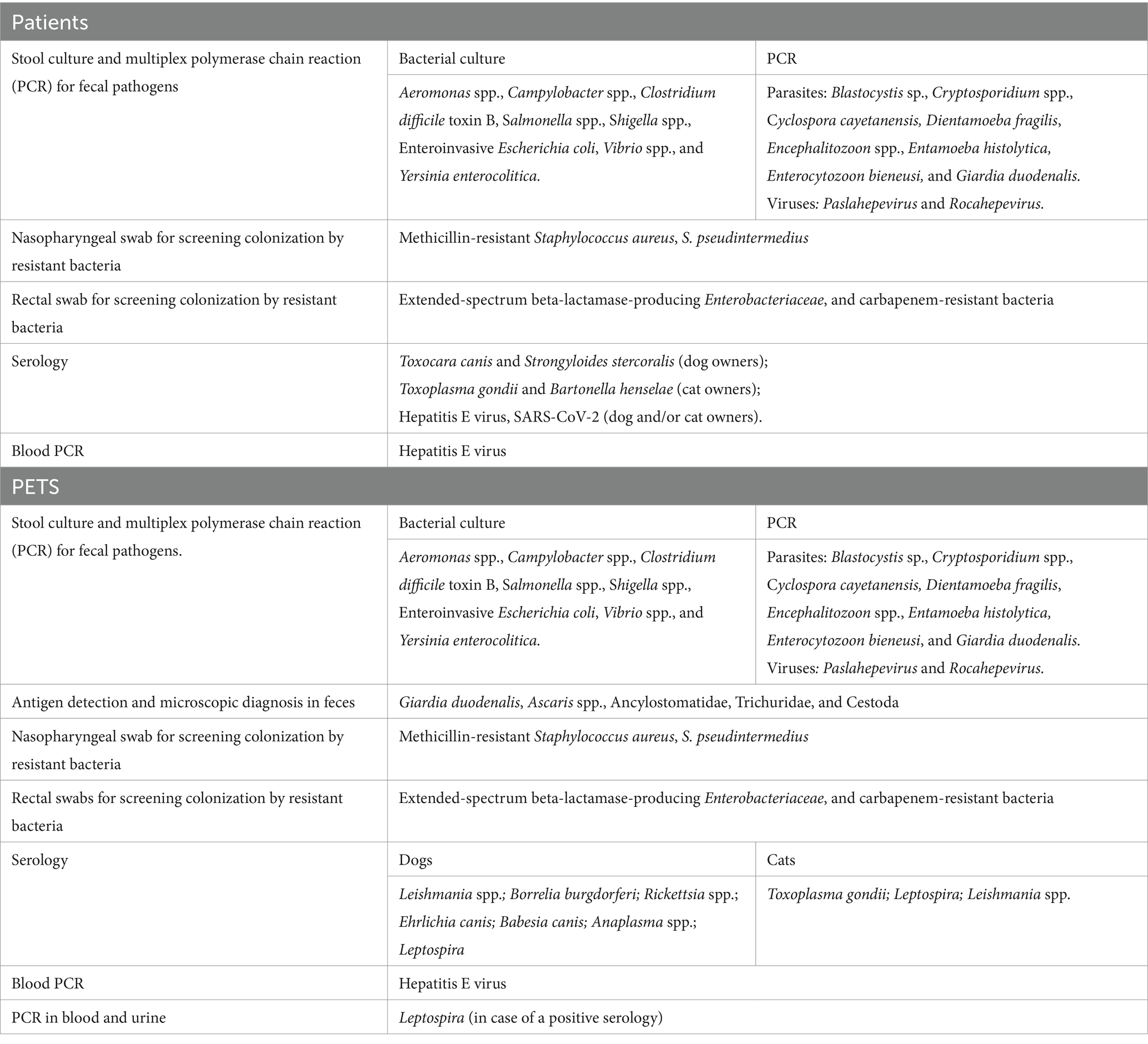
Table 1. Microbiological tests performed in patients and pets.
Stool samples from patients and pets were collected by their families. Patients’ swabs and blood samples were collected during scheduled hospital appointments. Pets’ swabs were collected by their owners following an explanatory sheet created for this purpose and their blood extraction was performed in veterinary clinics.
The statistical analysis was performed using the Statistical Package for the Social Sciences (IBM SPSS Statistics Version 21, IBM Inc., Chicago, IL, USA). Qualitative data were presented as absolute frequencies and percentages and quantitative variables were expressed as the main measures of centralization and dispersion (mean, standard deviation, median, minimum, maximum, interquartile range [IQR]).
For the study of risk factors for colonization by zoonotic agents, a univariate analysis was performed. Pearson’s chi-squared test (or Fisher’s exact test for 2×2 tables or likelihood ratio in mXn tables, if necessary) was used for qualitative variables; p-values under 0.05 were considered significant.
A total of 340 immunocompromised patients were contacted, 163 (47.9%) of whom owned a pet, mainly dogs and/or cats (135; 82.8%). Ultimately, 74 patients (51.3% female, median age 10.2 years [IQR 6.8–13.8]) and their 92 pets (63 dogs and 29 cats) were included in the study (Figure 1).
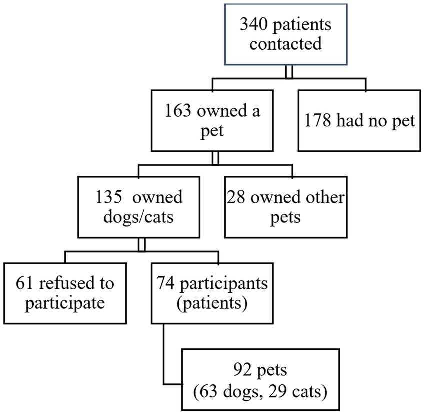
Figure 1. Flow chart of contacted and participating patients and pets.
Table 2 summarizes the clinical characteristics of the included patients and pets and Table 3 shows the pets’ main data, feeding and veterinary care (2, 19–22). Most (86.4%) owners took their pets to the veterinarian at least once a year and 96.5% of the pets were fed with commercially processed food. Up to 84.4% of newly acquired pets were puppies or kittens. Although 92.2% of the pets underwent intestinal deworming, only 4.9% underwent it monthly and 23.4% of owners reported having found ticks on their pets.
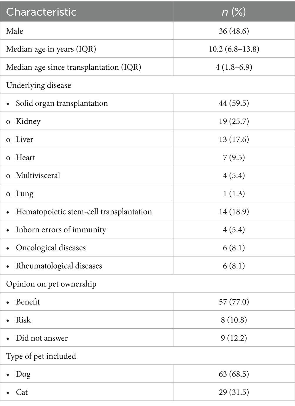
Table 2. Clinical characteristics of the included patients and pets.
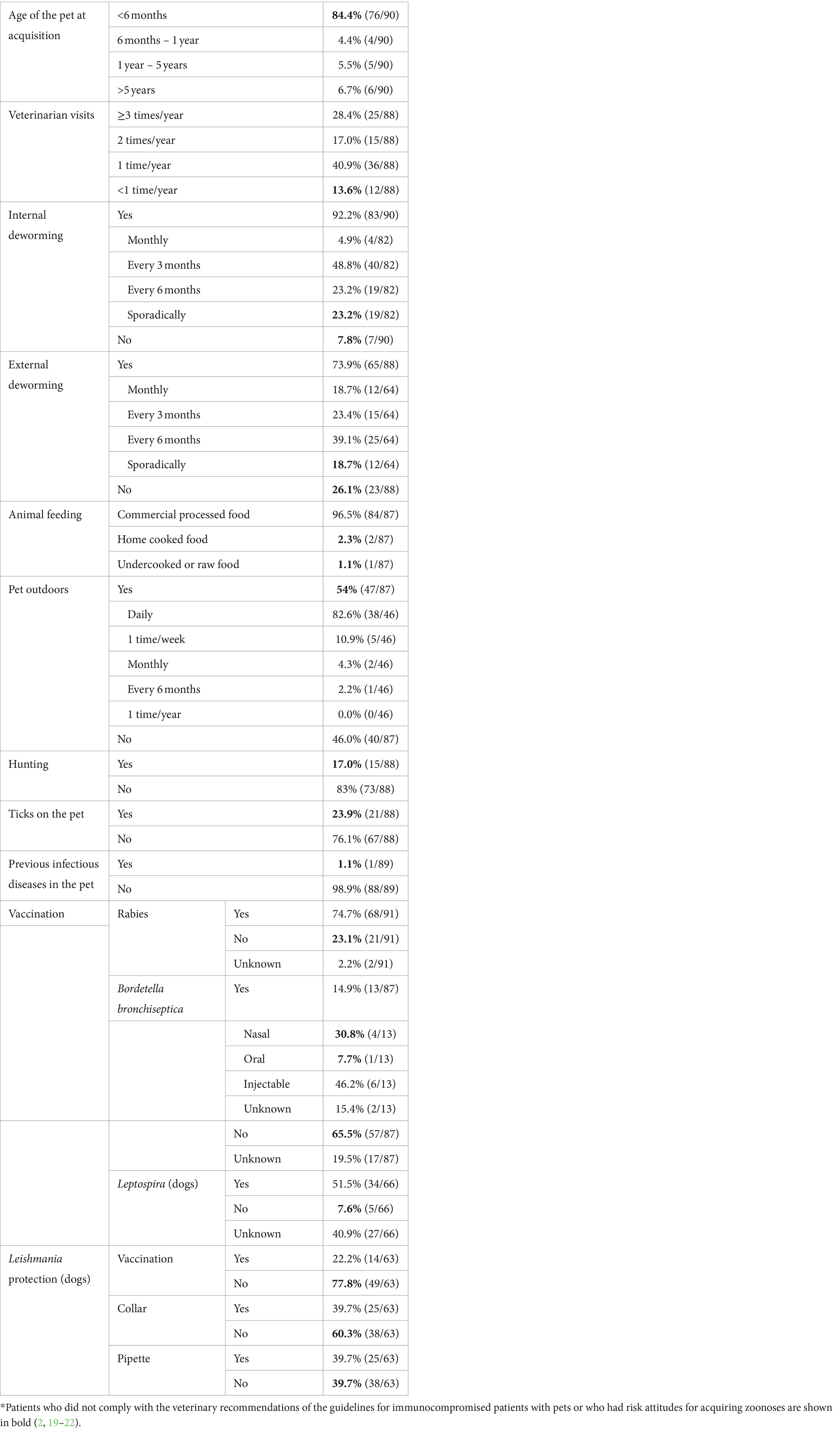
Table 3. Data on hygiene, feeding, and veterinary care of pets, and patients’ attitudes.
Regarding the risk–benefit balance of pet ownership, 77.0% (57/74) of the respondents believed that the benefits of pet ownership outweighed the risks, whereas 10.8% (8/74) thought that pet ownership was more risky than beneficial and 12.2% (9/74) did not answer this question.
Although not all samples were available from all participants and their pets, 33 (44.6%) patients had at least one positive result in the tests performed, including bacterial swabs (4.6%, 3/65), fecal samples (37.3%, 22/59) and blood serologies (22.5%, 16/71). Almost one-third of the pets (31.5%, 29/92) had positive results: 8.1% of nasopharyngeal swabs (7/86), 18.4% of fecal samples (16/87) and 26.3% of blood serologies (10/38). However, only one case of shared colonization involving Blastocystis was identified in stool samples (ST4 in a patient, unknown subtype in a dog) and no zoonotic transmission event could be demonstrated. No helminths were found in the stool tests of any pet, despite the presence of a high frequency of incorrect intestinal deworming regimens.
Specific results from nasopharyngeal and rectal bacterial swabs and from stool samples are summarized in Table 4, including the total number of samples in each category. Colonization by S. pseudintermedius was more common among pets (8%), compared to patients (0%). Up to 18.4% of pet fecal samples were positive, with the following microbiological findings: Cryptosporidium spp. (4.6%), E. bieneusi (3.4%), Campylobacter spp. (3.4%), G. duodenalis (2.3%), hepatitis E virus (2.3%), D. fragilis (1.1%), Blastocystis sp. (1.1%) and Encephalocytozoon spp. (1.1%). Among children, gastrointestinal microorganisms were found in 37.3% (primarily C. difficile, followed by Blastocystis sp. (6.8%), G. duodenalis (5.1%), D. fragilis (3.4%), hepatitis E virus (3.4%), Campylobacter spp. (1.7%), Y. enterocolitica (1.7%), Aeromonas spp. (1.7%) and Cryptosporidium spp. (1.7%). Results from serology and blood PCR are summarized in Table 5. Among patients, serological tests were positive for Strongyloides stercoralis (14.8%), Toxocara canis (3.2%), Bartonella henselae (19.1%) and hepatitis E (5.6%). In dogs, serologies were positive for Rickettsia spp. (22.6%) and Babesia canis (6.5%). One cat tested positive for Leishmania spp. and another cat tested positive for Toxoplasma spp. Supplementary File 3 summarizes the main molecular findings and sequencing data from pathogens found in feces.
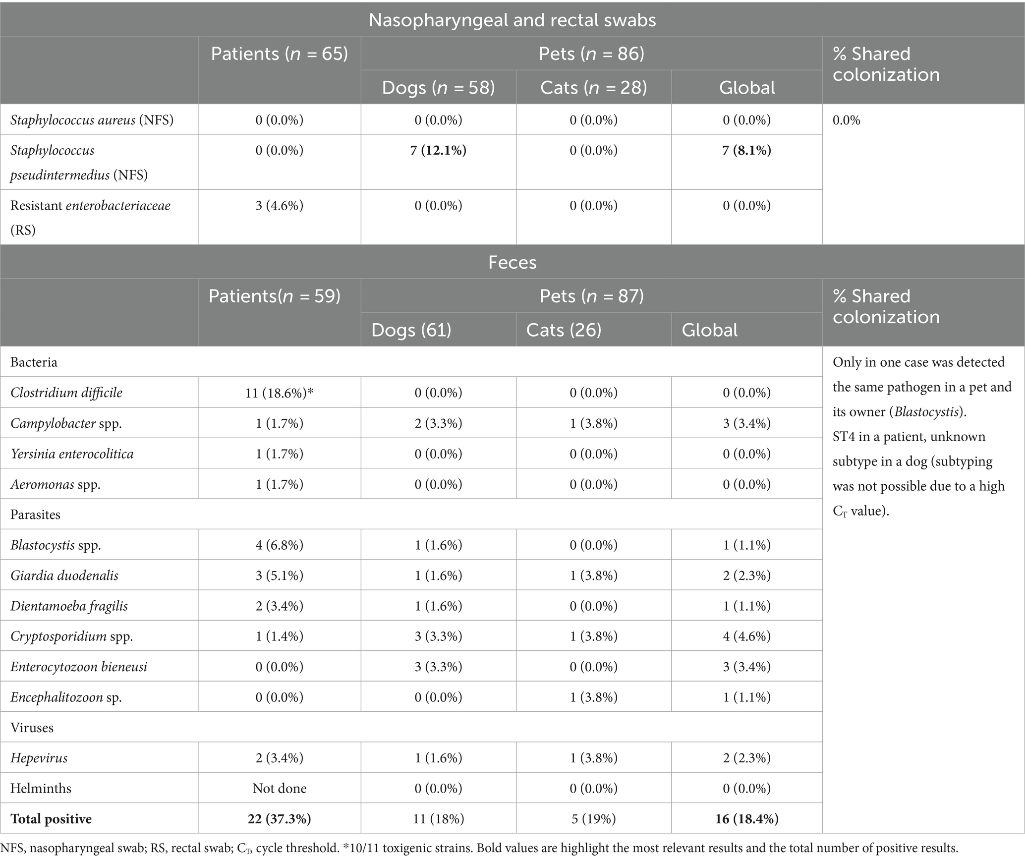
Table 4. Results from nasopharyngeal and rectal swabs and fecal samples.
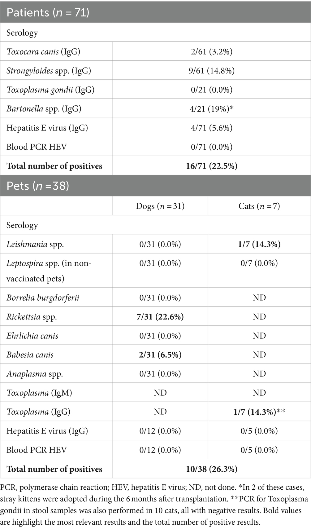
Table 5. Serological tests and blood PCR for hepatitis performed in patients and pets.
We then analyzed the association between the presence of microorganisms and all hygiene and diet habits. These included the pet’s age, number of veterinary visits, deworming frequency and compliance with recommendations, type of food, outdoor activities, have seen the pet eat or hunt another animal, presence of ticks and adequate vaccination schedule. None of the variables analyzed was associated with a higher presence of microorganisms in pets and/or patients (Table 6).
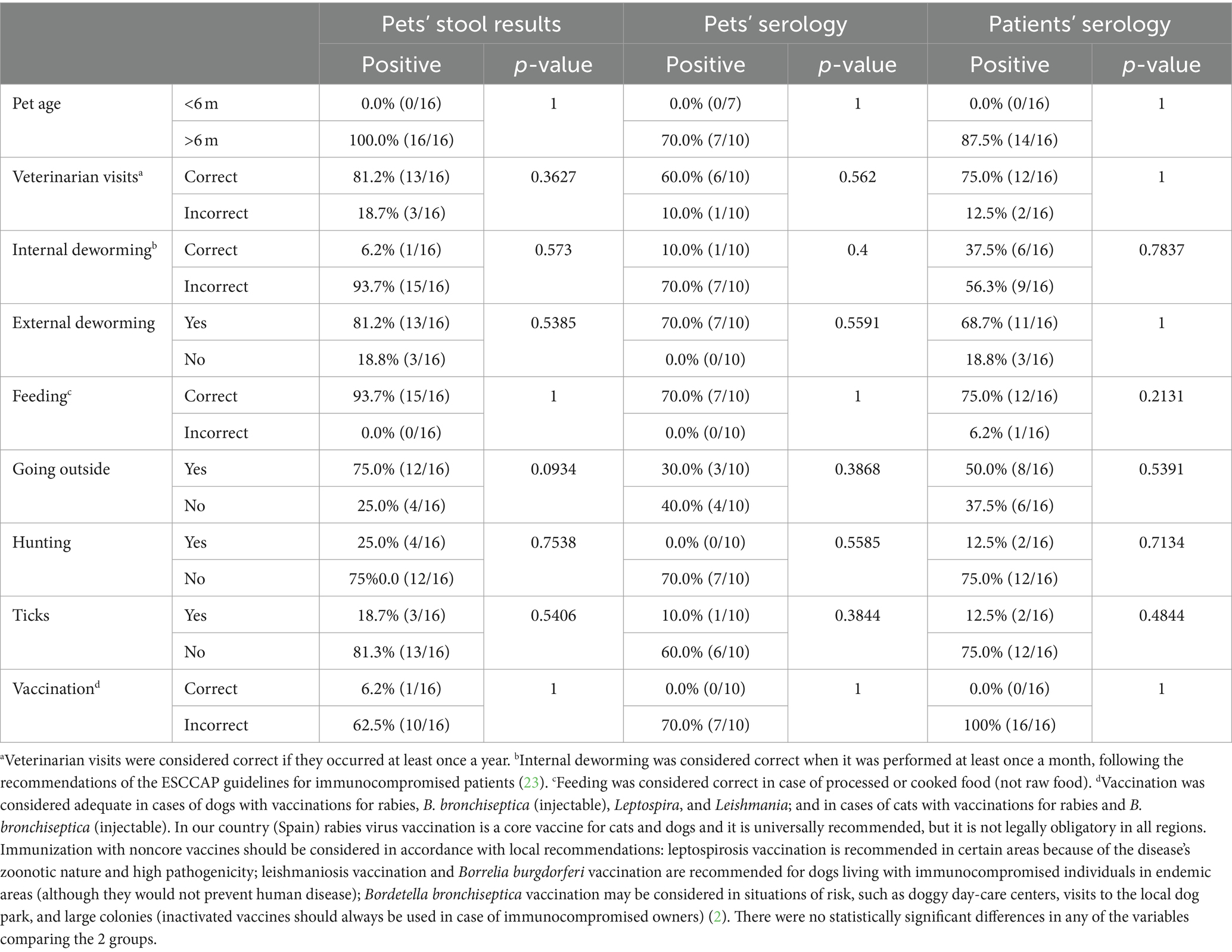
Table 6. Relationship between pets’ and patients’ positive results and pets’ epidemiological data.
This is one of the first studies, to our knowledge, assessing shared colonizations/infections in immunocompromised children and their pets aiming to analyze the role of dogs and cats as sources of zoonotic infections, including viral, bacterial and parasitic pathogens. Via a complete microbiological study, a noticeable number of microorganisms was identified in both patients and pets, with up to 44.6% of the patients and almost one-third of the pets testing positive for at least one microorganism under investigation. Although several potentially zoonotic agents were found in dogs and cats sharing a household with these patients, there was only one case of shared colonization (Blastocystis) and no zoonotic transmission could be demonstrated. Although gaps in preventive zoonotic measures were detected, no differences were found between pets with positive and negative zoonotic screening results and none of the studied factors was associated with a higher prevalence of colonization/infection among pets or children.
As described among human households (24), humans and pets can share microorganisms. However, the evidence remains scarce and the clinical implications are unknown. Correct deworming treatments in pets, adherence to scheduled immunization visits and following veterinary recommendations are strongly encouraged but might not cover the entire range of potential zoonotic pathogens that pets can harbor and the clinical impact in terms of zoonosis prevention has not been demonstrated (2). Immunocompromised hosts are more vulnerable to infections than their immunocompetent counterparts; therefore, the risks are presumably higher.
Our results reveal a high prevalence of pet ownership (47.9%), similar to previous data from our group (45.8%) (7) and from Europe (46%) (25). We found a considerable number of pathogens in our patients’ fecal samples (37.3%), whereas the number of pathogens in pets’ feces was lower (18.4%). Even so, potential zoonotic pathogens such as Cryptosporidium or Campylobacter were detected in pets’ feces. No helminths were found in pets’ feces, despite the high frequency of incorrect intestinal deworming practices (23) and considering that most of the pets were fed with commercially processed food. Clinicians should consider that routine deworming of pets involves anthelmintic drugs that are effective against cestodes and nematodes, but not against protists such as Giardia or Cryptosporidium. Previous molecular-based studies investigating the potential occurrence of zoonotic transmission events involving Giardia and Cryptosporidium among healthy individuals and their pets have failed to so demonstrate (26, 27). However, these surveys were hampered by transversal rather than longitudinal sampling designs and limited sample sizes; thus, to date, no previous studies, as well as our data, have demonstrated that these pathogens are a source of gastrointestinal cross-infections/colonizations.
Giardia duodenalis was detected in 5.6% of the studied patients in our cohort and in only 1.6% of the dogs. This is an unexpectedly low percentage, especially among pets, according to previously published data (26, 28–31). In Spain, previous epidemiological studies in the pediatric population have demonstrated the presence of G. duodenalis in 3–25% of asymptomatic children (32, 33). Among pets, the presence of Giardia in feces has been reported in 17.3–40.9% of owned and sheltered dogs (26, 28–31) and in 5.9 and 9.2% of owned and sheltered cats, respectively (26, 29). A potential explanation for this discrepancy is that families of immunocompromised children might be more aware of the risks associated with pet ownership and provide better care of their pets’ health compared with the general population. However, previous studies regarding pet ownership among immunocompromised young patients have revealed non-compliance with basic veterinary recommendations and risky exposures for acquiring zoonoses (7, 34). Most of our human and animal Giardia-positive samples yielded high (>35) cycle threshold (CT) values, indicative of low parasite loads. The only isolate successfully genotyped (assigned to zoonotic assemblage B) was identified in a human sample with a CT value of 33.7.
Another remarkable parasite encountered was Blastocystis sp., which is probably the most common enteric parasite in humans globally (35), although its pathogenicity remains controversial (35). It was present in 6.8% of participants but only 1 dog. The role of companion animals as reservoirs of human Blastocystis infections is uncertain (35). A Spanish study conducted in Northern Spain found Blastocystis in 35.2% of the human stool samples analyzed, but not in any of the canine or feline fecal specimens investigated, suggesting that these pets play a negligible role as natural reservoirs of human Blastocystis infection (35). In our cohort, 4 human fecal samples were Blastocystis-positive. Three were successfully subtyped, allowing the identification of the subtypes ST1, ST2, and ST4, all common in European human populations. However, only one of the canine fecal samples tested positive for the parasite, although our molecular analyses failed to determine the subtype involved. Blastocystis was the only case of shared colonization (ST4 in a patient, unknown subtype in a dog) in our cohort. Therefore, pet dogs and cats do not appear to have a relevant role as reservoirs of human Blastocystis infections.
On the other hand, emerging pathogens are becoming increasingly relevant, such as Enterocytozoon bieneusi. This fungi-related pathogen is considered an emerging infectious agent, with the most common Microsporidia species contributing to human microsporidiosis; it is an opportunistic pathogen infecting immunocompromised individuals (36, 37). Some commonly reported human genotypes have been found in animals, raising the question of whether human-animal contact could play a role in its transmission to humans (12). Enterocytozoon bieneusi has been detected in 0.8% of owned dogs and 3% of owned cats in Northern Spain (38) and in 0.4% of dogs in the Madrid area (Central Spain) (30). In our series, the prevalence of E. bieneusi in dogs and cats was 3.3 and 0.0%, respectively, and there were no positive results in humans. In addition, Encephalitozoon intestinalis DNA was identified in a feline fecal sample. This is the first report of the presence of E. intestinalis in this host in Spain. Taken together, these data indicate that companion animals might act as a potential source of human microsporidiosis.
Regarding bacterial findings in feces, no C. difficile isolates were identified in our canine and feline populations, although the owners were highly colonized, probably due to a high number of previous hospitalizations and frequent use of antibiotherapy. A recent review of several studies in various countries worldwide on the prevalence and molecular epidemiology of C. difficile in dogs and cats revealed variable colonization rates (39). In healthy dogs, the colonization rate was shown to be 3–5.5% and this percentage increased to 12% in dogs with gastrointestinal diseases. Similarly, C. difficile was isolated in 2.5–9.4% of healthy and diarrheal cats (39). These studies have shown that pets carry strains genetically identical to that of their owners, suggesting inter-species transmission (39, 40). Similarly to our results, few pets were infected in a recent small prospective study conducted in the USA in patients with diarrhea and their pets (owned dogs and cats) (40). Only in 2 households was C. difficile detected in both the owner and pet, although these strains were different (40). Two studies conducted in veterinary clinics from the Madrid region (Central Spain) reported prevalence of C. difficile in feces from owned dogs and owned cats of 4.8 and 0.0%, respectively (41), and of 6.7% in diarrheic dogs (42). These data suggest low probability of cross-transmission.
Taking into account serological tests, our results show previous exposure to several zoonotic agents in both patients and pets.
Strongyloides stercoralis can lead to severe hyperinfection and disseminated strongyloidiasis in immunocompromised patients (43). Its prevalence in this specific clinical population is not well documented and recent studies have reported prevalence rates of approximately 3–5% (43). It should be noted that reported prevalence rates were based on a limited number of heterogeneous studies that differ in the study regions and the diagnostic methods used (43). The results from our patients are in contrast to those published by other authors, with a much higher Strongyloides seroprevalence (14.8%). Although all infected patients in our cohort lived with dogs, the patients could have been infected by walking barefoot or by playing with soil (44). To date, it remains unclear whether dogs act as a suitable reservoir for human infections.
Toxocariasis is another neglected zoonotic infection, dogs and cats being the natural definitive hosts (45). Given that the majority of the infected individuals remain asymptomatic (45), its prevalence can be underestimated. In a previous study conducted in our center, we found a seroprevalence for toxocariasis of 5.3% among migrant and internationally adopted children (45). In our series, 2 asymptomatic patients were seropositive (3.2%). However, severe forms such as ocular or cerebral toxocariasis could occur in immunocompromised hosts (46); thus, screening based on serology should be performed in immunocompromised patients.
Bartonella henseale poses a notable risk to cat owners. Infected individuals may experience symptoms such as cat scratch disease, fever, lymphadenopathy, fatigue or muscle pain. While the infection typically remains mild, severe complications such as pulmonary nodules, pneumonia, ocular and skin lesions, osteomyelitis, hepatosplenic disease, bacillary angiomatosis or encephalitis can occur, especially in immunocompromised individuals (2). Up to 19% (4/21) of positive results for Bartonella henselae were observed among cat owners. Interestingly, half of these positive patients (n = 2) adopted stray cats a few months after transplantation, confirming important gaps in zoonotic risk knowledge in this population (7). In our country, the prevalence of Bartonella varies between series: 8.7% in healthy people from Catalonia (Northern Spain) (47), 22.3% among patients with HIV from the same region (48) and up to 37.1% among the veterinary worker population (49). Specifically in cat owners, a seroprevalence of 6.07% was estimated in a Chilean study (50), lower than our results.
None of the cats’ stools tested positive for Toxoplasma gondii and no patient had a positive serology. Although toxoplasmosis has traditionally been linked to contact with cats, the majority of infections in Europe occur by other means of transmission (51). A recent meta-analysis has observed that although the pooled prevalence of oocysts in European domestic cats’ feces is as low as 1.2%, their presence in soil is found in up to 16% (52). The risk is extremely low for indoor urban domestic cats (52). These findings highlight the lack of evidence supporting most recommendations to prevent zoonoses.
Many families (23.9%) reported having found ticks on their pets and a relevant percentage of dogs (29.0%) presented positive serology for microorganisms such as Rickettsia spp. or Babesia canis. A previous Chilean study revealed the presence of ectoparasites in nearly 50% of dogs and cats (3). Ticks can be vectors of serious infections in the USA and in Europe, such as Lyme disease, borreliosis, Central European encephalitis, or Crimean Congo hemorrhagic fever. The geographical distribution of this tick species has been expanding and an increase in tick-borne infections has recently been reported (53–55). Curiously, although all dogs tested were negative, one case of positive serology for Leishmania was detected in a cat. A recent study performed in our country found that 2% of stray cats were seropositive for Leishmania (56); thus, although infrequent, these felines could also be infected.
HEV and ratHEV (Rocahepevirus ratti) are 2 emerging viruses affecting humans for which cats and dogs might serve as hosts, as shown in previous studies (11, 57). A study in southern Spain reported a prevalence of anti-HEV antibodies in dogs and cats of 10 and 2.8%, respectively (11), suggesting that these species might play a potential role in the HEV zoonotic cycle. Similarly, this study provides evidence of ratHEV circulation in these species, indicating that cats and dogs might serve as reservoirs. This potential susceptibility was confirmed in a study conducted in Hong Kong, which reported that 1.2% of dogs and 1.5% of cats in the area exhibited IgG antibodies against ratHEV (57). However, the risk of zoonotic transmission from pets to humans was deemed minimal, given that none of the studies found evidence of viral RNA. Our study is the first to report the presence of HEV in these species (feces from 1 dog and 1 cat), both of which harbored strains capable of zoonotic transmission, such as HEV-3 f. Similarly, we report for the first time the presence of ratHEV in cats and dogs, suggesting that these species could also be susceptible to infection by this recently described zoonotic virus. Although the source of HEV infection cannot be definitively identified, the most plausible route could be through the consumption of raw or undercooked meat, because it constitutes the most efficient transmission pathway. In the case of ratHEV, although the primary host of this virus appears to be rodents, the route of infection between animal species and zoonotic transmission remains unknown. In fact, the dog in our sample with ratHEV identified in stool samples consumed raw or undercooked food on a monthly basis. The absence of infection in the children owning these animals reinforces the idea that the risk of transmission from these species through direct contact could be minimal, and thus, they are likely play a limited role in the epidemiology of these viruses.
Bidirectional bacterial transmission between owners and pets has already been reported (24, 58). According to a previous study by our group, up to 16% of children with complex chronic conditions are S. aureus colonized, with up to 27% of them colonized by multidrug-resistant Enterobacteriaceae (59). We hypothesize that pets living with immunocompromised children might be more frequently colonized by multidrug-resistant pathogens, and that pets could act as reservoirs, maintaining transmission in the community. However, the unexpectedly low colonization rates observed in our patients and pets did not allow us to observe possible cross-colonization. The prevalence of S. pseudintermedius colonization in the nasopharyngeal swabs (12%) of the screened dogs was also lower than expected; previous studies have reported colonization rates in dogs from 43 to 92% (13). Dogs can be persistent or intermittent carriers, so collecting more than one sample at various time points could have increased our ability to detect colonizations (13, 60).
Despite the high number of global positive results among both pets and patients, we found no association with pet age, veterinary visits, vaccination, deworming, hunting, presence of ticks, or feeding compared with the pets with negative results. However, the small sample size has limited the analysis. Nonetheless, we detected a few interesting findings related to zoonosis risk, such as stray cat ownership a few months after transplantation in half of the children with positive serology for Bartonella, or the consumption of raw or undercooked meat in one dog with ratHEV identified in stool samples. These findings are indicative of important gaps in zoonotic risk knowledge among this vulnerable population.
Some 77% of the surveyed patients considered pet ownership a benefit. Facing a life-threatening condition requiring long-term treatment has significant emotional implications and animal contact can offer substantial mental health benefits (2). Taking into account our results and considering that most of these zoonoses could be prevented, the balance between the psychological benefits and health risks for these patients appears to lean in favor of benefits, as long as basic veterinary recommendations are followed. However, our findings have limitations and deserve cautious interpretation. Close collaboration between veterinary and medical doctors as well as an enhanced role of veterinarians is required and patients should receive evidence-based information (8).
Our study has several limitations. It was a single-center study; thus, the number of patients and pets analyzed is relatively low and it might not be generalizable to other populations. Patient recruitment was complex and not all samples were collected for all participants, especially those from pets. In addition, its transversal design and the lower than expected number of individuals colonized impaired the identification of shared colonizations and/or zoonotic transmission events in our series. Samples were collected at a single time point; thus, zoonosis transmission could not be demonstrated.
This is one of the first studies addressing the presence of colonization and zoonotic infections among immunocompromised children and their pets. We found that many pets living with immunocompromised children are infected by zoonotic pathogens and we observed previous exposure to zoonotic agents in both patients and pets. However, shared colonization was rare and could not be explained by diet/hygiene habits; thus, larger studies are warranted in order to address the role of pets as zoonosis reservoirs. In the meantime, our data are reassuring, because no additional risk was identified for immunocompromised children having pets (dogs and/or cats). Given that pets have important socio-emotional benefits, defining the potential risks and effective preventive interventions is very much needed to increase the quality of life of immunocompromised patients.
The original contributions presented in the study are included in the article/Supplementary material, further inquiries can be directed to the corresponding author.
The studies involving humans and animals were approved by Clinical Research Ethics Committee of La Paz University Hospital (PI-4770). The studies were conducted in accordance with the local legislation and institutional requirements. Written informed consent for participation in this study was provided by the participants' legal guardians/next of kin. Written informed consent was obtained from the owners for the participation of their animals in this study.
PG-S: Conceptualization, Data curation, Formal analysis, Investigation, Methodology, Writing – original draft, Writing – review & editing. DR-T: Conceptualization, Data curation, Formal analysis, Investigation, Methodology, Writing – review & editing. IF-R: Data curation, Resources, Writing – review & editing, Methodology. PN: Data curation, Resources, Writing – review & editing, Methodology. GR-C: Conceptualization, Data curation, Resources, Writing – review & editing, Methodology. DC: Data curation, Resources, Supervision, Writing – review & editing, Methodology. MC: Data curation, Investigation, Resources, Writing – review & editing, Methodology. AR-J: Data curation, Investigation, Resources, Writing – review & editing, Methodology. LM: Conceptualization, Data curation, Methodology, Resources, Writing – review & editing. JR: Data curation, Investigation, Methodology, Resources, Writing – review & editing. FE: Investigation, Resources, Writing – review & editing, Data curation. BP-H: Conceptualization, Data curation, Formal analysis, Investigation, Methodology, Writing – review & editing. RS-L: Data curation, Investigation, Methodology, Writing – review & editing. JH-G: Data curation, Investigation, Methodology, Writing – review & editing. SA: Conceptualization, Data curation, Formal analysis, Investigation, Software, Writing – review & editing. TS: Supervision, Writing – review & editing, Formal analysis. CC: Supervision, Writing – review & editing, Formal analysis. AM-E: Conceptualization, Funding acquisition, Investigation, Methodology, Resources, Supervision, Writing – review & editing, Formal analysis.
The author(s) declare that financial support was received for the research, authorship, and/or publication of this article. This study was supported by the Spanish Pediatrics Association (AEP) 2021 Research Grant, by the MAPFRE Foundation (“Research grants by Ignacio H. de Larramendi 2021”), by the Spanish Ministry of Science and Innovation – Instituto de Salud Carlos III, and Fondos FEDER of the UE, Grant N° PI23/00917 and PI22/01098 [Fondo de Investigaciones Sanitarias-Spanish Health Research Fund (ISCIII)] and, TS has been funded by a Springboard Award 2023 by the European Society for Pediatric Infectious Diseases (ESPID). BP-H has been funded by CIBERINFEC. RS-L has been funded by Programa Investigo 2022 (reference A113), Community of Madrid Government (Spain). AR-J is supported by a contract from the Spanish Junta de Andalucía (Nicolas Monardes program: C1-0001-2023). MC is the recipient of a PFIS predoctoral grant (FI22/00180) from the Carlos III Health Institute and co-funded by Fondos FEDER of the European Union. The funding bodies did not have a role in the design or conduct of the study, the analysis and interpretation of the results, the writing of the report, or the decision to publish.
The authors declare that the research was conducted in the absence of any commercial or financial relationships that could be construed as a potential conflict of interest.
The author(s) declared that they were an editorial board member of Frontiers, at the time of submission. This had no impact on the peer review process and the final decision.
All claims expressed in this article are solely those of the authors and do not necessarily represent those of their affiliated organizations, or those of the publisher, the editors and the reviewers. Any product that may be evaluated in this article, or claim that may be made by its manufacturer, is not guaranteed or endorsed by the publisher.
The Supplementary material for this article can be found online at: https://www.frontiersin.org/articles/10.3389/fvets.2024.1425870/full#supplementary-material
1. Christian, H, Mitrou, F, Cunneen, R, and Zubrick, SR. Pets are associated with fewer peer problems and emotional symptoms, and better prosocial behavior: findings from the longitudinal study of Australian children. J Pediatr. (2020) 220:200–206.e2. doi: 10.1016/j.jpeds.2020.01.012
2. García Sánchez, P, Iglesias, I, Falces-Romero, I, Serrano-Villar, M, Calvo, C, Alcolea, S, et al. Balancing the risks and benefits of pet ownership in pediatric transplant recipients. Transplantation. (2023) 107:855–66. doi: 10.1097/TP.0000000000004419
3. Peña, A, Abarca, K, Weitzel, T, Gallegos, J, Cerda, J, García, P, et al. One health in practice: a pilot project for integrated Care of Zoonotic Infections in immunocompromised children and their pets in Chile. Zoonoses Public Health. (2016) 63:403–9. doi: 10.1111/zph.12241
4. Agarwal, L, Singh, H, Jani, C, Banankhah, P, Abdalla, M, Kurman, JS, et al. A wolf in sheep’s clothing: dogs confer an unrecognized risk for their immunocompromised master. Respir Med Case Rep. (2022) 38:101672. doi: 10.1016/j.rmcr.2022.101672
5. Bretón-Martínez, JR, Alcolea, A, Quintero-García, D, Méndez-Echevarria, A, Ramos, E, Bueno, F, et al. Non-wild-type cryptococcosis in a child with multivisceral organ transplant who owned bird pets. Transpl Infect Dis. (2021) 23:e13558. doi: 10.1111/tid.13558
6. Ner, Z, Ross, LA, Horn, MV, Keens, TG, MacLaughlin, EF, Starnes, VA, et al. Bordetella bronchiseptica infection in pediatric lung transplant recipients. Pediatr Transplant. (2003) 7:413–7. doi: 10.1034/j.1399-3046.2003.00074.x
7. Garcia-Sanchez, P, Aguilar-Valero, E, Sainz, T, Calvo, C, Iglesias, I, Bueno, D, et al. Immunocompromised children and Young patients living with pets: gaps in knowledge to avoid zoonosis. Transbound Emerg Dis. (2023) 2023:e2151761:1–10. doi: 10.1155/2023/2151761
8. Garcia-Sanchez, P, Romero-Trancón, D, Sainz, T, Calvo, C, Iglesias, I, Perez-Hernando, B, et al. The role of veterinarians in zoonosis prevention: advising families of immunocompromised children with pets. One Health. (2023) 18:100662. doi: 10.1016/j.onehlt.2023.100662
9. Platero, L, Garcia-Sanchez, P, Sainz, T, Calvo, C, Iglesias, I, Esperon, F, et al. Pets for pediatric transplant recipients: to have or not to have. Front Vet Sci. (2022) 9:974665. doi: 10.3389/fvets.2022.974665
10. Roussel, C, Drake, J, and Ariza, JM. French national survey of dog and cat owners on the deworming behaviour and lifestyle of pets associated with the risk of endoparasites. Parasit Vectors. (2019) 12:480. doi: 10.1186/s13071-019-3712-4
11. Caballero-Gómez, J, Rivero-Juarez, A, Jurado-Tarifa, E, Jiménez-Martín, D, Jiménez-Ruiz, E, Castro-Scholten, S, et al. Serological and molecular survey of hepatitis E virus in cats and dogs in Spain. Transbound Emerg Dis. (2022) 69:240–8. doi: 10.1111/tbed.14437
12. Zhang, Y, Koehler, AV, Wang, T, Cunliffe, D, and Gasser, RB. Enterocytozoon bieneusi genotypes in cats and dogs in Victoria, Australia. BMC Microbiol. (2019) 19:183. doi: 10.1186/s12866-019-1563-y
13. Bannoehr, J, and Guardabassi, L. Staphylococcus pseudintermedius in the dog: taxonomy, diagnostics, ecology, epidemiology and pathogenicity. Vet Dermatol. (2012) 23:e51–2. doi: 10.1111/j.1365-3164.2012.01046.x
14. Hernandez, BG, Vinithakumari, AA, Sponseller, B, Tangudu, C, and Mooyottu, S. Prevalence, colonization, epidemiology, and public health significance of Clostridioides difficile in companion animals. Front Vet Sci. (2020) 7:512551. doi: 10.3389/fvets.2020.512551
15. Meropol, SB, Haupt, AA, and Debanne, SM. Incidence and outcomes of infections caused by multidrug-resistant Enterobacteriaceae in children, 2007-2015. J Pediatric Infect Dis Soc. (2018) 7:36–45. doi: 10.1093/jpids/piw093
16. Nomoto, H, Kutsuna, S, Nakamura, K, Nakamoto, T, Shimomura, A, Hirakawa, T, et al. Totally implantable venous access port infection caused by Staphylococcus pseudintermedius: possible transmission from a companion dog to a human. J Infect Chemother. (2020) 26:1305–8. doi: 10.1016/j.jiac.2020.07.011
17. De Giusti, M, Barbato, D, Lia, L, Colamesta, V, Lombardi, AM, Cacchio, D, et al. Collaboration between human and veterinary medicine as a tool to solve public health problems. Lancet Planet Health. (2019) 3:e64–5. doi: 10.1016/S2542-5196(18)30250-X
18. Wallace, BI, Kenney, B, Malani, PN, Clauw, DJ, Nallamothu, BK, and Waljee, AK. Prevalence of immunosuppressive drug use among commercially insured US adults, 2018-2019. JAMA Netw Open. (2021) 4:e214920. doi: 10.1001/jamanetworkopen.2021.4920
19. Avery, RK, and Michaels, MG. AST infectious diseases Community of Practice. Strategies for safe living following solid organ transplantation-guidelines from the American Society of Transplantation infectious diseases Community of Practice. Clin Transpl. (2019) 33:e13519. doi: 10.1111/ctr.13519
20. Day, MJ, Horzinek, MC, Schultz, RD, and Squires, RA. Vaccination guidelines group (VGG) of the world small animal veterinary association (WSAVA). WSAVA guidelines for the vaccination of dogs and cats. J Small Anim Pract. (2016) 57:E1–E45. doi: 10.1111/jsap.2_12431
21. Hemsworth, S, and Pizer, B. Pet ownership in immunocompromised children--a review of the literature and survey of existing guidelines. Eur J Oncol Nurs. (2006) 10:117–27. doi: 10.1016/j.ejon.2005.08.001
22. Tomblyn, M, Chiller, T, Einsele, H, Gress, R, Sepkowitz, K, Storek, J, et al. Guidelines for preventing infectious complications among hematopoietic cell transplantation recipients: a global perspective. Biol Blood Marrow Transplant. (2009) 15:1143–238. doi: 10.1016/j.bbmt.2009.06.019
23. ESCCAP . (2021). ESCCAP Guideline 01 Sixth edition May 2021-Warm control in dogs and cats. Available at: https://www.esccap.org/guidelines (Accessed February 20, 2024)
24. Abdullahi, IN, Lozano, C, Zarazaga, M, Saidenberg, ABS, Stegger, M, and Torres, C. Clonal relatedness of coagulase-positive staphylococci among healthy dogs and dog-owners in Spain. Detection of multidrug-resistant-MSSA-CC398 and novel linezolid-resistant-MRSA-CC5. Front Microbiol. (2023) 14:1121564. doi: 10.3389/fmicb.2023.1121564
25. FEDIAF . (2023). Annual Report 2023. Available at: https://europeanpetfood.comingsoon.site/wp-content/uploads/2023/07/FEDIAF_Annual-Report_2023.pdf (Accessed April 4, 2024)
26. de Lucio, A, Bailo, B, Aguilera, M, Cardona, GA, Fernández-Crespo, JC, and Carmena, D. No molecular epidemiological evidence supporting household transmission of zoonotic Giardia duodenalis and Cryptosporidium spp. from pet dogs and cats in the province of Álava, northern Spain. Acta Trop. (2017) 170:48–56. doi: 10.1016/j.actatropica.2017.02.024
27. Rehbein, S, Klotz, C, Ignatius, R, Müller, E, Aebischer, A, and Kohn, B. Giardia duodenalis in small animals and their owners in Germany: a pilot study. Zoonoses Public Health. (2019) 66:117–24. doi: 10.1111/zph.12541
28. Drake, J, Sweet, S, Baxendale, K, Hegarty, E, Horr, S, Friis, H, et al. Detection of Giardia and helminths in Western Europe at local K9 (canine) sites (DOGWALKS study). Parasit Vectors. (2022) 15:311. doi: 10.1186/s13071-022-05440-2
29. Gil, H, Cano, L, de Lucio, A, Bailo, B, de Mingo, MH, Cardona, GA, et al. Detection and molecular diversity of Giardia duodenalis and Cryptosporidium spp. in sheltered dogs and cats in northern Spain. Infect Genet Evol. (2017) 50:62–9. doi: 10.1016/j.meegid.2017.02.013
30. Mateo, M, Montoya, A, Bailo, B, Köster, PC, Dashti, A, Hernández-Castro, C, et al. Prevalence and public health relevance of enteric parasites in domestic dogs and cats in the region of Madrid (Spain) with an emphasis on Giardia duodenalis and Cryptosporidium sp. Vet Med Sci. (2023) 9:2542–58. doi: 10.1002/vms3.1270
31. Sanchez-Thevenet, P, Carmena, D, Adell-Aledón, M, Dacal, E, Arias, E, Saugar, JM, et al. High prevalence and diversity of zoonotic and other intestinal parasites in dogs from eastern Spain. Vector Borne Zoonotic Dis. (2019) 19:915–22. doi: 10.1089/vbz.2019.2468
32. Mateo, M, Mateo, M, Montoya, A, Bailo, B, Saugar, JM, Aguilera, M, et al. Detection and molecular characterization of Giardia duodenalis in children attending day care centers in Majadahonda, Madrid, Central Spain. Medicine (Baltimore). (2014) 93:e75. doi: 10.1097/MD.0000000000000075
33. Muadica, AS, Köster, PC, Dashti, A, Bailo, B, Hernández-de-Mingo, M, Reh, L, et al. Molecular diversity of Giardia duodenalis, Cryptosporidium spp. and Blastocystis sp. in asymptomatic school children in Leganés, Madrid (Spain). Microorganisms. (2020) 8:466. doi: 10.3390/microorganisms8040466
34. Abarca, VK, López Del, PJ, Peña, DA, and López, GJC. Pet ownership and health status of pets from immunocompromised children, with emphasis in zoonotic diseases. Rev Chilena Infectol. (2011) 28:205–10. doi: 10.4067/S0716-10182011000300001
35. Paulos, S, Köster, PC, de Lucio, A, Hernández-de-Mingo, M, Cardona, GA, Fernández-Crespo, JC, et al. Occurrence and subtype distribution of Blastocystis sp. in humans, dogs and cats sharing household in northern Spain and assessment of zoonotic transmission risk. Zoonoses Public Health. (2018) 65:993–1002. doi: 10.1111/zph.12522
36. Galván, AL, Sánchez, AMM, Valentín, MAP, Henriques-Gil, N, Izquierdo, F, Fenoy, S, et al. First cases of microsporidiosis in transplant recipients in Spain and review of the literature. J Clin Microbiol. (2011) 49:1301–6. doi: 10.1128/JCM.01833-10
37. Chozas, M, Dashti, A, Prieto-Pérez, L, Pérez-Tanoira, R, Cobo, E, Bailo, B, et al. Enterocytozoon bieneusi and Encephalitozoon intestinalis (microsporidia) in HIV-positive patients in Central Spain. Med Mycol. (2023) 61:myad039. doi: 10.1093/mmy/myad039
38. Dashti, A, Santín, M, Cano, L, de Lucio, A, Bailo, B, de Mingo, MH, et al. Occurrence and genetic diversity of Enterocytozoon bieneusi (microsporidia) in owned and sheltered dogs and cats in northern Spain. Parasitol Res. (2019) 118:2979–87. doi: 10.1007/s00436-019-06428-1
39. Tsai, CS, Hung, YP, Lee, JC, Syue, LS, Hsueh, PR, and Ko, WC. Clostridioides difficile infection: an emerging zoonosis? Expert Rev Anti-Infect Ther. (2021) 19:1543–52. doi: 10.1080/14787210.2021.1967746
40. Redding, LE, Habing, GG, Tu, V, Bittinger, KL, O’Day, J, Pancholi, P, et al. Infrequent intrahousehold transmission of Clostridioides difficile between pet owners and their pets. Zoonoses Public Health. (2023) 70:341–51. doi: 10.1111/zph.13032
41. Álvarez-Pérez, S, Blanco, JL, Harmanus, C, Kuijper, EJ, and García, ME. Data from a survey of Clostridium perfringens and Clostridium difficile shedding by dogs and cats in the Madrid region (Spain), including phenotypic and genetic characteristics of recovered isolates. Data Brief. (2017) 14:88–100. doi: 10.1016/j.dib.2017.07.029
42. Andrés-Lasheras, S, Martín-Burriel, I, Mainar-Jaime, RC, Morales, M, Kuijper, E, Blanco, JL, et al. Preliminary studies on isolates of Clostridium difficile from dogs and exotic pets. BMC Vet Res. (2018) 14:77. doi: 10.1186/s12917-018-1402-7
43. Barkati, S, Naeem, F, Hales, L, Quan, C, and Libman, M. Strongyloides stercoralis prevalence in solid-organ and haematopoietic stem cell transplant candidates and recipients: a systematic review and meta-analysis protocol. BMJ Open. (2022) 12:e057649. doi: 10.1136/bmjopen-2021-057649
44. Bustamante, J, Pérez-Muñoz, S, Sainz, T, Lopez-Hortelano, MG, Montero-Vega, D, and Mellado, MJ. Is there autochthonous strongyloidiasis in Spanish children? Eur J Pediatr. (2021) 180:1641–5. doi: 10.1007/s00431-021-03928-0
45. Bustamante, J, Sainz, T, Pérez, S, Rodríguez-Molino, P, Montero Vega, D, Mellado, MJ, et al. Toxocariasis in migrant children: a 6 years’ experience in a reference pediatric unit in Spain. Travel Med Infect Dis. (2022) 47:102288. doi: 10.1016/j.tmaid.2022.102288
46. Henke, K, Ntovas, S, Xourgia, E, Exadaktylos, AK, Klukowska-Rötzler, J, and Ziaka, M. Who let the dogs out? Unmasking the neglected: a semi-systematic review on the enduring impact of Toxocariasis, a prevalent zoonotic infection. Int J Environ Res Public Health. (2023) 20:6972. doi: 10.3390/ijerph20216972
47. Pons, I, Sanfeliu, I, Cardeñosa, N, Nogueras, MM, Font, B, and Segura, F. Serological evidence of Bartonella henselae infection in healthy people in Catalonia, Spain. Epidemiol Infect. (2008) 136:1712–6. doi: 10.1017/S0950268808000368
48. Pons, I, Sanfeliu, I, Nogueras, MM, Sala, M, Cervantes, M, Amengual, MJ, et al. Seroprevalence of Bartonella spp. infection in HIV patients in Catalonia, Spain. BMC Infect Dis. (2008) 8:58. doi: 10.1186/1471-2334-8-58
49. Oteo, JA, Maggi, R, Portillo, A, Bradley, J, García-Álvarez, L, San-Martín, M, et al. Prevalence of Bartonella spp. by culture, PCR and serology, in veterinary personnel from Spain. Parasit Vectors. (2017) 10:553. doi: 10.1186/s13071-017-2483-z
50. Sepúlveda-García, P, Alabi, A, Álvarez, K, Rojas, L, Mella, A, Gonçalves, LR, et al. Bartonella spp. in households with cats: risk factors for infection in cats and human exposure. One Health. (2023) 16:100545. doi: 10.1016/j.onehlt.2023.100545
51. Cook, AJ, Gilbert, RE, Buffolano, W, Zufferey, J, Petersen, E, Jenum, PA, et al. Sources of toxoplasma infection in pregnant women: European multicentre case-control study. European research network on congenital toxoplasmosis. BMJ. (2000) 321:142–7. doi: 10.1136/bmj.321.7254.142
52. Hatam-Nahavandi, K, Calero-Bernal, R, Rahimi, MT, Pagheh, AS, Zarean, M, Dezhkam, A, et al. Toxoplasma gondii infection in domestic and wild felids as public health concerns: a systematic review and meta-analysis. Sci Rep. (2021) 11:9509. doi: 10.1038/s41598-021-89031-8
53. Donnelly, SC . Tick-borne encephalitis-a substantial increase in cases in Western Europe. QJM. (2023) 116:971. doi: 10.1093/qjmed/hcad269
54. Melis, S, Batisti Biffignandi, G, Olivieri, E, Galon, C, Vicari, N, Prati, P, et al. High-throughput screening of pathogens in Ixodes ricinus removed from hosts in Lombardy, northern Italy. Ticks Tick Borne Dis. (2024) 15:102285. doi: 10.1016/j.ttbdis.2023.102285
55. Sormunen, JJ, Sääksjärvi, IE, Vesterinen, EJ, and Klemola, T. Crowdsourced tick observation data from across 60 years reveals major increases and northwards shifts in tick contact areas in Finland. Sci Rep. (2023) 13:21274. doi: 10.1038/s41598-023-48744-8
56. Villanueva-Saz, S, Martínez, M, Nijhof, AM, Gerst, B, Gentil, M, Müller, E, et al. Molecular survey on vector-borne pathogens in clinically healthy stray cats in Zaragoza (Spain). Parasit Vectors. (2023) 16:428. doi: 10.1186/s13071-023-06046-y
57. Shun, EHK, Situ, J, Tsoi, JYH, Wu, S, Cai, J, Lo, KHY, et al. Rat hepatitis E virus (Rocahepevirus ratti) exposure in cats and dogs, Hong Kong. Emerg Microbes Infect. (2024) 13:2337671. doi: 10.1080/22221751.2024.2337671
58. Lenart-Boroń, A, Stankiewicz, K, Czernecka, N, Ratajewicz, A, Bulanda, K, Heliasz, M, et al. Wounds of companion animals as a habitat of antibiotic-resistant Bacteria that are potentially harmful to humans-phenotypic, proteomic and molecular detection. Int J Mol Sci. (2024) 25:3121. doi: 10.3390/ijms25063121
59. Agud, M, de Medrano, I, Mendez-Echevarria, A, Sainz, T, Román, F, Ruiz Carrascoso, G, et al. Risk factors for antibiotic-resistant bacteria colonisation in children with chronic complex conditions. Sci Rep. (2022) 12:7223. doi: 10.1038/s41598-022-11295-5
60. Hartmann, FA, White, DG, West, SEH, Walker, RD, and Deboer, DJ. Molecular characterization of Staphylococcus intermedius carriage by healthy dogs and comparison of antimicrobial susceptibility patterns to isolates from dogs with pyoderma. Vet Microbiol. (2005) 108:119–31. doi: 10.1016/j.vetmic.2005.03.006
Keywords: children, colonization, emerging pathogens, immunocompromised, infection, pets, zoonoses
Citation: Garcia-Sanchez P, Romero-Trancón D, Falces-Romero I, Navarro Carrera P, Ruiz-Carrascoso G, Carmena D, Casares Jiménez M, Rivero-Juárez A, Moya L, Rodón J, Esperón F, Pérez-Hernando B, Sánchez-León R, Hurtado-Gallego J, Alcolea S, Sainz T, Calvo C and Méndez-Echevarría A (2024) Zoonosis screening in Spanish immunocompromised children and their pets. Front. Vet. Sci. 11:1425870. doi: 10.3389/fvets.2024.1425870
Edited by:
Bedaso Edao, Addis Ababa University, EthiopiaReviewed by:
Sonia Almeria, United States Food and Drug Administration, United StatesCopyright © 2024 Garcia-Sanchez, Romero-Trancón, Falces-Romero, Navarro Carrera, Ruiz-Carrascoso, Carmena, Casares Jiménez, Rivero-Juárez, Moya, Rodón, Esperón, Pérez-Hernando, Sánchez-León, Hurtado-Gallego, Alcolea, Sainz, Calvo and Méndez-Echevarría. This is an open-access article distributed under the terms of the Creative Commons Attribution License (CC BY). The use, distribution or reproduction in other forums is permitted, provided the original author(s) and the copyright owner(s) are credited and that the original publication in this journal is cited, in accordance with accepted academic practice. No use, distribution or reproduction is permitted which does not comply with these terms.
*Correspondence: Paula Garcia-Sanchez, cGF1bGEuZ2Fyc2FAZ21haWwuY29t
†ORCID: Paula Garcia-Sanchez, orcid.org/0000-0003-4866-6656
Disclaimer: All claims expressed in this article are solely those of the authors and do not necessarily represent those of their affiliated organizations, or those of the publisher, the editors and the reviewers. Any product that may be evaluated in this article or claim that may be made by its manufacturer is not guaranteed or endorsed by the publisher.
Research integrity at Frontiers
Learn more about the work of our research integrity team to safeguard the quality of each article we publish.