 Vicente Micó
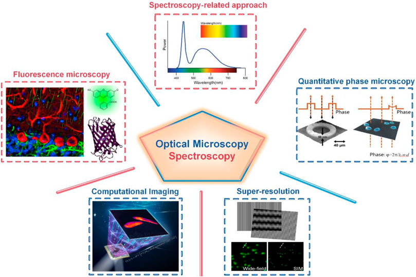

94% of researchers rate our articles as excellent or good
Learn more about the work of our research integrity team to safeguard the quality of each article we publish.
Find out more
EDITORIAL article
Front. Phys. , 06 September 2021
Sec. Optics and Photonics
Volume 9 - 2021 | https://doi.org/10.3389/fphy.2021.752435
This article is part of the Research Topic Optical Microscopic and Spectroscopic Techniques Targeting Biological Applications View all 24 articles
Editorial on the Research Topic
Optical Microscopic and Spectroscopic Techniques Targeting Biological Applications
Optical microscopy and spectroscopy are two mainstream tools for the exploration of an unknown microworld, especially in biomedical fields. Specifically, optical microscopy uses visible light and a system of lenses to magnify images of small samples [1]. It is non-invasive to live samples and has the capability to visualize specific structures once employing fluorescent labeling strategies [2]. Meanwhile, optical spectroscopy allows investigation of chemical, physical and electronic structures of matter at atomic, molecular, and macro scales [3]. In the past decades, remarkable developments of optical microscopy and spectroscopy have been witnessed, mainly in hardware implementation, algorithm performance, and innovative approaches [4–8].
We are glad to see that this special issue collects 23 articles, which report both the latest technological advances and the applications of optical microscopy and spectroscopy. Conceptually, these articles can be categorized into five classes according to the specific techniques (Figure 1).
The first class is fluorescence microscopy and correlation spectroscopy. These techniques can visualize the structures or the bio-molecular dynamics of samples by tagging them with fluorescence markers. Specifically, Li et al. review 3D imaging with dual-lens fluorescence microscopy, Qin and Xia present simultaneous two-photon fluorescence microscopy with two endogenous fluorescent coenzymes, entitled NADH and FAD. Yu et al. reviews fluorescence correlation spectroscopy (FCS), a powerful technique for quantification of molecular dynamics. Cui et al. fabricated a novel near-infrared fluorescent nanoparticles (NPs) for high-contrast and high-penetration in-vivo imaging.
The second class is quantitative phase microscopy (QPM). These techniques explore the phase of the light passing through or being reflected by a sample, providing quantitative information of 3D profiles or refractive index distributions of the sample. Picazo-Bueno et al., Guo et al., and Karako et al. propose new types of digital holographic microscopy (DHM) to quantify the thickness and refractive index distributions of samples. Meanwhile, Lima and Cojoc utilize DHM for the assessment of human neutrophil differentiation from myeloid cells. Zhou et al. propose a non-interference QPM strategy, recovering the phase from a stack of through-focus intensity images. Soto et al. present a partially coherent illumination based optical diffraction tomography (ODT) approach, allowing 3D refractive-index imaging of dynamic samples. Wu et al. propose a lens-free on-chip microscopy incorporated with a high-precision autofocusing algorithm for pixel-super-resolved QPM imaging of the sample. Sahu and Mazumder summarize some of the application of adaptive optics (AO) in two-photon fluorescence (TPF) microscopy for wavefront corrections in brain imaging and ophthalmoscopy.
The third class is super-resolution optical microscopy. The techniques of this class are far-field, minimally invasive, and they can image samples with a resolution surpassing the diffraction limit. Yong et al. present a high-dynamic-range structured illumination microscopy (HDR-SIM) using a multi-exposure acquisition strategy. With HDR-SIM, microspheres and vesicles with 420 intensity levels can be visualized in the same scene. Wen et al. demonstrate a digital micromirror device (DMD) based optical microscopic apparatus for dual-modality imaging, including quantitative differential phase contrast (qDIC) imaging and coherent SIM imaging. Zhao et al. review recent advances in high-speed SIM that include both hardware and software improvements, including reduction of the number of raw images, GPU acceleration, deep learning, and spatial domain reconstruction. Chen et al. present a super-resolution optical microscopy entitled expansion microscopy (ExM). This technique immobilizes the fluorescent molecules of interest in a polyacrylamide hydrogel. Then, the structure of the sample is spatially amplified as the hydrogel physically expands isotopically, thus indirectly improving resolution. This paper also present the applications of super resolution expansion microscopy in yeast.
The fourth class is deep-learning based computational imaging techniques. These techniques brought about new revolutionary computational power in optical microscopy. Zhang et al. present deep learning (DL) based adaptive optics (AO) to correct optical aberrations. They utilize a revised ResNet-34 network to infer the phase distortions of all the 224 valid zones on a SLM in one shot. Zhang et al. present a new deep-learning-based approach for recovering halo-free white-light diffraction phase images. The neural network-based method can accurately and rapidly remove the halo artifacts, not relying on any a-priori knowledge. Di et al. present an optimized structural convolution neural network PhaseNet for the reconstruction of digital holograms for DHM.
The fifth class is spectroscopy-related techniques. These techniques can non-invasively probe the structure, properties, and dynamics of molecules in different environments or different physico-chemical conditions. Hence, they are widely used in physics, astronomy, chemistry, biology, and related research fields. Here, Li et al. review the principle, configuration, and applications of coherent anti-stokes Raman scattering (CARS) microscopy, which can provide high-resolution, high-sensitivity, and non-invasive imaging of specific biomolecules without fluorescent labeling. Poulen et al. review the application of CARS in the identification and characterization of myelin in the mammalian nervous system of different species. Duadi et al. investigate near-infrared (NIR) measurements of turbid media using different size detectors at different positions. García-Martínez et al. propose a supercontinuum (SC) laser source from 450 to 1,600 nm with programmable spectrum by using liquid-crystal on silicon (LCoS) spatial light modulators (SLM).
To sum up, this research topic features 23 excellent articles encompassing the start-of-art developments and applications of optical microscopy and spectroscopy. This collection should be of interest to readers in the areas of optics, biophysics, and chemistry orientated subjects. It is our hope that this special research topic will contribute to the scientific advancements of optical microscopy and spectroscopy, as well as to their practical applications in biology and medicine.
All authors listed have made a substantial, direct, and intellectual contribution to the work, and approved it for publication.
The authors declare that the research was conducted in the absence of any commercial or financial relationships that could be construed as a potential conflict of interest.
All claims expressed in this article are solely those of the authors and do not necessarily represent those of their affiliated organizations, or those of the publisher, the editors and the reviewers. Any product that may be evaluated in this article, or claim that may be made by its manufacturer, is not guaranteed or endorsed by the publisher.
2. Dean KM, Palmer AE. Advances in Fluorescence Labeling Strategies for Dynamic Cellular Imaging. Nat Chem Biol (2014) 10:512–23. doi:10.1038/nchembio.1556
3. Atkin JM, Raschke MB. Optical Spectroscopy Goes Intramolecular. Nature (2013) 498:44–5. doi:10.1038/498044a
4. Bacia K, Kim SA, Schwille P. Fluorescence Cross-Correlation Spectroscopy in Living Cells. Nat Methods (2006) 3:83–9. doi:10.1038/nmeth822
5. Cheng J-X, Volkmer A, Xie XS. Theoretical and Experimental Characterization of Coherent Anti-Stokes Raman Scattering Microscopy. J Opt Soc Am B (2002) 19:1363–75. doi:10.1364/JOSAB.19.001363
6. Lippincott-Schwartz J, Manley S. Putting Super-Resolution Fluorescence Microscopy to Work. Nat Methods (2009) 6:21–3. doi:10.1038/nmeth.f.233
7. Lichtman JW, Conchello J-A. Fluorescence Microscopy. Nat Methods (2005) 2:910–9. doi:10.1038/nmeth817
8. Micó V, Zheng J, Garcia J, Zalevsky Z, Gao P. Resolution Enhancement in Quantitative Phase Microscopy. Adv Opt Photon (2019) 11:135–214. doi:10.1364/AOP.11.000135
9. Kemper B, von Bally G. Digital Holographic Microscopy for Live Cell Applications and Technical Inspection. Appl Opt (2008) 47:A52–A61. doi:10.1364/AO.47.000A52
10. Lyncée tec. Available at: https://www.lynceetec.com/types/downloads/
Keywords: optical microscopy, spectroscopy, super-resolution, deep-learning, quantitative phase imaging, fluorescent probe, algorithms
Citation: Micó V, Pedrini G, Lei M, Zuo C and Gao P (2021) Editorial: Optical Microscopic and Spectroscopic Techniques Targeting Biological Applications. Front. Phys. 9:752435. doi: 10.3389/fphy.2021.752435
Received: 03 August 2021; Accepted: 10 August 2021;
Published: 06 September 2021.
Edited and Reviewed by:
Lorenzo Pavesi, University of Trento, ItalyCopyright © 2021 Micó, Pedrini, Lei, Zuo and Gao. This is an open-access article distributed under the terms of the Creative Commons Attribution License (CC BY). The use, distribution or reproduction in other forums is permitted, provided the original author(s) and the copyright owner(s) are credited and that the original publication in this journal is cited, in accordance with accepted academic practice. No use, distribution or reproduction is permitted which does not comply with these terms.
*Correspondence: Peng Gao, peng.gao@xidian.edu.cn
Disclaimer: All claims expressed in this article are solely those of the authors and do not necessarily represent those of their affiliated organizations, or those of the publisher, the editors and the reviewers. Any product that may be evaluated in this article or claim that may be made by its manufacturer is not guaranteed or endorsed by the publisher.
Research integrity at Frontiers
Learn more about the work of our research integrity team to safeguard the quality of each article we publish.