- 1Department of Nutrition and Health Sciences, University of Nebraska, Lincoln, NE, United States
- 2School of Computing, University of Nebraska, Lincoln, NE, United States
Small extracellular vesicles (sEVs) and their RNA cargo in milk are bioavailable in humans, pigs, and mice, and their dietary depletion and supplementation elicits phenotypes. Little is known about the content and biological activity of sEVs in foods of animal origin other than milk. Here we tested the hypothesis that sEVs in chicken eggs (Gallus gallus) facilitate the transfer of RNA cargo from an avian species to humans and mice, and their dietary depletion elicits phenotypes. sEVs were purified from raw egg yolk by ultracentrifugation and authenticated by transmission electron microscopy, nano-tracking device, and immunoblots. The miRNA profile was assessed by RNA-sequencing. Bioavailability of these miRNAs in humans was assessed by egg feeding study in adults, and by culturing human peripheral blood mononuclear cells (PBMCs) with fluorophore-labeled egg sEVs ex vivo. To further assess bioavailability, fluorophore-labeled miRNAs, encapsulated in egg sEVs, were administered to C57BL/6 J mice by oral gavage. Phenotypes of sEV RNA cargo depletion were assessed by feeding egg sEV and RNA-defined diets to mice and using spatial learning and memory in the Barnes and water mazes as experimental readouts. Egg yolk contained 6.30 × 10¹⁰ ± 6.06 × 10⁹ sEVs/mL, which harbored eighty-three distinct miRNAs. Human PBMCs internalized sEVs and their RNA cargo. Egg sEVs, loaded with fluorophore-labeled RNA and administered orally to mice, accumulated primarily in brain, intestine and lungs. Spatial learning and memory (SLM) was compromised in mice fed on egg sEV- and RNA-depleted diet compared to controls. Egg consumption elicited an increase of miRNAs in human plasma. We conclude that egg sEVs and their RNA cargo probably are bioavailable. The human study is registered as a clinical trial and accessible at https://www.isrctn.com/ISRCTN77867213.
Introduction
Extracellular vesicles (EVs) are detectable in most, if not all, body fluids including saliva, plasma, urine, and milk (1, 2). Five classes of EVs may be distinguished based on biogenesis, size, and cargo: apoptotic bodies, microvesicles, exosomes, exomeres, and midbody remnants (3–6). Exosomes have attracted more attention than other EVs, because they facilitate cell-to-cell communication, e.g., by transferring miRNA cargo from exosome donor cells to adjacent and distant recipient cells (4). MiRNAs have been implicated in the regulation of approximately 60% of human genes and loss of miRNA maturation in Dicer knockout mice is embryonic lethal (7, 8). In this manuscript, we will refer to exosomes as small EVs (sEVs).
Food-borne sEVs and their RNA cargo have attracted considerable attention ever since it was reported that sEVs and miRNAs do not originate exclusively in endogenous synthesis but may also be absorbed from milk in humans, pigs, and mice (9–13). Milk sEVs and their miRNA cargo meet the definition of bioactive food compounds by the National Cancer Institute, which is “A type of chemical found in small amounts in plants and certain foods (such as fruits, vegetables, nuts, oil and whole grains). Bioactive compounds have actions in the body that may promote good health. They are being studied in the prevention of cancer, heart disease, and other diseases” (14). For example, human milk contains approximately 2.18 × 10¹¹ sEVs/mL that harbor more than 200 distinct miRNAs (12). Milk sEVs accumulated primarily in the small intestinal mucosa, liver, spleen, heart, kidneys, and brain (11). Dietary depletion of milk sEVs and miRNAs impaired spatial learning and memory (SLM) (11), moderately decreased muscle strength (15), decreased fecundity and postnatal survival (16), and altered neurotransmitter metabolism (L-glutamate, tryptophan and purines), and bacterial communities in the cecum in humans and mice (17), whereas supplementation with milk sEVs decreased the severity of necrotizing enterocolitis in neonate mice (13) and improved bone health in mice models of osteoporosis (18).
Here, we sought to fill the knowledge gap whether milk is unique as a source of dietary sEVs and miRNA, or whether sEVs and miRNA in other animal-borne foods also have biological activity (19). We chose chicken eggs as model because, unlike milk miRNAs, chickens (Gallus gallus) express miRNAs with nucleotide sequences that can be distinguished from endogenous miRNAs in humans and mice, thereby serving as endogenous tracers of dietary miRNAs (20, 21). Specifically, we pursued the following aims. (1) Determine whether chicken eggs contain sEVs and assess the miRNA content of egg sEVs. (2) Determine bioavailability of chicken miRNA in humans and whether human peripheral blood mononuclear cells (PBMCs) internalize fluorophore-labeled egg sEVs and their RNA cargo ex vivo. (3) Assess the bioavailability and tissue distribution of fluorophore-labeled egg sEVs and miRNAs in mice. (4) Determine whether mice fed an egg sEV and miRNA sufficient (ERS) diet perform better than mice fed an egg sEV and miRNA-depleted (ERD) diet in tests of SLM.
Materials and methods
Small RNA sequencing analysis of eggs
Chicken eggs were purchased from a local grocery store (Hyvee®). Egg yolk and albumen were separated manually. Samples (50 ug) were mixed with 5 uL of proteinase K (Thermo Fisher) and incubated at 37°C with light shaking for 60 min (22). RNA was isolated from egg yolk and albumen by using Trizol. Sequencing was done on an Illumina-Hiseq 2,500 platform with a single-end 50-basepair protocol (BGI, Inc.; Hong Kong, China) using NEBNext® Multiplex Small RNA Library Prep Set for illumina® Set 1. The CAP-miSeq Analysis pipeline and miRDeep2 was used for analysis (23). A minimum of five sequencing reads was used as threshold for miRNAs and only miRNAs that were detected in both yolk samples are reported in this paper. The reads were mapped to chicken miRNAs using miRbase 22 (24). A threshold of minimum five sequencing reads were used to identify egg miRNAs and miRNAs detected in both yolks were reported.
Isolation and characterization of egg sEVs
Egg yolks were mixed with ten volumes of phosphate-buffered saline (PBS). sEVs were isolated by sequential ultracentrifugation as previously described (25). Some samples were further purified by iodixanol density gradient centrifugation (26). The protein concentration in gradient fractions was measured using the BCA Protein Assay Kit (Thermo Fisher). Fraction 6 contained the highest concentration of protein and was selected for further analysis. Fraction 6 was diluted with 10 mL PBS and centrifuged at 120,000 g at 4°C for 2 h. The sEV pellet was dissolved in 1 mL PBS and characterized following EV community standards (27). In immunoblots, CD63, TSG101 and Alix were used as positive markers for extracellular vesicles, while integrin β1 was used as marker for microvesicles (28). The following antibodies and dilutions were used: mouse anti-Alix (Abcam, ab117600, Cambridge, United Kingdom, 1000-fold dilution), rabbit anti-TSG101 (Abcam, ab30871, 100-fold dilution), mouse anti-CD63 (sc-5,275 Santa Cruz, 1,000-fold dilution), and rabbit anti-integrin β1 (cat. no. 4706 Cell Signaling, Leiden, The Netherlands; 1,000-fold dilution). IRDye® 680RD goat anti-rabbit IgG and IRDye® 800CW goat anti-mouse IgG (LI-COR, 10,000-fold dilution) were used as secondary antibodies. Transmission electron microscopy (Hitachi H7500) and nanoparticle tracking analysis (NTA, Nanosight NS300; Malvern Instruments Ltd., United Kingdom) were conducted as previously described.
MiRNA bioavailability in humans
Five apparently healthy men, ages 26–35 years, consumed four hard-boiled eggs within less than 10 min. Blood was collected in EDTA tubes before and at timed intervals after egg consumptions. Subjects did not consume poultry products 24 h before study began until after the last sample was collected. Plasma and Peripheral Blood Mononuclear Cells (PBMCs) were separated using density centrifugation (Histopaque-1,077®) as previously described (29). RNA was isolated from plasma by using Trizol and analyzed by RNA-sequencing analysis (BGI, Inc.; Cambridge, MA, United States) as described above.
RNA was extracted from PBMCs by using the RNAeasy Mini Kit (Qiagen). mRNAs were analyzed by RNA-sequencing analysis (paired-end 150-bp reads; Illumina HiSeq 2,500) in the DNA Sequencing Core at the University of Nebraska Medical Center (Omaha, NE) and selected miRNAs were analyzed by quantitative reverse transcriptase PCR (qRT-PCR) (Supplementary Table S1) (30). All human research in this paper was approved by the Institutional Review Board in the University of Nebraska-Lincoln (protocols no. 14585 and 16566).
Internalization of sEVs and RNA cargo by human PBMCs ex vivo
PBMCs were cultured in RPMI-1640 media supplemented with exosome-depleted fetal bovine serum. Exo-Glow labeled sEVs (2.5 × 10¹²) were added to PBMC cultures (106 cells in 3 mL media) from four subjects (ages 26–35 years) and cultured at 37°C for 9 h. Controls were cultured with unlabeled sEVs. Extracellular sEVs were removed by washing PBMCs in PBS (1,500 g, 10 min). The final PBMC pellet was resuspended in 3 mL PBS and fluorescence was measured using an EVOS FL Cell Imaging System (AMG, United States).
RNA cargo distribution studies in mice
We used the following strategies for assessing the bioavailability distribution of egg sEV RNA in mice: In the first strategy, the entire pool of single-stranded RNA in egg sEVs was labeled with Exo-Glow following the manufacturer’s recommendations (Exo-Glow Red kit, System Biosciences, Inc., EXOR100A-1). Exo-Glow labeled sEVs were administered to C57/BL6 age 6 weeks (Jackson Laboratories, stock number 000664) by oral gavage (1 × 10¹²/g body weight). Controls received unlabeled sEVs and free Exo-Glow. Note that the vendor has replaced the Exo-Glow Red kit with a new kit that labels polyA tails and therefore cannot be used to label miRNAs. Tissues were harvested 12 and 24 h after gavage, rinsed and perfused with PBS, and fluorescence was assessed in a LI-COR Odyssey® CLx. In another strategy, sEVs were loaded with two miRNAs found in egg yolk (Supplementary Table S2), i.e., synthetic IRDye-labeled miR-9 and miR-29a (IDTDNA, Inc). The miRNAs were chosen because they are implicated in SLM (31, 32). miR-9 is associated with neuronal development and miR-29 has been previously implicated in synapse formation and plasticity (33, 34). sEVs were loaded with IRDye-labeled miRNAs and extra-exosomal miRNAs were removed as previously described (10). Loaded sEVs were used within 2 h of preparation. sEVs (1012 sEVs /per g body weight) were administered by oral gavage to C57/BL6 mice age 6 weeks. Controls received unloaded sEVs. Tissues were harvested 12 h after gavage, rinsed and perfused with PBS, and fluorescence was assessed in a LI-COR Odyssey® CLx. All animal experiments were approved by the Institutional Animal Care Program at the University of Nebraska-Lincoln (protocol 1229).
Gene expression in the murine hippocampus
We conducted an egg sEV and miRNA feeding study to assess whether dietary depletion elicits gene expression changes in the hippocampus, i.e., a region in the brain that is crucial for SLM (35, 36). The pelleted rodent diets represent a variation of our milk sEV and RNA-defined diets and are based on the AIN-93G formulation (37, 38). Briefly, in the egg sEV and RNA-sufficient (ERS) diet, the equivalent of four egg yolks consumed by an adult per day is substituted for 80 g lyophilized yolk (Supplementary Table S3). In the egg sEV and RNA-depleted (ERD) diet, the egg yolks were ultrasonicated for 90 min (VWR Aquasonic 250 T) and incubated at 37°C for 60 min prior to pelleting. Ultrasonication led to a decrease of 62.1 ± 3.0, 81.3% ± 1.7, 82.4% ± 1% and 92.1 ± 1.5% of miR-30a-5p, miR-92-3p, miR-181-5p and miR-221-3p, respectively, compared to non-ultrasonicated yolks. Previous studies showed that ultrasonication leads to an irregular sEV surface morphology and a decrease in sEV count (37). All other ingredients are identical in ERS and ERD diets. Male and female mice were fed experimental diets from age 3 to 9 weeks with free access to food and water (12-h light–dark cycle; 22°) when ERS/ERS and ERD/ERD pairs were mated. Offspring continued on parental diets until age 22 weeks when hippocampi were harvested.
RNA was purified using the miRNAeasy Mini Kit (Qiagen). RNA integrity was confirmed in the DNA Sequencing Core at the University of Nebraska Medical Center by using a Fragment Analyzer Automated CE System (Advanced Analytical Technologies, Inc.). Libraries were prepared by using the TrueSeq RNA Library Prep Kit v2 (Illumina, Inc.) and sequenced using an Illumina HiSeq 2,500 platform and a 150 base-pair paired-end protocol (RNA-seq). The sequencing reads were annotated to the mouse reference genome [GRCm38, mm10] using RSEM (39).
Spatial learning and memory
Barnes and Morris water mazes were used to assess SLM. Tests were performed in C57BL/6 breeders fed ERS or ERD diets (ERS x ERS and ERD x ERD), and in F1 pups born to ERS and ERD parents and fed parental diets (n = 8). In the Barnes maze, breeders were studied at age 12 weeks and F1 offspring was studied at age 16 weeks as previously described (40). Mice were trained three times per day for six consecutive days, followed by a performance test on day 7. The ANY-maze video tracking system (Stoelting Co., Wood Dale, IL, United States) was used to record the time until mice entered the escape hole. The Morris water maze tests were conducted as previously described at a water temperature of 26°C (41).
Statistical analysis
Data variances were homogenous as per Bartlett’s test. Effect of eggs consumption on mRNA abundance in PBMCs was analyzed by the paired, two-sided t-test. miRNA concentrations in plasma were analyzed by the Wilcoxon signed-rank test. GraphPad Prism 6 (GraphPad Software Inc.) was used for statistical analyses. Differentially expressed transcripts were identified by DESeq2 (42) using the paired-sample design and multiple test correction (adjusted value of p < 0.05 for multiple comparisons using Benjamini’s method). Differences were considered statistically significant at value of p < 0.05.
Results
sEV in eggs
When sEVs were isolated by sequential centrifugation, the concentration was 6.30 × 10¹⁰ ± 6.06 × 10⁹ sEVs per mL yolk with a diameter of 110.0 ± 20.5 nm (mean ± SD) (Figure 1A). Consistent with the previous studies of sEVs (11), egg sEVs showed a spherical shape (Figure 1B). Iodixanol density centrifugation was used for further purification of egg sEVs. Column fraction 6 contained 7.90 × 10⁹ ± 0.19 × 10³ sEVs (Figure 1C). The diameter measured 99.5 ± 30.1 nm (mean ± SD) for sEVs purified by a combination of ultracentrifugation and iodixanol gradient (Figure 1D), which is the size expected for these vesicles (11). TEM analysis revealed a spherical shape (Figure 1E). Immunoblots detected exosome markers (CD63, TSG101 and Alix) in egg sEVs, whereas a microvesicle marker (integrin β1) was not detectable (Figure 1F).
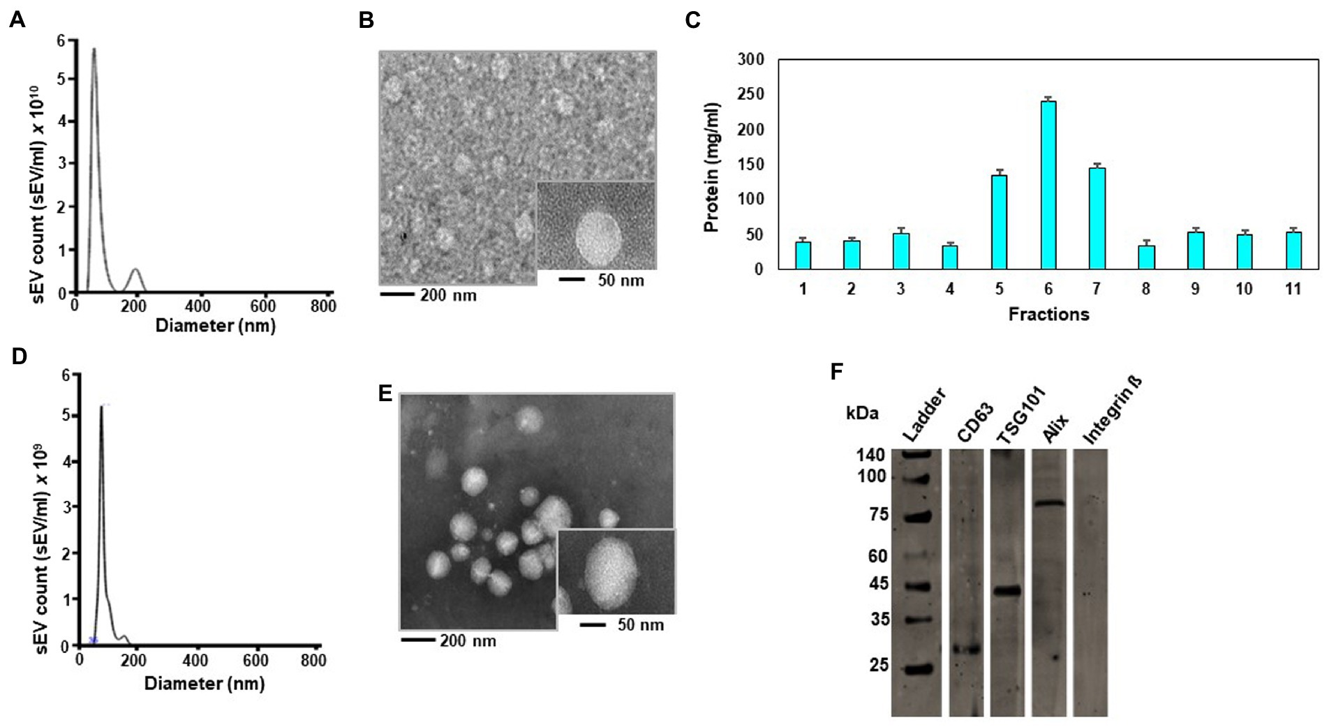
Figure 1. Authentication of small extracellular vesicles (sEVs) from chicken egg yolk. Size distribution of sEVs isolated by ultracentrifugation without subsequent iodixanol gradient centrifugation analyzed in a Nanosight NS300 instrument (A). Transmission electron microscope (TEM) image of sEV suspension isolated by ultracentrifugation without subsequent iodixanol gradient centrifugation. The large-field image was obtained at 20,000-fold magnification and the insert depicts an sEV at 50,000-fold magnification (B). Protein concentration in iodixanol gradient fractions (C). Size distribution of sEVs in iodixanol gradient fraction 6 (D). TEM image of sEVs in iodixanol gradient fraction 6. The large-field image was obtained at 20,000-fold magnification and the insert depicts an sEV at 50,000-fold magnification (E). Immunoblot of chicken egg sEVs in iodixanol gradient fraction 6. Samples were run on the same gel. Lanes were cut for probing with antibodies and electronically re-assembled. Proteins were detected as follows: CD63 at 26 kDa, TSG101 at 44 kDa, Alix at 96 kDa and no Integrin ᵝ at 130 kDa (F).
miRNAs in egg sEVs
Two eggs yielded 0.141 and 0.319 ng RNA per μL yolk. Eighty-three miRNAs were identified in both yolks by RNA-sequencing analysis. Out of these eighty-three miRNAs, fifty-six were common in both yolk samples (Supplementary Table S2). Two eggs yielded only 0.012 and 0.017 ng RNA per μL albumen, which was insufficient for RNA-sequencing analysis.
Bioavailability of egg miRNAs in humans
Egg consumption elicited postprandial changes in plasma miRNA levels in dose–response studies in adults. Plasma miRNAs were analyzed before and 9 hours after consuming two to four eggs (n = 5). One hundred-seventeen miRNAs were identified by RNA-sequencing analysis. Twenty-six miRNAs were upregulated after egg consumption; the others were downregulated or remained unchanged (Figure 2, Supplementary Table S4). The nucleotide sequences of 75 miRNAs detected in plasma 9 h after egg consumption are identical in humans and chickens (43). Thirty-one miRNAs have near-identical sequences in chickens and humans with short extensions (1–4 base pairs) at the 5′-end or the 3′-end. Eleven miRNAs have nucleotide sequences that differed by 1 bp in chickens and humans.
These eleven chicken specific miRNAs were below detection limit in human plasma after egg meals (low RPKM). Their low abundance in plasma was attributed to their low abundance in egg yolk, e.g., miR-146c-5p (Supplementary Table S2).
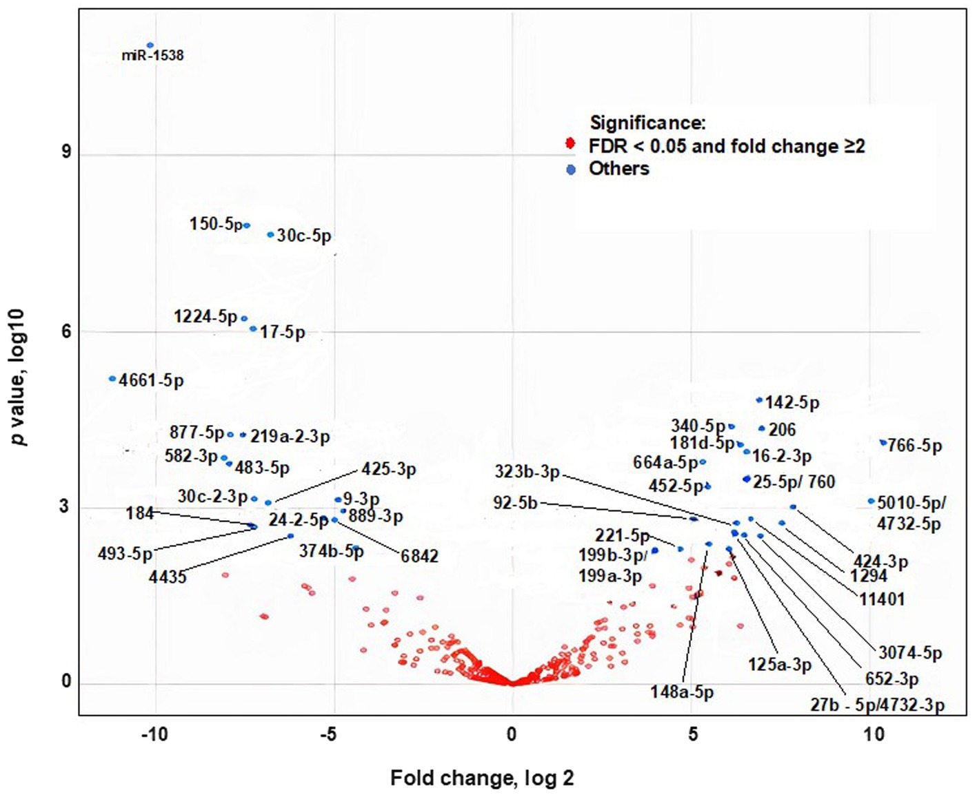
Figure 2. Effect of egg consumption of miRNAs expression in human plasma. Blue dots denote miRNAs for which the expression differs at least 2-fold before compared to 9 h after egg consumption (False Discovery Rate adjusted Value of p < 0.05).
miRNA levels and mRNA expression in human peripheral blood mononuclear cells
Egg consumption caused an increase in miRNA levels in PBMCs, and the increase was associated with changes in gene expression in vivo. We isolated PBMCs from human subjects before and after consumption of four eggs and analyzed the levels of three miRNAs by qRT-PCR. The levels of miR-142-5p, miR-181b-5p, and miR-30a-5p were more than 2-fold higher 9 hours after egg consumption compared to before consumption (Figure 3A).
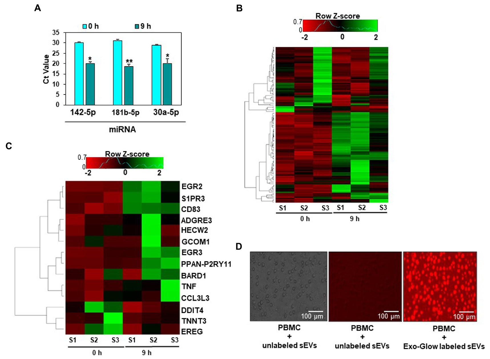
Figure 3. Egg small extracellular vesicles (sEVs) deliver miRNAs to human peripheral blood mononuclear cells (PBMCs). Expression of miRNAs detected in PBMCs 9 h after egg consumption. Data are presented as means ± standard deviations. *Value of p <0.05 (n = 5) for Student’s paired t-test (A). Heat map of the expression data of all genes in PBMCs (B). Heat map of fourteen genes for which the expression was at least 2-fold or more different after the egg meal. For each time point, samples (n = 3) were collected from the same subjects before and after the egg meal (C). Uptake of Exo-Glow labeled RNAs, encapsulated in egg sEVs by human PBMCs (D).
Eighty-eight genes were differentially expressed in PBMCs before and after egg consumption, as per RNA-sequencing analysis. For 14 mRNAs the difference was 2-fold or greater (Figures 3B,C, Supplementary Table S5). Using all differentially expressed mRNAs as input, twenty Kyoto Encyclopedia of Genes and Genomes (KEGG) pathways and two hundred and forty-one Gene Ontology (GO) terms were identified (Supplementary Table S6). mRNA-sequencing data are available in BioProject (PRJNA422156). The sEV-dependent changes in miRNA and mRNA expression in vivo are consistent with our observation that human PBMCs internalized Exo-Glow Red labeled egg sEVs ex vivo (Figure 3D).
Egg RNA distribution in mice
Fluorophore-labeled RNA, encapsulated in egg sEVs, was detected in the intestinal mucosa and peripheral tissues. Two RNA labeling strategies were used to assess sEV cargo distribution. In one of the strategies, the entire pool of single-stranded RNA in sEVs was labeled with Exo-Glow. Exo-Glow labeled RNA accumulated in brain and lungs when administered by oral gavage (Figures 4A–C). Using an alternative strategy, egg sEVs were loaded with IRDye-labeled synthetic miRNAs. Oral administration of the miRNA-loaded egg sEVs led to the accumulation of miRNAs (miR-9 and miR-29a) in the intestinal mucosa and brain (Figures 5A–C).

Figure 4. Distribution of Exo-Glow labeled small extracellular vesicles (sEVs) in C57BL/6 J mice. Free Exo-Glow, unlabeled egg sEVs and Exo-Glow labeled sEVs were administered by oral gavage. Mice were euthanized 12 h and 24 h after oral administration of sEVs (A). Densitometry analysis of background-normalized fluorescence in brain and lungs 12 h (B) and 24 h (C) after oral gavage. Data were normalized by using the fluorescence from tissue-free sections in the dish. Values are mean ± SEM (n = 3). Data were analyzed by Mann–Whitney U test (**Value of p < 0.01).
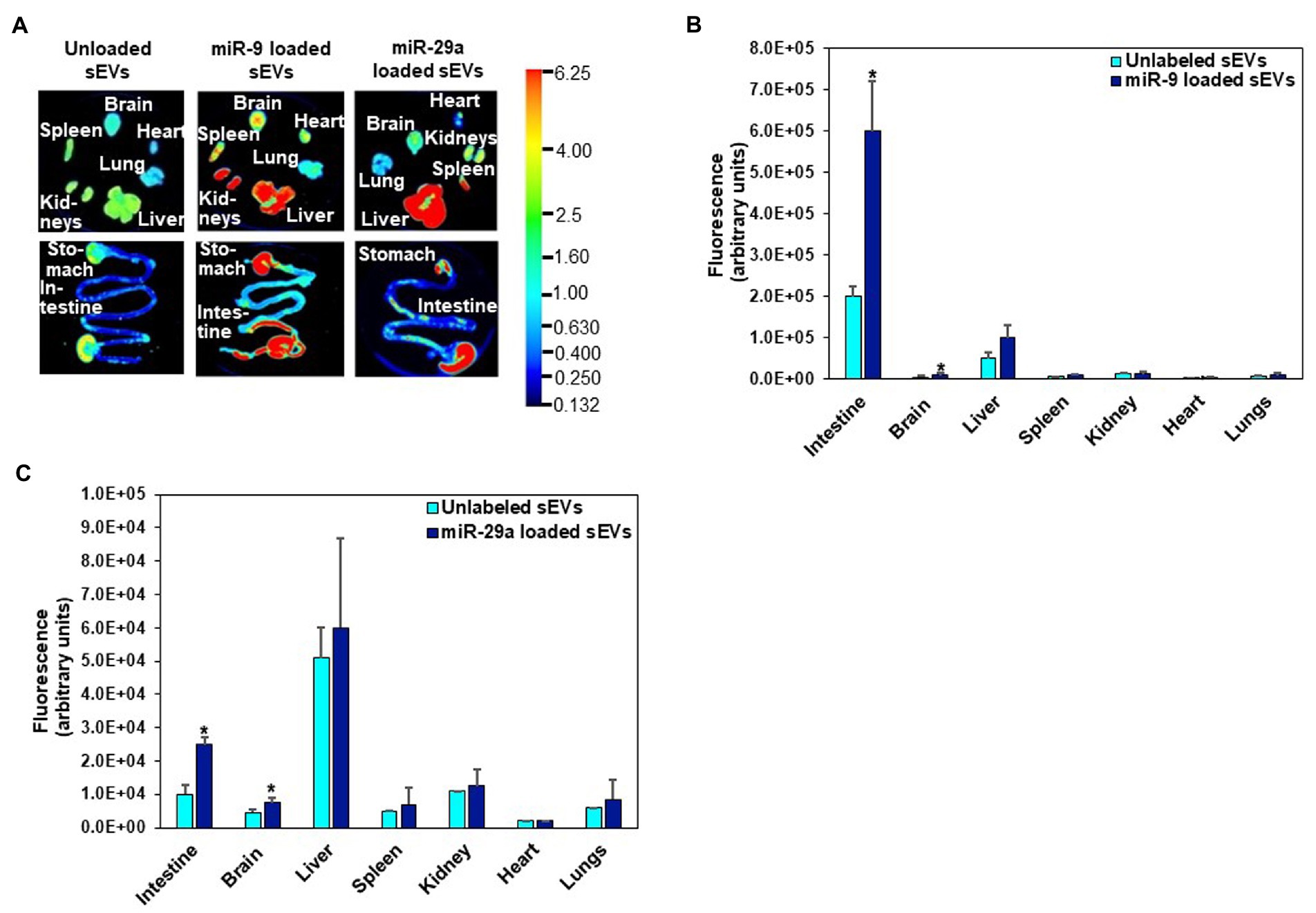
Figure 5. Distribution of egg sEVs loaded with IRDye-labeled miR-9 or miR-29a and delivered by oral gavage in C57BL/6 J mice (A). Densitometric analysis of background-normalized fluorescence in intestine, brain, liver, spleen, kidney, heart, and lungs showing accumulation of miR-9 (B) and miR-29a (C). Values are mean ± SEM (n = 3). Data were analyzed by Mann–Whitney U test (*Value of p < 0.05). Fluorescence is reported as arbitrary units (au) (B, C). Data were normalized by using the fluorescence from tissue-free sections in the dish.
Egg sEV-dependent gene expression changes in the brain and associated SLM phenotypes
The accumulation of RNA in egg sEVs in the brain was associated with changes in gene expression. When mice were fed egg sEV and RNA-deficient or egg sEV and RNA-sufficient diets, seven genes implicated in SLM were differentially expressed in the hippocampus (Figure 6), i.e., the region of the brain that regulates SLM (35, 44).
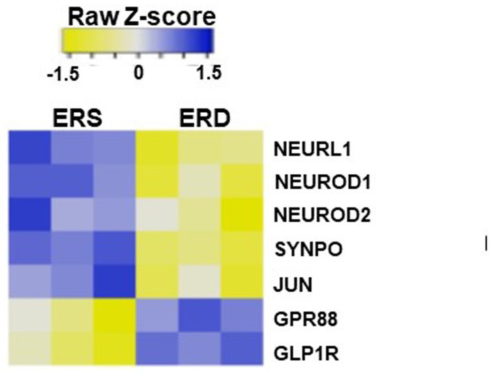
Figure 6. Heat map of differential expression in genes associated with spatial learning and memory in mice brain fed by sEV rich and sEV deficient diet.
Dietary depletion of egg sEVs and RNA impaired SLM, assessed by Barnes maze and Morris water maze in mice. Mice fed on ERD diet took more than ten times longer to locate and enter the escape hole in a Barnes maze compared to ERS-fed mice at 4, 8, 16, and 20 weeks of feeding (Figure 7A). Morris water maze test results showed that the mice fed on ERD diet took longer to locate the hidden platform, compared to ERS-fed mice at 6 and 11 weeks (Figure 7B).
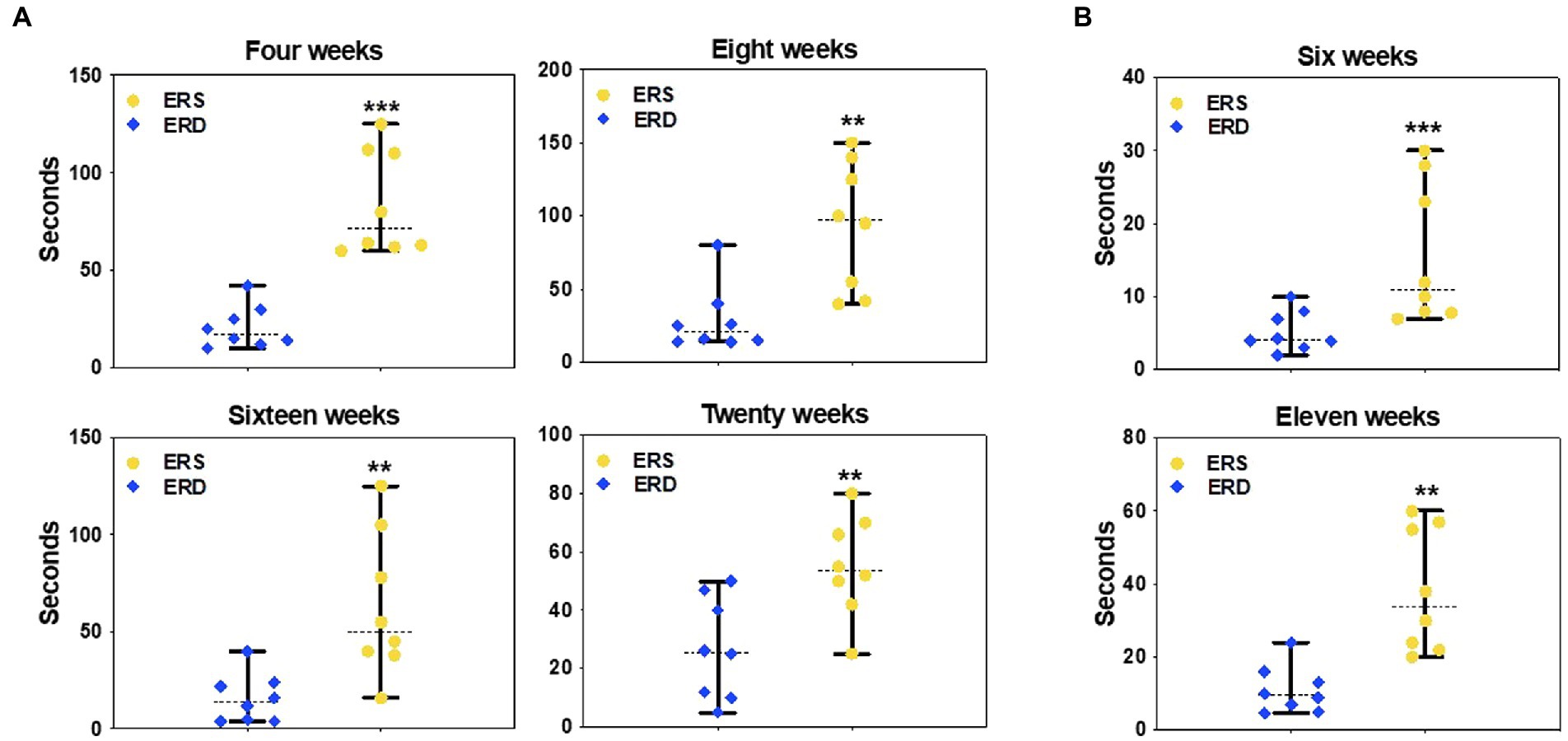
Figure 7. Effect of egg small EVs and RNA-sufficient (ERS) diet on spatial learning and memory in C57/BL6J mice. Latency to locate the escape platform in the Barnes maze after consuming sEV RNA rich and sEV RNA deficient diets for 4, 8, 16, and 20 weeks in C57BL/6 mice (A). Latency to locate the escape platform in the Morris Water maze after consuming ERS or ERD diets for 6 and 11 weeks in C57BL/6 mice (B). Data are presented as median with range (n = 8). The middle line denotes the median. Data were analyzed by Mann–Whitney test. Bars indicate the minimum and maximum values. *Value of p <0.05; **Value of p <0.01.
Discussion
The presence of extracellular vesicles (EVs) in diet such as edible plant juices and milk has been the center of attention for the past few years. Studies suggest that these vesicles and their cargo (including miRNA) can be absorbed through intestine and travel via blood stream affecting the health of various organs through gene-regulation and ultimately influencing consumer health. Here, in this study, we explored and validated the presence of sEVs and its miRNA cargo in chicken eggs particularly in yolk. Consumption of chicken eggs led to the presence of chicken miRNA in blood sera of consumers, which potentially were absorbed through intestine. Similarity of chicken and human miRNAs indicate their potential role in regulating immune response, metabolic and vital cellular processes in human consumers.
This study provides compelling evidence that chicken egg yolk, but not albumen, contains detectable amounts of sEVs with features expected of exosomes, including morphology, size, marker proteins, and miRNA cargo. Yolk content is the sole source of nutrition for the avian embryo (45, 46) and it is reasonable to propose that yolk sEVs and their RNA cargo are crucial for normal embryonic development similar to what has been reported for milk sEVs and postnatal development in mammals (16).
The intent of this study was to assess the biological activity of egg sEVs and miRNA cargo in non-avian species. After the initial demonstration that egg yolk contains sEVs and documenting the miRNA content of the sEVs, we focused on two aspects that are crucial when assessing the relevance in nutrition, namely bioavailability and phenotypes of depletion. Evidence is strong that human PBMCs internalize egg sEVs and their RNA cargo ex vivo, and egg miRNAs accumulate in murine tissues, predominantly in the gastrointestinal mucosa, brain, and lungs. In contrast, we cannot state with a high degree of confidence that avian miRNAs entered human circulation. While postprandial changes in plasma miRNA levels were readily apparent in humans after egg consumption, the changes were limited to miRNAs for which the nucleotide sequence is conserved in humans and chickens. We attributed the absence of a postprandial increase in chicken-specific miRNAs in human plasma to their low content in yolk. For example, gga-miR-146c-5p was one of the least abundant miRNAs in yolk and not detected in human plasma. We acknowledge the limitation that we did not collect a 9-h sample from subjects who did not consume eggs, but used the baseline sample as control. Approximately a third of plasma miRNAs exhibit a diurnal variation (47).
We identified two explanations for postprandial changes in miRNA levels in plasma and PBMCs. In one scenario, egg consumption increases or decreases the endogenous synthesis of miRNAs, i.e., the miRNAs do not originate in eggs. In the other scenario, egg miRNAs are bioavailable. An alternative, not mutually exclusive, explanation is that humans absorbed miRNAs from egg sEVs, which led to changes in endogenous miRNA levels. Unfortunately, chicken-specific miRNAs are rare in egg sEVs. A limitation of the bioavailability arm of this study is that one cannot determine the effects of boiling on the content of sEVs and miRNAs in eggs because it proved impossible to purify sEVs and RNA from hard-boiled eggs (data not shown).
As for phenotypes of sEV and miRNA depletion, we focused on SLM because loss of SLM was among the strongest phenotypes of (milk) sEV and RNA depletion observed in previous studies in mice (11). In the previous studies of milk sEVs, the latency for entering the escape hole in the Barnes maze was 9-fold greater in adolescent mice fed a milk sEV-depleted diet compared to controls fed a milk sEV-sufficient diet. Diet effects were similar in this study of egg sEVs and SLM. The number of hippocampal genes that were differentially expressed in sEV and miRNA-defined diets was greater for bovine milk (219 genes) than eggs (86 genes). We speculate that the differential effects of milk and egg sEVs in the hippocampus might be due to a higher degree of evolutionary conservation in cow and mice compared to chicken and mice.
We conclude that studies of milk sEVs are a more fruitful line of investigation than studies of chicken sEVs, primarily because of the unique role of milk in infant nutrition and the use of milk sEVs in the delivery of therapeutics (48, 49). Because of the low content of chicken-specific miRNAs in eggs, we could not detect such transcripts in human plasma with satisfactory confidence. We shall point out a unique opportunity afforded by egg sEVs. Hens deposit in eggs what is crucial for early embryonic development, and it would be of interest to study the time courses of miRNA biogenesis in developing avian embryos. Such studies could lead to the identification of miRNAs that are essential during the early stages in embryonic development. Secondly, the study opens a new avenue of research in egg nutrition and its short- and long-term effects on human health.
Data availability statement
The datasets presented in this study can be found in online repositories. The names of the repository/repositories and accession number(s) can be found at: https://www.ncbi.nlm.nih.gov/, PRJNA422156.
Ethics statement
The studies involving human participants were reviewed and approved by Institutional Review Board in the University of Nebraska-Lincoln (protocols no. 14585 and 16566). The patients/participants provided their written informed consent to participate in this study. The animal study was reviewed and approved by Institutional Animal Care Program at the University of Nebraska-Lincoln (protocol 1229).
Author contributions
JZ conceived the project. DF, KH, and SB collected data. JM, DF, JS, KH, and JC analyzed data. JM and DF wrote the draft manuscript. JZ wrote the final manuscript. All authors contributed to the article and approved the submitted version.
Funding
This study was supported by the National Institutes of Health (NIH 1P20GM104320), the National Institute of Food and Agriculture (NIFA 2016–67001-25301), the Egg Nutrition Center, United States Department of Agriculture (Hatch NEB-36-087 and W4002).
Acknowledgments
Samples were analyzed in the Biomedical and Obesity Research Core, which is supported by the National Institutes of Health (NIH 1P20GM104320).
Conflict of interest
The authors declare that the research was conducted in the absence of any commercial or financial relationships that could be construed as a potential conflict of interest.
Publisher’s note
All claims expressed in this article are solely those of the authors and do not necessarily represent those of their affiliated organizations, or those of the publisher, the editors and the reviewers. Any product that may be evaluated in this article, or claim that may be made by its manufacturer, is not guaranteed or endorsed by the publisher.
Supplementary material
The Supplementary material for this article can be found online at: https://www.frontiersin.org/articles/10.3389/fnut.2023.1162679/full#supplementary-material
References
1. Lasser, C, Alikhani, VS, Ekstrom, K, Eldh, M, Paredes, PT, Bossios, A, et al. Human saliva, plasma and breast milk exosomes contain RNA: uptake by macrophages. J Transl Med. (2011) 9:9. doi: 10.1186/1479-5876-9-9
2. Huebner, AR, Somparn, P, Benjachat, T, Leelahavanichkul, A, Avihingsanon, Y, Fenton, RA, et al. Exosomes in urine biomarker discovery. Adv Exp Med Biol. (2015) 845:43–58. doi: 10.1007/978-94-017-9523-4_5
3. Gyorgy, B, Szabo, TG, Pasztoi, M, Pal, Z, Misjak, P, Aradi, B, et al. Membrane vesicles, current state-of-the-art: emerging role of extracellular vesicles. Cell Mol Life Sci. (2011) 68:2667–88. doi: 10.1007/s00018-011-0689-3
4. OBrien, K, Breyne, K, Ughetto, S, Laurent, LC, and Breakefield, XO. RNA delivery by extracellular vesicles in mammalian cells and its applications. Nat Rev Mol Cell Biol. (2020) 21:585–606. doi: 10.1038/s41580-020-0251-y
5. Zhang, H, Freitas, D, Kim, HS, Fabijanic, K, Li, Z, Chen, H, et al. Identification of distinct nanoparticles and subsets of extracellular vesicles by asymmetric flow field-flow fractionation. Nat Cell Biol. (2018) 20:332–43. doi: 10.1038/s41556-018-0040-4
6. Rai, A, Greening, DW, Xu, R, Chen, M, Suwakulsiri, W, and Simpson, RJ. Secreted midbody remnants are a class of extracellular vesicles molecularly distinct from exosomes and microparticles. Commun Biol. (2021) 4:400. doi: 10.1038/s42003-021-01882-z
7. Friedman, RC, Farh, KK, Burge, CB, and Bartel, DP. Most mammalian mRNAs are conserved targets of microRNAs. Genome Res. (2009) 19:92–105. doi: 10.1101/gr.082701.108
8. Bernstein, E, Kim, SY, Carmell, MA, Murchison, EP, Alcorn, H, Li, MZ, et al. Dicer is essential for mouse development. Nat Genet. (2003) 35:215–7. doi: 10.1038/ng1253
9. Baier, SR, Nguyen, C, Xie, F, Wood, JR, and Zempleni, J. MicroRNAs are absorbed in biologically meaningful amounts from nutritionally relevant doses of cow milk and affect gene expression in peripheral blood mononuclear cells, HEK-293 kidney cell cultures, and mouse livers. J Nutr. (2014) 144:1495–500. doi: 10.3945/jn.114.196436
10. Manca, S, Upadhyaya, B, Mutai, E, Desaulniers, AT, Cederberg, RA, White, BR, et al. Milk exosomes are bioavailable and distinct microRNA cargos have unique tissue distribution patterns. Sci Rep. (2018) 8:11321. doi: 10.1038/s41598-018-29780-1
11. Zhou, F, Ebea, P, Mutai, E, Wang, H, S, S, Navazesh, SE, et al. Small extracellular vesicles in milk cross the blood-brain barrier in murine cerebral cortex endothelial cells and promote dendritic complexity in the hippocampus and brain function in C57BL/6J mice. Front Nutr. (2022) 9:838543. doi: 10.3389/fnut.2022.838543
12. Leiferman, A, Shu, J, Upadhyaya, B, Cui, J, and Zempleni, J. Storage of extracellular vesicles in human milk, and microRNA profiles in human milk exosomes and infant formulas. J Pediatr Gastroenterol Nutr. (2019) 69:235–8. doi: 10.1097/mpg.0000000000002363
13. Li, B, Hock, A, Wu, RY, Minich, A, Botts, SR, Lee, C, et al. Bovine milk-derived exosomes enhance goblet cell activity and prevent the development of experimental necrotizing enterocolitis. PLoS One. (2019) 14:e0211431. doi: 10.1371/journal.pone.0211431
14. National Cancer Institute: NCI Dictionary of Cancer Terms. Available at: https://www.cancer.gov/publications/dictionaries/cancer-terms/def/bioactive-compound (Accessed July 6, 2014).
15. Leiferman, AL, Aguilar, A, Mutai, E, Adamec, J, and Zempleni, J. Dietary depletion of bovine milk exosomes elicits changes in amino acid metabolism in C57BL/6 mice. FASEB J. (2017) 31:135.3 [peer-reviewed abstract]. doi: 10.1096/fasebj.31.1_supplement.135.3
16. Sadri, M, Shu, J, Kachman, SD, Cui, J, and Zempleni, J. Milk exosomes and microRNAs cross the placenta and promote embryo survival in mice. Reproduction. (2020) 160:501–9. doi: 10.1530/REP-19-0521
17. Zhou, F, Paz, HA, Sadri, M, Cui, J, Kachman, SD, Fernando, SC, et al. Dietary bovine bilk exosomes elicit changes in bacterial communities in C57BL/6 mice. Am J Physiol Gastrointest Liver Physiol. (2019) 317:G618–24. doi: 10.1152/ajpgi.00160.2019
18. Yun, B, Maburutse, BE, Kang, M, Park, MR, Park, DJ, Kim, Y, et al. Short communication: dietary bovine milk-derived exosomes improve bone health in an osteoporosis-induced mouse model. J Dairy Sci. (2020) 103:7752–60. doi: 10.3168/jds.2019-17501
19. Izumi, H, Tsuda, M, Sato, Y, Kosaka, N, Ochiya, T, Iwamoto, H, et al. Bovine milk exosomes contain microRNA and mRNA and are taken up by human macrophages. J Dairy Sci. (2015) 98:2920–33. doi: 10.3168/jds.2014-9076
20. Kozomara, A, and Griffiths-Jones, S. miRBase: integrating microRNA annotation and deep-sequencing data. Nucleic Acids Res. (2011) 39:D152–7. doi: 10.1093/nar/gkq1027
21. Kozomara, A, Birgaoanu, M, and Griffiths-Jones, S. miRBase: from microRNA sequences to function. Nucleic Acids Res. (2019) 47:D155–62. doi: 10.1093/nar/gky1141
22. Willems, E, Hu, T-T, Vasco, LS, Buyse, J, Decuypere, E, Arckens, L, et al. Embryonic protein undernutrition by albumen removal programs the hepatic amino acid and glucose metabolism during the perinatal period in an avian model. PLoS One. (2014) 9:e94902. doi: 10.1371/journal.pone.0094902
23. Friedlander, MR, Mackowiak, SD, Li, N, Chen, W, and Rajewsky, N. miRDeep2 accurately identifies known and hundreds of novel microRNA genes in seven animal clades. Nucleic Acids Res. (2012) 40:37–52. doi: 10.1093/nar/gkr688
24. Kozomara, A, and Griffiths-Jones, S. miRBase: annotating high confidence microRNAs using deep sequencing data. Nucleic Acids Res. (2014) 42:D68–73. doi: 10.1093/nar/gkt1181
25. Zhou, F, and Zempleni, J. Maternal dicer knockout impairs growth and gut health in nursing wild-type pups in mice. Curr Dev Nutr. (2019) 3:nzz048.P11. doi: 10.1093/cdn/nzz048.P11-137-19
26. Li, K, Wong, DK, Hong, KY, and Raffai, RL. Cushioned-density gradient ultracentrifugation (C-DGUC): a refined and high performance method for the isolation, characterization, and use of exosomes. Methods Mol Biol. (2018) 1740:69–83. doi: 10.1007/978-1-4939-7652-2_7
27. Théry, C, Witwer, KW, Aikawa, E, Alcaraz, MJ, Anderson, JD, Andriantsitohaina, R, et al. Minimal information for studies of extracellular vesicles 2018 (MISEV2018): a position statement of the International Society for Extracellular Vesicles and update of the MISEV2014 guidelines. J Extracell Vesicles. (2018) 7:1535750. doi: 10.1080/20013078.2018.1535750
28. Muralidharan-Chari, V, Clancy, J, Plou, C, Romao, M, Chavrier, P, Raposo, G, et al. ARF6-regulated shedding of tumor cell-derived plasma membrane microvesicles. Curr Biol. (2009) 19:1875–85. doi: 10.1016/j.cub.2009.09.059
29. Zempleni, J, and Mock, DM. Uptake and metabolism of biotin by human peripheral blood mononuclear cells. Am J Physiol Cell Physiol. (1998) 275:C382–8. doi: 10.1152/ajpcell.1998.275.2.C382
30. Wang, L, Sadri, M, Giraud, D, and Zempleni, J. RNase H2-dependent polymerase chain reaction and elimination of confounders in sample collection, storage, and analysis strengthen evidence that microRNAs in bovine milk are bioavailable in humans. J Nutr. (2018) 148:153–9. doi: 10.1093/jn/nxx024
31. Parsons, MJ, Grimm, CH, Paya-Cano, JL, Sugden, K, Nietfeld, W, Lehrach, H, et al. Using hippocampal microRNA expression differences between mouse inbred strains to characterise miRNA function. Mamm Genome. (2008) 19:552–60. doi: 10.1007/s00335-008-9116-y
32. Wang, W, Kwon, EJ, and Tsai, LH. MicroRNAs in learning, memory, and neurological diseases. Learn Mem. (2012) 19:359–68. doi: 10.1101/lm.026492.112
33. Coolen, M, Katz, S, and Bally-Cuif, L. miR-9: a versatile regulator of neurogenesis. Front Cell Neurosci. (2013) 7:220. doi: 10.3389/fncel.2013.00220
34. Roshan, R, Shridhar, S, Sarangdhar, MA, Banik, A, Chawla, M, Garg, M, et al. Brain-specific knockdown of miR-29 results in neuronal cell death and ataxia in mice. RNA. (2014) 20:1287–97. doi: 10.1261/rna.044008.113
35. Shrager, Y, Bayley, PJ, Bontempi, B, Hopkins, RO, and Squire, LR. Spatial memory and the human hippocampus. Proc Natl Acad Sci U S A. (2007) 104:2961–6. doi: 10.1073/pnas.0611233104
36. Tsien, JZ, Huerta, PT, and Tonegawa, S. The essential role of hippocampal CA1 NMDA receptor-dependent synaptic plasticity in spatial memory. Cells. (1996) 87:1327–38. doi: 10.1016/S0092-8674(00)81827-9
37. Sukreet, S, Pereira Braga, C, An, TT, Cui, AJJ, and Zempleni, J. Ultrasonication of milk decreases the content of exosomes and microRNAs in an exosome-defined rodent diet. J Nutr. (2022) 152:961–70. doi: 10.1093/jn/nxab452
38. Reeves, PG, Nielsen, FH, and Fahey, GC Jr. AIN-93 purified diets for laboratory rodents: final report of the American Institute of Nutrition ad hoc writing committee on the reformulation of the AIN-76A rodent diet. J Nutr. (1993) 123:1939–51. doi: 10.1093/jn/123.11.1939
39. Li, B, and Dewey, CN. RSEM: accurate transcript quantification from RNA-Seq data with or without a reference genome. BMC Bioinformatics. (2011) 12:323. doi: 10.1186/1471-2105-12-323
40. Rosenfeld, CS, and Ferguson, SA. Barnes maze testing strategies with small and large rodent models. J Vis Exp. (2014) 84:e51194. doi: 10.3791/51194
42. Love, MI, Huber, W, and Anders, S. Moderated estimation of fold change and dispersion for RNA-seq data with DESeq2. Genome Biol. (2014) 15:550. doi: 10.1186/s13059-014-0550-8
43. Zhang, H, Vieira Resende, ESB, and Cui, J. miRDis: a web tool for endogenous and exogenous microRNA discovery based on deep-sequencing data analysis. Brief Bioinform. (2017) 19:bbw140–424. doi: 10.1093/bib/bbw140
44. Bird, CM, and Burgess, N. The hippocampus and memory: insights from spatial processing. Nat Rev Neurosci. (2008) 9:182–94. doi: 10.1038/nrn2335
45. Liu, H, Ding, P, Tong, Y, He, X, Yin, Y, Zhang, H, et al. Metabolomic analysis of the egg yolk during the embryonic development of broilers. Poult Sci. (2021) 100:101014. doi: 10.1016/j.psj.2021.01.036
46. Uni, Z, Yadgary, L, and Yair, R. Nutritional limitations during poultry embryonic development. J Appl Poult Res. (2012) 21:175–84. doi: 10.3382/japr.2011-00478
47. Heegaard, NHH, Carlsen, AL, Lilje, B, Ng, KL, Rønne, ME, Jørgensen, HL, et al. Diurnal variations of human circulating cell-free micro-RNA. PLoS One. (2016) 11:e0160577. doi: 10.1371/journal.pone.0160577
48. Ngu, A, Wang, S, Wang, H, Khanam, A, and Zempleni, J. Milk exosomes in nutrition and drug delivery. Am J Physiol Cell Physiol. (2022) 322:C865–74. doi: 10.1152/ajpcell.00029.2022
Keywords: bioavailability, chicken eggs, extracellular vesicles, gene expression, miRNA, spatial learning and memory
Citation: Fratantonio D, Munir J, Shu J, Howard K, Baier SR, Cui J and Zempleni J (2023) The RNA cargo in small extracellular vesicles from chicken eggs is bioactive in C57BL/6 J mice and human peripheral blood mononuclear cells ex vivo. Front. Nutr. 10:1162679. doi: 10.3389/fnut.2023.1162679
Edited by:
Yan Huang, University of Arkansas, United StatesReviewed by:
Jianli Zhang, Quanterix Corporation, United StatesXin Nie, Purdue University, United States
Copyright © 2023 Fratantonio, Munir, Shu, Howard, Baier, Cui and Zempleni. This is an open-access article distributed under the terms of the Creative Commons Attribution License (CC BY). The use, distribution or reproduction in other forums is permitted, provided the original author(s) and the copyright owner(s) are credited and that the original publication in this journal is cited, in accordance with accepted academic practice. No use, distribution or reproduction is permitted which does not comply with these terms.
*Correspondence: Janos Zempleni, anplbXBsZW5pMkB1bmwuZWR1
†These authors have contributed equally to this work and share first authorship
 Deborah Fratantonio
 Javaria Munir
 Jiang Shu2
 Janos Zempleni