
95% of researchers rate our articles as excellent or good
Learn more about the work of our research integrity team to safeguard the quality of each article we publish.
Find out more
REVIEW article
Front. Neurosci. , 26 May 2023
Sec. Brain Imaging Methods
Volume 17 - 2023 | https://doi.org/10.3389/fnins.2023.1191999
This article is part of the Research Topic Generative AI for Brain Imaging and Brain Network Construction View all 10 articles
 Richu Jin1,2*
 Yongning Cai1Shiyang Zhang2Ting Yang2Haibo Feng2
 Hongyang Jiang2
 Xiaoqing Zhang2Yan Hu2Jiang Liu1,2,3*
Optic never fibers in the visual pathway play significant roles in vision formation. Damages of optic nerve fibers are biomarkers for the diagnosis of various ophthalmological and neurological diseases; also, there is a need to prevent the optic nerve fibers from getting damaged in neurosurgery and radiation therapy. Reconstruction of optic nerve fibers from medical images can facilitate all these clinical applications. Although many computational methods are developed for the reconstruction of optic nerve fibers, a comprehensive review of these methods is still lacking. This paper described both the two strategies for optic nerve fiber reconstruction applied in existing studies, i.e., image segmentation and fiber tracking. In comparison to image segmentation, fiber tracking can delineate more detailed structures of optic nerve fibers. For each strategy, both conventional and AI-based approaches were introduced, and the latter usually demonstrates better performance than the former. From the review, we concluded that AI-based methods are the trend for optic nerve fiber reconstruction and some new techniques like generative AI can help address the current challenges in optic nerve fiber reconstruction.
The visual pathway is a general name for a series of brain tissues including the optic nerve (ON), optic chiasm (OC), optic tract (OT), lateral geniculate nucleus (LGN), optic radiation (OR), and visual cortex (VC) (Smith and Strottmann, 2001; Jäger, 2005). In the visual pathway, ON, OC and OT are formed by the axons of the retinal ganglion cells (Becker et al., 2010), while OR is formed by another type of optic nerve fibers. These two types of optic nerve fibers are connected at the LGN, which is a relay station of optic signals (Fujita et al., 2001). The optic never fibers along the visual pathway are responsible for the conduction of optic signals from the retina to the visual cortex and play significant roles in vision formation.
Optic nerve fibers can be affected by various ophthalmological diseases, e.g., glaucoma (Hernowo et al., 2011; Tellouck et al., 2016; Haykal et al., 2022), age-related macular degeneration (Prins et al., 2016; Yoshimine et al., 2018) and optic neuritis (Yamamoto et al., 2005; Spierer et al., 2010; Zhao et al., 2018), and neurological diseases, e.g., multiple sclerosis (MS) and Alzheimer's disease (AD) (Reich et al., 2010; Klistorner et al., 2015; Mutlu et al., 2018; Wang et al., 2021). For different types of diseases, optic nerve fibers would represent varied symptoms like edema, demyelination, atrophy and degeneration at different locations along the visual pathway, which would change the original morphological and even structural characteristics of the optic nerve fibers. Also, the severity of the symptoms is highly relevant to disease progression. Dysfunction of optic nerve fibers would cause serious vision problems; describing the status of optic nerve fibers in morphology and structure can help determine a patient's condition and choose the appropriate treatment strategy.
In addition, it is not uncommon that optic nerve fibers get compressed or damaged due to tumors and traumas (Romano et al., 2007, 2009; Chamberland et al., 2018). It requires a clear delineation of the morphological and structural status of the optic nerve fibers to evaluate the damage. Meanwhile, the accurate locations of the optic nerve fibers in the brain play significant roles in neurosurgery for compression release and damage repair. The location information of the optic nerve fibers is also crucial for radiation therapy to protect the optic nerve fibers from radiation (Isambert et al., 2008; Dai et al., 2021).
Currently, there are several imaging techniques that can provide an in-vivo delineation of the optic nerve fiber in the visual pathway. Particularly, computed tomography (CT) and magnetic resonance imaging (MRI) are used to reveal the optic nerve fibers at the anterior visual pathway, i.e., from the optic disc to the LGN (Tamraz et al., 1999; Wichmann and Müller-Forell, 2004), while diffusion tensor imaging (DTI) is usually applied to delineate the optic nerve fibers at the OR (Dayan et al., 2015b). These imaging techniques make it possible to evaluate the morphological and structural status of the optic nerve fibers and target their locations in the brain via in-vivo approaches, and reconstructing the optic nerve fibers from medical images can further facilitate these approaches.
Manual optic nerve fiber reconstruction is difficult and time-consuming, thus computational approaches for automated optic nerve fiber reconstruction are developed. These computational approaches can be divided into two categories, i.e., image segmentation and fiber tracking. The former is used for CT/MRI images, while the latter is performed for DTI data. Despite the difference in implementation, these approaches face the same challenge, i.e., the thin-long structure of the optic nerve fibers. The thin-long structure makes the optic nerve fibers easily affected by the partial volume effect (PVE) (Mansoor et al., 2016). PVE can decrease the image contrast to neighboring tissues, increasing the difficulty of image segmentation (Cabezas et al., 2011); also, it enables multi-orientations in each voxel, raising the complexity of orientation estimation for fiber tracking (Alexander et al., 2001; Jeurissen et al., 2019). Though various computational approaches are proposed for this challenge in optic nerve fiber reconstruction, it has not been well addressed yet.
In recent years, some advanced techniques such as generative artificial intelligence (AI) have been developed and these techniques exhibit their potential in handling this challenge. Generative AI has demonstrated its power in image super-resolution and multi-modal image synthesis (Hu et al., 2020a,b, 2021; You et al., 2022). The major cause of PVE is the low image resolution, thus higher image resolution can help get it alleviated. Multi-modal image fusion is another way to resist PVE. Multi-modal images can provide consistent and complementary information to release the confusion caused by PVE. However, it is not common to see multi-modal approaches for optic nerve fiber reconstruction as the acquisition of multi-modal data would be expensive and time-consuming in clinical practice. Multi-modal image synthesis provides a cheap and efficient way to acquire multi-modal images (Hu et al., 2021), removing the biggest barrier that hinders multi-modal research on optic nerve fiber reconstruction.
To apply generative AI and other new techniques in optic nerve fiber reconstruction, it is better to gain a comprehensive understanding of the task and the existing methods. However, to the best of our knowledge, a comprehensive review of the computational approaches for the reconstruction of optic nerve fibers from medical images is still lacking. Therefore, we performed such a review in this paper. We started with the anatomy of the visual pathway and imaging techniques of the optic nerve fibers. Then, we described both the two strategies, i.e., image segmentation and fiber tracking, for optic nerve fiber reconstruction. For each strategy, both conventional and AI-based methods were introduced. Finally, we discussed the selection rules and future challenges to performing optic nerve fiber reconstruction, providing guidance for clinical application and future studies. More details can be viewed in the following sections.
The visual pathway consists of the ON, OC, OT, LGN, OR, and VC (Tamraz et al., 1999; Smith and Strottmann, 2001; Wichmann and Müller-Forell, 2004; Jäger, 2005), as shown in Figure 1. The ON is the first part of the visual pathway. It is a thin-long myelinated fiber bundle formed by the axons of the retinal ganglion cells. There is a pair of ONs, which start from the optic disks of each eye, pass through the orbit and optic canals, and finally get crossed at the OC. Based on the locations, the ON can be further divided into four segments, i.e., the intraocular, intraorbital, intracanalicular, and intracranial segments. The lengths for the four segments are about 1 mm, 30 mm, 6 mm, and 10 mm, respectively. The OC is a flat x-shape structure located at the junction of the floor and the anterior wall of the third ventricle. It is just situated anteriorly to the pituitary stalk. In OC, only the optic nerve fibers from the medial retina (nasal side) would get crossed, while those from the lateral retina (temporal side) remain uncrossed. Then, the optic nerve fibers at each side keep going from the posterolateral angle of the OC and form the left and right OTs. The optic nerve fibers in OTs run backward and lateralward of the OC and wind around the midbrain. Most of these optic nerve fibers get terminated at the LGN, while there are also some passing over the LGN and reaching the superior colliculus to coordinate eye movements. The LGN is located in the lateral geniculate body, which is the posterior-inferior aspect of the thalamus. The LGN consists of alternating gray and white matter layers and serves as a relay station in the visual pathway (Fujita et al., 2001). The LGN projects the visual signal from the retina to the VC, and the optic nerve fiber connecting the LGN and VC form the optic radiation. The OR can be divided into three major fiber bundles, i.e., the dorsal, lateral, and ventral bundles. The dorsal and lateral bundles pass through the temporal and parietal lobes posteriorly and terminate at the occipital lobe; the ventral bundle runs anteriorly and laterally into the temporal lobe and bypasses the temporal horn of the lateral ventricle, generating the Meyer's loop (Tamraz et al., 1999; Dayan et al., 2015b). The VC is also called the striate cortex. It is located at the superior and inferior lips of the calcarine fissure.
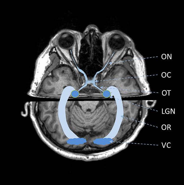
Figure 1. Schematic of anatomy of visual pathway. ON, optic nerve; OC, optic chiasm; OT, optic tract; LGN, lateral geniculate nucleus; OR, optic radiation; VC, visual cortex.
It is seen that the major components of the visual pathway are optic nerve fibers. Separated by the LGN, the two types of optic nerve fibers share similar symmetric curved thin-long structures but vary from each other in length, diameter, and curvature. Also, they are located with different neighboring tissues. The optic nerve fibers in the anterior visual pathway, i.e., from the optic disc to the LGN, are mostly located around muscle, fat, cranium, and blood vessels, while those in the OR are next to the brain's gray and white matters. The differences in these anatomical characteristics lead to quite different representations in medical images. More details on imaging would be introduced in the next section.
Currently, CT, MRI and DTI are the common imaging techniques for in-vivo delineation of the optic nerve fibers in the visual pathway. CT and MRI images are usually used to assess the optic nerve fibers in the anterior visual pathway (Smith and Strottmann, 2001; Becker et al., 2010). In general, MRI is superior in imaging the optic nerve fibers than CT as it can achieve high contrast among soft tissues. In comparison with CT, it can more easily differentiate the optic nerve fibers from the complex adjacent tissues in the orbit and sellar regions. Also, MRI is free from radiation and is safer than CT. Nevertheless, CT has its advantage in revealing bony tissues and foreign bodies. It can reveal the damages to the visual pathway caused by orbital or optic canal trauma as well as the calcification of the optic nerve fibers due to the tumor compression. In addition, CT is less affected by motion artifacts and can be applied to people with metal implants, making it a better choice than MRI in some special clinical scenarios. Besides CT and MRI, DTI can also be applied to reconstruct the optic nerve fibers in the anterior visual pathway; and, it can provide more details such as the fiber crossing at the OC (He et al., 2021). But, DTI takes much longer scanning time than MRI and CT, making it less practical in clinical scenarios. Instead, DTI is more frequently applied to reconstruct the optic nerve fibers in the OR (Dayan et al., 2015a; Schurr et al., 2018). The three fiber bundles in OR are located very close to other white matter tracts; the subtle variations in white matter signal make it difficult to reveal the anatomical heterogeneity in OR from CT and conventional MR images (Yogarajah et al., 2009; Winston et al., 2012). DTI is a technique to monitor the motion of water molecules in the human body by collecting multi-gradient MRI images. As the motion of water molecules is bounded by the nerve fibers, its speed and direction can be used to describe the structure and orientation of nerve fibers. Unlike MRI and CT images, the structure of optic radiation cannot be directly viewed in raw DTI images. There is a need to calculate the DTI metrics or perform fiber tracking to reveal the structure of the OR. It is seen that the three imaging techniques have their unique advantages and their own application scenarios. Also, the different representations of the optic nerve fibers in images of different modalities require different reconstruction methods. Usually, image segmentation is applied to CT and MRI images where optic nerve fibers exhibit a certain image contrast to neighboring tissues, while fiber tracking is performed to DTI data to exploit the structural and orientational information for more precise delineation of the optic nerve fibers. Both image segmentation and fiber tracking approaches can be further classified as conventional and AI-based methods. In the following two sections, we would describe more details of both the conventional and AI-based methods using the two reconstruction strategies.
Image segmentation is usually used to reconstruct optic nerve fibers from CT and MRI images. Conventionally, there are various methods to perform image segmentation, such as thresholding, boundary-based, region-based, model-based, atlas-based, etc (Despotović et al., 2015; Wang et al., 2016). The thresholding methods are not suitable for the segmentation of the optic nerve fibers given their poor image contrast with neighboring tissues at some segments of the visual pathway. Also, their thin-long structures make it difficult to perform boundary-based and region-based methods. It is found that most methods for optic nerve fiber reconstruction are model-based or atlas-based (Table 1).
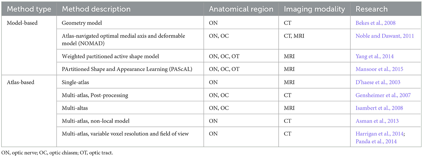
Table 1. Conventional image segmentation methods for optic nerve fiber reconstruction.
Model-based methods would first define a model based on the prior information on the shape and appearance of the tissue to be segmented and then fit the model to the new images (Heimann and Meinzer, 2009). The models can be either fixed geometry models or deformable models. For the fixed geometry models, they can be easily fitted via an explicit parameter estimation based on selected landmarks. The deformable models such as active shape models, active appearance models and level-set are usually fitted with searching or optimization procedures. Particularly, Bekes et al. (2008) approximate the ON in a CT image as a cone and cylinder and fit the cone and cylinder using a semi-automatic way. This fixed-model-based approach is simple but its reproducibility is doubted. Noble and Dawant (2011) applied an atlas-navigated optimal medial axis and deformable model (NOMAD) to segment the ON and OC based on paired CT and T1-weighted MRI images. The exploitation of multi-modal images and hybrid methods (model- and atlas-based) enhances the segmentation results, but the paired CT and MRI images are not always available in clinical practice. Yang et al. (2014) proposed a weighted partitioned active shape model to segment the anterior visual pathway from T1-weighted MRI images. This method is also capable to segment the OT, which is believed as a more challenging task than ON and OC segmentation before this study. Mansoor et al. (2015) developed a method entitled PArtitioned Shape and Appearance Learning (PAScAL) to segment the anterior visual pathway from MRI images. This method can also be applied to the pathological anterior visual pathway.
Atlas-based methods treat the segmentation problem as a registration problem, i.e., aligning the new image and the atlas (Cabezas et al., 2011). Usually, an atlas contains two image volumes, one intensity image (template) and one segmented image (label). Image registration is used to build the geometrical connection between the new image and the template; then, the segmentation can be achieved by propagating the label to the image space via the geometrical connection. D'haese et al. (2003) manually drew an atlas that includes the ON based on visually selected MRI images and segmented the ON with the atlas. Gensheimer et al. (2007) extended single-atlas segmentation to multi-atlas segmentation and performed additional post-processing procedures including a ray casting algorithm, reshaping of unreasonable cross sections, and surface fitting to further modify the inaccurate contours. Isambert et al. (2008) applied a multi-atlas segmentation method to segment ON and OC from MRI images under clinical conditions. Asman et al. (2013) developed a non-local model to perform multi-atlas segmentation for the ON based on CT images. Harrigan et al. (2014) and Panda et al. (2014) paid attention to the robustness of the atlas-based segmentation for the ON and proposed an improved registration procedure.
AI-based methods are data-driven approaches, which learn the rules from the data. Such approaches reduce manual operations like predefining models or atlases and are more easily implemented in practice. AI-based methods usually treat the segmentation procedure as a pixel/voxel-wise classification or clustering task. In the beginning, the classification/clustering is performed using conventional machine learning algorithms based on hand-crafted features. For example, Dolz et al. (2015) extracted features from neighborhood information and applied the support vector machine (SVM) to conduct the classification. With the occurrence and development of deep learning techniques, it becomes possible to integrate the feature extraction procedure into the learning process, further simplifying the procedure to segment the optic nerve fibers.
The studies on deep learning methods for optic nerve fiber segmentation from CT/MRI images have passed through three periods (Table 2). In the early period, deep learning methods are only used for feature extraction and segmentation is still implemented by conventional methods. For example, Mansoor et al. (2016) used a stacked auto-encoder to learn new feature representations for a model-based segmentation procedure. After this early period, deep learning is also used for pixel/voxel classification. At this stage, the network is usually formed by two network modules, e.g., a convolutional neural network (CNN) and a fully connected network, responsible for feature extraction and pixel/voxel classification, respectively. Based on this basic network structure, Ren et al. (2018) extended the original CNN to an interleaved structure for joint segmentation of optic nerve and chiasm; Dolz et al. (2017) replaced the CNN with a stacked denoised auto-encoders to learn a compact representation of the hand-crafted features; Duanmu et al. (2020) modified the CNN using a multi-resolution path approach to combine multi-scale features. Recently, a more powerful network, i.e., the U-Net, has been developed (Ronneberger et al., 2015). U-Net is composed of a down-sampling branch and an up-sampling branch. The down-sampling and up-sampling branches are made up of paired encoders and decoders, respectively. The down-sampling procedure can help extract the context information and the up-sampling procedure is used for fine localization. Also, there are skip connections between the encoders and decoders. As there might be information loss during the up-sampling procedure, the skip connections make it possible to combine the up-sampling results with the original information before the down-sampling procedure. With the skip connections, the localization can be more accurate. Compared with the two-module network, U-Net further integrates the feature extraction and pixel/voxel classification procedures.
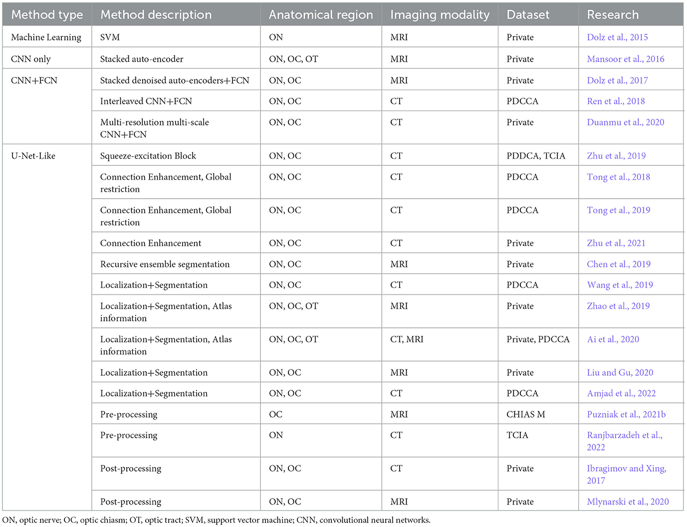
Table 2. AI-based image segmentation methods for optic nerve fiber reconstruction.
The state-of-the-art (SOTA) methods for optic nerve fiber reconstruction from CT/MRI images are mostly based on the U-Net. Particularly, some researchers tried to modify the encoders and decoders as well as their connections to enhance context information exploitation. For example, Zhu et al. (2019) added squeeze-excitation blocks into the down-sampling and up-sampling approaches; Tong et al. (2019) and Zhu et al. (2021) tried DenseNet and V-Net, which enhance the connections among encoders and decoders, to segment the ON and OC from CT and MRI images. Also, some researchers tried to add global loss restrictions to avoid irregular segmentation results due to the pixel/voxel-wise segmentation strategy. Specifically, Tong et al. (2018, 2019) added a latent shape restriction as well as an adversarial restriction to guarantee the global shape of the segmented ON and OC. Besides the modification of network blocks and losses, some researchers paid attention to the training strategies. Chen et al. proposed a recursive ensemble organ segmentation framework. In this framework, the organs that are easily segmented, e.g., the eyeballs, would be first segmented; and then, the segmentation results are fed to the network together with the original inputs for the segmentation of more complicated organs like ON and OC (Chen et al., 2019). Wang et al. proposed a hybrid network containing two U-Nets for localization and segmentation, respectively. The U-Net for localization was named “LocNet” and used to localize the region of the ON, while the one for segmentation was named “SegNet” and applied only in the extracted region to exclude other interference (Wang et al., 2019). Zhao et al. adopted a similar strategy but replaced the LocNet with an atlas-based approach, i.e., performing registration between the atlas and a new image to localize the ON. They also generated a spatial probabilistic distribution map using the atlas to assist the segmentation (Zhao et al., 2019; Ai et al., 2020). Differently, Liu and Gu (2020) and Amjad et al. (2022) replaced the SegNet with a two-module network, where the CNN adopted a multi-resolution structure.
In addition to the deep learning networks, researchers also tried to enhance the segmentation results using proper pre-processing and post-processing approaches. For the pre-processing, Puzniak et al. (2021b) applied a data-augmentation strategy to train a 3D U-Net. Ranjbarzadeh et al. (2022) pre-processed the input images by combining a fuzzy C-mean clustering algorithm, histogram equalization, and a texture descriptor based on the local directional number. For post-processing, Ibragimov and Xing (2017) proposed a post-processing procedure based on Markov random fields. Mlynarski et al. (2020) developed a graph-based post-processing approach to guarantee the connectivity between the eyes and OC.
Fiber tracking, also called fiber tractography, is a computational procedure to reconstruct nerve fibers from DTI images. Although there is a debate on the reliability of fiber tracking in delineating the true brain nerve fibers, it has been widely applied in both medical research and clinical practice. There are also plenty of studies focusing on the reconstruction of optic nerve fibers, especially for the OR, using fiber tracking.
Fiber tracking would estimate a series of streamlines to delineate the global fiber tractography using deterministic, probabilistic, or global algorithms (Jeurissen et al., 2019; Li et al., 2020). Deterministic algorithms are proposed based on the assumption that there is a predominant orientation in each voxel of DTI images. Common deterministic algorithms include streamlines tracking (STT) (Basser, 1998; Basser et al., 2000), fiber assignment by continuous tracking (FACT) (Mori et al., 1999; Chao et al., 2008), Tensor-lines (Weinstein et al., 1999), tensor deflection (TEND) (Lazar et al., 2003), and vector criterion tracking (VCT) (Kim et al., 2004). These algorithms usually select the diffusion tensor as the model to describe fibers' microstructures at each voxel. But, the diffusion signal would be inevitably distorted by noise and artifacts, affecting the certainty of voxel orientation inferred from the diffusion tensor (Jones, 2010). The assumption of one orientation per voxel is also doubted due to the existence of crossing fibers (Behrens et al., 2007). The existence of these problems raises concerns about the deterministic algorithms; the probabilistic algorithms are then proposed. To cope with the uncertainty, the probabilistic algorithms use the probability density functions (PDF) (Behrens et al., 2003) and fiber orientation distribution (FOD) (Tournier et al., 2004) to represent fibers' microstructures at each voxel. Based on these probabilistic models, the algorithms like probabilistic index of connectivity (PICo) (Parker et al., 2003), unscented Kalman filter (UKF) (Malcolm et al., 2010), probabilistic tracking with crossing fibers (PROBTRACKX) (Behrens et al., 2007), ConTrack (Sherbondy et al., 2008), particle filtering tractography (PFT) (Zhang et al., 2009), and 2nd-order Integration over Fiber Orientation Distributions (iFOD2) (Smith et al., 2012) are proposed. Compared with deterministic algorithms, probabilistic algorithms can delineate more complicated nerve fiber distributions; but, they would also cause a large number of false positive streamlines and suffer from heavy computational costs. Both the deterministic and probabilistic algorithms are based on local information, while global algorithms treat fiber tracking as a global optimization problem. The existing global algorithms can be mostly divided into two categories, i.e., graph-based algorithms (Iturria-Medina et al., 2007) and Gibbs algorithms. Graph-based algorithms should set the seeding and targeting regions, which is not necessary for Gibbs algorithms (Kreher et al., 2008). Global algorithms can avoid the error accumulation problem in local algorithms and reduce the number of false positive streamlines; but, their computational costs are much greater than local algorithms and convergent solutions are not guaranteed.
Besides the algorithm, there are also some key operations and settings to ensure an accurate fiber tracking procedure (Jacquesson et al., 2019; Jeurissen et al., 2019). For local algorithms and graph-based global algorithms, there is a need to determine the seeding and target regions of interest (ROIs). The seeding and target ROIs mean the two ends of the generated fibers by the tracking algorithms. Except for the whole-brain tracking, these two ROIs can be drawn in a manual way (Rossi-Espagnet et al., 2020; Haykal et al., 2022) or by projecting the labels in a built brain atlas (Karahan et al., 2019; Papadopoulou et al., 2021). In addition, the ROIs can also be acquired by other fiber tracking procedures (Davion et al., 2020). Besides these two types of ROIs, there are also inclusive and exclusive ROIs for the filtering of valid fibers (Horbruegger et al., 2019). In addition, some thresholds to constrain the fibers' lengths, curvatures/angles, and fractional anisotropy (FA)/fiber orientation distribution function (fODF) values are also set for the filtering process.
The specific methods for optic nerve fiber reconstruction are shown in Table 3. The reconstruction of the optic nerve fibers from DTI images follows the above fiber tracking frameworks; but, the selection of tracking algorithms, ROI drawing, and thresholds setting would change with the location of optic nerve fibers. Particularly, deterministic algorithms can be applied to the optic nerve fibers in the anterior visual pathway (Dasenbrock et al., 2011; De Blank et al., 2013; Takemura et al., 2017; Hofstetter et al., 2019; Jin et al., 2019) but they are not suggested for OR reconstruction (Yogarajah et al., 2009). The OR region is close to the neighboring white matter tracts and image voxels in this region are more likely to contain multiple orientations. The probabilistic algorithms can be applied for both the two types of optic nerve fibers (Dayan et al., 2015a; Kammen et al., 2016; Zolal et al., 2016; Backner et al., 2018; Yoshimine et al., 2018; Ather et al., 2019; Glick-Shames et al., 2019; Wu et al., 2019; Davion et al., 2020; Lacerda et al., 2020; Rossi-Espagnet et al., 2020; Reid et al., 2021; Haykal et al., 2022; Liu et al., 2022); but, there are still some differences. The probabilistic algorithms are proposed to handle the uncertainty and they can be classified into different categories based on the source of the uncertainty (Jeurissen et al., 2019). The reconstruction of optic nerve fibers at the anterior visual pathway and in the OR has different uncertainty sources. The former's uncertainty comes from the interference of the complicated skull base environment, which contains nerves, bone, air, soft tissue, and cerebrospinal fluid; the latter's uncertainty is mainly due to the multi-orientation problem. The difference in uncertainty sources would affect the selection of the probabilistic algorithms. In addition, the seeding ROIs for the ON and OT reconstruction are usually set as the end of the eyeballs and the OC, respectively, while those for the OR are set as the LGN. The target ROIs include the OC, LGN and VC for the reconstruction of the ON, OT and OR, respectively. The other settings like the inclusive and exclusive ROIs as well as the thresholds would be more task-specific.
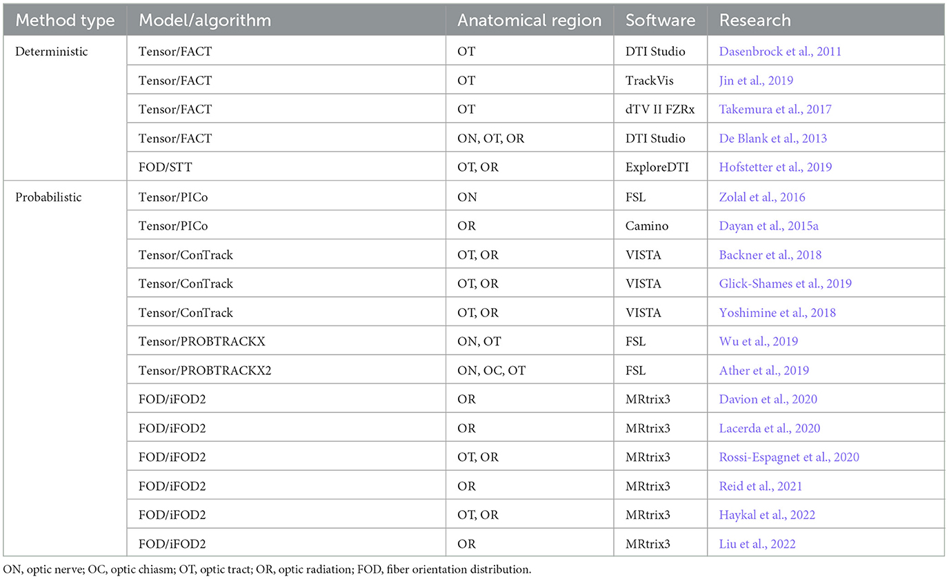
Table 3. Conventional fiber tracking approaches for optic nerve fiber reconstruction.
The conventional framework for fiber tracking is a complicated procedure containing the processes like pre-processing, seeding, tractography, and filtering of valid streamlines. Although several softwares integrate these processes (Table 3), the operations like ROI drawing, tracking algorithm selection, and threshold setting still require manual implementation. In recent years, AI technique has developed rapidly; researchers are trying to replace these manual operations with automated ways using AI technique (Table 4).
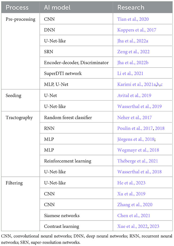
Table 4. AI-based fiber tracking approaches.
AI-based methods are applied first in the tractography process. Neher et al. (2017) tried to perform the tractography by machine learning. They applied a random forest classifier to learn multiple potential directions of a streamline from the raw diffusion signals and determined the streamline's progressing direction and termination using a neighborhood sampling strategy and a voting scheme, respectively. Poulin et al. treated the tractography as a regression problem and proposed the recurrent neural networks (RNN) to acquire the mapping between the diffusion signal and the streamlines' directions for both whole-brain and bundle-specific tractography (Poulin et al., 2017, 2018). The RNN can exploit both the new observations and the past seen information along the tracked streamlines. In addition to the diffusion signals, Jörgens et al. (2018) further pointed out the importance of the previous step directions for the tractography. They adopted an alternative way to predict the next step direction of a streamline via a multi-layer perceptron (MLP), whose input is a vector acquired by concatenating the diffusion signals and previous step directions. Wegmayr et al. (2018) also used an MLP to perform the tractography and further validated the significance of previous step directions; but, they changed the input of the MLP as a vector formed by a flattened data block and several incoming vectors. The tractography can also be implemented via reinforcement learning and Théberge et al. (2021) proposed a general framework for this strategy. Apart from these local tractography methods, Wasserthal et al. (2018) developed a U-Net-like network to directly reconstruct the fiber tracts from the fields of fODF peaks.
Recently, several AI-based methods have been applied in processes other than tractography. For the pre-processing approach, AI-based methods focus on two aspects, i.e., generating high-fidelity diffusion signals from low-quality input and building the diffusion model from the raw diffusion signals. Acquiring high-fidelity diffusion signals usually requires a certain number of diffusion-encoding directions and multi-shell acquisitions, which takes a long scanning time. Tian et al. (2020) proposed a 10-layer CNN to reduce the requirement on the number of diffusion-encoding directions, particularly limiting the number to the minimum level for diffusion tensor calculation. Koppers et al. (2017) and Jha et al. (2022a) reconstructed the multi-shell diffusion signals from single-shell acquisitions using DNN and U-Net-like network, respectively. Zeng et al. (2022) proposed a super-resolution network to enhance the FOD model that was built based on the single-shell acquisition, and Jha et al. (2022b) developed a more complicated network containing multiple encoder-decoder structures and discriminators. Mapping raw diffusion signals to diffusion models is also very challenging. It is quite difficult for conventional methods to estimate the fibers' number and orientations per voxel from raw diffusion signals. Li et al. (2021) demonstrated the advantages of AI-based methods in this challenging task. They proposed a SuperDTI network for diffusion model generation and the test results suggest that their model is less sensitive to noise and more robust to misregistration than conventional tensor fitting methods. Karimi et al. (2021a,b,c) further verified the superiority of AI-based methods via a series of explorations on diffusion metric map generation, fODF generation and fibers' number and orientations estimation. In addition to the pre-processing process, AI-based methods are also used to achieve automatic seeding. Avital et al. (2019) and Wasserthal et al. (2019) tried automated seeding using U-Net and U-Net-like network, respectively. There are also studies focusing on AI-based automated filtering of valid streamlines. Particularly, AI can be used to draw inclusive or exclusive ROIs, such as He's work (He et al., 2023). Also, AI models can be used to directly classify or cluster the reconstructed streamlines (Xu et al., 2019; Zhang et al., 2020; Chen et al., 2021; Xue et al., 2022, 2023).
Although multiple AI-based methods are proposed for fiber tracking, the application of these methods in optic nerve fiber reconstruction is still rare. To the best of our knowledge, Reid et al. (2021) applied a U-Net-like network to automatically draw seeding ROI at the optic tract. He et al. (2023) proposed a unified global tractography framework for automatic visual pathway reconstruction. Li et al. (2022) used a modified SupWMA network to cluster the streamlines in the anterior visual pathways. These methods demonstrate the feasibility and effectiveness of AI-based methods in optic nerve fiber reconstruction, while there is still room for further improvement.
Optic nerve fiber reconstruction is a common step to evaluate or project optic nerve fibers in clinical diagnosis and treatment. As shown in section 2, optic nerve fibers have thin-long structures and varying curvatures at different segments of the visual pathway, making them difficult to evaluate in either qualitative or quantitative ways without the reconstruction from the medical images. Also, manual delineation of the optic nerve fibers would be a tough task and costs a lot of time. As a result, computational methods are highly needed for clinical applications on optic nerve fibers. It is found that optic nerve fibers can be revealed in images of multiple modalities and there are different reconstruction strategies for each imaging modality. Also, each reconstruction strategy has both conventional and AI-based implementations. This paper reviews the existing computational methods to guide optic nerve fiber reconstruction in medical research and clinical practice and demonstrates the trend for future studies.
CT and MRI images are widely used for the visualization of the optic nerve fibers at the anterior visual pathway, i.e., from the end of the eyeballs to the LGN, while DTI can be used to visualize the optic nerve fibers along the entire visual pathway. Even though, DTI would not replace CT and MRI for optic nerve fiber reconstruction in clinical practice at the current stage. On one hand, DTI is with longer scanning time and lower image resolution than CT and MRI, making it less applicable in clinical practice. On the other hand, there are still debates on the consistency between the reconstructed fibers from DTI data and the real fibers in anatomy (Jeurissen et al., 2019), which limits its application scenarios such as the OAR drawing in radiation therapy.
Image segmentation and fiber tracking are two different reconstruction approaches for CT/MRI and DTI, respectively. Besides that, there are some other differences between these two approaches. Fiber tracking can achieve a more precise delineation of the optic nerve fibers than image segmentation, allowing the extraction of more accurate features to describe the morphological and structural changes of optic nerve fibers. For example, optic nerve fiber degeneration can be described by the volume change based on image segmentation results while it can be more precisely evaluated by the reduction in optic nerve fiber number based on fiber tracking results. Nevertheless, fiber tracking is time-consuming and its computational process is complicated and easily affected by noises and artifacts (Tournier et al., 2002; Lazar and Alexander, 2003). Also, it is not uncommon that there are false positive results and it requires abundant experience and enough knowledge of brain anatomy to ensure an accurate result (Jeurissen et al., 2019). These drawbacks restrict the scenarios where it can apply in clinical practice. In comparison to fiber tracking, image segmentation would be more efficient and robust; also, its results can be easily evaluated.
In comparison with conventional methods, AI-based methods are believed to be the trend for both image segmentation and fiber tracking. For image segmentation, AI-based methods are preferred to conventional model-based and atlas-based methods. Model-based methods require the design of complicated models to fit the thin-long structure of the optic nerve fiber; such models are difficult to estimate based on the complex background along the visual pathway and their robustness is doubted. Atlas-based methods require the registration between the target and template, while it is not easy to get two images fully aligned given the individual differences and interference from noises and artifacts. AI-based methods are data-driven approaches, which can automatically learn rules from complicated data. AI-based methods are more easily performed than those conventional methods and demonstrate much better segmentation accuracy and robustness. The only disadvantage of AI-based methods now is their high demand for fine-annotated labels.
For fiber tracking, the superiority of AI-based methods over conventional methods is not as great as image segmentation at the current stage. On one hand, AI-based methods are mostly proposed for one certain step of the fiber tracking procedure and a proper end-to-end AI-based fiber tracking approach is still lacking. On the other hand, the conventional methods for each fiber tracking step have been well integrated into toolboxes and software, decreasing their difficulty in implementation. Even though, it is seen that more and more studies on fiber tracking are trying to replace the conventional methods with AI-based ones.
It is also noticed that there are still some challenges in the reconstruction of optic nerve fibers from medical images with AI-based methods. These challenges point out the direction of future studies. The first challenge is the thin-long structure of the optic nerve fibers. The long optic nerve fibers pass through various brain regions that are formed by different brain tissues, yielding complicated contextual information. Meanwhile, the thin structure makes the signal intensities of the optic nerve fibers easily affected by their neighboring tissues due to the PVE, yielding varied signal intensities at different segments of the visual pathway. The existing image segmentation methods applied multi-scale, coarse-to-fine, or iterative strategies to handle the variations in signal intensity and contextual information; pre-processing and post-processing are also used to modify the false-positive and missing voxels. Even though measures are taken, it is seen that the improvement is far from satisfactory, suggesting that the current local voxel-based segmentation strategy would not be powerful enough to handle such a complicated problem. Also, the long optic nerve fibers have varied curvatures. In existing fiber tracking frameworks, the curvature is a significant sign for tracking termination and fiber selection. The varied curvatures increase the difficulty of setting these rules. Furthermore, it requires a large field of view to reveal the long optic nerve fibers in an image at the current stage. To achieve such a field of view, the image resolution has to be sacrificed to maintain an acceptable scanning time in clinical practice, increasing the PVE. Thus, more powerful segmentation and fiber tracking strategies are required to cope with the challenges brought by the thin-long structure.
The second challenge is the lacking of task-specific datasets. AI-based methods are data-driven methods and their performance highly depends on the quality of data. To the best of our knowledge, most existing studies on image segmentation and fiber tracking are based on private datasets, which are not available to the public. There are a minor number of publicly available datasets, such as PDDCA (Raudaschl et al., 2017), TCIA (Clark et al., 2013; Zhu et al., 2019), and CHIASM (Puzniak et al., 2021a,b) for image segmentation and HCP for fiber tracking. However, these datasets are not initially collected for optic nerve fiber reconstruction. Most of these datasets require further cleaning and annotation operations. Meanwhile, the imaging protocols and pre-processing steps in these datasets may not be consistent with those used in clinical practice. Also, in some of the datasets, the images only cover part of the optic nerve fibers and cannot be used to reconstruct the entire visual pathway. In addition, the situations like multi-modal images and disease-specific deformation are not fully considered in these existing datasets. Therefore, building a dataset specifically for optic nerve fiber reconstruction is in great need.
The third challenge is the control of computational cost. For image segmentation, more powerful segmentation networks are usually with more complicated network structures at the current stage. Also, the inputs are 3D brain images for the reconstruction of optic nerve fibers. These together indicate a high computational cost. For fiber tracking, the tractography is usually an iterative process and time-consuming for both conventional and AI-based methods. The high computational cost would reduce the value of clinical application. The way to balance the computational cost and reconstruction performance would be another challenge in future studies.
There are some new techniques such as generative AI that can help address these challenges. Generative AI has demonstrated its power in image super-resolution and image synthesis. Image super-resolution can be used to cope with the low-resolution problem caused by the large field of view. Also, image synthesis can be used to generate more data to get full exploitation of the existing datasets. In addition, it is realized that multi-modal fusion would be a possible way to enhance the performance of optic nerve fiber reconstruction. There are many other examples to support its effect on segmentation (Menze et al., 2014; Ibtehaz and Rahman, 2020; Wang et al., 2022). The combination of segmentation results and fiber tracking has once been explored (Reid et al., 2021; He et al., 2023). The segmentation results can be used as the seeds for tractography or the masks to filter valid streamlines. Therefore, developing new fusion and combination methods would be a feasible way to improve the reconstruction performance. Nevertheless, this kind of method would face the problem that multi-modal images are difficult to acquire in clinical practice. Generative AI provides a way for multi-modal image synthesis. Thus, in the future, we can try these new techniques in optic nerve fiber reconstruction.
In this paper, we provided a comprehensive review of the current SOTA computational methods for the reconstruction of optic nerve fibers. We described the difficulties to delineate or evaluate the optic nerve fibers directly from medical images, suggesting the necessity of optic nerve fiber reconstruction. We reviewed both the image segmentation and fiber tracking methods and the successful application of these methods in previous studies indicates the feasibility and effectiveness of computational methods in optic nerve fiber reconstruction. Also, we introduced both the conventional and AI-based implementations, and there is no doubt that AI-based methods are better choices for optic nerve fiber reconstruction. Meanwhile, we also pointed out the challenges for the existing AI methods, and future studies are needed to address these challenges.
RJ and JL contributed to conception and design of the reviewing process. YC, SZ, TY, and HF searched and sorted the literatures. HJ, YH, and XZ provided key suggestions on the anatomy of visual pathway, imaging techniques, and image segmentation methods. RJ wrote the first draft of the manuscript. All authors contributed to manuscript revision, read, and approved the submitted version.
This work was supported in part by the National Natural Science Foundation of China (Grant Nos. 62101236 and 82102189), General Program of National Natural Science Foundation of China (Grant No. 82272086), Guangdong Provincial Department of Education (Grant No. 2020ZDZX3043), Guangdong Provincial Key Laboratory (Grant No. 2020B121201001), Shenzhen Natural Science Fund (JCYJ20200109140820699), and the Stable Support Plan Program (20200925174052004).
The authors declare that the research was conducted in the absence of any commercial or financial relationships that could be construed as a potential conflict of interest.
All claims expressed in this article are solely those of the authors and do not necessarily represent those of their affiliated organizations, or those of the publisher, the editors and the reviewers. Any product that may be evaluated in this article, or claim that may be made by its manufacturer, is not guaranteed or endorsed by the publisher.
Ai, D., Zhao, Z., Fan, J., Song, H., Qu, X., Xian, J., et al. (2020). Spatial probabilistic distribution map-based two-channel 3D U-net for visual pathway segmentation. Pattern Recognit. Lett. 138, 601–607. doi: 10.1016/j.patrec.2020.09.003
Alexander, A. L., Hasan, K. M., Lazar, M., Tsuruda, J. S., and Parker, D. L. (2001). Analysis of partial volume effects in diffusion-tensor MRI. Magn. Reson. Med. 45, 770–780. doi: 10.1002/mrm.1105
Amjad, A., Xu, J., Thill, D., Lawton, C., Hall, W., Awan, M. J., et al. (2022). General and custom deep learning autosegmentation models for organs in head and neck, abdomen, and male pelvis. Med. Phys. 49, 1686–1700. doi: 10.1002/mp.15507
Asman, A. J., Delisi, M. P., Mawn, L. A., Galloway, R. L., and Landman, B. A. (2013). Robust Non-local Multi-atlas Segmentation of the Optic Nerve. Medical Imaging 2013: image Processing. Bellingham, WA: SPIE. p. 434–441.
Ather, S., Proudlock, F. A., Welton, T., Morgan, P. S., Sheth, V., Gottlob, I., et al. (2019). Aberrant visual pathway development in albinism: from retina to cortex. Hum. Brain Mapp. 40, 777–788. doi: 10.1002/hbm.24411
Avital, I., Nelkenbaum, I., Tsarfaty, G., Konen, E., Kiryati, N., and Mayer, A. (2019). Neural segmentation of seeding ROIs (sROIs) for pre-surgical brain tractography. IEEE Trans. Med. Imaging 39, 1655–1667. doi: 10.1109/TMI.2019.2954477
Backner, Y., Kuchling, J., Massarwa, S., Oberwahrenbrock, T., Finke, C., Bellmann-Strobl, J., et al. (2018). Anatomical wiring and functional networking changes in the visual system following optic neuritis. JAMA Neurol. 75, 287–295. doi: 10.1001/jamaneurol.2017.3880
Basser, P. J. (1998). Fiber-tractography via diffusion tensor MRI (DT-MRI)“, in: Proceedings of the 6th Annual Meeting ISMRM. Sydney, Australia.
Basser, P. J., Pajevic, S., Pierpaoli, C., Duda, J., and Aldroubi, A. (2000). In vivo fiber tractography using DT-MRI data. Magn. Reson. Med. 44, 625–632. doi: 10.1002/1522-2594(200010)44:4%3C625::AID-MRM17%3E3.0.CO;2-O
Becker, M., Masterson, K., Delavelle, J., Viallon, M., Vargas, M.-I., and Becker, C. D. (2010). Imaging of the optic nerve. Eur. J. Radiol. 74, 299–313. doi: 10.1016/j.ejrad.2009.09.029
Behrens, T. E., Berg, H. J., Jbabdi, S., Rushworth, M. F., and Woolrich, M. W. (2007). Probabilistic diffusion tractography with multiple fibre orientations: what can we gain? Neuroimage 34, 144–155. doi: 10.1016/j.neuroimage.2006.09.018
Behrens, T. E., Woolrich, M. W., Jenkinson, M., Johansen-Berg, H., Nunes, R. G., Clare, S., et al. (2003). Characterization and propagation of uncertainty in diffusion-weighted MR imaging. Magn. Reson. Med. 50, 1077–1088. doi: 10.1002/mrm.10609
Bekes, G., Mát,é, E., Nyúl, L. G., Kuba, A., and Fidrich, M. (2008). Geometrical model-based segmentation of the organs of sight on CT images. Med. Phys. 35, 735–743. doi: 10.1118/1.2826557
Cabezas, M., Oliver, A., Lladó, X., Freixenet, J., and Cuadra, M. B. (2011). A review of atlas-based segmentation for magnetic resonance brain images. Comput. Methods Programs Biomed. 104, e158–e177. doi: 10.1016/j.cmpb.2011.07.015
Chamberland, M., Tax, C. M. W., and Jones, D. K. (2018). Meyer's loop tractography for image-guided surgery depends on imaging protocol and hardware. Neuroimage-Clinical 20, 458–465. doi: 10.1016/j.nicl.2018.08.021
Chao, Y.-P., Chen, J.-H., Cho, K.-H., Yeh, C.-H., Chou, K.-H., and Lin, C.-P. (2008). A multiple streamline approach to high angular resolution diffusion tractography. Med. Eng. Phy. 30, 989–996. doi: 10.1016/j.medengphy.2008.01.010
Chen, H., Lu, W., Chen, M., Zhou, L., Timmerman, R., Tu, D., et al. (2019). A recursive ensemble organ segmentation (REOS) framework: application in brain radiotherapy. Phys. Med. Biol. 64, 025015. doi: 10.1088/1361-6560/aaf83c
Chen, Y., Zhang, C., Song, Y., Makris, N., Rathi, Y., Cai, W., et al. (2021). Deep fiber clustering: anatomically informed unsupervised deep learning for fast and effective white matter parcellation. In: Medical Image Computing and Computer Assisted Intervention–MICCAI 2021: 24th International Conference, Strasbourg, France, September 27–October 1, 2021, Proceedings, Part VII: Berlin: Springer. p. 497–507.
Clark, K., Vendt, B., Smith, K., Freymann, J., Kirby, J., Koppel, P., et al. (2013). The Cancer Imaging Archive (TCIA): maintaining and operating a public information repository. J. Digit. Imaging 26, 1045–1057. doi: 10.1007/s10278-013-9622-7
Dai, X., Lei, Y., Wang, T., Zhou, J., Curran, W. J., Liu, T., et al. (2021). Deep attention mask regional convolutional neural network for head-and-neck MRI multi-organ auto-delineation. In: Conference on Medical Imaging - Computer-Aided Diagnosis. eds M. A. Mazurowski and K. Drukker (International Society for Optics and Photonics, SPIE), 115971B. doi: 10.1117/12.2581131
Dasenbrock, H. H., Smith, S. A., Ozturk, A., Farrell, S. K., Calabresi, P. A., and Reich, D. S. (2011). Diffusion tensor imaging of the optic tracts in multiple sclerosis: association with retinal thinning and visual disability. J. Neuroimag. 21, e41–e49. doi: 10.1111/j.1552-6569.2010.00468.x
Davion, J.-B., Kuchcinski, G., Viard, R., Dumont, J., Pruvo, J.-P., Leclerc, X., et al. (2020). A fully automatic method for optic radiation tractography applicable to multiple sclerosis patients. Brain Topogr. 33, 533–544. doi: 10.1007/s10548-020-00771-8
Dayan, M., Kreutzer, S., and Clark, C. A. (2015a). Tractography of the optic radiation: a repeatability and reproducibility study. NMR Biomed. 28, 423–431. doi: 10.1002/nbm.3266
Dayan, M., Munoz, M., Jentschke, S., Chadwick, M. J., Cooper, J. M., Riney, K., et al. (2015b). Optic radiation structure and anatomy in the normally developing brain determined using diffusion MRI and tractography. Brain Struct. Funct. 220, 291–306. doi: 10.1007/s00429-013-0655-y
De Blank, P. M. K., Berman, J. I., Liu, G. T., Roberts, T. P. L., and Fisher, M. J. (2013). Fractional anisotropy of the optic radiations is associated with visual acuity loss in optic pathway gliomas of neurofibromatosis type 1. Neuro-oncology 15, 1088–1095. doi: 10.1093/neuonc/not068
Despotović, I., Goossens, B., and Philips, W. (2015). MRI segmentation of the human brain: challenges, methods, and applications. Comput. Math. Methods Med. 2015. doi: 10.1155/2015/450341
D'haese, P.-F., Duay, V., Li, R., Du Bois D'aische, A., Merchant, T. E., et al. (2003). Automatic segmentation of brain structures for radiation therapy planning. In: Medical Imaging 2003: Image Processing. Bellingham, WA: SPIE. p. 517–526.
Dolz, J., Leroy, H.-A., Reyns, N., Massoptier, L., and Vermandel, M. (2015). A fast and fully automated approach to segment optic nerves on MRI and its application to radiosurgery. In: 2015 IEEE 12th International Symposium on Biomedical Imaging (ISBI). Manhattan, NY: IEEE. p. 1102–1105.
Dolz, J., Reyns, N., Betrouni, N., Kharroubi, D., Quidet, M., Massoptier, L., et al. (2017). A deep learning classification scheme based on augmented-enhanced features to segment organs at risk on the optic region in brain cancer patients. arXiv. [Preprint]. doi: 10.48550/arXiv.1703.10480
Duanmu, H., Kim, J., Kanakaraj, P., Wang, A., Joshua, J., Kong, J., et al. (2020). Automatic brain organ segmentation with 3D fully convolutional neural network for radiation therapy treatment planning. In: 2020 IEEE 17th International Symposium on Biomedical Imaging (ISBI). Manhattan, NY: IEEE. p. 758–762.
Fujita, N., Tanaka, H., Takanashi, M., Hirabuki, N., Abe, K., Yoshimura, H., et al. (2001). Lateral geniculate nucleus: anatomic and functional identification by use of MR imaging. Am. J. Neuroradiol. 22, 1719–1726. Available online at: https://www.webofscience.com/wos/alldb/full-record/WOS:000171752400019
Gensheimer, M., Cmelak, A., Niermann, K., and Dawant, B. M. (2007). Automatic delineation of the optic nerves and chiasm on CT images. In: Medical Imaging 2007: Image Processing: Bellingham, WA: SPIE. p. 406–415.
Glick-Shames, H., Backner, Y., Bick, A., Raz, N., and Levin, N. (2019). The impact of localized grey matter damage on neighboring connectivity: Posterior cortical atrophy and the visual network. Brain Imag. Behav. 13, 1292–1301. doi: 10.1007/s11682-018-9952-7
Harrigan, R. L., Panda, S., Asman, A. J., Nelson, K. M., Chaganti, S., Delisi, M. P., et al. (2014). Robust optic nerve segmentation on clinically acquired computed tomography. J. Med. Imag. 1, 034006–034006. doi: 10.1117/1.JMI.1.3.034006
Haykal, S., Invernizzi, A., Carvalho, J., Jansonius, N., and Cornelissen, F. (2022). Microstructural visual pathway white matter alterations in primary open-angle glaucoma: a neurite orientation dispersion and density imaging study. Am. J. Neuroradiol. 43, 756–763. doi: 10.3174/ajnr.A7495
He, J., Yao, S., Zeng, Q., Chen, J., Sang, T., Xie, L., et al. (2023). A unified global tractography framework for automatic visual pathway reconstruction. NMR Biomed. 34, e4904. doi: 10.1002/nbm.4904
He, J., Zhang, F., Xie, G., Yao, S., Feng, Y., Bastos, D. C., et al. (2021). Comparison of multiple tractography methods for reconstruction of the retinogeniculate visual pathway using diffusion MRI. Hum. Brain Mapp. 42, 3887–3904. doi: 10.1002/hbm.25472
Heimann, T., and Meinzer, H.-P. (2009). Statistical shape models for 3D medical image segmentation: a review. Med. Image Anal. 13, 543–563. doi: 10.1016/j.media.2009.05.004
Hernowo, A. T., Boucard, C. C., Jansonius, N. M., Hooymans, J. M., and Cornelissen, F. W. (2011). Automated morphometry of the visual pathway in primary open-angle glaucoma. Invest. Ophthalmol. Visual Sci. 52, 2758–2766. doi: 10.1167/iovs.10-5682
Hofstetter, S., Sabbah, N., Mohand-Saïd, S., Sahel, J.-A., Habas, C., Safran, A. B., et al. (2019). The development of white matter structural changes during the process of deterioration of the visual field. Sci. Rep. 9, 2085. doi: 10.1038/s41598-018-38430-5
Horbruegger, M., Loewe, K., Kaufmann, J., Wagner, M., Schippling, S., Pawlitzki, M., et al. (2019). Anatomically constrained tractography facilitates biologically plausible fiber reconstruction of the optic radiation in multiple sclerosis. NeuroImage Clin. 22, 101740. doi: 10.1016/j.nicl.2019.101740
Hu, S., Lei, B., Wang, S., Wang, Y., Feng, Z., and Shen, Y. (2021). Bidirectional mapping generative adversarial networks for brain MR to PET synthesis. IEEE Trans. Med. Imaging 41, 145–157. doi: 10.1109/TMI.2021.3107013
Hu, S., Shen, Y., Wang, S., and Lei, B. (2020a). Brain MR to PET synthesis via bidirectional generative adversarial network. In: Medical Image Computing and Computer Assisted Intervention–MICCAI 2020: 23rd International Conference, Lima, Peru, October 4–8, 2020 Proceedings, Part II 23. Berlin: Springer. p. 698–707.
Hu, S., Yu, W., Chen, Z., and Wang, S. (2020b). Medical image reconstruction using generative adversarial network for Alzheimer disease assessment with class-imbalance problem. In: 2020 IEEE 6th International Conference on Computer and Communications: Manhattan, NY: IEEE. p. 1323–1327.
Ibragimov, B., and Xing, L. (2017). Segmentation of organs-at-risks in head and neck CT images using convolutional neural networks. Med. Phys. 44, 547–557. doi: 10.1002/mp.12045
Ibtehaz, N., and Rahman, M. S. (2020). MultiResUNet: Rethinking the U-Net architecture for multimodal biomedical image segmentation. Neural networks 121, 74–87. doi: 10.1016/j.neunet.2019.08.025
Isambert, A., Dhermain, F., Bidault, F., Commowick, O., Bondiau, P.-Y., Malandain, G., et al. (2008). Evaluation of an atlas-based automatic segmentation software for the delineation of brain organs at risk in a radiation therapy clinical context. Radiother. Oncol. 87, 93–99. doi: 10.1016/j.radonc.2007.11.030
Iturria-Medina, Y., Canales-Rodríguez, E. J., Melie-García, L., Valdés-Hernández, P. A., Martínez-Montes, E., Alemán-Gómez, Y., et al. (2007). Characterizing brain anatomical connections using diffusion weighted MRI and graph theory. Neuroimage 36, 645–660. doi: 10.1016/j.neuroimage.2007.02.012
Jacquesson, T., Frindel, C., Kocevar, G., Berhouma, M., Jouanneau, E., Atty,é, A., et al. (2019). Overcoming challenges of cranial nerve tractography: a targeted review. Neurosurgery 84, 313–325. doi: 10.1093/neuros/nyy229
Jäger, H. (2005). Loss of vision: imaging the visual pathways. Eur. Radiol. 15, 501–510. doi: 10.1007/s00330-004-2638-0
Jeurissen, B., Descoteaux, M., Mori, S., and Leemans, A. (2019). Diffusion MRI fiber tractography of the brain. NMR Biomed. 32, e3785. doi: 10.1002/nbm.3785
Jha, R. R., Jaswal, G., Bhavsar, A., and Nigam, A. (2022a). Single-shell to multi-shell dMRI transformation using spatial and volumetric multilevel hierarchical reconstruction framework. Magn. Reson. Imaging 87, 133–156. doi: 10.1016/j.mri.2021.12.011
Jha, R. R., Pathak, S. K., Nath, V., Schneider, W., Kumar, B. R., Bhavsar, A., et al. (2022b). VRfRNet: Volumetric ROI fODF reconstruction network for estimation of multi-tissue constrained spherical deconvolution with only single shell dMRI. Magn. Reson. Imaging 90, 1–16. doi: 10.1016/j.mri.2022.03.004
Jin, Z., Bao, Y., Wang, Y., Li, Z., Zheng, X., Long, S., et al. (2019). Differences between generalized Q-sampling imaging and diffusion tensor imaging in visualization of crossing neural fibers in the brain. Surg. Radiol. Anat. 41, 1019–1028. doi: 10.1007/s00276-019-02264-1
Jones, D. K. (2010). Challenges and limitations of quantifying brain connectivity in vivo with diffusion MRI. Imaging Med. 2, 341. doi: 10.2217/iim.10.21
Jörgens, D., Smedby, Ö., and Moreno, R. (2018). Learning a single step of streamline tractography based on neural networks. In: Computational Diffusion MRI: MICCAI Workshop, Québec, Canada, September 2017. Berlin: Springer. p. 103–116.
Kammen, A., Law, M., Tjan, B. S., Toga, A. W., and Shi, Y. (2016). Automated retinofugal visual pathway reconstruction with multi-shell HARDI and FOD-based analysis. Neuroimage 125, 767–779. doi: 10.1016/j.neuroimage.2015.11.005
Karahan, E., Costigan, A. G., Graham, K. S., Lawrence, A. D., and Zhang, J. (2019). Cognitive and white-matter compartment models reveal selective relations between corticospinal tract microstructure and simple reaction time. J. Neurosci. 39, 5910–5921. doi: 10.1523/JNEUROSCI.2954-18.2019
Karimi, D., Jaimes, C., Machado-Rivas, F., Vasung, L., Khan, S., Warfield, S. K., et al. (2021a). Deep learning-based parameter estimation in fetal diffusion-weighted MRI. Neuroimage 243, 118482. doi: 10.1016/j.neuroimage.2021.118482
Karimi, D., Vasung, L., Jaimes, C., Machado-Rivas, F., Khan, S., Warfield, S. K., et al. (2021b). A machine learning-based method for estimating the number and orientations of major fascicles in diffusion-weighted magnetic resonance imaging. Med. Image Anal. 72, 102129. doi: 10.1016/j.media.2021.102129
Karimi, D., Vasung, L., Jaimes, C., Machado-Rivas, F., Warfield, S. K., and Gholipour, A. (2021c). Learning to estimate the fiber orientation distribution function from diffusion-weighted MRI. Neuroimage 239, 118316. doi: 10.1016/j.neuroimage.2021.118316
Kim, K.-H., Ronen, I., Formisano, E., Goebel, R., Ugurbil, K., and Kim, D.-S. (2004). Robust fiber tracking method by vector selection criterion in diffusion tensor images. In: The 26th Annual International Conference of the IEEE Engineering in Medicine and Biology Society. Manhattan, NY: IEEE. p. 1080–1083.
Klistorner, A., Vootakuru, N., Wang, C., Yiannikas, C., Graham, S. L., Parratt, J., et al. (2015). Decoding diffusivity in multiple sclerosis: analysis of optic radiation lesional and non-lesional white matter. PLoS ONE 10, e0122114. doi: 10.1371/journal.pone.0122114
Koppers, S., Haarburger, C., and Merhof, D. (2017). Diffusion MRI signal augmentation: from single shell to multi shell with deep learning. In: Computational Diffusion MRI: MICCAI Workshop, Athens, Greece, October 2016 19. Berlin: Springer. p. 61–70.
Kreher, B., Mader, I., and Kiselev, V. (2008). Gibbs tracking: a novel approach for the reconstruction of neuronal pathways. Magn. Reson. Med. 60, 953–963. doi: 10.1002/mrm.21749
Lacerda, L. M., Clayden, J. D., Handley, S. E., Winston, G. P., Kaden, E., Tisdall, M., et al. (2020). Microstructural investigations of the visual pathways in pediatric epilepsy neurosurgery: insights from multi-shell diffusion magnetic resonance imaging. Front. Neurosci. 14, 269. doi: 10.3389/fnins.2020.00269
Lazar, M., and Alexander, A. L. (2003). An error analysis of white matter tractography methods: synthetic diffusion tensor field simulations. Neuroimage 20, 1140–1153. doi: 10.1016/S1053-8119(03)00277-5
Lazar, M., Weinstein, D. M., Tsuruda, J. S., Hasan, K. M., Arfanakis, K., Meyerand, M. E., et al. (2003). White matter tractography using diffusion tensor deflection. Hum. Brain Mapp. 18, 306–321. doi: 10.1002/hbm.10102
Li, H., Liang, Z., Zhang, C., Liu, R., Li, J., Zhang, W., et al. (2021). SuperDTI: Ultrafast DTI and fiber tractography with deep learning. Magn. Reson. Med. 86, 3334–3347. doi: 10.1002/mrm.28937
Li, S., He, J., Xue, T., Xie, G., Yao, S., Chen, Y., et al. (2022). DeepRGVP: A Novel Microstructure-Informed Supervised Contrastive Learning Framework for Automated Identification Of The Retinogeniculate Pathway Using dMRI Tractography. arXiv. [Preprint]. doi: 10.48550/arXiv.2211.08119
Li, M., He, J., and Feng, Y. (2020). Research progress of neural fiber tracking. J. Image Graph. 25, 1513–1528. doi: 10.11834/jig.190519
Liu, M., Lerma-Usabiaga, G., Clascá, F., and Paz-Alonso, P. M. (2022). Reproducible protocol to obtain and measure first-order relay human thalamic white-matter tracts. Neuroimage 262, 119558. doi: 10.1016/j.neuroimage.2022.119558
Liu, Y., and Gu, X. (2020). Evaluation and comparison of global-feature-based and local-feature-based segmentation algorithms in intracranial visual pathway delineation. In: 2020 42nd Annual International Conference of the IEEE Engineering in Medicine and Biology Society (EMBC). Manhattan, NY: IEEE. p. 1766–1769.
Malcolm, J. G., Shenton, M. E., and Rathi, Y. (2010). Filtered multitensor tractography. IEEE Trans. Med. Imaging 29, 1664–1675. doi: 10.1109/TMI.2010.2048121
Mansoor, A., Cerrolaza, J. J., Avery, R. A., and Linguraru, M. G. (2015). Partitioned shape modeling with on-the-fly sparse appearance learning for anterior visual pathway segmentation. In: Clinical Image-Based Procedures. Translational Research in Medical Imaging: 4th International Workshop, CLIP 2015, Held in Conjunction with MICCAI 2015, Munich, Germany, October 5, 2015. Revised Selected Papers 4. Berlin: Springer. p. 104–112.
Mansoor, A., Cerrolaza, J. J., Idrees, R., Biggs, E., Alsharid, M. A., Avery, R. A., et al. (2016). Deep learning guided partitioned shape model for anterior visual pathway segmentation. IEEE Trans. Med. Imaging 35, 1856–1865. doi: 10.1109/TMI.2016.2535222
Menze, B. H., Jakab, A., Bauer, S., Kalpathy-Cramer, J., Farahani, K., Kirby, J., et al. (2014). The multimodal brain tumor image segmentation benchmark (BRATS). IEEE Trans. Med. Imaging 34, 1993–2024. doi: 10.1109/TMI.2014.2377694
Mlynarski, P., Delingette, H., Alghamdi, H., Bondiau, P.-Y., and Ayache, N. (2020). Anatomically consistent CNN-based segmentation of organs-at-risk in cranial radiotherapy. J. Med. Imag. 7, 014502–014502. doi: 10.1117/1.JMI.7.1.014502
Mori, S., Crain, B. J., Chacko, V. P., and Van Zijl, P. C. (1999). Three-dimensional tracking of axonal projections in the brain by magnetic resonance imaging. Annals Neurol. 45, 265–269. doi: 10.1002/1531-8249(199902)45:2<265::aid-ana21>3.0.co;2-3
Mutlu, U., Ikram, M. K., Roshchupkin, G. V., Bonnemaijer, P. W. M., Colijn, J. M., Vingerling, J. R., et al. (2018). Thinner retinal layers are associated with changes in the visual pathway: A population-based study. Hum. Brain Mapp. 39, 4290–4301. doi: 10.1002/hbm.24246
Neher, P. F., Côt,é, M.-A., Houde, J.-C., Descoteaux, M., and Maier-Hein, K. H. (2017). Fiber tractography using machine learning. Neuroimage 158, 417–429. doi: 10.1016/j.neuroimage.2017.07.028
Noble, J. H., and Dawant, B. M. (2011). An atlas-navigated optimal medial axis and deformable model algorithm (NOMAD) for the segmentation of the optic nerves and chiasm in MR and CT images. Med. Image Anal. 15, 877–884. doi: 10.1016/j.media.2011.05.001
Panda, S., Asman, A. J., Delisi, M. P., Mawn, L. A., Galloway, R. L., and Landman, B. A. (2014). Robust optic nerve segmentation on clinically acquired CT. In: Medical Imaging 2014: Image Processing. Bellingham, WA: SPIE. p. 362–371.
Papadopoulou, A., Oertel, F. C., Chien, C., Kuchling, J., Zimmermann, H. G., Siebert, N., et al. (2021). Lateral geniculate nucleus volume changes after optic neuritis in neuromyelitis optica: a longitudinal study. NeuroImage Clin. 30, 102608. doi: 10.1016/j.nicl.2021.102608
Parker, G. J., Haroon, H. A., and Wheeler-Kingshott, C. A. (2003). A framework for a streamline-based probabilistic index of connectivity (PICo) using a structural interpretation of MRI diffusion measurements. J. Magn. Reson. Imag. 18, 242–254. doi: 10.1002/jmri.10350
Poulin, P., Côt,é, M.-A., Houde, J.-C., Petit, L., Neher, P. F., Maier-Hein, K. H., et al. (2017). Learn to track: deep learning for tractography. In: Medical Image Computing and Computer Assisted Intervention– MICCAI 2017: 20th International Conference, Quebec City, QC, Canada, September 11-13, 2017, Proceedings, Part I 20. Berlin: Springer. p. 540–547.
Poulin, P., Rheault, F., St-Onge, E., Jodoin, P.-M., and Descoteaux, M. (2018). Bundle-wise deep tracker: Learning to track bundle-specific streamline paths. Proc. of the Int. Society for Magnetic Resonance in medicine ISMRM-ESMRMB. Paris, France.
Prins, D., Hanekamp, S., and Cornelissen, F. W. (2016). Structural brain MRI studies in eye diseases: are they clinically relevant? A review of current findings. Acta Ophthalmol. 94, 113–121. doi: 10.1111/aos.12825
Puzniak, R. J., Mcpherson, B., Ahmadi, K., Herbik, A., Kaufmann, J., Liebe, T., et al. (2021a). CHIASM, the human brain albinism and achiasma MRI dataset. Scientific data 8, 308. doi: 10.1038/s41597-021-01080-w
Puzniak, R. J., Prabhakaran, G. T., and Hoffmann, M. B. (2021b). Deep learning-based detection of malformed optic chiasms from MRI images. Front. Neurosci. 15, 1332. doi: 10.3389/fnins.2021.755785
Ranjbarzadeh, R., Dorosti, S., Jafarzadeh Ghoushchi, S., Safavi, S., Razmjooy, N., Tataei Sarshar, N., et al. (2022). Nerve optic segmentation in CT images using a deep learning model and a texture descriptor. Comp. Intel. Syst. 8, 3543–3557. doi: 10.1007/s40747-022-00694-w
Raudaschl, P. F., Zaffino, P., Sharp, G. C., Spadea, M. F., Chen, A., Dawant, B. M., et al. (2017). Evaluation of segmentation methods on head and neck CT: auto-segmentation challenge 2015. Med. Phys. 44, 2020–2036. doi: 10.1002/mp.12197
Reich, D. S., Ozturk, A., Calabresi, P. A., and Mori, S. (2010). Automated vs. conventional tractography in multiple sclerosis: variability and correlation with disability. Neuroimage 49, 3047–3056. doi: 10.1016/j.neuroimage.2009.11.043
Reid, L. B., Martinez-Heras, E., Manjon, J. V., Jeffree, R. L., Alexander, H., Trinder, J., et al. (2021). Fully automated delineation of the optic radiation for surgical planning using clinically feasible sequences. Hum. Brain Mapp. 42, 5911–5926. doi: 10.1002/hbm.25658
Ren, X., Xiang, L., Nie, D., Shao, Y., Zhang, H., Shen, D., et al. (2018). Interleaved 3D-CNN s for joint segmentation of small-volume structures in head and neck CT images. Med. Phys. 45, 2063–2075. doi: 10.1002/mp.12837
Romano, A., D'andrea, G., Minniti, G., Mastronardi, L., Ferrante, L., Fantozzi, L. M., et al. (2009). Pre-surgical planning and MR-tractography utility in brain tumour resection. Eur. Radiol. 19, 2798–2808. doi: 10.1007/s00330-009-1483-6
Romano, A., Ferrante, M., Cipriani, V., Fasoli, F., Ferrante, L., D'andrea, G., et al. (2007). Role of magnetic resonance tractography in the preoperative planning and intraoperative assessment of patients with intra-axial brain tumours. Radiol. Med. 112, 906–920. doi: 10.1007/s11547-007-0181-1
Ronneberger, O., Fischer, P., and Brox, T. (2015). U-net: Convolutional networks for biomedical image segmentation. In: Medical Image Computing and Computer-Assisted Intervention–MICCAI 2015: 18th International Conference, Munich, Germany, October 5-9, 2015, Proceedings, Part III 18: Berlin: Springer. p. 234–241.
Rossi-Espagnet, M. C., Lucignani, M., Pasquini, L., Napolitano, A., Pro, S., Romano, A., et al. (2020). Visual pathways evaluation in Kearns Sayre syndrome: a diffusion tensor imaging study. Neuroradiology 62, 241–249. doi: 10.1007/s00234-019-02302-0
Schurr, R., Duan, Y., Norcia, A. M., Ogawa, S., Yeatman, J. D., and Mezer, A. A. (2018). Tractography optimization using quantitative T1 mapping in the human optic radiation. Neuroimage 181, 645–658. doi: 10.1016/j.neuroimage.2018.06.060
Sherbondy, A. J., Dougherty, R. F., Ben-Shachar, M., Napel, S., and Wandell, B. A. (2008). ConTrack: finding the most likely pathways between brain regions using diffusion tractography. J. Vis. 8, 15–15. doi: 10.1167/8.9.15
Smith, M. M., and Strottmann, J. M. (2001). Imaging of the optic nerve and visual pathways. In: Seminars in Ultrasound, CT and MRI. Amsterdam: Elsevier. 473–487.
Smith, R. E., Tournier, J.-D., Calamante, F., and Connelly, A. (2012). Anatomically-constrained tractography: improved diffusion MRI streamlines tractography through effective use of anatomical information. Neuroimage 62, 1924–1938. doi: 10.1016/j.neuroimage.2012.06.005
Spierer, O., Ben Sira, L., Leibovitch, I., and Kesler, A. (2010). MRI demonstrates restricted diffusion in distal optic nerve in atypical optic neuritis. J. Neuroophthalmol. 30, 31–33. doi: 10.1097/WNO.0b013e3181ce18eb
Takemura, M. Y., Hori, M., Yokoyama, K., Hamasaki, N., Suzuki, M., Kamagata, K., et al. (2017). Alterations of the optic pathway between unilateral and bilateral optic nerve damage in multiple sclerosis as revealed by the combined use of advanced diffusion kurtosis imaging and visual evoked potentials. Magn. Reson. Imaging 39, 24–30. doi: 10.1016/j.mri.2016.04.011
Tamraz, J. C., Outin-Tamraz, C., and Saban, R. (1999). MR imaging anatomy of the optic pathways. Radiol. Clin. North Am. 37, 1–36. doi: 10.1016/S0033-8389(05)70076-2
Tellouck, L., Durieux, M., Coupe, P., Cougnard-Gregoire, A., Tellouck, J., Tourdias, T., et al. (2016). Optic radiations microstructural changes in glaucoma and association with severity: a study using 3Tesla-magnetic resonance diffusion tensor imaging. Invest. Ophthalmol. Visual Sci. 57, 6539–6547. doi: 10.1167/iovs.16-19838
Théberge, A., Desrosiers, C., Descoteaux, M., and Jodoin, P.-M. (2021). Track-to-Learn: A general framework for tractography with deep reinforcement learning. Med. Image Anal. 72, 102093. doi: 10.1016/j.media.2021.102093
Tian, Q., Bilgic, B., Fan, Q., Liao, C., Ngamsombat, C., Hu, Y., et al. (2020). DeepDTI: High-fidelity six-direction diffusion tensor imaging using deep learning. Neuroimage 219, 117017. doi: 10.1016/j.neuroimage.2020.117017
Tong, N., Gou, S., Yang, S., Cao, M., and Sheng, K. (2019). Shape constrained fully convolutional DenseNet with adversarial training for multiorgan segmentation on head and neck CT and low-field MR images. Med. Phys. 46, 2669–2682. doi: 10.1002/mp.13553
Tong, N., Gou, S., Yang, S., Ruan, D., and Sheng, K. (2018). Fully automatic multi-organ segmentation for head and neck cancer radiotherapy using shape representation model constrained fully convolutional neural networks. Med. Phys. 45, 4558–4567. doi: 10.1002/mp.13147
Tournier, J.-D., Calamante, F., Gadian, D. G., and Connelly, A. (2004). Direct estimation of the fiber orientation density function from diffusion-weighted MRI data using spherical deconvolution. Neuroimage 23, 1176–1185. doi: 10.1016/j.neuroimage.2004.07.037
Tournier, J. D., Calamante, F., King, M., Gadian, D., and Connelly, A. (2002). Limitations and requirements of diffusion tensor fiber tracking: an assessment using simulations. Magn. Reson. Med. 47, 701–708. doi: 10.1002/mrm.10116
Wang, C., Barton, J., Kyle, K., Ly, L., Barnett, Y., Hartung, H.-P., et al. (2021). Multiple sclerosis: structural and functional integrity of the visual system following alemtuzumab therapy. Journal of Neurology Neurosurgery and Psychiatry 92, 1319–1324. doi: 10.1136/jnnp-2021-326164
Wang, L., Chitiboi, T., Meine, H., Günther, M., and Hahn, H. K. (2016). Principles and methods for automatic and semi-automatic tissue segmentation in MRI data. MAGMA 29, 95–110. doi: 10.1007/s10334-015-0520-5
Wang, X., Li, Z., Huang, Y., and Jiao, Y. (2022). Multimodal medical image segmentation using multi-scale context-aware network. Neurocomputing 486, 135–146. doi: 10.1016/j.neucom.2021.11.017
Wang, Y., Zhao, L., Wang, M., and Song, Z. (2019). Organ at risk segmentation in head and neck CT images using a two-stage segmentation framework based on 3D U-Net. IEEE Access 7, 144591–144602. doi: 10.1109/ACCESS.2019.2944958
Wasserthal, J., Neher, P., and Maier-Hein, K. H. (2018). TractSeg-Fast and accurate white matter tract segmentation. Neuroimage 183, 239–253. doi: 10.1016/j.neuroimage.2018.07.070
Wasserthal, J., Neher, P. F., Hirjak, D., and Maier-Hein, K. H. (2019). Combined tract segmentation and orientation mapping for bundle-specific tractography. Med. Image Anal. 58, 101559. doi: 10.1016/j.media.2019.101559
Wegmayr, V., Giuliari, G., Holdener, S., and Buhmann, J. (2018). Data-driven fiber tractography with neural networks. In: 2018 IEEE 15th International Symposium on Biomedical Imaging (ISBI 2018). Manhattan, NY: IEEE. p. 1030–1033.
Weinstein, D., Kindlmann, G., and Lundberg, E. (1999). Tensorlines: Advection-diffusion based propagation through diffusion tensor fields. In: Proceedings Visualization'99 (Cat. No. 99CB37067). Manhattan, NY: IEEE. p. 249–530.
Wichmann, W., and Müller-Forell, W. (2004). Anatomy of the visual system. Eur. J. Radiol. 49, 8–30. doi: 10.1016/j.ejrad.2003.11.001
Winston, G. P., Daga, P., Stretton, J., Modat, M., Symms, M. R., Mcevoy, A. W., et al. (2012). Optic radiation tractography and vision in anterior temporal lobe resection. Ann. Neurol. 71, 334–341. doi: 10.1002/ana.22619
Wu, C.-N., Duan, S.-F., Mu, X.-T., Wang, Y., Lan, P.-Y., Wang, X.-L., et al. (2019). Assessment of optic nerve and optic tract alterations in patients with orbital space-occupying lesions using probabilistic diffusion tractography. Int. J. Ophthalmol. 12, 1304. doi: 10.18240/ijo.2019.08.11
Xu, H., Dong, M., Lee, M.-H., O'hara, N., Asano, E., and Jeong, J.-W. (2019). Objective detection of eloquent axonal pathways to minimize postoperative deficits in pediatric epilepsy surgery using diffusion tractography and convolutional neural networks. IEEE Trans. Med. Imaging 38, 1910–1922. doi: 10.1109/TMI.2019.2902073
Xue, T., Zhang, F., Zhang, C., Chen, Y., Song, Y., Golby, A. J., et al. (2023). Superficial white matter analysis: An efficient point-cloud-based deep learning framework with supervised contrastive learning for consistent tractography parcellation across populations and dMRI acquisitions. Med. Image Anal. 85, 102759. doi: 10.1016/j.media.2023.102759
Xue, T., Zhang, F., Zhang, C., Chen, Y., Song, Y., Makris, N., et al. (2022). Supwma: consistent and efficient tractography parcellation of superficial white matter with deep learning. In: 2022 IEEE 19th International Symposium on Biomedical Imaging (ISBI): Manhattan, NY: IEEE. p. 1–5.
Yamamoto, T., Yamada, K., Nishimura, T., and Kinoshita, S. (2005). Prognostic value of diffusion tensor imaging in acute optic neuritis. Invest. Ophthalmol. Visual Sci. 46, 639.
Yang, X., Cerrolaza, J., Duan, C., Zhao, Q., Murnick, J., Safdar, N., et al. (2014). Weighted partitioned active shape model for optic pathway segmentation in MRI. In: Clinical Image-Based Procedures. Translational Research in Medical Imaging: Third International Workshop, CLIP 2014, Held in Conjunction with MICCAI 2014, Boston, MA, USA, September 14, 2014, Revised Selected Papers 3. Berlin: Springer. p. 109–117.
Yogarajah, M., Focke, N. K., Bonelli, S., Cercignani, M., Acheson, J., Parker, G., et al. (2009). Defining Meyer's loop–temporal lobe resections, visual field deficits and diffusion tensor tractography. Brain 132, 1656–1668. doi: 10.1093/brain/awp114
Yoshimine, S., Ogawa, S., Horiguchi, H., Terao, M., Miyazaki, A., Matsumoto, K., et al. (2018). Age-related macular degeneration affects the optic radiation white matter projecting to locations of retinal damage. Brain Struct. Funct. 223, 3889–3900. doi: 10.1007/s00429-018-1702-5
You, S., Lei, B., Wang, S., Chui, C. K., Cheung, A. C., Liu, Y., et al. (2022). Fine Perceptive GANs for Brain MR Image Super-Resolution in Wavelet Domain. Manhattan, NY: IEEE transactions on neural networks and learning systems.
Zeng, R., Lv, J., Wang, H., Zhou, L., Barnett, M., Calamante, F., et al. (2022). FOD-Net: A deep learning method for fiber orientation distribution angular super resolution. Med. Image Anal. 79, 102431. doi: 10.1016/j.media.2022.102431
Zhang, F., Hancock, E. R., Goodlett, C., and Gerig, G. (2009). Probabilistic white matter fiber tracking using particle filtering and von Mises–Fisher sampling. Med. Image Anal. 13, 5–18. doi: 10.1016/j.media.2008.05.001
Zhang, F., Karayumak, S. C., Hoffmann, N., Rathi, Y., Golby, A. J., and O'donnell, L. J. (2020). Deep white matter analysis (DeepWMA): fast and consistent tractography segmentation. Med. Image Anal. 65, 101761. doi: 10.1016/j.media.2020.101761
Zhao, Y., Tan, S., Chan, T. C. Y., Xu, Q., Zhao, J., Teng, D., et al. (2018). Clinical features of demyelinating optic neuritis with seropositive myelin oligodendrocyte glycoprotein antibody in Chinese patients. Br. J. Ophthalmol. 102, 1372–1377. doi: 10.1136/bjophthalmol-2017-311177
Zhao, Z., Ai, D., Li, W., Fan, J., Song, H., Wang, Y., et al. (2019). Spatial probabilistic distribution map based 3D FCN for visual pathway segmentation. In: Image and Graphics: 10th International Conference, ICIG 2019, Beijing, China, August 23–25, 2019, Proceedings, Part II 10. Berlin: Springer. p. 509–518.
Zhu, F., Gao, Z., Zhao, C., Zhu, Z., Tang, J., Liu, Y., et al. (2021). Semantic segmentation using deep learning to extract total extraocular muscles and optic nerve from orbital computed tomography images. Optik 244, 167551. doi: 10.1016/j.ijleo.2021.167551
Zhu, W., Huang, Y., Zeng, L., Chen, X., Liu, Y., Qian, Z., et al. (2019). AnatomyNet: deep learning for fast and fully automated whole-volume segmentation of head and neck anatomy. Med. Phys. 46, 576–589. doi: 10.1002/mp.13300
Keywords: optic nerve fiber, visual pathway, image segmentation, fiber tracking, artificial intelligence, medical image analysis
Citation: Jin R, Cai Y, Zhang S, Yang T, Feng H, Jiang H, Zhang X, Hu Y and Liu J (2023) Computational approaches for the reconstruction of optic nerve fibers along the visual pathway from medical images: a comprehensive review. Front. Neurosci. 17:1191999. doi: 10.3389/fnins.2023.1191999
Received: 22 March 2023; Accepted: 09 May 2023;
Published: 26 May 2023.
Edited by:
Shuqiang Wang, Chinese Academy of Sciences (CAS), ChinaReviewed by:
Chao Wang, Shenzhen University, ChinaCopyright © 2023 Jin, Cai, Zhang, Yang, Feng, Jiang, Zhang, Hu and Liu. This is an open-access article distributed under the terms of the Creative Commons Attribution License (CC BY). The use, distribution or reproduction in other forums is permitted, provided the original author(s) and the copyright owner(s) are credited and that the original publication in this journal is cited, in accordance with accepted academic practice. No use, distribution or reproduction is permitted which does not comply with these terms.
*Correspondence: Richu Jin, amlucmNAc3VzdGVjaC5lZHUuY24=; Jiang Liu, bGl1akBzdXN0ZWNoLmVkdS5jbg==
Disclaimer: All claims expressed in this article are solely those of the authors and do not necessarily represent those of their affiliated organizations, or those of the publisher, the editors and the reviewers. Any product that may be evaluated in this article or claim that may be made by its manufacturer is not guaranteed or endorsed by the publisher.
Research integrity at Frontiers
Learn more about the work of our research integrity team to safeguard the quality of each article we publish.