95% of researchers rate our articles as excellent or good
Learn more about the work of our research integrity team to safeguard the quality of each article we publish.
Find out more
CORRECTION article
Front. Neurosci. , 23 March 2023
Sec. Perception Science
Volume 17 - 2023 | https://doi.org/10.3389/fnins.2023.1170370
This article is part of the Research Topic Neuroscience and the Media View all 14 articles
This article is a correction to:
Differential neural reward reactivity in response to food advertising medium in children
 Dabin Yeum1,2*
 Courtney A. Jimenez3
 Jennifer A. Emond1,4
 Meghan L. Meyer5
 Reina K. Lansigan2
 Delaina D. Carlson2
 Grace A. Ballarino2
 Diane Gilbert-Diamond2,4,6†
 Travis D. Masterson7†
A corrigendum on
Differential neural reward reactivity in response to food advertising medium in children
by Yeum, D., Jimenez, C. A., Emond, J. A., Meyer, M. L., Lansigan, R. K., Carlson, D. D., Ballarino, G. A., and Masterson, T. D. (2023). Front. Neurosci. 17, 1052384. doi: 10.3389/fnins.2023.1052384
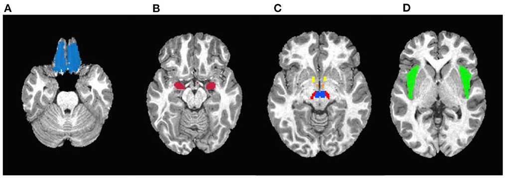
In the original article, there was an error in Figure 3 as published. An error was caught with the ventral tegmental area (VTA) masks. We have identified the MNI coordinates for VTA, created a mask, and updated the analysis.
The corrected Figure 3 and its caption appear below.
The corrected Figure 3 with caption:

Figure 3. Masks used in the region-of-interest (ROI) analysis. (A) Orbitofrontal cortex. (B) Amygdala. (C) Yellow: Nucleus accumbens; Orange: Hypothalamus; Red: Substantia nigra; Blue: Ventral tegmental area. (D) Insula.
Following the incorrect mask used for VTA, there was an error in Table 2/Supplementary Table 1 as published. Using the corrected mask, t-, p-, q-values for VTA changed. q-values (FDR-corrected statistical significance) for some other regions slightly changed because they are derived using the p-values of the multiple tests, but did not affect the interpretation of statistical significance. The VTA was statistically significantly associated with the dynamic advertising condition, but the statistical significance did not survive FDR correction.
The corrected Table 2/Supplementary Table 1 appears below.
The corrected Table 2:
The corrected Supplementary Table 1:
Three corrections have been made to the main text due to the error in the VTA mask.
1. A correction has been made to the abstract, Result, line 46.
This sentence previously stated:
“From the ROI analyses, the right and left hemispheres of the amygdala and insula, and the right hemisphere of the ventral tegmental area and substantia nigra showed significantly higher responses for the dynamic food ad medium after controlling for covariates and a false discovery rate correction.”
The corrected sentence appears below:
“From the ROI analyses, the right and left hemispheres of the amygdala and insula, and the right hemisphere of the substantia nigra showed significantly higher responses for the dynamic food ad medium after controlling for covariates and a false discovery rate correction.”
2. A correction has been made to the method, Region of Interest Analyses, paragraph 1, line 312.
This sentence previously stated:
“Masks of these bilateral regions were generated using the Talairach Daemon and Montreal Neurological Institute (MNI) atlas using AFNI (Analysis of Functional NeuroImages version: 21.0.06, (Cox and Hyde, 1997) and are shown in Figure 3.”
The corrected sentence appears below:
“Masks of these bilateral regions were generated using the Talairach Daemon and Montreal Neurological Institute (MNI) atlas using AFNI [Analysis of Functional NeuroImages version: 21.0.06 (Cox and Hyde, 1997)]. The mask of the ventral tegmental area was defined by the sphere with a radius of 5 mm centered at MNI coordinate [4, −16, −10] (Carter, 2009). The ROI masks are shown in Figure 3.”
3. A correction has been made to the results, ROI Analyses, paragraph 1, line 357.
This sentence previously stated:
“Specifically, in both unadjusted and adjusted models and after the FDR correction, the right and left amygdala, the right and left insula, right ventral tegmental area, and right substantia nigra showed statistically significant higher reward-related response to dynamic ads as compared to static ads.”
The corrected sentence appears below:
“Specifically, in both unadjusted and adjusted models and after the FDR correction, the right and left amygdala, the right and left insula, and right substantia nigra showed statistically significant higher reward-related response to dynamic ads as compared to static ads. The right ventral tegmental area and left substantia nigra showed significantly higher reward-related response to dynamic ads as compared to static ads before the FDR correction but not after.”
The authors apologize for this error and state that this does not change the scientific conclusions of the article in any way. The original article has been updated.
All claims expressed in this article are solely those of the authors and do not necessarily represent those of their affiliated organizations, or those of the publisher, the editors and the reviewers. Any product that may be evaluated in this article, or claim that may be made by its manufacturer, is not guaranteed or endorsed by the publisher.
Carter, R. M. (2009). Activation in the VTA and nucleus accumbens increases in anticipation of both gains and losses. Front. Behav. Neurosci. 3, 21. doi: 10.3389/neuro.08.021.2009
Keywords: food cues, fMRI, neural reactivity, visual stimuli, children, static ad, dynamic ad
Citation: Yeum D, Jimenez CA, Emond JA, Meyer ML, Lansigan RK, Carlson DD, Ballarino GA, Gilbert-Diamond D and Masterson TD (2023) Corrigendum: Differential neural reward reactivity in response to food advertising medium in children. Front. Neurosci. 17:1170370. doi: 10.3389/fnins.2023.1170370
Received: 20 February 2023; Accepted: 08 March 2023;
Published: 23 March 2023.
Edited and reviewed by: Celia Andreu-Sánchez, Autonomous University of Barcelona, Spain
Copyright © 2023 Yeum, Jimenez, Emond, Meyer, Lansigan, Carlson, Ballarino, Gilbert-Diamond and Masterson. This is an open-access article distributed under the terms of the Creative Commons Attribution License (CC BY). The use, distribution or reproduction in other forums is permitted, provided the original author(s) and the copyright owner(s) are credited and that the original publication in this journal is cited, in accordance with accepted academic practice. No use, distribution or reproduction is permitted which does not comply with these terms.
*Correspondence: Dabin Yeum, ZGFiaW4ueWV1bS5nckBkYXJ0bW91dGguZWR1
†These authors have contributed equally to this work and share senior authorship
Disclaimer: All claims expressed in this article are solely those of the authors and do not necessarily represent those of their affiliated organizations, or those of the publisher, the editors and the reviewers. Any product that may be evaluated in this article or claim that may be made by its manufacturer is not guaranteed or endorsed by the publisher.
Research integrity at Frontiers

Learn more about the work of our research integrity team to safeguard the quality of each article we publish.