 Solon Alberto Orlando1,2
 Katherine Paez Martinez3Ericka Sanchez1Carmen de la Cruz1Joselyn Calderon1Fabrizio Arcos3Pablo Torres-Lasso4Manuel Calvopiña5
 Miguel Angel Garcia-Bereguiain5*
- 1Instituto Nacional de Salud Pública e Investigación, Guayaquil, Ecuador
- 2Universidad Espiritu Santo, Guayaquil, Ecuador
- 3Facultad de Medicina Veterinaria y Zootecnia, Universidad Agraria del Ecuador, Guayaquil, Ecuador
- 4Facultad de Veterinaria, Universidad de Guayaquil, Guayaquil, Ecuador
- 5One Health Research Group, Universidad de Las Américas, Quito, Ecuador
Background: Leptospirosis is a zoonotic disease of worldwide distribution that affects humans and domestic and wild animals, and it is highly endemic in Ecuador. However, no reports of infections affecting horses have been published in the country.
Methods: This study evaluates the prevalence of anti-Leptospira spp. antibodies in racing horses from a breeding farm in the coastal Santa Elena province, southwest Ecuador. Sera were collected from 108 non-vaccinated horses and evaluated for 24 serovars of Leptospira spp. using the microscopic agglutination test (MAT).
Results: It was found that 100% of horses were reactive for Leptospira spp., most of them for multiple serovars. The most prevalent serovars were Leptospira kirschneri serovar Grippotyphosa (100%; 95% CI 99.9% to 100.1%); L. interrogans serovars Sejroe (96.3%; 95% CI 96.2% to 96.4%), Saxkoebing (95.4%; 95% CI 95.3% to 95.5%), Canicola (90.7%; 95% CI 90.5% to 90.9%), Icterohaemorrhagiae (80.5%; 95% CI 80.4% to 80.6%), Bataviae (73.1%; 95% CI 73.0% to 73.2%), Australis (75.0%; 95% CI 74.9% to 75.1%), and Bratislava (71.2%; 95% CI 71.1% to 71.4%); and L. borgpetersenii serovar Tarassovi (76.8%; 95% CI 76.6% to 77.0%).
Conclusions: We found a high prevalence of anti-Leptospira spp. seropositivity in racehorses from a breeding farm in Ecuador. This is the first serologic report for leptospirosis in horses in Ecuador. “One Health”-based sanitary practices for horse-breeding farms are recommended to improve animal and human health.
Introduction
Leptospirosis is a zoonotic disease with worldwide distribution although its prevalence is higher in the tropics and poorer regions, and it is endemic in South America (1, 2). Bacteria from the genus Leptospira are the causative agent and can infect almost all mammal species (3). Leptospirosis affects domestic and wild mammals, and cross-species transmission, including to humans, occurs through direct contact with urine from infected animals or indirect contact with contaminated soil and water where Leptospira spp. can survive for long periods (3, 4). Free-roaming dogs and rats are considered the main reservoir of the disease in urban areas (5), while livestock plays an important role in occupational leptospirosis transmission (6). Leptospirosis is estimated to cause 1.03 million human cases and 58,900 deaths each year worldwide. Although infected individuals can be asymptomatic, severe disease may produce renal or hepatic failure and pulmonary bleeding that can lead to death (7, 8).
Leptospira spp. that infects human and animal populations include pathogenic, intermediate pathogenic, and saprophytic clusters, defined further by the presence of serological characteristics, or serovars. The pathogenic, or interrogans, cluster comprises 16 strains across nine species: L. interrogans, L. kirschneri, L. noguchii, L. borgpetersenii, L. weilii, L. santarosai, L. alexanderi, L. kmetyi, and L. alstonii. The intermediate cluster comprises L. fainei, L. licerasiae, and L. wolffii, which have been associated with mild disease and chronic infections. The saprophytic, or biflexa, cluster comprises 14 non-pathogenic strains, of the species L. biflexa and L. wolbachii.
Although leptospirosis is mainly subclinical in horses, it can lead to abortion, stillbirth, and neonatal mortality (9, 10). Clinical signs of leptospirosis in horses include moderate fever, anorexia, jaundice, and pulmonary bleeding; death by interstitial nephritis has also been described as indicative of leptospirosis (10–13). Leptospirosis causes economic losses in the racehorse business due to the interruption of training, poor performance, and disqualification in competition, as well as the cost of treatment for sick horses (11). In addition, leptospirosis in racehorses is a threat for zoonotic transmission due to the closeness of horse–human contact. In addition, as racehorses are among the most expensive domestic animals, leptospirosis in racehorses could be considered as a paradigm for evaluating leptospirosis management and concern in a particular region.
Leptospirosis is a neglected tropical disease in Ecuador, with 1,279 human cases reported in 2012 (14–16). A total of 2,584 hospitalizations were recorded from 2000 to 2022 across 22 provinces in Ecuador (17). The few studies addressing leptospirosis in livestock in Ecuador report a high prevalence in cattle, pigs, and dogs (18, 19). Moreover, leptospirosis has never been studied in horses in Ecuador although it is considered endemic in South America, with prevalence values ranging from 4.5% to 90.7% (6). A recent publication from 2019 found a prevalence of 85% using a 24-serovar microscopic agglutination test (MAT) panel on horses from police departments in Colombia (20). There are no public health policies with a “One Health” perspective to address leptospirosis in Ecuador, and even livestock vaccination is scarce. Considering this situation, the aim of this study was to evaluate the seroprevalence of anti-Leptospira spp. antibodies in an exploited animal of high economic value, namely, horses from racehorse farms in the coastal region of Ecuador, where leptospirosis is endemic.
Methods
Study design and setting
This study was performed in a racehorse-breeding farm located in the province of Santa Elena, in the southwest coastal region of Ecuador. This is, to our knowledge, the only farm of this kind in the coastal region of Ecuador. The ecological features of this area allow exposure to Leptospira spp. through direct contact between horses and free-roaming dogs and cats and wild rodents, or through contaminated water sources.
For the present study, samples were collected from December 2016 to February 2017. None of the animals were vaccinated against leptospirosis. In addition, no signs of leptospirosis were found in any of the horses at the time of sample collection (we were allowed only one quick and superficial animal inspection). The farm veterinarians did not report any horses with signs of leptospirosis during the sample collection period. As this study uses samples collected from domestic animals for diagnosis, in accordance with animal research regulations in Ecuador, IRB approval was waived.
Blood sample collection
Horses were managed by certified veterinarians. Blood was collected from the jugular vein. The serum was separated by centrifugation (5,000 rpm for 5 min). A total of 108 samples of sera were collected from all horses older than 1 year present on the farm at the time of the study (Supplementary Material 1).
Microscopic agglutination test for anti-Leptospira spp.
The microscopic agglutination test (MAT) was performed using 24 live antigens. The Leptospira species, serogroups, serovars, and strains used for MAT are detailed in Supplementary Table 1. MAT was performed in the Laboratorio Nacional de Referencia para Zoonosis of the Instituto Nacional de Salud Pública e Investigación in Guayaquil. This laboratory focuses on human sample analysis and uses a MAT panel implemented following Pan American Health Organization guidelines.
The antigens were prepared from the reference strains detailed in Supplementary Table 1. For the screening of sera, a 1:200 dilution was used initially. Reactive samples were then examined with increasing dilutions from 1:200 to 1:3,200, taking the highest positive dilution to be the titer of the serum. The serum was considered reactive when at least 50% agglutination occurred at a magnification of 40× under the microscope.
Results
All the 108 horses tested were seropositive for 5–15 different Leptospira spp. serovars when a MAT titer cut-off value of 200 was set, with titers ranging from 200 to 1,600 (Tables 1, 2). The most prevalent serovars were Leptospira kirschneri serovar Grippotyphosa (100%; 95% CI 99.9% to 100.1%); Leptospira interrogans serovars Sejroe (96.3%; 95% CI 96.2% to 96.4%), Saxkoebing (95.4%; 95% CI 95.3% to 95.5%), Canicola (90.7%; 95% CI 90.5% to 90.9%), Icterohaemorrhagiae (80.5%; 95% CI 80.4% to 80.6%), Bataviae (73.1%; 95% CI 73.0% to 73.2%), Australis (75.0%; 95% CI 74.9% to 75.1%), and Bratislava (71.2%; 95% CI 71.1% to 71.4%); and Leptospira borgpetersenii serovar Tarassovi (76.8%; 95% CI 76.6% to 77.0%). Serogroup and serovar distribution and titers for all horses included in the study are detailed in Supplementary Table 2. No differences in serovar prevalence were found between horses of different ages. As no horses presented signs of leptospirosis at the time that we visited the farm, no association between serovars and signs could be addressed.

Table 1 Distribution of Leptospira spp. serogroups, serovars and strains in the 108 horses included in this study for microaglutination test (MAT) with a cut off titer value of 200.
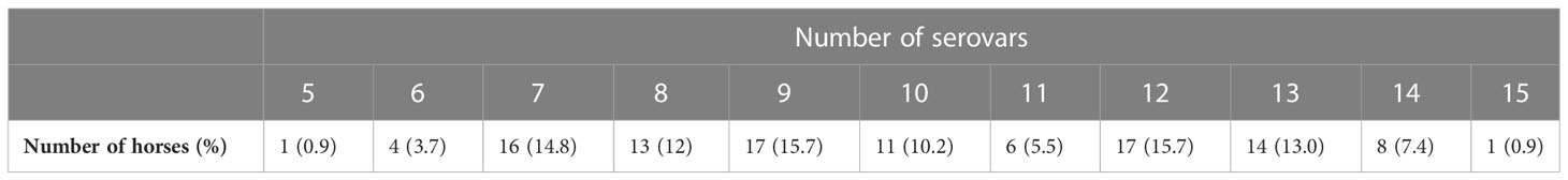
Table 2 Number and percentage of seropositive horses for multiple Leptospira spp. serovars.
We also addressed MAT seropositivity for the 108 horses using a MAT titer cut-off value of 800, as detailed in Table 3 and Supplementary Table 3. In this case, 55 out of 108 horses were positive for at least 1 of 11 different Leptospira spp. serovars, and the prevalence obtained was 50.9%. For this cut-off value of 800, the most prevalent serovars were Leptospira kirschneri serovar Grippotyphosa (12/108; 11.1%; 95% CI 11.0% to 11.2%), and Leptospira interrogans serovars Bataviae (22/108; 22.4%; 95% CI 21.9% to 22.1%) and Canicola (20/108; 18.5%; 95% CI 18.3% to 18.7%).
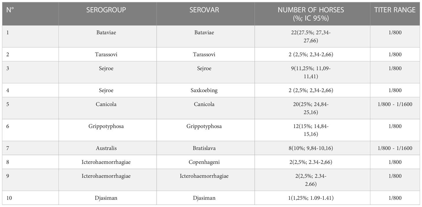
Table 3 Distribution of Leptospira spp. serogroups, serovars and strains in the 108 horses included in this study for microaglutination test (MAT) with a cut off titer value of 800.
Discussion
This study reports on the striking case of a racehorse-breeding farm in Ecuador, where 100% of the horses were seropositive for Leptospira spp. at a MAT cut-off titer value of 200. Furthermore, most horses were positive for multiple Leptospira spp. serovars. Cross-reactivity between different Leptospira spp. serovars has been described for the MAT technique; therefore, we also addressed anti-Leptospira spp. antibody seroprevalence using a MAT titer cut-off value of 800 (21). Although there was a remarkable reduction in seropositivity and serovar distribution (some serovars were not present at the 800 titer cut-off value), we cannot totally rule out the possibility that positive MAT results for titers ranging from 200 to 800 are due to cross-reactivity. Moreover, anti-Leptospira spp. antibody seroprevalence was over 50% even for a high specific cut-off titer value of 800. In addition, for a titer cut-off value of 800, the main infective serovars were Bataviae (20.4%) and Canicola (18.5%), which belong to the pathogenic cluster. We found six horses that were positive for both serovars, belonging to different serogroups, so either coinfection with multiple serovars or successive infections could have occurred.
We could not find any differences associated with age for Leptospira spp. serovar distribution. The differences in serovars between horses could be partially explained by the different origins of the horses (some of them were brought from neighboring countries such as Peru, Chile, and Argentina) or frequent travel to competitions. However, this information was not provided in detail by the farm and could not be evaluated. Another limitation of our study was the scarce information regarding signs of leptospirosis in the horses, as we were allowed only one quick evaluation of the horses prior to sample collection; furthermore, no clinical records were provided by the farm, and the veterinarians did not recall any horses with signs of leptospirosis. Therefore, no associations between signs of leptospirosis and specific serovars could be addressed.
The results of our study coincide with previous reports showing that horse leptospirosis is endemic in South America, with prevalence values ranging from 4.5% to 90.7% (6). In addition, a recent publication, from 2019, found a prevalence of 85% using a 24-serovar MAT panel on horses from police departments in Colombia (20). However, it is important to note that the most prevalent serovars in our study (Grippotyphosa, Bataviae, Sejroe, Canicola, Bratislava, and Tarassovi) differed from the most prevalent ones reported in South America (Icterohaemorrhagiae, Australis, and Pomona) (6) or in Colombia in particular (Djasiman) (20). These differences may be explained by environmental differences, such as the amount of rain or the duration of the rainy season, or by the presence of different transmission vectors depending on the area of study. Notably, the Leptospira spp. serovar MAT panels used by different countries in South America may differ, which could be an additional methodological reason for the difference in serovars reported in this study (22). Moreover, a recent study from New Zealand of 499 racing horses from 25 different breading farms found a Leptospira spp. seroprevalence of 25%, despite only five serovars being tested (23). These results indicate that leptospirosis is not just a problem for horse health in low- and middle-income countries.
The ultrahigh seroprevalence of anti-Leptospira spp. antibodies reported in our study coincide with values reported in previous publications regarding leptospirosis prevalence in livestock and domestic animals in Ecuador: 70% in dogs, 35.4%–74% in cattle, and 67% in pigs (16, 18, 19). Although those studies reported a high prevalence of leptospirosis, they were performed using PCR or a MAT panel comprising only eight serovars for diagnosis; these methodological differences could explain the higher prevalence found in our study. Moreover, a recent report from our laboratory using a MAT panel of 24 serovars for diagnosis also found 100% seroprevalence for Leptospira spp. in domestic and wild animals from a mixed-use rescue center in the coastal region of Ecuador (22). Although this is the first report of leptospirosis in horses from Ecuador, the Leptospira spp. serovars reported in those studies were also found in our study, suggesting the widespread distribution of serovars among livestock.
These horses were not vaccinated against Leptospira spp., despite the high economic value of this kind of horses in the market (some of the most prevalent serovars are included in the vaccine formulation, such as Grippotyphosa and Canicola). The farm lacked proper sanitary conditions, and free-roaming dogs, cats, and wild rodents were reported by the farm workers. Moreover, the farm is located in the coastal region of Ecuador, where seasonal floods are frequent and could facilitate leptospirosis transmission (18).
Future directions of our research will include a deeper “One Health” approach, identifying the Leptospira spp. serovars associated with free-roaming dogs, cats, and rodents found in farms, and also with water sources, to identify a potential transmission route. Finally, it is also necessary to increase awareness among the public health authorities of the risk of environmental exposure to Leptospira spp. in farm workers and other high-risk groups in Ecuador, in order to develop guidelines for leptospirosis surveillance and prevention according to the “One Health” concept.
Conclusions
Although the main limitation of our study was that only a single horse-breeding farm was included, a “One Health”-based management approach to horse breeding must be improved in an epidemiological context such as that in Ecuador, where anti-Leptospira spp. antibody seroprevalence in livestock and companion animals is extremely high, especially considering the close human–horse contact that racehorse breeding and training implies.
Data availability statement
The original contributions presented in the study are included in the article/Supplementary Material. Further inquiries can be directed to the corresponding author.
Ethics statement
The animal study was reviewed and approved by Universidad de Las Americas. Written informed consent was obtained from the owners for the participation of their animals in this study.
Author contributions
SO contributed to study conceptualization, logistics and funds allocation, data collection, experimental analysis, drafting the first manuscript, and reviewing the final version. KP contributed to data collection, experimental analysis, and reviewing the final version. ES contributed to data collection, experimental analysis, drafting of the first manuscript, and reviewing the final version. CC contributed to data collection, experimental analysis, and reviewing the final version. FA contributed to logistics and funds allocation, data collection, and reviewing the final version. PT-L contributed to logistics and funds allocation, data collection, and reviewing the final version. MG-B contributed to study conceptualization, logistics and funds allocation, publication funds allocation, data collection, experimental analysis, and drafting of the first and final version of the manuscript. All authors contributed to the article and approved the submitted version.
Acknowledgments
The authors of this work thank the staff from the horse-breeding farm for their support in carrying out this study.
Conflict of interest
The authors declare that the research was conducted in the absence of any commercial or financial relationships that could be construed as a potential conflict of interest.
Publisher’s note
All claims expressed in this article are solely those of the authors and do not necessarily represent those of their affiliated organizations, or those of the publisher, the editors and the reviewers. Any product that may be evaluated in this article, or claim that may be made by its manufacturer, is not guaranteed or endorsed by the publisher.
Supplementary material
The Supplementary Material for this article can be found online at: https://www.frontiersin.org/articles/10.3389/fitd.2023.1061038/full#supplementary-material
Abbreviations
MAT, microscopic agglutination test.
References
1. Bharti AR, Nally JE, Ricaldi JN, Matthias MA, Diaz MM, Lovett MA, et al. Peru-United states leptospirosis consortium, leptospirosis: a zoonotic disease of global importance. Lancet Infect Dis (2003) 3(12):757–71. doi: 10.1016/S1473-3099(03)00830-2
2. Schneider MC, Leone DG, Hamrick PN, de Caldas EP, Velásquez RT, Mendigaña Paez FA, et al. Leptospirosis in Latin America: exploring the first set of regional data. Rev Panam Salud Publica (2017) 41:e81. doi: 10.26633/RPSP.2017.81
3. Ellis WA. Animal leptospirosis. Curr Top Microbiol Immunol (2015) 387:99–137. doi: 10.1007/978-3-662-45059-8_6
4. Porto de Abreu JA, Krawczak FS, Barbosa Guedes I, de Souza-Filho AF, Oliveira de Souza G, de Campos Binder L, et al. Frequency of anti-leptospira spp. antibodies in dogs and wild small mammals from rural properties and conservation units in southern Brazil. One Health (2019) 8:100104. doi: 10.1016/j.onehlt.2019.100104
5. Costa F, Ribeiro GS, Felzemburgh RDM, Santos N, Barbosa Reis R, Santos AC, et al. Influence of household rat infestation on leptospira transmission in the urban slum environment. PloS Negl Trop Dis (2014) 8:e3338. doi: 10.1371/journal.pntd.0003338
6. Pinto PS, Libonati H, Lilenbaum W. A systematic review of leptospirosis on dogs, pigs, and horses in Latin America. Trop Anim Health Prod (2017) 49:231–8. doi: 10.1007/s11250-016-1201-8
7. Costa F, Hagan JE, Calcagno J, Kane M, Torgerson P, Martinez-Silveira MS, et al. Global morbidity and mortality of leptospirosis: a systematic review. PloS Negl Trop Dis (2015) 9:e0003898. doi: 10.1371/journal.pntd.0003898
8. Haake DA, Levett PN. Leptospirosis in humans. Curr Top Microbiol Immunol (2015) 387:65–97. doi: 10.1007/978-3-662-45059-8_5
9. Whitwell KE, Blunden AS, Miller J, Errington J. Two cases of equine pregnancy loss associated with leptospira infection in England. Vet Rec (2009) 165:377–8. doi: 10.1136/vr.165.13.377
10. Verma A, Stevenson B, Adler B. Leptospirosis in horses. Vet Microbiol (2013) 167:61–6. doi: 10.1016/j.vetmic.2013.04.012
11. Baverud V, Gunnarsson A, Engvall EO, Frazen PEA. Leptospira seroprevalence and association between seropositivity, clinical disease and host factors in horses. Acta Veterinaria Scandinavica (2009) 51:15. doi: 10.1186/1751-0147-51-15
12. Braga J, Hamond C, Martins G, Abreu RN, Lilenbaum W. Ophthalmic alterations in horses with leptospirosis by serovar icterohaemorrhagiae in Rio de Janeiro, Brazil. Pesquisa Veterinária Brasileira (2011) 31:147–50. doi: 10.1590/S0100-736X2011000200009
13. Hamond C, Pinna A, Martins G, Lilenbaum W. The role of leptospirosis in reproductive disorders in horses. Trop Anim Health Prod (2014) 46:1–10. doi: 10.1007/s11250-013-0459-3
14. Manock SR, Jacobsen KH, de Bravo NB, Russell KL, Negrete M, Olson JG, et al. Etiology of acute undifferentiated febrile illness in the Amazon basin of Ecuador. Am J Trop Med Hyg (2009) 81(1):146–51. doi: 10.4269/ajtmh.2009.81.146
15. Cartelle-Gestal M, Holban AM, Escalante S, Cevallos M. Epidemiology of tropical neglected diseases in Ecuador in the last 20 years. PloS One (2015) 10(9):e0138311. doi: 10.1371/journal.pone.0138311
16. Chiriboga J, Barragan V, Arroyo G, Sosa A, Birdsell DN, España K, et al. High prevalence of intermediate leptospira spp. DNA in febrile humans from urban and rural Ecuador. Emerg Infect Dis (2015) 21(12):2141–7. doi: 10.3201/eid2112.140659
17. Calvopiña M, Vásconez E, Coral-Almeida M, Romero-Alvarez D, Garcia-Bereguiain MA, Orlando A. Leptospirosis: morbidity, mortality, and spatial distribution of hospitalized cases in ecuador. a nationwide study 2000-2020. PloS Negl Trop Dis (2022) 16(5):e0010430. doi: 10.1371/journal.pntd.0010430
18. Barragan V, Chiriboga J, Miller E, Olivas S, Birdsell D, Hepp C, et al. High leptospira diversity in animals and humans complicates the search for common reservoirs of human disease in rural Ecuador. PloS Negl Trop Dis (2016) 13(10):e0004990. doi: 10.1371/journal.pntd.0004990
19. Burgos Macias DI, Perez Ruano M, Bulnes Goicochea CA, Zambrano Aguayo MD, Sandoval Valencia HP, Falconi Flores MA, et al. Determination of the seroprevalence of leptospira spp. and the main serovars circulating in cattle in the province of Manabí, Ecuador. Rev Sci Tech. Off Int Epiz. (2019) 38(3):1–17. doi: 10.1016/j.cimid.2020.101527
20. Calderon JC, Astudillo M, Romero M. Caracterización epidemiológica de la infección por leptospira spp. en caballos de trabajo y en personas ocupacionalmente expuestas en seis unidades de la policía nacional de Colombia. Biomédica (2019) 39:19–34.
21. Trimble AC, Blevins CA, Beard LA, Deforno AR, Davis EG. Seroprevalence, frequency of leptospiuria, and associated risk factors in horses in Kansas, Missouri, and Nebraska from 2016-2017. PloS One (2018) 13(10):e0206639. doi: 10.1371/journal.pone.0206639
22. Orlando SA, Perez A, Sanchez E, de la Cruz C, Rugel O, Garcia-Bereguiain MA. High seroprevalence of anti- leptospira spp. antibodies in domestic and wild mammals from a mixed use rescue center in Ecuador: lessons for "One health" based conservation strategies. One Health (2020) 10:100140. doi: 10.1016/j.onehlt.2020.100140
Keywords: Leptospira, MAT, leptospirosis, horses, Ecuador, One Health
Citation: Orlando SA, Paez Martinez K, Sanchez E, de la Cruz C, Calderon J, Arcos F, Torres-Lasso P, Calvopiña M and Garcia-Bereguiain MA (2023) Racehorses from a breeding farm in Tropical Ecuador have a high seroprevalence of anti-Leptospira spp. antibodies: a paradigm for leptospirosis management from a One Health perspective. Front. Trop. Dis 4:1061038. doi: 10.3389/fitd.2023.1061038
Received: 04 October 2022; Accepted: 17 April 2023;
Published: 09 May 2023.
Edited by:
Gabriel Motoa, Jackson Health System, United StatesReviewed by:
Shih Keng Loong, University of Malaya, MalaysiaGabriel Gustavo Varela Pensado, Universidad de la República, Uruguay
Copyright © 2023 Orlando, Paez Martinez, Sanchez, de la Cruz, Calderon, Arcos, Torres-Lasso, Calvopiña and Garcia-Bereguiain. This is an open-access article distributed under the terms of the Creative Commons Attribution License (CC BY). The use, distribution or reproduction in other forums is permitted, provided the original author(s) and the copyright owner(s) are credited and that the original publication in this journal is cited, in accordance with accepted academic practice. No use, distribution or reproduction is permitted which does not comply with these terms.
*Correspondence: Miguel Angel Garcia-Bereguiain, bWFnYmVyZWd1aWFpbkBnbWFpbC5jb20=