
Editors
2
Impact
Loading...
Review
19 September 2011
Gene replacement therapy by in vivo delivery of adeno-associated virus (AAV) is attractive as a potential treatment for a variety of genetic disorders. However, while AAV has been used successfully in many models, other experiments in clinical trials and in animal models have been hampered by undesired responses from the immune system. Recent studies of AAV immunology have focused on the elimination of transgene-expressing cells by the adaptive immune system, yet the innate immune system also has a critical role, both in the initial response to the vector and in prompting a deleterious adaptive immune response. Responses to AAV vectors are primarily mediated by the TLR9–MyD88 pathway, which induces the production of pro-inflammatory cytokines by activating the NF-κB pathways and inducing type I IFN production; self-complementary AAV vectors enhance these inflammatory processes. Additionally, the alternative NF-κB pathway influences transgene expression in cells transduced by AAV. This review highlights these recent discoveries regarding innate immune responses to AAV and discusses strategies to ablate these potentially detrimental signaling pathways.

Review
25 April 2011
David Kleinfeld
, 5 more and
Andy Y. Shih
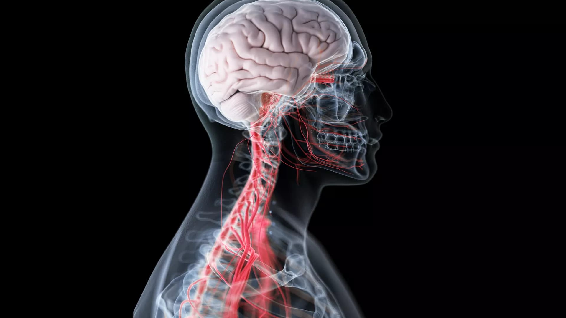
The neurovascular system may be viewed as a distributed nervous system within the brain. It transforms local neuronal activity into a change in the tone of smooth muscle that lines the walls of arterioles and microvessels. We review the current state of neurovascular coupling, with an emphasis on signaling molecules that convey information from neurons to neighboring vessels. At the level of neocortex, this coupling is mediated by: (i) a likely direct interaction with inhibitory neurons, (ii) indirect interaction, via astrocytes, with excitatory neurons, and (iii) fiber tracts from subcortical layers. Substantial evidence shows that control involves competition between signals that promote vasoconstriction versus vasodilation. Consistent with this picture is evidence that, under certain circumstances, increased neuronal activity can lead to vasoconstriction rather than vasodilation. This confounds naïve interpretations of functional brain images. We discuss experimental approaches to detect signaling molecules in vivo with the goal of formulating an empirical basis for the observed logic of neurovascular control.

Hypothesis and Theory
04 October 2010
Ute Lindauer
, 5 more and
Georg Royl
10,810 views
70 citations
Original Research
15 September 2010
Imaging Retinal Blood Flow with Laser Speckle Flowmetry
Anja I. Srienc
, 1 more and
Eric A. Newman
Laser speckle flowmetry (LSF) was initially developed to measure blood flow in the retina. More recently, its primary application has been to image baseline blood flow and activity-dependent changes in blood flow in the brain. We now describe experiments in the rat retina in which LSF was used in conjunction with confocal microscopy to monitor light-evoked changes in blood flow in retinal vessels. This dual imaging technique permitted us to stimulate retinal photoreceptors and measure vessel diameter with confocal microscopy while simultaneously monitoring blood flow with LSF. We found that a flickering light dilated retinal arterioles and evoked increases in retinal blood velocity with similar time courses. In addition, focal light stimulation evoked local increases in blood velocity. The spatial distribution of these increases depended on the location of the stimulus relative to retinal arterioles and venules. The results suggest that capillaries are largely unresponsive to local neuronal activity and that hemodynamic responses are mediated primarily by arterioles. The use of LSF to image retinal blood flow holds promise in elucidating the mechanisms mediating functional hyperemia in the retina and in characterizing changes in blood flow that occur during retinal pathology.

Hypothesis and Theory
27 September 2010
Noam Harel
, 3 more and
Essa Yacoub
9,527 views
26 citations

Original Research
27 August 2010
Mohamad Saka
, 1 more and
Myles Jones
9,486 views
27 citations

Review
19 August 2010
Fahmeed Hyder
, 6 more and
Douglas L. Rothman
8,537 views
47 citations
Review
16 August 2010
Jessica A. Filosa
10,477 views
37 citations
Original Research
11 August 2010
Sam Harris
, 2 more and
Jason Berwick
7,812 views
21 citations
8,160 views
27 citations
6,904 views
12 citations
Review
30 July 2010
Multi-Photon Nanosurgery in Live Brain
Anna Letizia Allegra Mascaro
, 1 more and
Francesco S. Pavone
In the last few years two-photon microscopy has been used to perform in vivo high spatial resolution imaging of neurons, glial cells and vascular structures in the intact neocortex. Recently, in parallel to its applications in imaging, multi-photon absorption has been used as a tool for the selective disruption of neural processes and blood vessels in living animals. In this review we present some basic features of multi-photon nanosurgery and we illustrate the advantages offered by this novel methodology in neuroscience research. We show how the spatial localization of multi-photon excitation can be exploited to perform selective lesions on cortical neurons in living mice expressing fluorescent proteins. This methodology is applied to disrupt a single neuron without causing any visible collateral damage to the surrounding structures. The spatial precision of this method allows to dissect single processes as well as individual dendritic spines, preserving the structural integrity of the main neuronal arbor. The same approach can be used to breach the blood-brain barrier through a targeted photo-disruption of blood vessels walls. We show how the vascular system can be perturbed through laser ablation leading toward two different models of stroke: intravascular clot and extravasation. Following the temporal evolution of the injured system (either a neuron or a blood vessel) through time lapse in vivo imaging, the physiological response of the target structure and the rearrangement of the surrounding area can be characterized. Multi-photon nanosurgery in live brain represents a useful tool to produce different models of neurodegenerative disease.
Original Research
30 July 2010
Janos Luckl
, 4 more and
Joel H. Greenberg
9,650 views
19 citations
Original Research
14 July 2010
Brain Specificity of Diffuse Optical Imaging: Improvements from Superficial Signal Regression and Tomography
Nicholas M. Gregg
, 3 more and
Joseph P. Culver
Functional near infrared spectroscopy (fNIRS) is a portable monitor of cerebral hemodynamics with wide clinical potential. However, in fNIRS, the vascular signal from the brain is often obscured by vascular signals present in the scalp and skull. In this paper, we evaluate two methods for improving in vivo data from adult human subjects through the use of high-density diffuse optical tomography (DOT). First, we test whether we can extend superficial regression methods (which utilize the multiple source–detector pair separations) from sparse optode arrays to application with DOT imaging arrays. In order to accomplish this goal, we modify the method to remove physiological artifacts from deeper sampling channels using an average of shallow measurements. Second, DOT provides three-dimensional image reconstructions and should explicitly separate different tissue layers. We test whether DOT’s depth-sectioning can completely remove superficial physiological artifacts. Herein, we assess improvements in signal quality and reproducibility due to these methods using a well-characterized visual paradigm and our high-density DOT system. Both approaches remove noise from the data, resulting in cleaner imaging and more consistent hemodynamic responses. Additionally, the two methods act synergistically, with greater improvements when the approaches are used together.
6,402 views
11 citations
Open for submission
Frontiers in Human Neuroscience
Exploring the Impact of Music Interventions on Brain Function, Behavior, and HealthEdited by Ying Liu, Jiancheng Hou, Guangyuan Liu, Jie Chen
Deadline
21 June 2025
Recommended Research Topics
Frontiers in Human Neuroscience
Exploring the Impact of Music Interventions on Brain Function, Behavior, and HealthEdited by Ying Liu, Jiancheng Hou, Guangyuan Liu, Jie Chen
Deadline
21 June 2025