Editorial
04 January 2024
Ainhoa Iglesias-Ara
and
Bart Westendorp

Objectives: Colorectal cancer (CRC) is one of the most common human malignancies. It was reported that the alterations in the DNA damage response (DDR) pathways are emerging as novel targets for treatment across different cancer types including CRC. RFWD3 plays a critical role in replication protein A (RPA)-mediated DNA damage in cancer cells. More importantly, RFWD3 can response to DNA damage by positively regulating p53 stability when the G1 cell cycle checkpoint is activated. However, the functional significance of RFWD3 in CRC has not been reported in the existing documents.
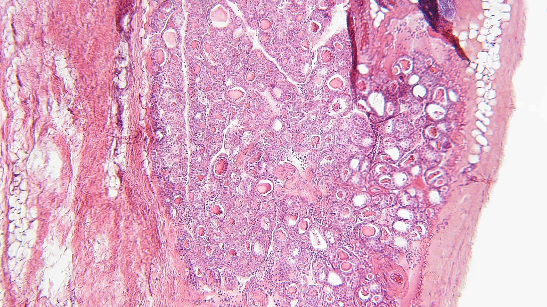
Materials and Methods: Here, we revealed high expression of RFWD3 in CRC tissues by IHC analysis and The Cancer Genome Atlas (TCGA) database. Besides, overexpression of RFWD3 in CRC cell lines was also confirmed by qRT-PCR and western blot assay. The Celigo cell counting method and wound-healing/transwell migration assay were applied to evaluate CRC cell proliferation and migration. The tumor growth indicators were quantified in nude mice xenografted with shRFWD3 and shCtrl RKO cells.
Results: The results indicated that RFWD3 knockdown restricted CRC development in vitro and in vivo. In exploring the downstream mechanism of RFWD3’s action, we found that RFWD3 could transcriptionally activate BIRC5 by interacting with E2F transcription factor 1 (E2F1). Accordingly, we identified BIRC5 as a downstream gene of RFWD3 regulating CRC. Subsequent loss- and gain- of function experiments demonstrated that upon overexpressing BIRC5 in RKO cells with down-regulated RFWD3, the inhibitory effects of cell proliferation, migration and colony formation could be reversed, while the capacity of cell apoptosis was ameliorated, suggesting that the effects of RFWD3 depletion was mainly due to BIRC5 suppression.
Conclusion: Taken together, this study revealed that RFWD3 participates in the occurrence and development of colorectal cancer via E2F1 transcriptional regulation of BIRC5.


Background: Quiescin Q6 sulfhydryl oxidase 2 (QSOX2), an enzyme that can be directly secreted into the extracellular space, is known to be associated with oxidative protein folding. However, whether QSOX2 is abnormally expressed in non-small cell lung cancer (NSCLC) and its role in tumor growth remains unclear.
Methods: Real-time quantitative PCR (qPCR), immunohistochemistry (IHC), bioinformatics analyses were applied to analyze the expression pattern and prognostic significance of QSOX2 in NSCLC. Xenografts model, enzyme-linked immunosorbent assays (ELISA), western blot analysis (WB), and IHC were preformed to examine in vivo tumor suppression and intracellular and extracellular expression of QSOX2. Flow cytometry, WB and qPCR analyses were used to elucidate the role of QSOX2 in cell cycle regulation. Chromatin immunoprecipitation assay (ChIP) assay and Dual-Luciferase reporter assay were employed to investigate transcriptional regulation of QSOX2 by E2F Transcription Factor 1 (E2F1).
Results: Quiescin sulfhydryl oxidase 2 was significantly overexpressed in NSCLC and associated with poor survival in advanced-stage patients. The intracellular and extracellular expression of QSOX2 by tumor cells markedly decreased after anti-cancer therapy in vitro, in vivo and in the clinic. Moreover, QSOX2 silencing in NSCLC cell lines resulted in inhibition of cancer cell proliferation, induction of apoptosis, and decreased expression of cell division-related genes (CENPF and NUSAP1) and Wnt pathway activators (PRRX2 and Nuc-β-catenin). Mechanistically, QSOX2 was expressed periodically during cell cycle and directly regulated by E2F1.
Conclusions: Our findings demonstrate that QSOX2 is directly regulated by E2F1 in the cell cycle, which is essential for the proliferation of NSCLC cells. Furthermore, QSOX2 is a prognostic indicator for NSCLC and may be developed into a biomarker for monitoring tumor burden and therapeutic progress.
Objective: The aim of this study was to investigate the role of KIF26A in breast cancer.
Method: qRT-PCR and immunohistochemistry were conducted to explore KIF26A expression and functional contribution to breast cancer development. MTS, EDU, colony formation assays, and flow cytometry analysis were conducted to assess cell proliferation characteristics and cell cycle progression. A series of 5′-flanking region deletion plasmids and mutating the binding site, with the luciferase reporter assay, were used to identify the core promotor region of KIF26A. The prediction by software and construction of the transcriptional factor plasmids were used to identify the transcriptional factor. Chromatin immunoprecipitation assay could demonstrate transcriptional factor directly binding to the KIF26A promoter. Human Genome Oligo Microarray Assay and gene ontology (GO) and pathway analyses were used to predict the downstream pathway.
Results: Our results showed that in breast cancer tissues, elevated KIF26A expression was significantly correlated with lymph node metastasis. KIF26A could promote proliferation and G0/G1 phase cell cycle progression in breast cancer cells. The core promoter region of the human KIF26A gene was located upstream of the transcription start site at position −395 to −385. The transcriptional factor E2F1 was shown to activate KIF26A expression. Furthermore, KIF26A was shown to inhibit the expression of p21, then activate CDK–RB–E2Fs pathway. The elevated E2F1 can activate the cell cycle progression and the KIF26A expression, forming feedback loop.
Conclusions: The present study demonstrated that KIF26A, directly upregulated by E2F1, promoted cell proliferation and cell cycle progression via CDK–RB–E2Fs feedback loop in breast cancer.