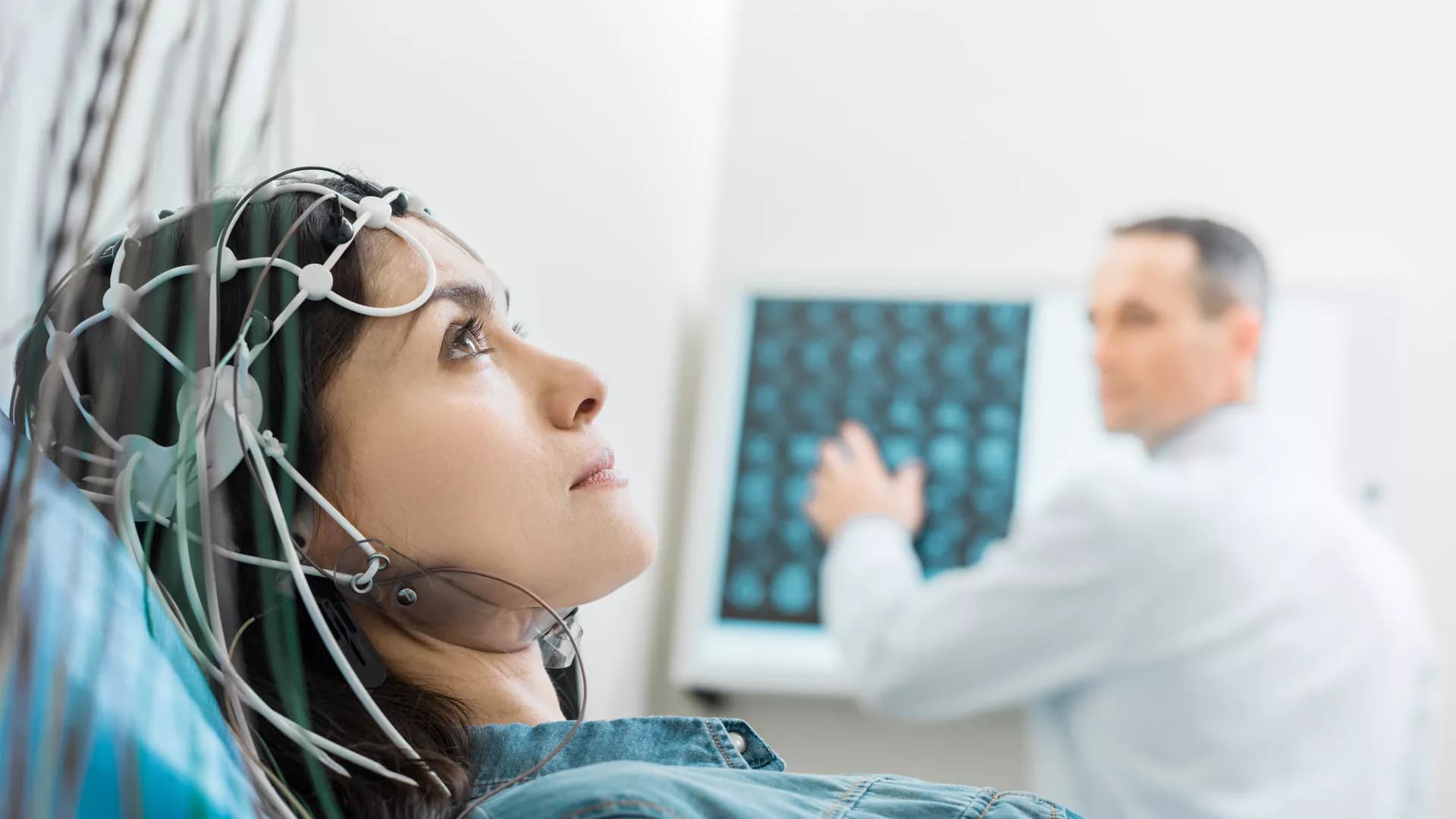
The aim of this study was to observe the clinical effects and brain electrical potential changes following acupuncture in the treatment of insomnia patients with mood disorders. Ninety patients with insomnia who met the inclusion criteria were randomly divided into the active acupuncture group (AA group, n = 44) and sham acupuncture group (SA group, n = 46) at a ratio of 1:1. The primary outcome was the total score of the Pittsburgh Sleep Quality Index (PSQI), and the secondary outcomes were the total effective rate, Self-Rating Anxiety Scale (SAS), Self-Rating Depression Scale (SDS) scores, and values of steady-state visual evoked potentials (SSVEP). The two groups received acupuncture or sham acupuncture 10 times (2 weeks). Finally, the total PSQI scores of the AA group and SA group were significantly different (p < 0.05) at 2 weeks (6.11 ± 2.33 vs. 10.37 ± 4.73), 6 weeks (6.27 ± 1.39 vs. 11.93 ± 3.07), 18 weeks (6.32 ± 2.84 vs. 11.78 ± 2.95) and 42 weeks (8.05 ± 3.14 vs. 12.54 ± 2.81). Further analysis found that AA group patients received acupuncture treatment at any age after the same effect (p > 0.05). The SAS and SDS scores of the AA group were also significantly different from those of the SA group at each assessment time point (p < 0.05). The total effective rate of the AA group was 81.82%, while that of the SA group was 30.43% (p < 0.05). There was no significant difference between the AA group and SA group only in the brain potential of the parietal lobe (F4), left temporal lobe (C3) and right temporal lobe (T8) (P > 0.05), but there was a significant difference between other brain regions (P < 0.05). In addition, correlation analysis showed that there was a certain positive correlation between the total PSQI score, SAS score, efficacy level, and SSVEP value in the AA group as follows: C4 and the total PSQI score (r = 0.595, P = 0.041), F3 and SAS score (r = 0.604, P = 0.037), FPz and efficiency level of the frontal lobe (r = 0.581, P = 0.048), and O2 and efficiency level of the occipital lobe (r = 0.704, P = 0.011). Therefore, acupuncture have a good clinical effect on patients with insomnia and emotional disorders and have a significant regulatory effect on abnormally excited brain potentials.
Objective: The aim of this study was to investigate the pattern of volume changes in neurofunctional hippocampal subfields in patients with insomnia and their associations with risk of development of insomnia.
Methods: A total of 120 patients with insomnia (78 females, 42 males; mean age ± standard deviation, 43.74 ± 13.02 years) and 120 good sleepers (67 females, 53 males; mean age, 42.69 ± 12.24 years) were recruited. The left hippocampus was segmented into anterior (L1), middle (L2), and posterior (L3) subregions. The right hippocampus was segmented into top anterior (R1), second top anterior (R2), middle (R3), posterior (R4), and last posterior (R5) subregions. Multivariate logistic regression was used to evaluate the associations of hippocampal volume (HV) of each subfield with the risk of the development of insomnia. Mediation analyses were performed to evaluate mediated associations among post-insomnia negative emotion, insomnia severity, and HV atrophy. A visual easy-to-deploy risk nomogram was used for individual prediction of risk of development of insomnia.
Results: Hippocampal volume atrophy was identified in the L1, R1, and R2 subregions. L1 and R2 volume atrophy each predisposed to an ~3-fold higher risk of insomnia (L1, odds ratio: 2.90, 95% confidence intervals: [1.24, 6.76], p = 0.014; R2, 2.72 [1.19, 6.20], p = 0.018). Anxiety fully mediates the causal path of insomnia severity leading to R1 volume atrophy with a positive effect. We developed a practical and visual competing risk-nomogram tool for individual prediction of insomnia risk, which stratifies individuals into different levels of insomnia risk with the highest prediction accuracy of 97.4% and an average C-statistic of 0.83.
Conclusion: Hippocampal atrophy in specific neurofunctional subfields was not only found to be associated with insomnia but also a significant risk factor predicting development of insomnia.
Idiopathic rapid eye movement sleep behavior disorder (iRBD) is an important non-motor complication of Parkinson's disease. At the same time, iRBD is considered to be the prodromal stage of α-synucleinopathy. This high risk of conversion suggests that iRBD becomes a nerve It is a window for early research on degenerative diseases and is the best candidate for neuroprotection trials. A wide range of neuroimaging techniques has improved our understanding of iRBD as a prodromal stage of the disease. In addition, neuroimaging of abnormal iRBD is expected to be a potential biomarker for predicting clinical phenotypic transformation. This article reviews the research progress of neuromolecular imaging in patients with iRBD from the perspective of iRBD transforming synucleinopathies.


