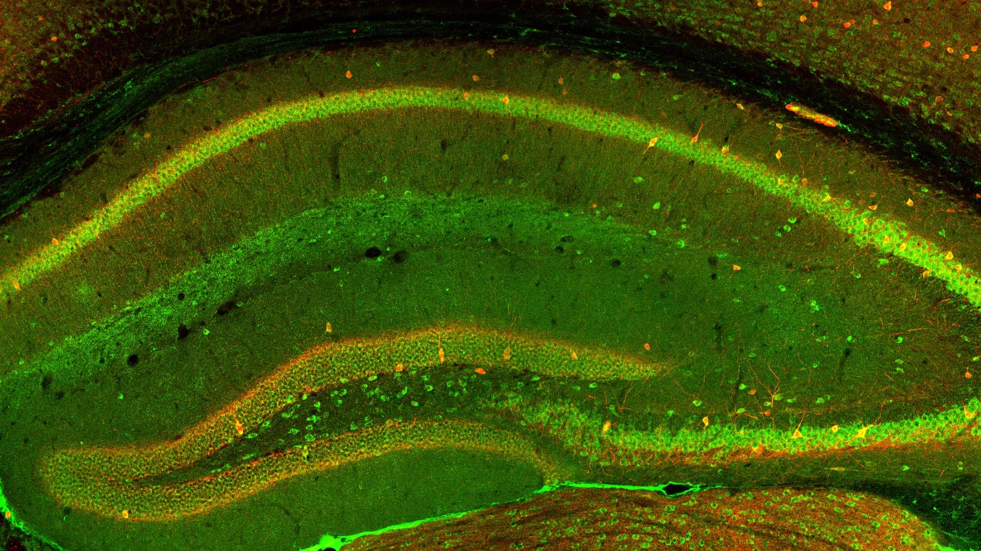
In addition to genetic information, environmental factors play an important role in the structure and function of nervous system and the occurrence and development of some nervous system diseases. Enriched environment (EE) can not only promote normal neural development through enhancing neuroplasticity but also play a nerve repair role in restoring functional activities during CNS injury by morphological and cellular and molecular adaptations in the brain. Different stages of development after birth respond to the environment to varying degrees. Therefore, we systematically review the pro-developmental and anti-stress value of EE during pregnancy, pre-weaning, and “adolescence” and analyze the difference in the effects of EE and its sub-components, especially with physical exercise. In our exploration of potential mechanisms that promote neurodevelopment, we have found that not all sub-components exert maximum value throughout the developmental phase, such as animals that do not respond to physical activity before weaning, and that EE is not superior to its sub-components in all respects. EE affects the developing and adult brain, resulting in some neuroplastic changes in the microscopic and macroscopic anatomy, finally contributing to enhanced learning and memory capacity. These positive promoting influences are particularly prominent regarding neural repair after neurobiological disorders. Taking cerebral ischemia as an example, we analyzed the molecular mediators of EE promoting repair from various dimensions. We found that EE does not always lead to positive effects on nerve repair, such as infarct size. In view of the classic issues such as standardization and relativity of EE have been thoroughly discussed, we finally focus on analyzing the essentiality of the time window of EE action and clinical translation in order to devote to the future research direction of EE and rapid and reasonable clinical application.
One reason that many central nervous system injuries, including those arising from traumatic brain injury, spinal cord injury, and stroke, have limited recovery of function is that neurons within the adult mammalian CNS lack the ability to regenerate their axons following trauma. This stands in contrast to neurons of the adult mammalian peripheral nervous system (PNS). New evidence, provided by single-cell expression profiling, suggests that, following injury, both mammalian central and peripheral neurons can revert to an embryonic-like growth state which is permissive for axon regeneration. This “redevelopment” strategy could both facilitate a damage response necessary to isolate and repair the acute damage from injury and provide the intracellular machinery necessary for axon regrowth. Interestingly, serotonin neurons of the rostral group of raphe nuclei, which project their axons into the forebrain, display a robust ability to regenerate their axons unaided, counter to the widely held view that CNS axons cannot regenerate without experimental intervention after injury. Furthermore, initial evidence suggests that norepinephrine neurons within the locus coeruleus possess similar regenerative abilities. Several morphological characteristics of serotonin axon regeneration in adult mammals, observable using longitudinal in vivo imaging, are distinct from the known characteristics of unaided peripheral nerve regeneration, or of the regeneration seen in the spinal cord and optic nerve that occurs with experimental intervention. These results suggest that there is an alternative CNS program for axon regeneration that likely differs from that displayed by the PNS.
Significant stress exposure and psychiatric depression are associated with morphological, biochemical, and physiological disturbances of astrocytes in specific brain regions relevant to the pathophysiology of those disorders, suggesting that astrocytes are involved in the mechanisms underlying the vulnerability to or maintenance of stress-related neuropathology and depression. To understand those mechanisms a variety of studies have probed the effect of various modalities of stress exposure on the metabolism, gene expression and plasticity of astrocytes. These studies have uncovered the participation of various cellular pathways, such as those for intracellular calcium regulation, neuroimmune responses, extracellular ionic regulation, gap junctions-based cellular communication, and regulation of neurotransmitter and gliotransmitter release and uptake. More recently epigenetic modifications resulting from exposure to chronic forms of stress or to early life adversity have been suggested to affect not only neuronal mechanisms but also gene expression and physiology of astrocytes and other glial cells. However, much remains to be learned to understand the specific role of those and other modifications in the astroglial contribution to the vulnerability to and maintenance of stress-related disorders and depression, and for leveraging that knowledge to achieve more effective psychiatric therapies.
Frontiers in Cellular Neuroscience
Spatial Microglial Identities and Functions in the Mammalian CNS: Unveiling Cellular Diversity and Functional Roles


