Editorial
18 January 2023
Roberto Esposito
, Nicoletta Cera
, Fernando Barbosa
and
Filippo Cieri


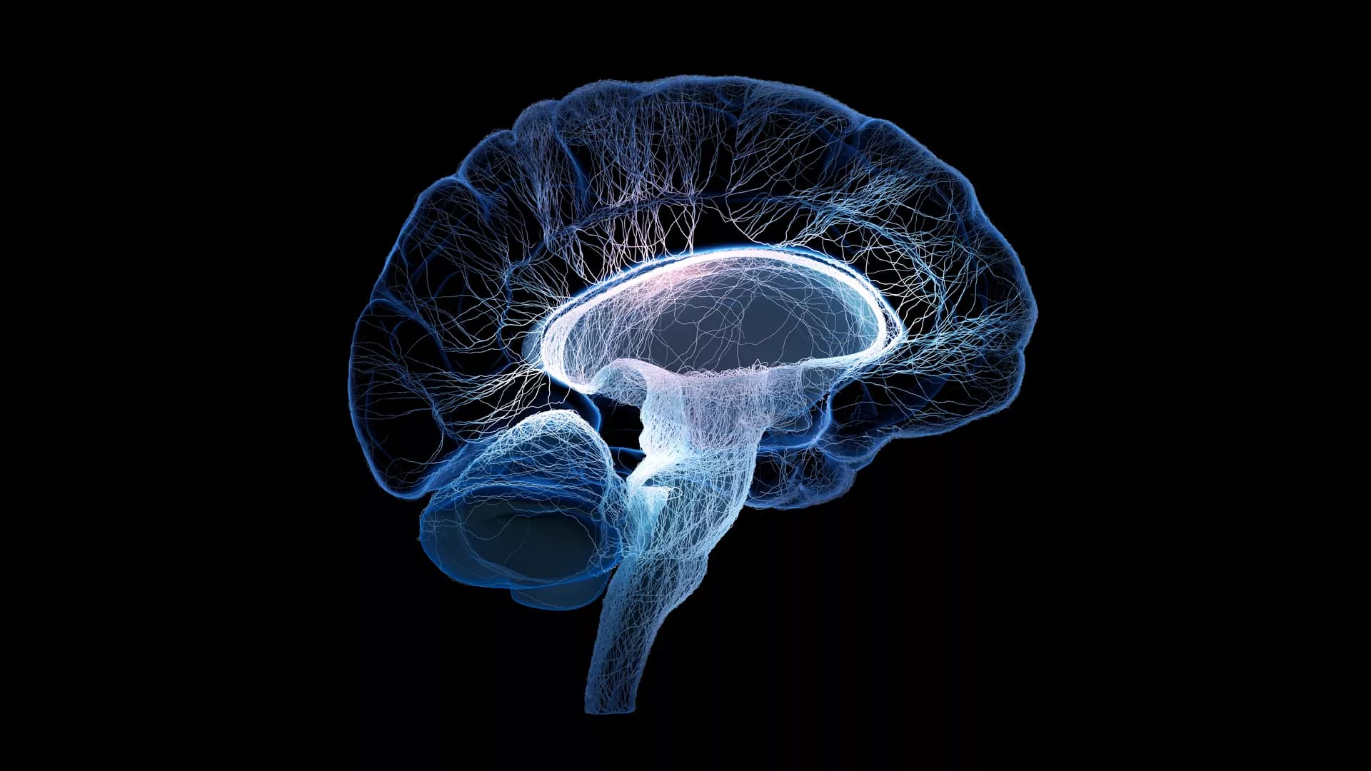
![Spatial maps of the 48 independent components identified as RSNs categorized by domain [auditory (AUD), default mode network (DMN), executive control (ECN), salience (SAL), sensorimotor (SEN), subcortical (SBC), attentional (ATT), and visual (VIS)] and component number.](https://www.frontiersin.org/_rtmag/_next/image?url=https%3A%2F%2Fwww.frontiersin.org%2Ffiles%2FArticles%2F971201%2Ffnimg-01-971201-HTML%2Fimage_m%2Ffnimg-01-971201-g001.jpg&w=3840&q=75)
Background: Textural features of the hippocampus in structural magnetic resonance imaging (sMRI) images can serve as potential diagnostic biomarkers for Alzheimer’s disease (AD), while exhibiting a relatively poor discriminant performance in detecting early AD, such as amnestic mild cognitive impairment (aMCI). In contrast to sMRI, functional magnetic resonance imaging (fMRI) can identify brain functional abnormalities in the early stages of cerebral disorders. However, whether the textural features reflecting local functional activity in the hippocampus can improve the diagnostic performance for AD and aMCI remains unclear. In this study, we combined the textural features of the amplitude of low frequency fluctuation (ALFF) in the slow-5 frequency band and structural images in the hippocampus to investigate their diagnostic performance for AD and aMCI using multimodal radiomics technique.
Methods: Totally, 84 AD, 50 aMCI, and 44 normal controls (NCs) were included in the current study. After feature extraction and feature selection, the radiomics models incorporating sMRI images, ALFF values and their combinations in the bilateral hippocampus were established for the diagnosis of AD and aMCI. The effectiveness of these models was evaluated by receiver operating characteristic (ROC) analysis. The radiomics models were further validated using the external data from the Alzheimer’s Disease Neuroimaging Initiative (ADNI) database.
Results: The results of ROC analysis showed that the radiomics models based on structural images in the hippocampus had a better diagnostic performance for AD compared with the models using ALFF, while the ALFF-based model exhibited better discriminant performance for aMCI than the models with structural images. The radiomics models based on the combinations of structural images and ALFF were found to exhibit the highest accuracy for distinguishing AD from NCs and aMCI from NCs.
Conclusion: In this study, we found that the textural features reflecting local functional activity could improve the diagnostic performance of traditional structural models for both AD and aMCI. These findings may deepen our understanding of the pathogenesis of AD, contributing to the early diagnosis of AD.

Idiopathic generalized epilepsy (IGE) was characterized by 3–6 Hz generalized spike-wave discharges (GSWDs), and extensive altered interactions in subcortical-cortical circuit. However, the dynamics and the causal relationship among these interactions were less studied. Using resting-state functional magnetic resonance imaging (fMRI) data, the abnormal connections in the subcortical-cortical pathway in IGE were examined. Then, we proposed a novel method of granger causal analysis based on the dynamic functional connectivity, and the predictive effects among these abnormal connections were calculated. The results showed that the thalamus, and precuneus were key regions representing abnormal functional network connectivity (FNC) in the subcortical-cortical circuit. Moreover, the connectivity between precuneus and adjacent regions had a causal effect on the widespread dysfunction of the thalamocortical circuit. In addition, the connection between the striatum and thalamus indicated the modulation role on the cortical connection in epilepsy. These results described the causality of the widespread abnormality of the subcortical-cortical circuit in IGE in terms of the dynamics of functional connections, which provided additional evidence for understanding the potential modulation pattern of the abnormal epileptic pathway.

Postpartum depression (PPD) is a major public health concern with significant consequences for mothers, their children, and their families. However, less is known about its underlying neuropathological mechanisms. The voxel-based degree centrality (DC) analysis approach provides a new perspective for exploring the intrinsic dysconnectivity pattern of whole-brain functional networks of PPD. Twenty-nine patients with PPD and thirty healthy postpartum women were enrolled and received resting-state functional magnetic resonance imaging (fMRI) scans in the fourth week after delivery. DC image, clinical symptom correlation, and seed-based functional connectivity (FC) analyses were performed to reveal the abnormalities of the whole-brain functional network in PPD. Compared with healthy controls (HCs), patients with PPD exhibited significantly increased DC in the right hippocampus (HIP.R) and left inferior frontal orbital gyrus (ORBinf.L). The receiver operating characteristic (ROC) curve analysis showed that the area under the curve (AUC) of the above two brain regions is all over 0.7. In the seed-based FC analyses, the PPD showed significantly decreased FC between the HIP.R and right middle frontal gyrus (MFG.R), between the HIP.R and left median cingulate and paracingulate gyri (DCG.L), and between the ORBinf.L and the left fusiform (FFG.L) compared with HCs. The PPD showed significantly increased FC between the ORBinf.L and the right superior frontal gyrus, medial (SFGmed.R) compared with HCs. Mean FC between the HIP.R and DCG.L positively correlated with EDPS scores in the PPD group. This study provided evidence of aberrant DC and FC within brain regions in patients with PPD, which was associated with the default mode network (DMN) and limbic system (LIN). Identification of these above-altered brain areas may help physicians to better understand neural circuitry dysfunction in PPD.