Editorial
13 September 2022
Abdul Malik Tyagi
and
Sadiq Umar


Background: Sex hormones are recognized to play a significant role in increasing bone mineral density (BMD) and promoting bone maturation during adolescence. The purpose of our study was to use a database with large population data to evaluate the association of BMD with sex hormones (including testosterone and estradiol) and sex hormone-binding globulin (SHBG) in adolescent boys and girls aged 12–19 years.
Methods: The data for our study were taken from the National Health and Nutrition Examination Survey 2013-2016, and we used weighted multiple linear regression models to assess the relationship between testosterone, estradiol, and SHBG and total BMD. We use weighted generalized additive models and smooth curve fitting to discover underlying nonlinear relationships.
Results: A total of 1648 teenagers (853 boys, 795 girls) were selected for the final analysis. In boys, testosterone and estradiol levels were positively associated with total BMD, whereas SHBG levels were negatively associated with total BMD after adjusting for covariates [P < 0.05; 95% confidence interval (CI)]. In addition, there was a point between estradiol and total BMD, after which the positive correlation between estradiol and total BMD was relatively insignificant in boys. In girls, there was a positive association between estradiol and total BMD (P < 0.05; 95% CI), but there was no significant association between the testosterone (β 0.0004; 95% CI -0.0001 to 0.0008) or SHBG (β -0.0001; 95% CI -0.0002 to 0.0001) levels and total BMD. We also found an inverted U-shaped association between testosterone and total BMD with the inflection point at 25.4 ng/dL of testosterone.
Conclusions: We found differences in the association of sex hormones with total BMD in boys and girls. Based on our findings, an appropriate increase in serum testosterone levels may be beneficial for skeletal development in girls because of the inverted U-shaped relationship (with the inflection point at 25.4 ng/dL of testosterone), and a high testosterone level might be detrimental to BMD. Furthermore, keeping estradiol levels below a certain level in boys (24.3 pg/mL) may be considered.
Purpose: The aim of this review is to assess the current evidence regarding the impact of relaxin on incidence of soft tissue hip injuries in women.
Methods: A trained research librarian assisted with searches of PubMed, Embase, CINAHL, and SPORTDiscus, with a preset English language filter. The review was completed per the Joanna Briggs Institute (JBI) Manual for Evidence Synthesis methodology. Included studies required assessment of relaxin effects on musculoskeletal health, pelvic girdle stability, or hip joint structures in human subjects. Letters, texts, and opinion papers were excluded.
Results: Our screen yielded 82 studies. Molecularly, relaxin activates matrix metalloproteinases (MMPs) including collagenases MMP-1/-13 and gelatinases MMP-2/-9 to loosen pelvic ligaments for parturition. However, relaxin receptors have also been detected in female periarticular tissues, such as the anterior cruciate ligament, which tears significantly more often during the menstrual cycle peak of relaxin. Recently, high concentrations of relaxin-activated MMP-9 receptors have been found on the acetabular labrum; their expression upregulated by estrogen.
Conclusions: Menstrual cycle peaks of relaxin activate MMPs, which locally degrade collagen and gelatine. Women have relaxin receptors in multiple joints including the hip and knee, and increased relaxin correlates with increased musculoskeletal injuries. Relaxin has paracrine effects in the female pelvis on ligaments adjacent to hip structures, such as acetabular labral cells which express high levels of relaxin-targeted MMPs. Therefore, it is imperative to investigate the effect of relaxin on the hip to determine if increased levels of relaxin are associated with an increased risk of acetabular labral tears.

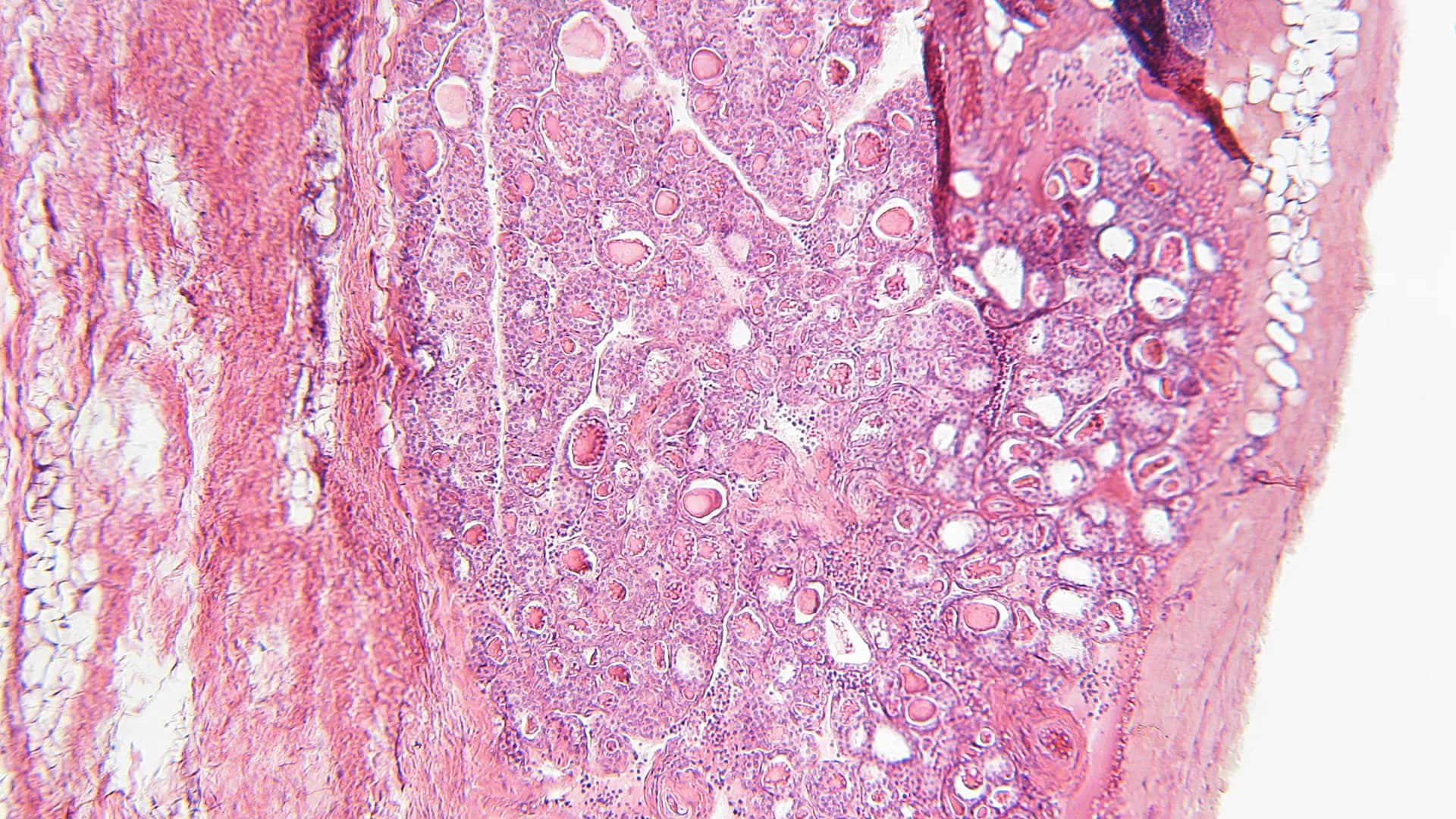
Osteoporosis is a common systemic bone disease caused by the imbalance between osteogenic activity and osteoclastic activity. Aged women are at higher risk of osteoporosis, partly because of estrogen deficiency. However, the underlying mechanism of how estrogen deficiency affects osteoclast activity has not yet been well elucidated. In this study, GSE2208 and GSE56815 datasets were downloaded from GEO database with 25 PreH BMD women and 25 PostL BMD women in total. The RRA algorithm determined 38 downregulated DEGs and 30 upregulated DEGs. Through GO analysis, we found that downregulated DEGs were mainly enriched in myeloid cell differentiation, cytokine-related functions while upregulated DEGs enriched in immune-related biological processes; pathways like Notch signaling and MAPK activation were found in KEGG/Rectome pathway database; a PPI network which contains 66 nodes and 91 edges was constructed and three Modules were obtained by Mcode; Correlation analysis helped us to find highly correlated genes in each module. Moreover, three hub genes FOS, PTPN6, and CTSD were captured by Cytohubba. Finally, the hub genes were further confirmed in blood monocytes of ovariectomy (OVX) rats by real-time PCR assay. In conclusion, the integrative bioinformatics analysis and real-time PCR analysis were utilized to offer fresh light into the role of monocytes in premenopausal osteoporosis and identified FOS, PTPN6, and CTSD as potential biomarkers for postmenopausal osteoporosis.
Breast cancer and osteoporosis are common diseases that affect the survival and quality of life in postmenopausal women. Women with breast cancer are more likely to develop osteoporosis than women without breast cancer due to certain factors that can affect both diseases simultaneously. For instance, estrogen and the receptor activator of nuclear factor-κB ligand (RANKL) play important roles in the occurrence and development of these two diseases. Moreover, chemotherapy and hormone therapy administered to breast cancer patients also increase the incidence of osteoporosis, and in recent years, neuropeptide Y (NPY) has also been found to impact breast cancer and osteoporosis.Y1 and Y5 receptors are highly expressed in breast cancer, and Y1 and Y2 receptors affect osteogenic response, thus potentially highlighting a potential new direction for treatment strategies. In this paper, the relationship between breast cancer and osteoporosis, the influence of NPY on both diseases, and the recent progress in the research and treatment of these diseases are reviewed.