Editorial
24 February 2022
Timo Uphaus
, Heinrich J. Audebert
, Michael W. Graner
, Steffen Tiedt
and
Robert G. Kowalski

Background: A reliable distinction between ischemic stroke (IS) and intracerebral hemorrhage (ICH) is required for diagnosis-specific treatment and effective secondary prevention in patients with stroke. However, in resource-limited settings brain imaging, which is the current diagnostic gold standard for this purpose, is not always available in time. Hence, an easily accessible and broadly applicable blood biomarker-based diagnostic test differing stroke subtypes would be desirable. Using an explorative proteomics approach, this pilot study aimed to identify novel blood biomarker candidates for distinguishing IS from ICH.
Material and Methods: Plasma samples from patients with IS and ICH were drawn during hospitalization and were analyzed by using liquid chromatography/mass spectrometry. Proteins were identified using the human reference proteome database UniProtKB, and label-free quantification (LFQ) data were further analyzed using bioinformatic tools.
Results: Plasma specimens of three patients with IS and four patients with ICH with a median National Institute of Health Stroke Scale (NIHSS) of 12 [interquartile range (IQR) 10.5–18.5] as well as serum samples from two healthy volunteers were analyzed. Among 495 identified protein groups, a total of 368 protein groups exhibited enough data points to be entered into quantitative analysis. Of the remaining 22 top-listed proteins, a significant difference between IS and ICH was found for Carboxypeptidase N subunit 2 (CPN2), Coagulation factor XII (FXII), Plasminogen, Mannan-binding lectin serine protease 1, Serum amyloid P-component, Paraoxonase 1, Carbonic anhydrase 1, Fibulin-1, and Granulins.
Discussion: In this exploratory proteomics-based pilot study, nine candidate biomarkers for differentiation of IS and ICH were identified. The proteins belong to the immune system, the coagulation cascade, and the apoptosis system, respectively. Further investigations in larger cohorts of patients with stroke using additional biochemical analysis methods, such as ELISA or Western Blotting are now necessary to validate these markers, and to characterize diagnostic accuracy with regard to the development of a point-of-care-system for use in resource-limited areas.


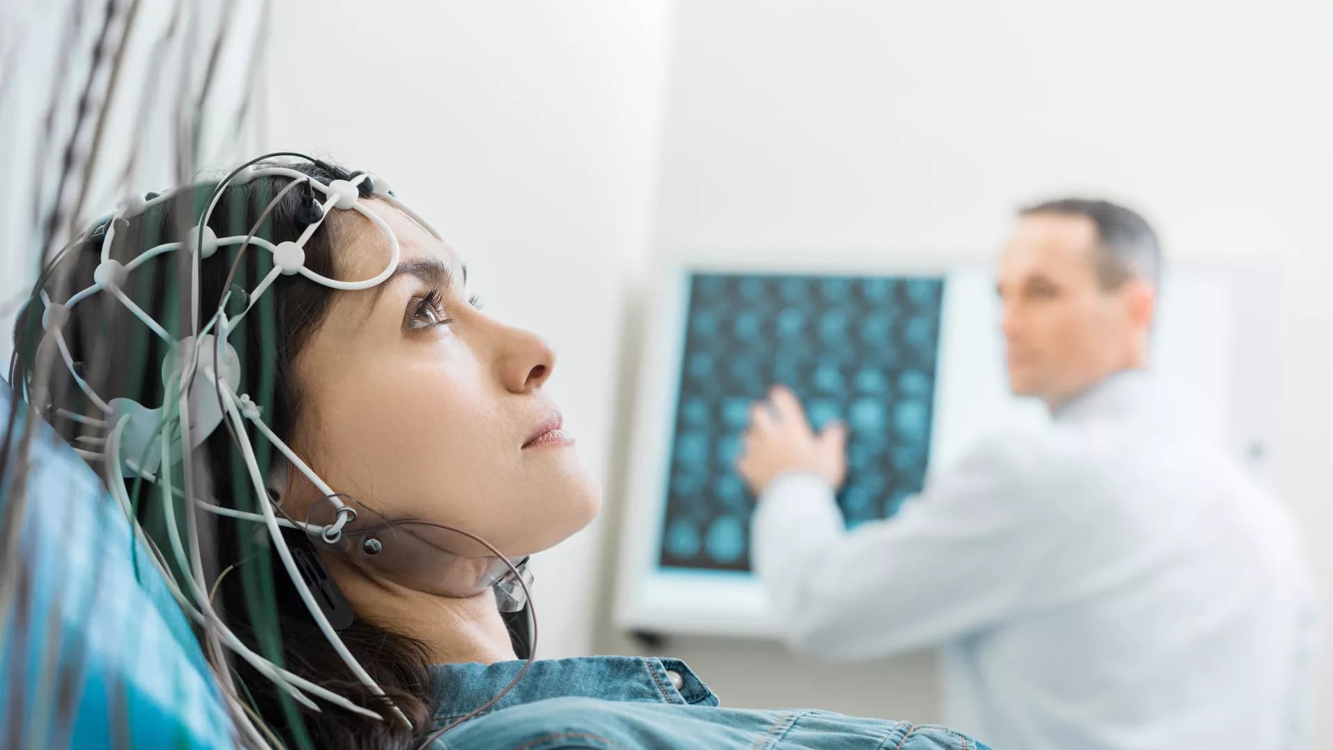
Background and Purpose: In the setting of acute ischemic stroke, increased blood-brain barrier permeability (BBBP) as a sign of injury is believed to be associated with increased risk of poor outcome. Pre-clinical studies show that selected serum biomarkers including C-reactive protein (CRP), interleukin-6 (IL-6), tumor necrosis factor-alpha (TNFα), matrix metallopeptidases (MMP), and vascular endothelial growth factors (VEGFs) may play a role in BBBP post-stroke. In the subacute phase of stroke, increased BBBP may also be caused by regenerative mechanisms such as vascular remodeling and therefore may improve functional recovery. Our aim was to investigate the evolution of BBBP in ischemic stroke using contrast-enhanced (CE) magnetic resonance imaging (MRI) and to analyze potential associations with blood-derived biomarkers as well as functional recovery in subacute ischemic stroke patients.
Methods: This is an exploratory analysis of subacute ischemic stroke patients enrolled in the BAPTISe study nested within the randomized controlled PHYS-STROKE trial (interventions: 4 weeks of aerobic fitness training vs. relaxation). Patients with at least one CE-MRI before (v1) or after (v2) the intervention were eligible for this analysis. The prevalence of increased BBBP was visually assessed on T1-weighted MR-images based on extent of contrast-agent enhancement within the ischemic lesion. The intensity of increased BBBP was assessed semi-quantitatively by normalizing the mean voxel intensity within the region of interest (ROI) to the contralateral hemisphere (“normalized CE-ROI”). Selected serum biomarkers (high-sensitive CRP, IL-6, TNF-α, MMP-9, and VEGF) at v1 (before intervention) were analyzed as continuous and dichotomized variables defined by laboratory cut-off levels. Functional outcome was assessed at 6 months after stroke using the modified Rankin Scale (mRS).
Results: Ninety-three patients with a median baseline NIHSS of 9 [IQR 6–12] were included into the analysis. The median time to v1 MRI was 30 days [IQR 18–37], and the median lesion volume on v1 MRI was 4 ml [IQR 1.2–23.4]. Seventy patients (80%) had increased BBBP visible on v1 MRI. After the trial intervention, increased BBBP was still detectable in 52 patients (74%) on v2 MRI. The median time to v2 MRI was 56 days [IQR 46–67]. The presence of increased BBBP on v1 MRI was associated with larger lesion volumes and more severe strokes. Aerobic fitness training did not influence the increase of BBBP evaluated at v2. In linear mixed models, the time from stroke onset to MRI was inversely associated with normalized CE-ROI (coefficient −0.002, Standard Error 0.007, p < 0.01). Selected serum biomarkers were not associated with the presence or evolution of increased BBBP. Multivariable regression analysis did not identify the occurrence or evolution of increased BBBP as an independent predictor of favorable functional outcome post-stroke.
Conclusion: In patients with moderate-to-severe subacute stroke, three out of four patients demonstrated increased BBB permeability, which decreased over time. The presence of increased BBBP was associated with larger lesion volumes and more severe strokes. We could not detect an association between selected serum biomarkers of inflammation and an increased BBBP in this cohort. No clear association with favorable functional outcome was observed.
Trial registration: NCT01954797.

Intracerebral hemorrhage (ICH) accounts for ~15% of all strokes and is associated with high mortality and disability rates. The systemic inflammation response index (SIRI) is a novel systemic inflammatory marker based on peripheral neutrophil, monocyte, and lymphocyte counts. This study aimed to evaluate the prognostic significance of admission SIRI in patients with spontaneous ICH and compare its predictive ability with that of the neutrophil-to-lymphocyte ratio (NLR). This retrospective study was conducted based on a prospectively collected database of patients with ICH between June 2016 and January 2019. Propensity score matching (PSM) was conducted to adjust for potential imbalances in the clinical parameters. A total of 403 patients were included in the original cohort. The optimal SIRI cut-off value was 2.76. After 1:1 PSM based on potential confounding variables, a new cohort containing 262 patients was established for further analysis. In the original cohort, SIRI served as an independent predictor of 3-month functional outcome [odds ratio (OR), 1.302; 95% CI, 1.120–1.512; p = 0.001] and 1-month mortality (OR, 1.072; 95% CI, 1.020–1.126; p = 0.006), while NLR was independently associated with only 3-month functional outcomes (OR, 1.051; 95% CI, 1.004–1.100; p = 0.031) and not 1-month mortality. The same applied to the PSM cohort. Receiver operating characteristic analyses and predictive models indicated that in most instances, SIRI was superior to NLR and their components in predicting the outcomes of patients with ICH. Our study found that SIRI is determined to be an independent predictive indicator for ICH patients in 3-month functional outcomes and 1-month mortality. The prognostic predictive ability of SIRI was stronger than that of NLR.

Purpose: Stroke-associated infection (SAI) is associated with adverse outcomes in patients with acute ischemic stroke (AIS). In this study, we aimed to evaluate the association between neutrophil percentage-to-albumin ratio (NPAR) and SAI occurrence in patients with AIS.
Methods: We retrospectively analyzed all AIS patients who were admitted to the Neurology ward of The Second Hospital of Tianjin Medical University from November 2018 to October 2020. The relationship between NPAR and SAI was analyzed by multivariable analysis. The receiver operating characteristic (ROC) curve was used to compare the predicted value of albumin, neutrophil percentage, neutrophil-to-lymphocyte ratio (NLR), and NPAR.
Results: We included 379 AIS patients out of which 51 (13.5%) developed SAI. The NPAR was independently associated with increased risk of SAI adjusting for confounders [adjusted odds ratio (aOR) = 10.52; 95% confidence interval (CI), 3.33–33.28; P <0.001]. The optimal cutoff value of NPAR for predicting SAI incidence was 1.64, with sensitivity and specificity of 90.2 and 55.8%, respectively. The area under the curve (AUC) value of NPAR [0.771 (0.725–0.812)] was higher than that of albumin [0.640 (0.590–0.689)], neutrophil percentage [0.747 (0.700–0.790)], and NLR [0.736 (0.689–0.780)], though the statistical significance appeared only between NPAR and albumin.
Conclusions: We demonstrated that a higher NPAR could predict the occurrence of SAI. Thus, NPAR might be a more effective biomarker to predict SAI compared with albumin, neutrophil percentage, and NLR.


A serious complication of acute ischemic stroke (AIS) after mechanical thrombectomy (MT) is hemorrhagic transformation (HT), which is potentially associated with clinical deterioration. This study examined predictors of HT following MT in AIS patients. Patients with AIS due to large artery occlusion in the anterior circulation, treated with MT and successfully recanalized (modified Thrombolysis in Cerebral Infarction score 2b/3), were studied retrospectively. HT was evaluated by computed tomography (CT) 24 h after MT and was diagnosed and classified into parenchymal hematoma (PH) and hemorrhagic infarction (HI). Multivariate logistic regression models were used to determine the risk factors for HT. Receiver operating characteristic (ROC) curve analysis was performed to determine the predictive utility of risk factors for HT. We enrolled 135 patients: 49 in the HT group and 86 in the non-HT group. The two groups differed significantly in baseline fibrinogen levels (p = 0.003) and platelet counts (p = 0.006). Multivariate logistic regression analyses showed that lower fibrinogen levels [odds ratio (OR), 0.41; 95% CI, 0.23–0.72; p = 0.002] and platelet counts (OR, 0.58; 95% CI, 0.33–0.99; p = 0.048) were independently associated with a higher risk of HT. Together, the binary variates fibrinogen and platelets well-predicted HT (area under the curve, 0.703; specificity, 77.9%; sensitivity, 55.1%). The combination of fibrinogen <2.165 g/L and platelets <171.5 × 10⁹/L was the strongest predictor of HT (OR, 23.17; 95% CI, 5.75–126.80; p < 0.0001). Our study suggests that lower baseline fibrinogen levels and platelet counts may be risk factors for HT in AIS patients following MT and reperfusion. Specifically, the combination of fibrinogen level and platelet count may predict the risk of HT after MT in these patients.


Background: Inflammatory markers, such as C-reactive Protein (CRP), Interleukin-6 (IL-6), tumor necrosis factor (TNF)-alpha and fibrinogen, are upregulated following acute stroke. Studies have shown associations of these biomarkers with increased mortality, recurrent vascular risk, and poor functional outcome. It is suggested that physical fitness training may play a role in decreasing long-term inflammatory activity and supports tissue recovery.
Aim: We investigated the dynamics of selected inflammatory markers in the subacute phase following stroke and determined if fluctuations are associated with functional recovery up to 6 months. Further, we examined whether exposure to aerobic physical fitness training in the subacute phase influenced serum inflammatory markers over time.
Methods: This is an exploratory analysis of patients enrolled in the multicenter randomized-controlled PHYS-STROKE trial. Patients within 45 days of stroke onset were randomized to receive either four weeks of aerobic physical fitness training or relaxation sessions. Generalized estimating equation models were used to investigate the dynamics of inflammatory markers and the associations of exposure to fitness training with serum inflammatory markers over time. Multiple logistic regression models were used to explore associations between inflammatory marker levels at baseline and three months after stroke and outcome at 3- or 6-months.
Results: Irrespective of the intervention group, high sensitive CRP (hs-CRP), IL-6, and fibrinogen (but not TNF-alpha) were significantly lower at follow-up visits when compared to baseline (p all ≤ 0.01). In our cohort, exposure to aerobic physical fitness training did not influence levels of inflammatory markers over time. In multivariate logistic regression analyses, increased baseline IL-6 and fibrinogen levels were inversely associated with worse outcome at 3 and 6 months. Increased levels of hs-CRP at 3 months after stroke were associated with impaired outcome at 6 months. We found no independent associations of TNF-alpha levels with investigated outcome parameters.
Conclusion: Serum markers of inflammation were elevated after stroke and decreased within 6 months. In our cohort, exposure to aerobic physical fitness training did not modify the dynamics of inflammatory markers over time. Elevated IL-6 and fibrinogen levels in early subacute stroke were associated with worse outcome up to 6-months after stroke.
Clinical Trial Registration: ClinicalTrials.gov, NCT01953549.


Introduction: We explored whether higher preoperative serum levels of lactate dehydrogenase (LDH) predicted outcome 3 months after surgery in patients with aneurysmal subarachnoid hemorrhage (aSAH) treated using microsurgical clipping in our institution.
Methods: Patients with aSAH treated at our institution between 2010 and 2018 were enrolled. The following parameters were recorded: age, sex, smoking and drinking history, medical history, Hunt–Hess and Fisher grades, aneurysm location, aneurysm size, surgical treatment, delayed cerebral ischemia (DCI), intracranial infection, hydrocephalus, pneumonia, and preoperative serum LDH levels within 24 h of aSAH. We investigated whether preoperative serum LDH levels were associated with Hunt–Hess grade, Fisher grade, and functional neurological outcome.
Results: In total, 2,054 patients with aSAH were enrolled, 874 of whom were treated using microsurgical clipping. The average serum LDH level (U/L) was significantly lower in the good outcome group (180.096 ± 50.237) than in the poor outcome group (227.554 ± 83.002; p < 0.001). After propensity score matching, the average serum LDH level (U/L) was still lower in the good outcome group (205.356 ± 76.785) than in the poor outcome group (227.119 ± 86.469; p = 0.029). The area under the receiver operating characteristic (ROC) curve was 0.702 (95% confidence interval [CI]: 0.650–0.754; p < 0.001). Based on the ROC curve, the optimal cutoff value for serum LDH levels as a predictor of poor 3-month outcome (modified Rankin Scale score > 2) was 201.5 U/L. The results revealed that Hunt–Hess grade, Fisher grade, DCI, pneumonia, and serum LDH (>201.5 U/L) were significantly associated with poor outcome. After propensity score matching, serum LDH levels > 201.5 U/L were still considered an independent risk factor for poor outcome (odds ratio: 2.426, 95% CI = 1.378–4.271, p = 0.002). Serum LDH levels were associated with Hunt–Hess and Fisher grades and were correlated with functional neurological outcomes (p < 0.001).
Conclusions: Our findings showed that higher preoperative serum levels of LDH correlated with Hunt–Hess grade, Fisher grade, and neurological functional outcome, and predicted the outcome of aSAH treated by microsurgical clipping at 3 months, which was involved in the related mechanisms of early brain injury and showed its potential clinical significance in patients with aSAH.


Background: Uric acid (UA) is proposed as a potential risk factor for stroke in adult, yet the results from published studies are not generally accordant.
Method: We included prospective studies that explored the relationship between serum UA (SUA) and strokes. In this study, strokes include ischemic stroke and hemorrhagic stroke, which consists of intracerebral hemorrhage and subarachnoid hemorrhage. The effect-size estimates were expressed as hazard ratio (HR) and 95% confidence interval (CI). Sensitivity and subgroup analyses were performed to assess the robustness of the pooled estimation and potential sources of heterogeneity between studies.
Results: We meta-analyzed 19 prospective cohort articles, which involve 37,386 males and 31,163 females. Overall analyses results showed a significant association between a 1 mg/dl increase in high levels of SUA and the risk of total stroke (HR = 1.13; 95% CI: 1.09–1.18; P < 0.001), ischemic stroke (HR = 1.15; 95% CI: 1.10–1.21; P < 0.001), and hemorrhagic stroke (HR = 1.07; 95% CI: 1.00 to 1.15; P = 0.046). No significant difference was found between ischemic stroke and hemorrhagic stroke. In the subgroup analyses, the association of high SUA levels and the risk of total stroke was statistically significant in females (HR = 1.19; 95% CI: 1.12–1.26; P < 0.001) and males (HR = 1.11; 95% CI: 1.05–1.17; P < 0.001). Coincidentally, the association was also statistically significant for ischemic stroke, both in females (HR = 1.26; 95% CI: 1.17–1.36; P < 0.001) and in males (HR = 1.12; 95% CI: 1.06–1.19; P < 0.001). However, for hemorrhagic stroke, it was only statistically significant in females (HR = 1.19; 95% CI: 1.04–1.35; P = 0.01). Our dose–response research indicated the J-shaped trend between the ascending SUA levels and the higher risk of suffering from a stroke.
Conclusions: Our findings indicate that elevated SUA is a significant risk factor for adult stroke, both for ischemic stroke and hemorrhagic stroke, and especially in females.