Editorial
18 February 2021
Ronald L. Rabin
and
Mark R. Walter

Like most plasma membrane proteins, type I interferon (IFN) receptor (IFNAR) traffics from the outer surface to the inner compartments of the cell. Long considered as a passive means to simply control subunits availability at the plasma membrane, an array of new evidence establishes IFNAR endocytosis as an active contributor to the regulation of signal transduction triggered by IFN binding to IFNAR. During its complex journey initiated at the plasma membrane, the internalized IFNAR complex, i.e. IFNAR1 and IFNAR2 subunits, will experience post-translational modifications and recruit specific effectors. These finely tuned interactions will determine not only IFNAR subunits destiny (lysosomal degradation vs. plasma membrane recycling) but also the control of IFN-induced signal transduction. Finally, the IFNAR system perfectly illustrates the paradigm of the crosstalk between membrane trafficking and intracellular signaling. Investigating the complexity of IFN receptor intracellular routes is therefore necessary to reveal new insight into the role of IFNAR membrane dynamics in type I IFNs signaling selectivity and biological activity.

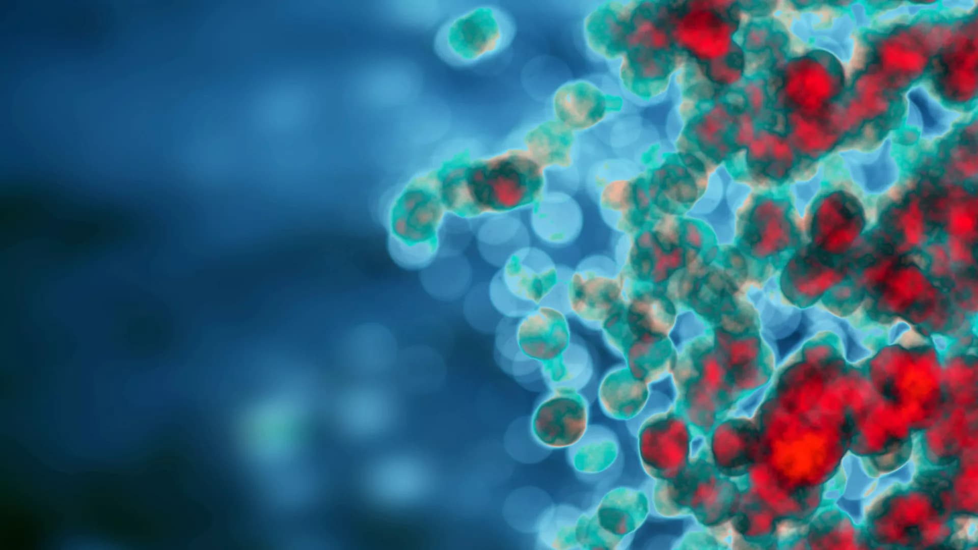
Interferons (IFNs) constitute the first line of defense against microbial infections particularly against viruses. They provide antiviral properties to cells by inducing the expression of hundreds of genes known as interferon-stimulated genes (ISGs). The two most important IFNs that can be produced by virtually all cells in the body during intrinsic innate immune response belong to two distinct families: the type I and type III IFNs. The type I IFN receptor is ubiquitously expressed whereas the type III IFN receptor’s expression is limited to epithelial cells and a subset of immune cells. While originally considered to be redundant, type III IFNs have now been shown to play a unique role in protecting mucosal surfaces against pathogen challenges. The mucosal specific functions of type III IFN do not solely rely on the restricted epithelial expression of its receptor but also on the distinct means by which type III IFN mediates its anti-pathogen functions compared to the type I IFN. In this review we first provide a general overview on IFNs and present the similarities and differences in the signal transduction pathways leading to the expression of either type I or type III IFNs. By highlighting the current state-of-knowledge of the two archetypical mucosal surfaces (e.g. the respiratory and intestinal epitheliums), we present the differences in the signaling cascades used by type I and type III IFNs to uniquely induce the expression of ISGs. We then discuss in detail the role of each IFN in controlling pathogen infections in intestinal and respiratory epithelial cells. Finally, we provide our perspective on novel concepts in the field of IFN (stochasticity, response heterogeneity, cellular polarization/differentiation and tissue microenvironment) that we believe have implications in driving the differences between type I and III IFNs and could explain the preferences for type III IFNs at mucosal surfaces.

Interferon (IFN) signaling induces the expression of a wide array of genes, collectively referred to as IFN-stimulated genes (ISGs) that generally function to inhibit viral replication. RNA viruses are frequently targeted by ISGs through recognition of viral replicative intermediates and molecular features associated with viral genomes, or the lack of molecular features associated with host mRNAs. The ISGs reviewed here primarily inhibit viral replication in an RNA-centric manner, working to sense, degrade, or repress expression of viral RNA. This review focuses on dissecting how these ISGs exhibit multiple antiviral mechanisms, often through use of varied co-factors, highlighting the complexity of the type I IFN response. Specifically, these ISGs can mediate antiviral effects through viral RNA degradation, viral translation inhibition, or both. While the OAS/RNase L pathway globally degrades RNA and arrests translation, ISG20 and ZAP employ targeted RNA degradation and translation inhibition to block viral replication. Meanwhile, SHFL targets translation by inhibiting -1 ribosomal frameshifting, which is required by many RNA viruses. Finally, a number of E3 ligases inhibit viral transcription, an attractive antiviral target during the lifecycle of negative-sense RNA viruses which must transcribe their genome prior to translation. Through this review, we aim to provide an updated perspective on how these ISGs work together to form a complex network of antiviral arsenals targeting viral RNA processes.
For several decades there has been accumulating evidence implicating type I interferons (IFNs) as key elements of the immune response. Therapeutic approaches incorporating different recombinant type I IFN proteins have been successfully employed to treat a diverse group of diseases with significant and positive outcomes. The biological activities of type I IFNs are consequences of signaling events occurring in the cytoplasm and nucleus of cells. Biochemical events involving JAK/STAT proteins that control transcriptional activation of IFN-stimulated genes (ISGs) were the first to be identified and are referred to as “canonical” signaling. Subsequent identification of JAK/STAT-independent signaling pathways, critical for ISG transcription and/or mRNA translation, are denoted as “non-canonical” or “non-classical” pathways. In this review, we summarize these signaling cascades and discuss recent developments in the field, specifically as they relate to the biological and clinical implications of engagement of both canonical and non-canonical pathways.
Interferons (IFNs) are a family of cytokines with the unique ability to induce cell intrinsic programs that enhance resistance to viral infection. Induction of an antiviral state at the cell, tissue, organ, and organismal level is performed by three distinct IFN families, designated as Type-I, Type-II, and Type-III IFNs. Overall, there are 21 human IFNs, (16 type-I, 12 IFNαs, IFNβ, IFNϵ, IFNκ, and IFNω; 1 type-II, IFNγ; and 4 type-III, IFNλ1, IFNλ2, IFNλ3, and IFNλ4), that induce pleotropic cellular activities essential for innate and adaptive immune responses against virus and other pathogens. IFN signaling is initiated by binding to distinct heterodimeric receptor complexes. The three-dimensional structures of the type-I (IFNα/IFNAR1/IFNAR2), type-II (IFNγ/IFNGR1/IFNGR2), and type-III (IFNλ3/IFNλR1/IL10R2) signaling complexes have been determined. Here, we highlight similar and unique features of the IFNs, their cell surface complexes and discuss their role in inducing downstream IFN signaling responses.
Type I interferons (IFN-I) were first discovered over 60 years ago in a classical experiment by Isaacs and Lindenman, who showed that IFN-Is possess antiviral activity. Later, it became one of the first approved protein drugs using heterologous protein expression systems, which allowed its large-scale production. It has been approved, and widely used in a pleiotropy of diseases, including multiple-sclerosis, hepatitis B and C, and some forms of cancer. Preliminary clinical data has supported its effectiveness against potential pandemic pathogens such as Ebola and SARS. Still, more efficient and specific drugs have taken its place in treating such diseases. The COVID-19 global pandemic has again lifted the status of IFN-Is to become one of the more promising drug candidates, with initial clinical trials showing promising results in reducing the severity and duration of the disease. Although SARS-CoV-2 inhibits the production of IFNβ and thus obstructs the innate immune response to this virus, it is sensitive to the antiviral activity of externally administrated IFN-Is. In this review I discuss the diverse modes of biological actions of IFN-Is and how these are related to biophysical parameters of IFN-I–receptor interaction and cell-type specificity in light of the large variety of binding affinities of the different IFN-I subtypes towards the common interferon receptor. Furthermore, I discuss how these may guide the optimized use IFN-Is in combatting COVID-19.