
94% of researchers rate our articles as excellent or good
Learn more about the work of our research integrity team to safeguard the quality of each article we publish.
Find out more
ORIGINAL RESEARCH article
Front. Virol., 01 April 2025
Sec. Emerging and Reemerging Viruses
Volume 5 - 2025 | https://doi.org/10.3389/fviro.2025.1548475
 Selma Mejri1,2*†
 Saida Emna Ayari Fakhfakh1†Makrem Ourabi3Amira Abid4Rahma Zaghouani5
 Marouène Nouassri6Samir Mhiri7Sahbi Gallah7
 Nouha Habboubi5Kaddour Hosni6Noura Abidi8Nadia Braiki9Wiem Mouelhi10
 Imed Ben Slimene10Ines Ghodhbene4Hager Mhamdi11Mansour Ksontini12Zoubeida Landolsi13Nesrine Zribi14Mohamed Hamdouni4Hayet Gamdou10Fethi Zammel6Arbi Hlel5Nahed Ben Harrath6Sihem Sbai6Sara Thabet1Hatem Ouled Ahmed15
 Soufien Sghaier16Roukaya Khorchani17
 Tirumala Bharani Kumar Settypalli18
 Irene Kasindi Meki18
 Charles Euloge Lamien18Aida Tlatli19
Lumpy Skin Disease (LSD) is an emerging bovine vector-borne disease of important economic impact on the cattle industry. Since its first identification in 1929, the disease was restricted for decades, to Sub-Saharan regions before its spread into new areas. In 2023 and 2024, LSD cases were identified for the first time in north African countries, Libya and Algeria, respectively. From June 2024, many LSD suspected cases were investigated in Tunisia. From June to October 2024, one hundred and twenty-one samples were investigated. Most of samples consist of blood samples, nasal and oral swabs from 49 suspected cattle from different parts of Tunisia. All samples were tested using Real-Time PCR and High Resolution Melting assay (HRM). On August 7, 2024, we reported the first LSD case in Tunisia. Two months later, other positive cases were confirmed by the two molecular techniques. The HRM technique allow the identification of a positive Bovine Papular Stomatitis animal presenting LSD clinical signs. Among the 49 tested cattle, eighteen were confirmed LSD positive. Most of LSD cases were from north western regions, close to Algerian border. The number of positive cases highly increased from October, period corresponding to increased LSD vectors’ activity. This is the first report on the identification of LSD in Tunisian cattle. Our findings confirm the progressive spread of LSD into new areas, and highlight the need of the implementation of control and surveillance measures to face such diseases.
Lumpy Skin Disease (LSD) is a viral transboundary disease affecting large ruminants, especially cattle and water buffaloes (1). Clinical signs of the disease include fever and lachrymation, followed by skin nodules appearing primarily on the head, neck, and abdomen, before covering all the body (2). The morbidity rate varies between 5% and 45%, while the mortality rate is generally lower than 3% (1). Due to its significant economic impact on cattle industry and its rapid spread, LSD is listed as a notifiable disease by the World Organization for Animal Health (WOAH) (3). LSD is caused by Lumpy Skin Disease virus (LSDV) which belongs to the family Poxviridae, genus Capripoxvirus, and is genetically related to the sheeppox virus (SPPV) and goatpox virus (GTPV) within the same genus. Its genome consists of a large double-stranded DNA of about 150Kb (4). The main source of LSDV are skin lesions, and the virus is transmitted mechanically by blood-sucking insects (4, 5).
LSD was first described in 1929 in Zambia from where it spread to other South African countries and became endemic in most of sub-Saharan regions (6, 7). The disease was restricted to these areas for decades, then it slowly extended and reached Egypt and other western African countries during the 1980s (8, 9). Later on, LSD spread out of Africa into the Arabian Peninsula, Turkey, some European countries and more recently into Asia (3, 10–13).
Until 2023, the only African countries considered as LSD free were Morocco, Algeria, Libya and Tunisia (11). In June 2023, LSD cases were identified in Libya (14). One year later in June 2024, LSD cases were detected in Algeria (15). Subsequently, multiple suspected cases were reported in Tunisia, with the first positive case detected on August 07, 2024.
Tunisian cattle herd is of 412,000 female units, almost 60% of which are imported breeds mainly Holstein, Pie noire and Swiss brown (16). Due to the increase of feed price, the size of national cattle herd has gradually decreased. Three livestock feeding systems are practiced in Tunisia: based on grazing (mainly in the north), intensive integrated system (in the center) and mixed farming system (in the south) (16). The highest prevalence of Tunisian cattle is reported in the governorate of Jendouba in the northwest (17), a region of important agricultural activity. In Tunisia, cattle industry provides almost 98.4% of dairy production, and about 13.5% of meat. This demonstrates the importance of livestock farming activity and the need to prevent the incursion and spread of diseases, such as LSD, with negative impact on cattle health.
This study aims to describe the first confirmed LSD cases detected in Tunisia using two molecular techniques: Real-Time PCR and High-Resolution Melting (HRM) assay.
The Virology laboratory at the Institute of Veterinary Research of Tunisia started receiving samples from cattle presenting at least one LSD clinical sign. From June to October 2024, 122 samples were collected from 49 cattle with clinical signs such as hyperthermia, anorexia and skin nodules. Tested animals were from 42 different herds located in 11 different governorates of Tunisia (Jendouba, Kasserine, Nabeul, Ben Arous, Sfax, Bizerte, Siliana, Kef, Monastir, Tozeur and Sidi Bouzid). Most of sampled cattle were from smallholder farms (less than 5 animals) with feeding system based on grazing (Table 1). Collected samples include, anticoagulated whole blood, oral and nasal swabs, skin nodules (swabs of burst nodules) and scabs. Samples were taken from animals in compliance with local and international ethics regulations.
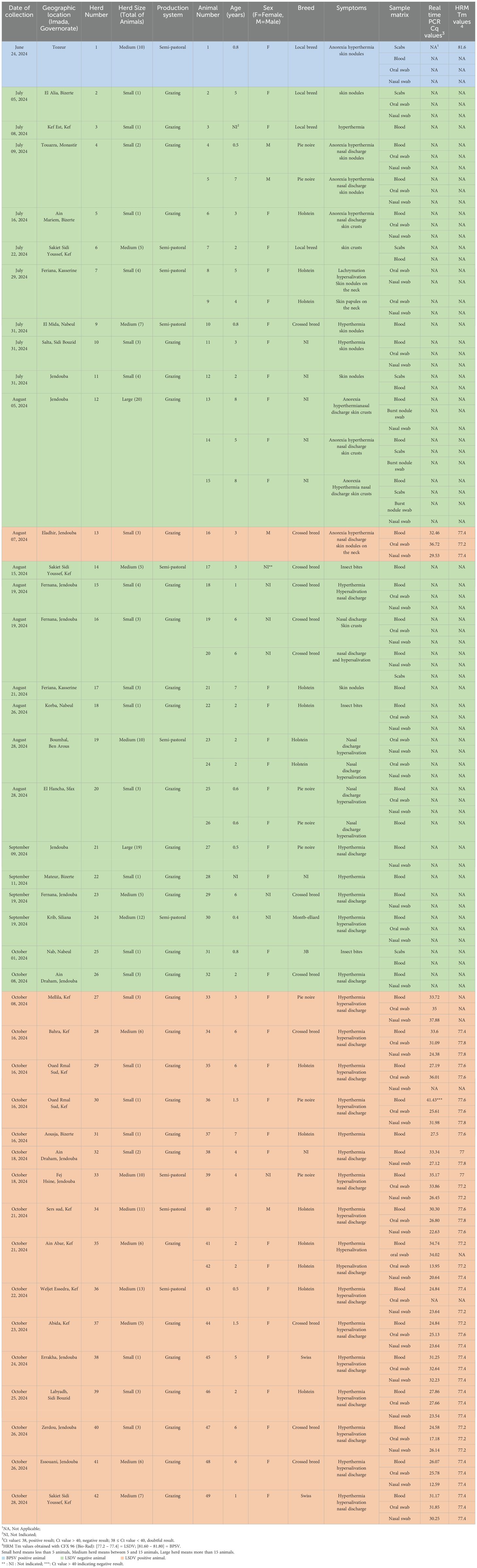
Table 1. Description of LSD outbreak in Tunisia between June and October 2024; herds, animals and results of LSD tested samples.
Viral DNA was extracted from all samples using the DNeasy Blood and Tissue kit (QIAGEN, Germany) according to the manufacturer’s instructions. The DNA was eluted in 35µl of elution buffer and directly analyzed using molecular techniques.
Each extracted DNA sample was tested for the presence of Capripoxvirus genome using the Bowden et al. procedure (18), with minor modifications. The PCR reactions were performed using the iQsupermix kit (Biorad, USA). Amplification was conducted in a reaction volume of 20µl containing 2X buffer, 400 nm each of the forward and reverse primers, 250 nm of the probe, and 2µl of template DNA. The PCR program consisted of an initial denaturation at 95°C for 10 min, followed by 45 cycles at 95°C for 15 s and 60°C for 1 min, with the fluorescence reading at the end.
Each DNA sample was also tested for coinfection of LSDV and other Poxviruses using an HRM assay. It is a sensitive technique allowing an accurate differentiation between closely related Poxviruses.
This technique consists of a multiplex Real-time PCR assay for detection and differentiation of Poxviruses belonging to Orthopoxvirus, Capripoxvirus, and Parapoxvirus based on the GC content, fragment lengths and the melting temperature (Tm) of the PCR products (19). Briefly, the PCR was set up in a reaction volume of 20µl, containing 1X of SsoFast EvaGreen Supermix (Bio-Rad, Hercules, CA, USA), 200 nm of each of the reverse and forward primers (Table 2), and 2µl of DNA sample. The PCR conditions consisted of an initial denaturation step at 95°C for 4 min, followed by 40 cycles at 95°C for 1 s, 59°C for 5 s and 70°C for 5 s with fluorescence reading at the end, followed by 95°C for 30 s, 65°C for 1 min and melting between 65°C and 85°C at 10 s/0.2°C with fluorescence reading at each °C, then 37°C for 1 min. The HRM assay was performed using the CFX 96 (Bio-Rad) instrument, and the amplification plots were analyzed using the Bio-Rad CFX Maestro™ Software version 1.0.

Table 2. List of oligonucleotide primers used in HRM technique for amplification of Poxviruses (Orthopoxvirus, Capripoxvirus, and Parapoxvirus).
In each HRM run, nine controls were included: one negative control and eight positive controls corresponding to eight Poxviruses: Cowpox virus (CPXV) and Camelpox virus (CMLV) (of the Orthopoxvirus genus), GTPV, SPPV and LSDV (of the Capripoxvirus genus), Orf virus (OrfV), Pseudocowpox virus (PCPV) and Bovine papular stomatitis virus (BPSV) (of the Parapoxvirus genus). These positive controls were kindly provided by the Animal Production and Health Laboratory, Joint FAO/IAEA Centre, Department of Nuclear Sciences and Applications, International Atomic Energy Agency, Vienna, Austria
Statistical analysis of qualitative variables was performed using the Fisher Test, to check if age and sex have a statistical impact on LSD positivity at the level of significance α = 0.05. A p value less than 0.05 is considered statistically significant. The variable sex could not be statistically analyzed because of limited data.
Among the 49 suspected cattle, 18 (36.7%) were confirmed LSDV positive based on the Real-Time PCR results. LSDV DNA was detected in 46/49 samples collected from the 18 positive animals. Positive samples consisted of 16 blood samples, 15 oral swabs and 15 nasal swabs. No LSDV DNA was detected in one blood sample, two oral swabs and two nasal swabs collected from three positive animals (Table 1). All scabs and skin nodule samples were detected negative. Indeed, all of them were from negative animals tested before the detection of the first LSD positive case. We did not receive such kind of samples from confirmed LSD positive animals. A sample was considered positive if its Cq value was lower than 38. Real-Time PCR results showed that Cq values of positive samples ranged between 12.59 to 37.88. Low Cq values (≤ 25), implying a high viral load, were observed in 12 samples (Table 1), most of them were nasal swabs (N = 7). An animal was considered LSD positive if at least one of its samples showed viral amplification by Real-Time PCR technique.
Results of HRM technique allow the identification of Poxviruses according to Tm values (19). Analysis of samples using the HRM assay showed that, one suspected animal that was negative for LSDV by the Real-Time PCR, was positive for BPSV (Tm = 81.60) (Animal Number 1 in Table 1) (Figure 1). This animal presented skin nodules suggestive of an LSD infection (Figure 2). A sample is considered LSD positive by HRM technique if its Tm value ranged between 77.2 and 77.4. HRM results (Figure 3) confirmed LSDV positivity of 42 samples out of the 43 positives detected by the Real-Time PCR.
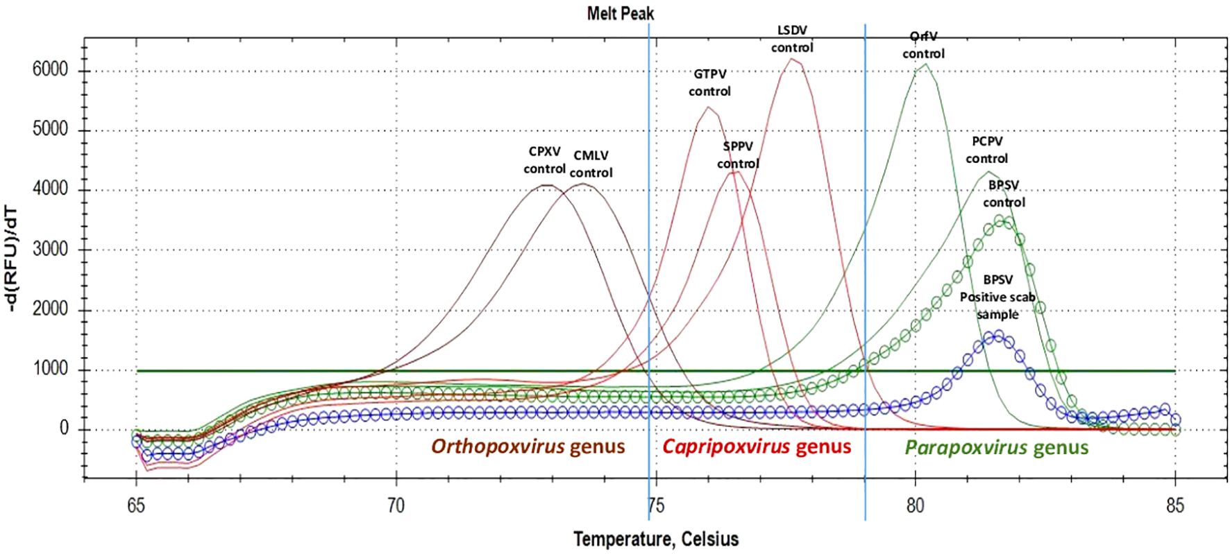
Figure 1. HRM result of the BPSV positive animal (animal number 1 in Table 2).
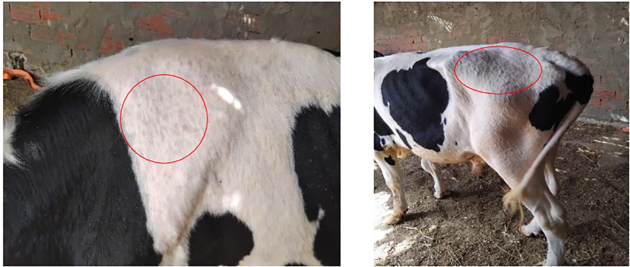
Figure 2. Nodules on the skin of a suspected case, detected LSD negative and BPSV positive (Photo: Dr Makrem OURABI).
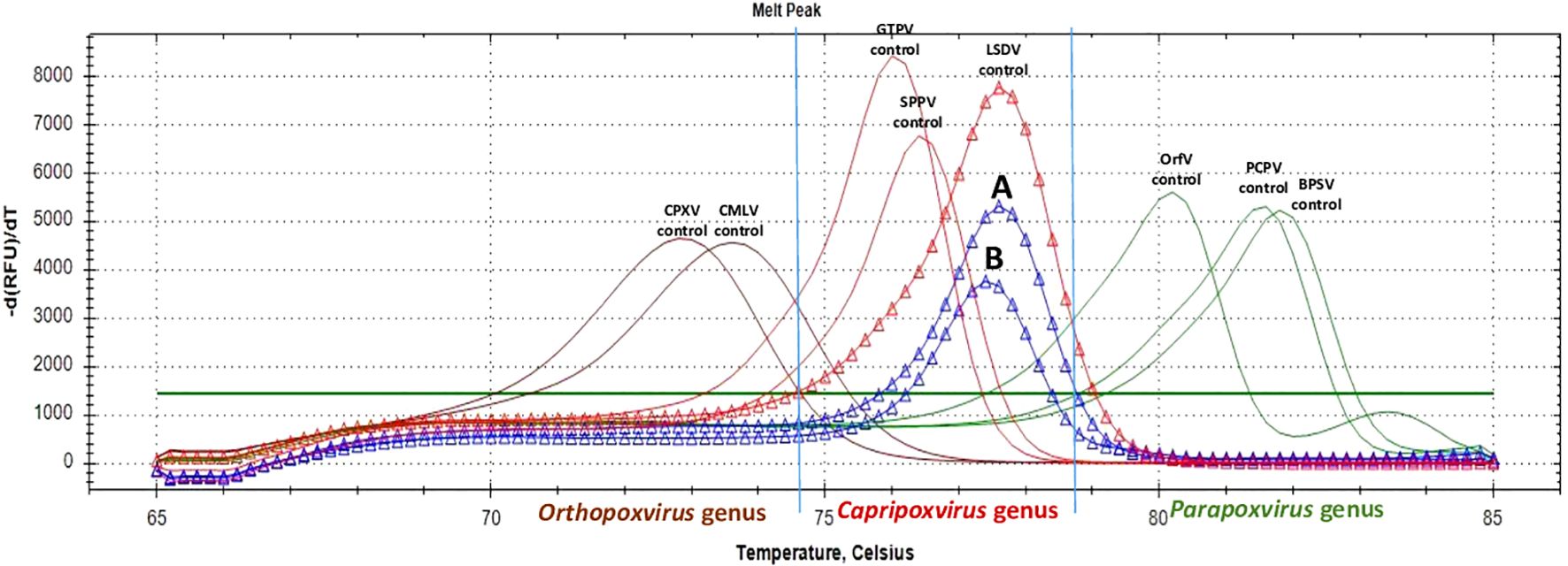
Figure 3. HRM results of first LSD positive case (animal number 16 in Table 2). (A) HRM positive result of nasal swab from animal number 16 in Table 2. (B) HRM positive result of blood sample from animal number 16 in Table 2.
Before the confirmation of the first LSD positive case on August 7, 2024, fifteen suspected cattle with clinical signs were tested both by Real-Time PCR and HRM and showed negative results. The first LSD positive animal was a three-year-old bull, used for insemination and presenting typical clinical signs of LSD (Figure 4). This animal was from Jendouba (northwest Tunisia), a region very close to the Algerian border. No other LSD cases were diagnosed for the following two months until October 8, when positive LSDV cases were increasingly detected (Figure 5).

Figure 4. Nodules on the skin of the first LSD positive case (Photo: Dr Imed BEN SLIMENE). (A) Skin nodules on the neck. (B) Skin nodule on the abdomen. (C) Oedema of limbs.
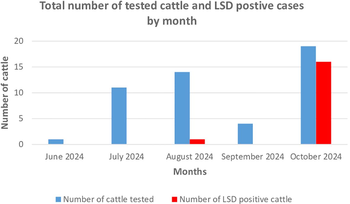
Figure 5. Repartition of tested cattle and detected LSD positive cases by month.
Most of tested cattle were females, and among the 18 LSD positive animals, 15 were females. Statistical analysis showed that the age of positive animals varied from 6 months to 7 years, with a mean of 3.75 years. A higher prevalence of LSD infection was found in animals aged more than one year (50%) than in animals aged one year or less (12.5%). However, this finding is not statistically significant (p value=1.00). It’s important to note that most reported symptoms among LSD positive animals were: hyperthermia, nasal discharge and hypersalivation recorded in 94.4%, in 89% and in 83% of positive animals, respectively. The presence of skin nodules was reported in only one positive case, representing 5.5% of total positive cases. The morbidity, mortality and lethality rates were estimated at 22%, 4% and 17%, respectively. According to veterinarians involved in LSD outbreak monitoring, infected animals recovered after a period of about one month.
Geographical location of positive cases (Figure 6) showed that LSD positive cattle were distributed in 18 different foci. Most of affected areas were in the northwestern part of Tunisia (Kef and Jendouba).
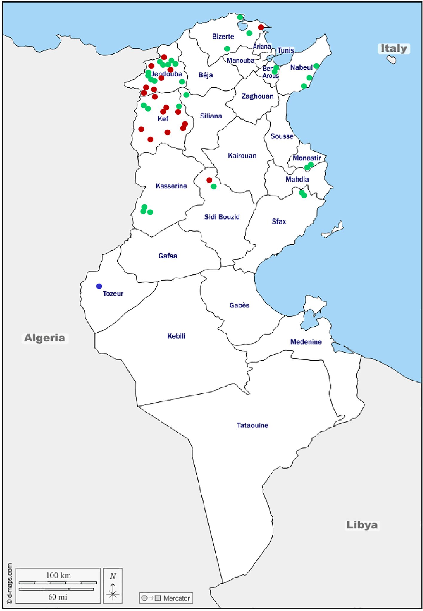
Figure 6. Geographic location of investigated animals. ● LSD positive animal; ● BPS positive animal; ● LSD and BPS negative animal.
For decades, LSD was restricted to south African regions, before spreading into new areas, even outside Africa (4). In 2023 and 2024, LSD positive cases were identified for the first time in north African countries (Libya and Algeria).
This article describes the first detection of LSDV in Tunisia in August 2024, event that was notified to the World Organisation for Animal Health (20). The appearance of LSD in Tunisia occurred one year after its identification in Libya and only 2 months later its incursion in Algeria. LSDV cases, described herein, were identified using two molecular techniques: Real-time PCR and HRM assay.
Real-Time PCR technique is a robust assay widely used in the molecular screening of Capripoxviruses, especially LSDV (18, 21–23). The HRM assay is a sensitive and specific technique that allows not only the detection and differentiation of LSDV from other Capripoxviruses but also from other poxviruses (19). Since its development, this technique is increasingly used in the diagnosis of LSD cases (24, 25).
A total of 43 samples (blood, nasal and oral swabs) collected from 18 cattle were confirmed LSD positive. Although skin lesions and scabs are known to be the main sources of LSDV, in the present study, very few skin lesion samples were collected from suspected animals. This is because collecting skin samples can be traumatic and painful for animals, due to the lack of anesthetic products. For some animals, skin nodules have burst and nodule swabs were collected and tested, all of them were LSD negative. In this study, we demonstrate that other type of samples, especially, nasal swabs could be suitable for LSDV screening, since they presented high rate of viral amplification.
Before the confirmation of the first LSD case, fifteen animals presented clinical signs, mainly skin lesions, suggestive of the disease (Figure 2). All of these animals were detected negative for LSDV both by Real-Time PCR and by HRM assay. One of these suspected cattle was detected positive for BPSV by the HRM assay (Table 1; Figure 1). This result could be explained by the fact that some other diseases cause almost the same cutaneous clinical signs. Among these diseases there are, Hypodermosis, Bovine Leukosis, Lymph node Tuberculosis, parapoxvirus infection etc., which are common in Tunisia (26–29). All these LSD negative results could also be explained by the vigilance of Tunisian veterinarians who paid attention to a lesser suspicion in cattle since the identification of LSD cases in Algeria. The first positive case was identified on August 7, 2024 (20), and no other LSD case was confirmed for almost two months. Since October 8, there has been a significant increase in LSD positive cases number. This is probably related to vectors’ activity period. In fact, in Tunisia, the autumn season (corresponding to the months of September, October and November) is known to be the period of high abundance of blood-sucking vectors (30, 31), especially this year, with the high temperatures and rainfall that occurred in late September and early October.
Most of the infected animals were from geographical regions close to the Algerian border (Figure 6). This finding supports the hypothesis of illegal introduction of infected animals from Algeria, considering the high permeability and uncontrolled animal movements between the two countries. In order to limit the spread of such diseases, strict surveillance and control measures targeting border regions must be implemented to stop illegal animal movements and animal products trade.
The mean age of infected animals was 3.75 years, a relatively young age during which a cow is at the maximum of its productivity. Results also demonstrated that LSD infection was more prevalent in animals aged more than one year, even it was not statistically significant. This finding is in agreement with a previous study reporting that adult cattle were more likely to be LSD infected than calves aged less than one year (32). However, other studies confirmed that higher LSD prevalence was detected in young animals than in adult ones (12, 33). Regarding the factor sex and although the data could not be statistically analyzed, we found that LSD was more detected in female animals. This finding confirms results of previous published works reporting higher LSD prevalence in females compared to males (2, 34). While other studies suggested that male animals are more susceptible to LSD infection because of their exposure to stress factors such as hard work (35, 36). The morbidity, mortality and lethality rates reported in the present work are in accordance with those reported before (1, 37, 38). The negative impact of the disease on cattle industry is mainly due to the high morbidity rate, while the economic losses are due to drop in milk production, decrease of growth rate in beef cattle, infertility and abortion (2, 4).
In Tunisia, cattle are not vaccinated against LSD. A commission regarding the implementation of a massive vaccination campaign and the choice of the appropriate vaccine has been appointed and its work will be shortly completed in order to proceed as soon as possible with the vaccination of susceptible animals in Tunisia.
Each suspected animal was isolated from the herd, however this measure did not limit the spread of the disease. To prevent other LSD outbreak in Tunisia, it is critical to perform an epidemiological investigation across the country. Results of such study are important to determine factors to be considered in the implementation of a national strategy for LSD surveillance and control and to implement an appropriate vaccination program. Additionally, strict measures are needed to limit LSD spread, such as controlling illegal animal movements between Tunisia and neighboring countries and restriction of infected animals’ movements between regions. Moreover, an effective control of blood-feeding vectors must be implemented and maintained to prevent LSD spread.
Further molecular studies are needed to characterize LSDV strains circulating in Tunisia. The results will be useful for improving diagnostic tools and understanding LSD epidemiology at least at the Mediterranean and African level.
The original contributions presented in the study are included in the article/supplementary material. Further inquiries can be directed to the corresponding author.
Ethical approval was not required for the study involving animals in accordance with the local legislation and institutional requirements because Ethical approval was not required for the study involving animals in accordance with the local legislation and institutional requirements because samples were taken by veterinarians for diagnostic purposes and in compliance with local and international regulations. In addition, samples consist of blood samples, nasal and oral swabs and crusts that do not require invasive procedures for the animals.
SMe: Conceptualization, Writing – original draft, Formal analysis, Resources. SF: Conceptualization, Formal analysis, Resources, Writing – original draft. MO: Investigation, Resources, Writing – review & editing. AA: Investigation, Resources, Writing – review & editing. RZ: Investigation, Resources, Writing – review & editing. MN: Investigation, Resources, Writing – review & editing. SMh: Investigation, Resources, Writing – review & editing. SG: Investigation, Resources, Writing – review & editing. NH: Investigation, Resources, Writing – review & editing. KH: Investigation, Resources, Writing – review & editing. NA: Investigation, Resources, Writing – review & editing. NB: Investigation, Resources, Writing – review & editing. WM: Investigation, Resources, Writing – review & editing. IS: Investigation, Resources, Writing – review & editing. IG: Investigation, Resources, Writing – review & editing. HM: Investigation, Resources, Writing – review & editing. MK: Investigation, Resources, Writing – review & editing. ZL: Investigation, Resources, Writing – review & editing. NZ: Investigation, Resources, Writing – review & editing. MH: Investigation, Resources, Writing – review & editing. HG: Investigation, Resources, Writing – review & editing. FZ: Investigation, Resources, Writing – review & editing. AH: Investigation, Resources, Writing – review & editing. NBH: Investigation, Resources, Writing – review & editing. SSb: Investigation, Resources, Writing – review & editing. ST: Investigation, Resources, Writing – review & editing. HA: Investigation, Resources, Writing – review & editing. SSg: Investigation, Writing – review & editing. RK: Investigation, Methodology, Resources, Writing – review & editing. TS: Funding acquisition, Investigation, Software, Writing – review & editing. IM: Data curation, Investigation, Methodology, Writing – review & editing. CL: Funding acquisition, Investigation, Methodology, Resources, Writing – review & editing. AT: Methodology, Writing – review & editing.
The author(s) declare that financial support was received for the research and/or publication of this article. This research was supported by the VETLAB Network initiative of the Joint FAO/IAEA Centre of Nuclear Techniques in Food and Agriculture, funded through the Peaceful Uses Initiative (PUI) by Japan and the United States of America.
The authors declare that the research was conducted in the absence of any commercial or financial relationships that could be construed as a potential conflict of interest.
The author(s) declare that no Generative AI was used in the creation of this manuscript.
All claims expressed in this article are solely those of the authors and do not necessarily represent those of their affiliated organizations, or those of the publisher, the editors and the reviewers. Any product that may be evaluated in this article, or claim that may be made by its manufacturer, is not guaranteed or endorsed by the publisher.
1. Coetzer JAW. Lumpy skin disease” in Infectious diseases of livestock. In: Coetzer JAW, Tustin RC, editors, 2nd edition. South Africa: University Press Southern Africa (2004). p. 1268–76.
2. Tuppurainen ESM, Oura CAL. Lumpy skin disease: an emerging threat to europe, the middle east and asia. Trans Emerg Dis. (2012) 59:40–8. doi: 10.1111/j.1865-1682.2011.01242.x
3. Bianchini J, Simons X, Humblet MF, Saegerman C. Lumpy skin disease: A systematic review of mode of transmission, risk of emergence and risk entry pathway. Viruses. (2023) 15:1622. doi: 10.3390/v15081622
4. Namazi F, Tafti AK. Lumpy skin disease, an emerging transboundary viral disease: A review. Vet Med Sci. (2021) 7:888–96. doi: 10.1002/vms3.434
5. MacLachlan NJ, Dubovi EJ. Fenner’s veterinary virology, 4th ed. Amsterdam, Netherlands: Academic Press (2011).
6. Morris JPA. Pseudo-urticaria. Northern rhodesia department of animal health, annual report 1930. (1931). p. 12
7. Davies FG. Lumpy skin disease, an African capripox virus disease of cattle. Br Vet J. (1991) 147:489–503. doi: 10.1016/0007-1935(91)90019-J
8. Ali AA, Esmat M, Attia H, Selim A, Abdelhamid YM. Clinical and pathological studies on lumpy skin disease in Egypt. Vet Rec. (1990) 127:549–50.
9. Gari G, Grosbois V, Waret-szkuta A, Babiuk S, Jacquiet P, Roger F. Lumpy skin disease in Ethiopia: seroprevalence study across diferent agro-climate zones. Acta Tropica. (2012) 123:101–6. doi: 10.1016/j.actatropica.2012.04.009
10. Ratyotha K, Prakobwong S, Piratae S. Lumpy skin disease: A newly emerging disease in Southeast Asia. Vet World. (2022) 15:2764–71. doi: 10.14202/vetworld.2022.2764-2771
11. Whittle L, Chapman R, Williamson AL. Lumpy skin disease—An emerging cattle disease in europe and asia. Vaccines. (2023) 11:578. doi: 10.3390/vaccines11030578
12. Sevik M, Dogan M. Epidemiological and molecular studies on lumpy skin disease outbreaks in Turkey during 2014-2015. Transbound Emerg Dis. (2017) 64:1268–79. doi: 10.1111/tbed.12501
13. Anwar A, Na-Lampang K, Preyavichyapugdee N, Punyapornwithaya V. Lumpy skin disease outbreaks in africa, europe, and asia, (2005– 2022): multiple change point analysis and time series forecast. Viruses. (2022) 14:2203. doi: 10.3390/v14102203
14. World Organisation for Animal Health. (2024). Available online at: https://wahis.woah.org//in-review/5091?fromPage=event-dashboard-url (Accessed November 09, 2024).
15. World Organisation for Animal Health. (2024). Available online at: https://wahis.woah.org//inreview/5936?reportId=169684&fromPage=event-dashboard-url (Accessed Novembre 08, 2024).
16. Brahmi E, Souli A, Soltani N, Saidani F, Ben Attia M, Ayadi M. Evaluation of the productive and reproductive performance of Holstein dairy cows reared in a warm Tunisian (Mediterranean) climate. J New sciences Agric Biotechnol. (2023) 91:5139–49. doi: 10.55416/sunb.jns01.2302.09101
17. Observatoire National de l’Agriculture. (2022). Available online at: http://www.onagri.nat.tn/uploads/Etudes/Annuaire-Statistique-2022.pdf (Accessed November 09, 2024).
18. Bowden TR, Babiuk SL, Parkyn GR, Copps JS, Boyle DB. Capripoxvirus tissue tropism and shedding: a quantitative study in experimentally infected sheepand goats. Virology. (2008) 371:380–93. doi: 10.1016/j.virol.2007.10.002
19. Gelaye E, Mach L, Kolodziejek J, Grabherr R, Loitsch A, Achenbach JE, et al. A novel HRM Assay for the simultaneous detection and differenciation of eight Poxviruses of medical and veterinary importance. Sci Rep. (2017) 7:42892. doi: 10.1038/srep42892
20. World Organisation for Animal Health. (2024). Available online at: https://wahis.woah.org//in-review/5814 (Accessed September 25, 2024).
21. Molini U, Boshoff E, Niel AP, Phillips J, Khaiseb S, Settypalli TBK, et al. Detection of lumpy skin disease virus in an asymptomatic eland (Taurotragus oryx) in Namibia. J Wildl Dis. (2021) 57:708–11. doi: 10.7589/JWD-D-20-00181
22. Maw MT, Khin MM, Hadrill D, Meki IK, Settypalli TBK, Kyin MM, et al. First report of lumpy skin disease in Myanmar and molecular analysis of the field virus isolates. Microorganisms. (2022) 10:897. doi: 10.3390/microorganisms10050897
23. Tsai KJ, Tu YC, Wu CH, Huang CW, Ting LJ, Huang YL, et al. First detection and phylogenetic analysis of lumpy skin disease virus from Kinmen Island, Taiwan in 2020. J Vet Med Sci. (2022) 84:1093–100. doi: 10.1292/jvms.21-0649
24. Ziba MK, Chitala C, Settypalli TBK, Mumba M, Cattoli G, Fandamu P, et al. First detection and molecular characterisation of pseudocowpox virus in a cattle herd in Zambia. Virol J. (2020) 17:152. doi: 10.1186/s12985-020-01426-7
25. Modise BM, Settypalli TBK, Kgotlele T, Xue D, Ntesang K, Kumile K, et al. First molecular characterization of poxviruses in cattle, sheep, and goats in Botswana. Virol J. (2021) 18:167. doi: 10.1186/s12985-021-01634-9
26. Kilani MA, Jaballah M, Franc M, Dorchies PH. Observation sur I’infestation des bovins par Hypoperma specie au Cap-Bon en Tunisie. Rev Med Veterinary. (1986) 137:681–4.
27. Hassan M, Khan MN, Abubakar M, Waheed HM, Iqbal Z, Hussain M. Bovine hypodermosis-A global aspect. Trop Anim Health Prod. (2010) 42:1615–25. doi: 10.1007/s11250-010-9634-y
28. Lamine-Khemiri H, Martínez R, García-Jiménez WL, Benítez-Medina JM, Cortés M, Hurtado I, et al. Genotypic characterization by spoligotyping and VNTR typing of Mycobacterium bovis and Mycobacterium caprae isolates from cattle of Tunisia. Trop Anim Health Prod. (2014) 46:305–11. doi: 10.1007/s11250-013-0488-y
29. Fakhfakh E, Le Goff C, Albina E, Zekri S, Seghaier C, Odisseev C, et al. Isolement et étude moléculaire de souches des virus de la clavelée et de l’ecthyma contagieux en Tunisie. Rev Élev. Méd. vét. Pays Trop. (2005) 58:7–14. doi: 10.19182/remvt.9943
30. Lysyk TJ. Temperature and population density effects on feeding activity of stomoxys calcitrans (Diptera: muscidae) on cattle. J Med Entomol. (1995) 32:508–14. doi: 10.1093/jmedent/32.4.508
31. Ducheyne E, Tran Minh NH, Haddad N, Bryssinckx W, Buliva E, Simard F, et al. Current and future distribution of Aedes aEgypti and Aedes albopictus (Diptera: Culicidae) in WHO Eastern Mediterranean Region. Int J Health Geogr. (2018) 17:4. doi: 10.1186/s12942-018-0125-0
32. Albayrak H, Ozan EM, Kadi H, Cavunt A, Tamer C, Tutuncu M. Molecular detection and seasonal distribution of lumpy skin disease virus in cattle breeds in Turkey. Med Weter. (2018) 74. doi: 10.21521/mw.6081
33. Ahmed WM, Zaher KS. Observations on lumpy skin disease in local Egyptian cows with emphasis on its impact on ovarian function. Afr J Microbiol Res. (2008) 2:252–7. doi: 10.5897/AJMR.9000531
34. Sethi RS, Senapati SK, Selim AM, Acharya AP, Mishra C, Das M, et al. Molecular epidemiology of first lumpy skin disease outbreak in odisha, India. Res Square. (2021). doi: 10.21203/rs.3.rs-359508/v1
35. Gari G, Waret-Szkuta A, Grosbois V, Jacquiet P, Roger F. Risk factors associated with observe clinical lumpy skin disease in Ethiopia. Epidemiol Infect J. (2010) 138:1657–66. doi: 10.1017/S0950268810000506
36. Abera Z, Degefu H, Gari G, Ayana Z. Review on epidemiology and economic importance of lumpy skin disease. Int J Basic Appl Virol. (2015) 4:08–21. doi: 10.5829/idosi.ijbav.2015.4.1.9117
37. Tasioudi KE, Antoniou SE, Iliadou P, Sachpatzidis A, Plevraki E, Agianniotaki EI, et al. Emergence of lumpy skin disease in Greece 2015. Trans Emerg Dis. (2016) 63:260–5. doi: 10.1111/tbed.12497
Keywords: Lumpy Skin Disease, Lumpy Skin Disease virus, Capripoxvirus, first circulation, Tunisia
Citation: Mejri S, Ayari Fakhfakh SE, Ourabi M, Abid A, Zaghouani R, Nouassri M, Mhiri S, Gallah S, Habboubi N, Hosni K, Abidi N, Braiki N, Mouelhi W, Ben Slimene I, Ghodhbene I, Mhamdi H, Ksontini M, Landolsi Z, Zribi N, Hamdouni M, Gamdou H, Zammel F, Hlel A, Harrath NB, Sbai S, Thabet S, Ahmed HO, Sghaier S, Khorchani R, Settypalli TBK, Meki IK, Lamien CE and Tlatli A (2025) First detection of Lumpy Skin Disease virus in Tunisia. Front. Virol. 5:1548475. doi: 10.3389/fviro.2025.1548475
Received: 19 December 2024; Accepted: 10 March 2025;
Published: 01 April 2025.
Edited by:
Federica Di Profio, Università degli Studi di Teramo, ItalyReviewed by:
Murat Şevik, Necmettin Erbakan University, TürkiyeCopyright © 2025 Mejri, Ayari Fakhfakh, Ourabi, Abid, Zaghouani, Nouassri, Mhiri, Gallah, Habboubi, Hosni, Abidi, Braiki, Mouelhi, Ben Slimene, Ghodhbene, Mhamdi, Ksontini, Landolsi, Zribi, Hamdouni, Gamdou, Zammel, Hlel, Harrath, Sbai, Thabet, Ahmed, Sghaier, Khorchani, Settypalli, Meki, Lamien and Tlatli. This is an open-access article distributed under the terms of the Creative Commons Attribution License (CC BY). The use, distribution or reproduction in other forums is permitted, provided the original author(s) and the copyright owner(s) are credited and that the original publication in this journal is cited, in accordance with accepted academic practice. No use, distribution or reproduction is permitted which does not comply with these terms.
*Correspondence: Selma Mejri, c2VsbWFfbWVqcmlAeWFob28uZnI=
†These authors have contributed equally to this work and share first authorship
Disclaimer: All claims expressed in this article are solely those of the authors and do not necessarily represent those of their affiliated organizations, or those of the publisher, the editors and the reviewers. Any product that may be evaluated in this article or claim that may be made by its manufacturer is not guaranteed or endorsed by the publisher.
Research integrity at Frontiers
Learn more about the work of our research integrity team to safeguard the quality of each article we publish.