 Marco Aurélio Gallina1,2
 Monike Willemin Quirino1
 Rafael Frandoloso3,4Yuso Henrique Tutida5Adriano Norenberg5Arlei Coldebella6Ivan Bianchi1
 Jalusa Deon Kich1,6*
- 1Curso de Pós-Graduação em Produção e Sanidade Animal, Instituto Federal Catarinense, Araquari, SC, Brazil
- 2MSD Saúde Animal, São Paulo, SP, Brazil
- 3Laboratório de Microbiologia e Imunologia Avançada, Escola de Ciências Agrárias, Inovação e Negócios, Universidade de Passo Fundo, Passo Fundo, RS, Brazil
- 4AFK Imunotech, Passo Fundo, RS, Brazil
- 5Pamplona Alimentos, Rio do Sul, SC, Brazil
- 6Embrapa Suínos e Aves, Concórdia, SC, Brazil
Introduction: This study evaluated vaccination and prophylactic use of antimicrobials as strategies to prevent Porcine Proliferative Enteropathy (PPE) during nursery and growth-finishing phases.
Methods: Three hundred weaned piglets (~ 29 days old) were distributed into groups: NVMED – no vaccinated against Lawsonia intracellularis but in-feed medicated with antimicrobials (amoxicillin, florfenicol, lincomycin, spectinomycin and tilmicosin); VMED – vaccinated and in-feed medicated; VNMED – vaccinated but no in-feed medicated. Piglets were vaccinated at weaning (Porcilis® Ileitis, MSD Animal Health). The following variables were assessed: growth and health performance, anti-L. intracellularis IgG levels, L. intracellularis fecal shedding, Pneumonia and Pleurisy Index (PPI) at slaughter, antimicrobial consumption and costs, and vaccination expenses.
Results: Average daily gain (ADG) at the nursery phase was lower in VNMED group (p < 0.01); however, there was no treatment effect on feed conversion, ADG, and body weight at growth-finishing phase (p ≥ 0.23). Similar anti-L. intracellularis IgG levels were found for VMED and VNMED groups at all evaluated moments (p = 0.01). L. intracellularis was only detected in feces samples from 4/90 tested piglets and no difference in health performance was found (p > 0.05). Groups presented PPI < 0.89. In-feed antimicrobial consumption and related costs were 3 to 3.5-fold higher for NVMED and VMED groups compared to VNMED group.
Discussion: The prophylactic administration of antimicrobials used in this study did not affect the serological performance post-vaccination against L. intracellularis. Additionally, vaccine use to prevent PPE reduced the antimicrobial consumption and related costs by ~70%, with no impairments on production outputs.
1 Introduction
Porcine Proliferative Enteropathy is an economically important endemic enteric disease for swine production (1) with a high herd-level prevalence worldwide [varying from 48 to 100%; (2–6)]. The etiological agent, Lawsonia intracellularis, can affect piglets older than 4 months, resulting in the acute clinical form (Porcine Hemorrhagic Enteropathy), causing high mortality rates (up to 50%) due to hemorrhagic diarrhea. In contrast, the agent can also infect young piglets (6–20 weeks old) leading to the chronic clinical form (Porcine Intestinal Adenomatosis), which despite resulting in a low mortality rate (~1%) causes significant losses in growth performance due to diarrhea and lesions observed at enterocytes level (6–8). Still, the infection can occur in a silent course (subclinical form), which also causes significant losses in growth performance, being the most common form observed on farms (5).
One of the control approaches for Porcine Proliferative Enteropathy is the use of licensed vaccines (9–16). Currently, three licensed antigen delivery platforms are available to immunize pigs: an attenuated strain of L. intracellularis administrated through drinking water or directly into the oral cavity (Enterisol® Ileitis; Boehringer Ingelheim) and two inactivated vaccines administered either intramuscularly (Porcilis® Ileitis & Porcilis® Lawsonia; MSD Animal Health) or intradermally (Porcilis® Lawsonia ID; MSD Animal Health). Porcine Proliferative Enteropathy can also be controlled through the prophylactic use of antimicrobials, which are included in the feed supplied to animals (17, 18), and nowadays the association of vaccines against L. intracellularis and prophylactic antimicrobial treatment is widely applied in swine herds (9–16).
In this scenario it is important to understand better how the interaction between antimicrobials and vaccines occurs since, beyond their antibiotic effects, antimicrobials may also modulate the immune response, as already shown for chickens and pigs (19–22). The therapeutic use of antimicrobials in pigs concurrently with the vaccination protocol against viral diseases, such as influenza and Aujeszky, either suppressed the post-vaccinal humoral immune response (21) or negatively affected the cell-mediated response (20). It was also shown that vaccination of pigs against erysipelas during the therapeutic use of antimicrobials decreased or enhanced the production of specific antibodies, according to the antibiotic type (23).
Regardless of the etiological agent, data concerning the possible interactions between the immune system post-vaccination and the prophylactic antimicrobial therapy in swine production are scarce. In respect to L. intracellularis, the few information available showed that Anti-L. intracellularis IgG levels 5 days after vaccination were lower in piglets medicated with three different antimicrobials than in piglets only vaccinated (24). Nonetheless, no data concerning the animals’ clinical and zootechnical performance was reported. In this sense, several studies reported that piglets vaccinated against L. intracellularis and submitted to prophylactic antimicrobial therapy over nursery and growth-finishing phases have shown greater performance compared to pigs that only received the prophylactic therapy (9–15, 25). Nevertheless, these investigations neither assessed piglets’ immune response nor considered a group of piglets vaccinated but not submitted to prophylactic antimicrobial therapy.
It is also worth noting that the ‘One Health’ concept raised important concerns regarding the antimicrobials’ prolonged usage in animal production, especially as prophylactic strategy, due to its contribution to the rise of antimicrobial resistance (26, 27). Around 73% of all antimicrobials sold globally have been used in food-producing animals (28) and the global antimicrobial use for cattle, sheep, chicken, and pigs in 2020 was estimated at 99,502 tones of active ingredients (29). In this sense, the animal food-producing sector, including the swine industry, is facing the challenge of practicing the prudent use of antimicrobials (30), particularly in countries with large herds, such as Brazil. Brazil is the fourth-largest producer and exporter of pork in the world as well as the second-largest consumer of antimicrobials for animal production, presenting increased rates of antimicrobial resistance rates in pig production in the last 20 years (29, 31). This scenario reinforces the necessity and relevance of studies further investigating the possible interactions between immune response after vaccination and prophylactic antimicrobial therapy. Nevertheless, the few studies investigating vaccination to prevent Porcine Proliferative Enteropathy as a strategy to reduce antimicrobial use did not address this topic since their methodology did not consider piglets receiving the vaccine as well as the prophylactic use of antimicrobials or piglets non-vaccinated and receiving the prophylactic use of antimicrobials (16, 32).
Against this background, this study aimed to evaluate the prophylactic use of antimicrobials and vaccination to prevent Porcine Proliferative Enteropathy in a Brazilian swine herd. It compared three groups of piglets: non-vaccinated and medicated, vaccinated and medicated, and vaccinated and non-medicated, evaluating the associated costs and the effects on piglets’ growth, health, and serological performance over the nursery and growth-finishing phases.
2 Materials and methods
2.1 Ethics statement
The study was approved by the Institutional Committee for Ethical Use of Animals of the Instituto Federal Catarinense – Campus Araquari (protocol no. 396/2022) and followed the Brazilian College of Animal Experimentation guidelines. The experiment was conducted on commercial pig farms located in the southern region of Brazil.
2.2 Experimental design
A total of 900 male piglets of the same genetic background from three different commercial sow farms were used in this study. During the suckling period, piglets were vaccinated against Mycoplasma hyopneumoniae, porcine circovirus type 2, Actinobacillus pleuropnemoniae, Glaesserella parasuis, and Pasteurella multocida D, as described in Table 1.
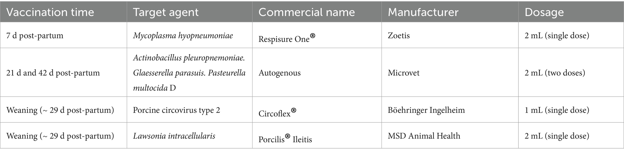
Table 1. Vaccination protocol applied during the suckling and nursery phases for all piglets used in this study.
At weaning, piglets were identified with ear tags, individually weighted and distributed into three groups according to their body weight: NVMED (n = 301) – no intramuscular vaccination against Lawsonia intracellularis but in-feed medication (antimicrobials with activity against L. intracellularis and other agents) over the nursery and growth-finishing phases; VMED (n = 297) – intramuscular vaccination against L. intracellularis and in-feed medication over the nursery and growth-finishing phases; VNMED (n = 302) – intramuscular vaccination against L. intracellularis but no in-feed medication over the nursery and growth-finishing phases. The vaccine against Lawsonia intracellularis (Porcilis® Ileitis, MSD Animal Health) was administered at weaning (when piglets were ~ 29 d-old) by a single intramuscular injection (2 mL) in the neck region, as recommended by the manufacturer. Stainless steel needles of 20 G × 0.9 mm were used. After weaning, piglets were transferred to a commercial nursery facility (27.27371 S, 49.82817 W) and later moved to a growth-finishing facility (27.14133 S, 49.76669 W).
The nursery and growth-finishing facilities presented the following biosecurity measures: vegetation features, fence around the farm, parking outside the farm, sanitary ford (showers and dressing room), bird-proof nets, and rodent and insect control plans. Furthermore, both farms were positive (molecular detection – qPCR) for L. intracellularis.
2.3 Management, housing, and feeding – nursery phase
In the nursery phase, piglets were randomly housed in 18 pens (50 piglets/pen; six pens/treatment) for 37 days. During the period, natural ventilation was applied using double curtains on both sides of each nursery room, and each pen was equipped with slatted plastic floors and five nipple drinkers to provide water ad libitum. The stocking density was 0.32 m2 per animal. Piglets had ad libitum access to feed, which was manually offered and the amount loaded was registered. Before housing, the cleaning and disinfection process of the facility was performed using glutaraldehyde- and quaternary ammonium-based disinfectant, followed by a downtime of 5 days.
Animals were fed according to their treatment group and production phase (Table 2). The feed formulation was obtained from the company hosting the study, and the antimicrobial inclusion was performed over the manufacturing process (Table 2). The feed provided to the VNMED group was stored in an exclusive silo. Throughout the study, the use of injectable medications (antibiotic and anti-inflammatory) and number of dead or fallout animals (piglets that were much lighter than their littermates) were recorded. Necropsy was performed on all dead or euthanized piglets during the phase.
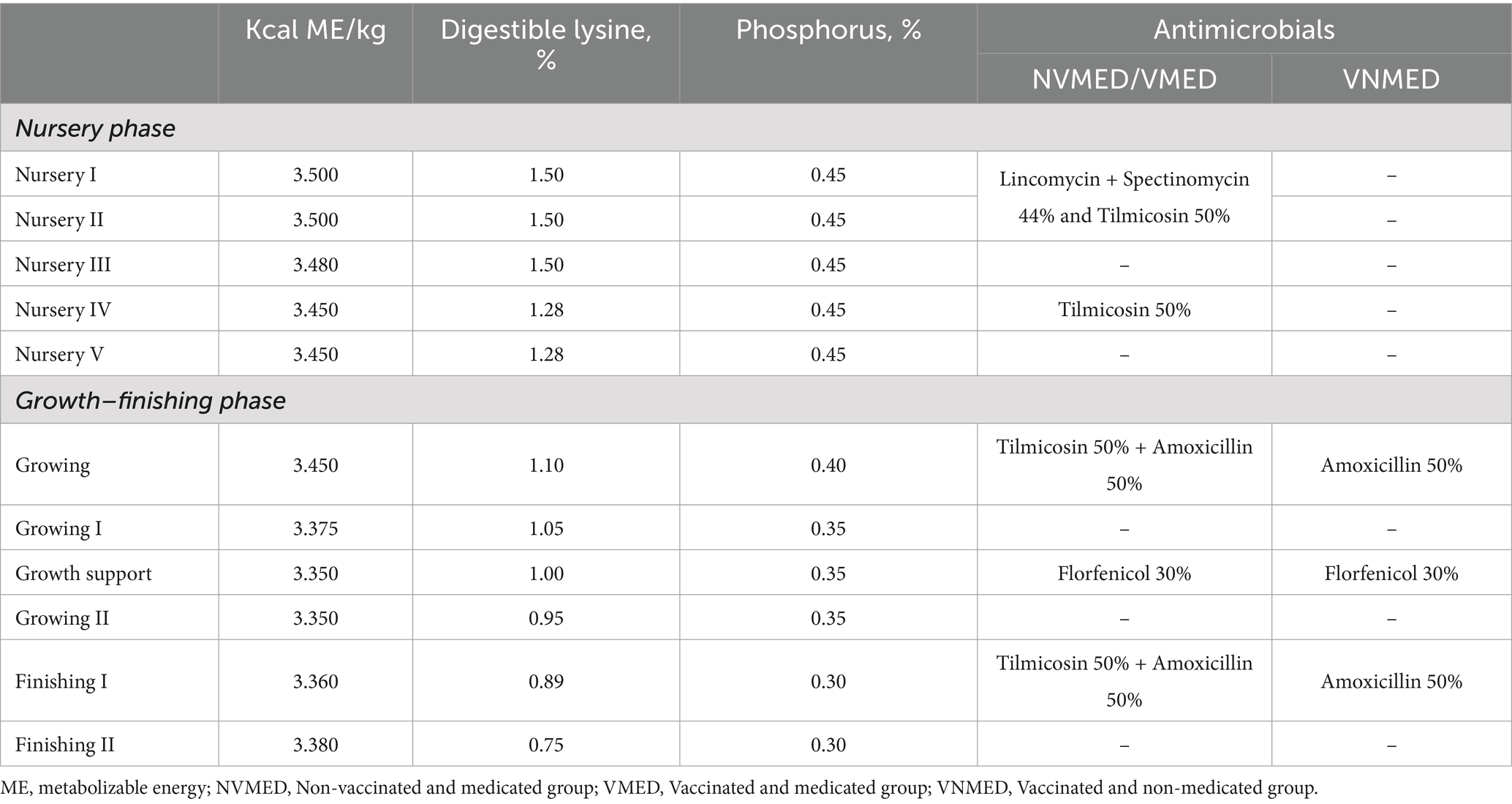
Table 2. Feeding protocol including nutritional composition and antimicrobial program of diets provided to piglets during the study period.
At the last housing day, piglets were weighted and 868 piglets presenting the highest weight were selected to be housed in a growth-finishing facility.
2.4 Management, housing, and feeding – growth and finishing phase
The 868 selected piglets were transferred to a growing and finishing facility and housed in 36 pens. Animals from each nursery pen were divided into two growing and finishing pens; therefore, piglets were housed according to their treatment group (12 pens/treatment) with no mixing of animals from different treatments. The stocking density was 1 m2 per pig, and pens had semi-compact concrete flooring along 2/3 of the pen with slatted concrete flooring in the remaining area, equipped with one nipple drinker for every 12 pigs and stainless steel feeder. Before housing, the facility was submitted to the cleaning and disinfection process using glutaraldehyde- and quaternary ammonium-based disinfectants, followed by a downtime period of 5 days.
Diets were also formulated and provided by the company housing the study (Table 1). The food used to feed the VNMED group was stored in an exclusive silo. Pigs were housed for 100 days and during this period the use of injectable medications (antibiotic and anti-inflammatory) and the number of dead or fallout animals were recorded. Necropsy was also performed on all dead or euthanized piglets. Before transporting the animals to the slaughterhouse, they were individually weighted.
2.5 Detection of serum anti-Lawsonia intracellularis IgG
A total of 30 pigs per treatment was randomly selected and paired blood samples were collected from pigs at different ages (29, 49, 65, 98, 128, and 175 days old). The sample collection was performed by puncture of the jugular vein using polypropylene tubes containing a clotting activator. Blood samples were centrifuged at 500 × g for 10 min, and serum samples were collected and stored at −80°C. The detection of anti-L. intracellularis IgG was conducted by Flow Cytometry Antibody Test as described by Baldasso et al. (33).
2.6 Quantification of Lawsonia intracellularis fecal shedding
To evaluate the excretion of L. intracellularis, feces samples were collected from the same pigs selected for blood samples collection. Samples were collected directly from the rectal ampoule using a sterile plastic bag and were kept at 2–8°C until arrival at the laboratory. The fecal samples were individually diluted (1 g into 9 mL of PBS pH 7.2) and the total genomic DNA was extracted using the MagMax CORE kit following the manufacturer’s recommendation (ThermoFisher Scientific, USA). The quantitative PCR (qPCR) was conducted according to Stahl et al. (34).
2.7 Pneumonia index (PI) assessment
During the slaughter, the severity index of pneumonic lesions was assessed from 150 animals per treatment. Each pulmonary lobe was evaluated and contributed to the scoring based on its proportion related to the total lung area. Lobes were scored according to the extent of pulmonary consolidation lesion: 0%; 0.1–11%; 11.1–21%; 21.1–31%; 31.1–41%; 41.1–51%; 51.1–100% (35). The values of the PPI were classified as pneumonia-free (0.0–0.55); pneumonia with no risk in the herd (0.56–0.89); and high risk of pneumonia in the herd (>0.9).
2.8 Economic analysis of vaccination and antimicrobial costs
The total amount of antimicrobials consumed (kg) per group was calculated based on the feed consumption, registered during the nursery and growth-finishing phases, and the amount of antimicrobials (active ingredient) included in the feed. Similarly, the amount of antimicrobials consumed (mg) per kg of live weight in each phase was also estimated, dividing the antimicrobial consumption by the total live weight of the piglets at the end of the nursery and growth-finishing phases. The cost of antimicrobials (per phase; US$) was calculated based on the antimicrobial consumption and the cost of antimicrobials (active ingredient) reported by the feed manufacturing. Additionally, the expenses related to the vaccination against Lawsonia intracellularis were estimated considering only the vaccine cost (number of piglets vaccinated × cost of vaccine’s dose; US$), since there was no additional service cost (Porcilis® ileitis was administered simultaneously with another vaccine – against porcine circovirus type 2 – following the sanitary protocol of the farm.
2.9 Statistical analysis
Data were analyzed using the Statistical Analysis System© software (SAS, 2012). The pen was the experimental unit and significant differences were considered when p ≤ 0.05. Quantitative responses (body weight – BW, average daily gain – ADG, and feed conversion – FC) were checked regarding residual normality and then analyzed through variance analysis (GLM procedure). Means were compared by the protected t-test. For responses number of dead piglets, number of fallout piglets, and number of piglets treated with injectable drugs a binomial distribution was fitted, and a logistic regression was applied considering the treatment effect (LOGISTIC procedure).
The levels of anti-L. intracellularis IgG were analyzed by repeated measures, using the MIXED procedure, selecting the adequate covariance structure based on the lowest value of Akaike Information Criterion (AIC). The treatment, sample moment (piglets’ age), and their interaction were included as fixed effects, comparing the values by the protected t-test.
3 Results
3.1 Average daily gain, feed conversion ratio, and health performance
The growth performance of piglets from the three groups is presented in Table 3. Feed conversion during the nursery phase was similar amongst the groups (1.46 ± 0.02; p = 0.56). Nevertheless, the ADG and the BW at the end of the nursery period were lower for piglets from the VNMED group (350.07 ± 5.79 g, 21.34 ± 0.23 kg; respectively) compared to NVMED (387.46 ± 7.13 g, 22.79 ± 0.26 kg; respectively) and VMED groups (385.04 ± 5.59 g, 22.70 ± 0.21 kg; respectively; p < 0.01). Despite that, piglets from VNMED group presented similar BW at the end of the growth and finishing phase compared to piglets from groups NVMED and VMED (131.4 ± 0.69; p = 0.75), with no differences in FC or ADG over the growth and finishing phase (p = 0.37).
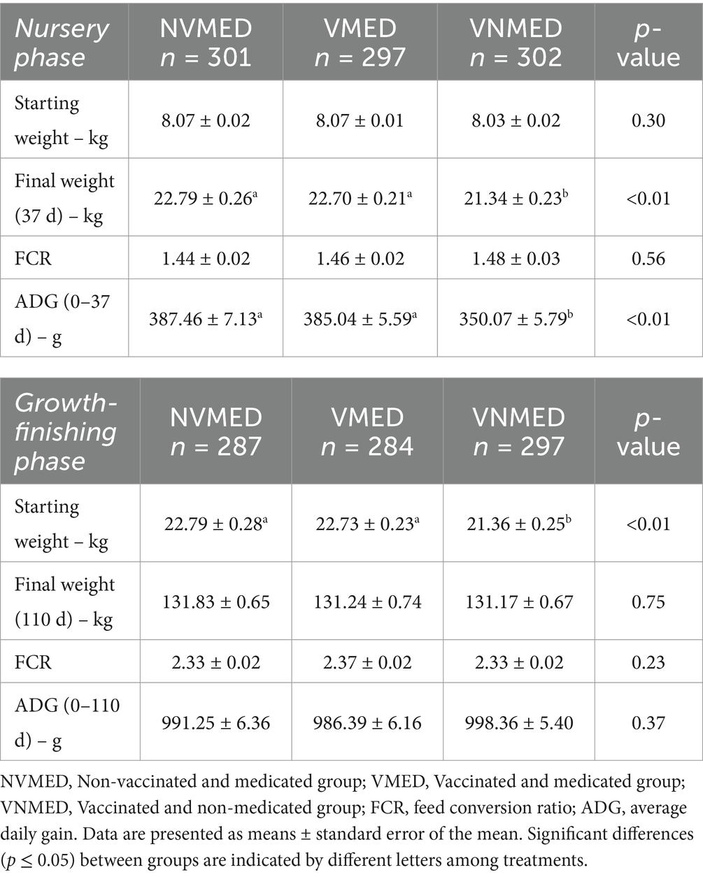
Table 3. Zootechnical performance of piglets in the nursery and growth-finishing phases during the study period.
The number of animals treated with injectable medication, the number of fallout piglets, and piglet mortality did not differ amongst groups, regardless of the phase (p ≥ 0.10; Table 4).
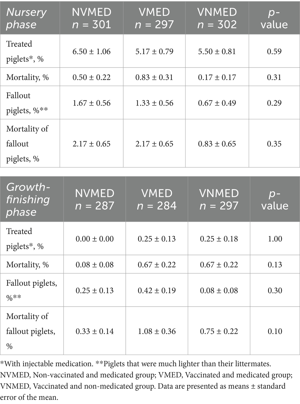
Table 4. Clinical performance of piglets in the nursery and growth-finishing phases during the study period.
3.2 Kinetics of the anti-Lawsonia intracellularis antibody response
As illustrated in Figure 1A, all piglets have detectable levels of maternally derived antibodies at 29 days of age. The levels of systemic anti-L. intracellularis IgG observed in vaccinated animals remained stable until day 49 and increased significantly (p < 0.05) from day 65 onwards, regardless of the presence or absence of antibiotics in the feed (Figure 1A). On the other hand, levels of anti-L. intracellularis IgGs observed in the unvaccinated group decreased from day 29 and at 49 days, only a few animals still had antibodies of maternal origin. Interestingly, we observed two peaks of antibody increase in this group, the first at 65 days of age, and the second at 128 days of age, which indicates that the animals were naturally exposed to L. intracellularis. As illustrated in Figure 1B, after application of the vaccine, the antibody curve observed in vaccinated animals compared to unvaccinated animals was completely different. Significant differences (p < 0.05) between the vaccinated and non-vaccinated groups were observed from day 49 of life (20 days after vaccine application) until the end of this study.
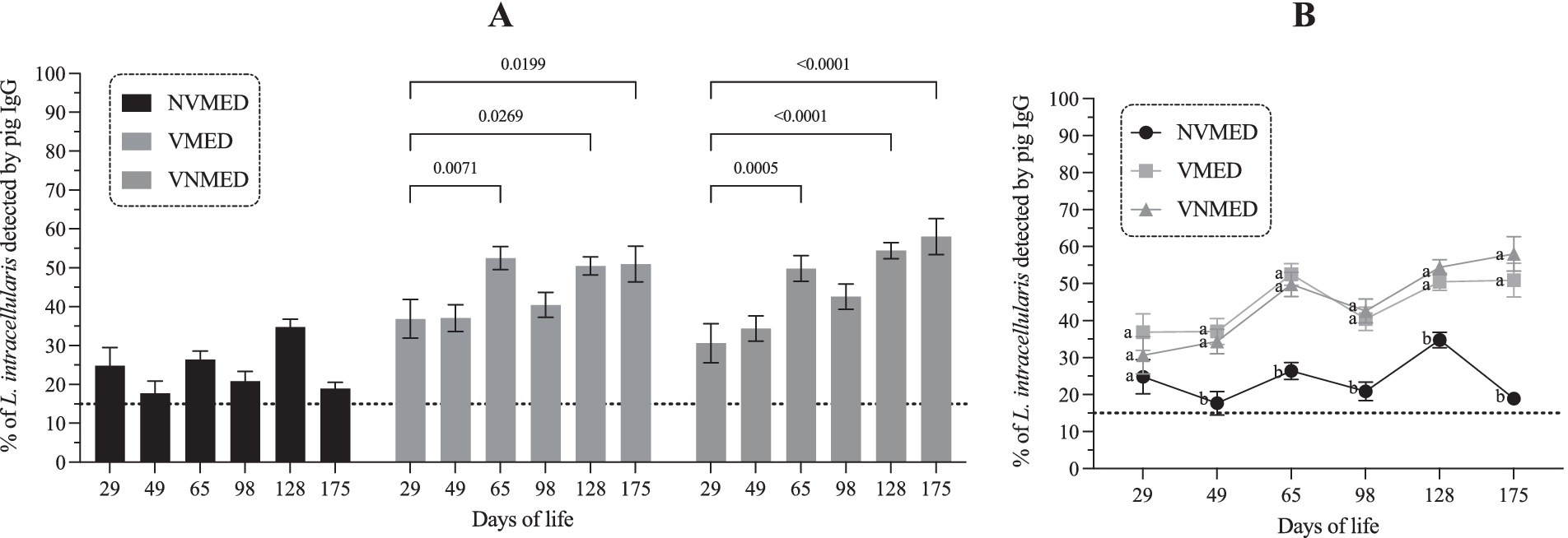
Figure 1. Profile of systemic anti-Lawsonia intracellularis IgG in piglets over the study period. (A) Antibody response of vaccinated and unvaccinated groups in the presence or absence of antibiotics in the feed. (B) Comparison of the antibody curve between the experimental groups. (A): absolute value of “p”; (B): different letters indicate significant difference; p < 0.05. NVMED, Non-vaccinated and medicated group; VMED, Vaccinated and medicated group; VNMED, Vaccinated and non-medicated group.
3.3 Shedding profile of Lawsonia intracellularis in fecal samples and lung inspection at slaughterhouse
The bacterial agent was only detected in feces samples from four pigs, two from the VMED group (at 49 and 175 days of life) and two from the VNMED group (at 128 and 175 days of life). According to evaluations performed at slaughter, all groups presented animals with pneumonia, showing similar prevalence: 68% (NVMED), 67.3% (VMED), and 70% (VNMED). For all groups, the PPI was <0.89, presenting no risk to the herd (0.73; 0.72; and 0.79, respectively).
3.4 Antimicrobial consumption and expenses with antimicrobial use and vaccination
Antimicrobial consumption and expenses with antimicrobial use and vaccination are presented in Table 5 and Figure 2. Total in-feed antimicrobial consumption (mg/kg of live weight) for NVMED and VMED piglets was ~3.5-fold greater than the consumption for VNMED piglets: 781.7 vs. 790.6 vs. 227.7, respectively; leading to a decrease of 554–563 mg of antimicrobial/kg of live weight (71%). The expenses with antimicrobials throughout the nursery and growth-finishing phases for NVMED and VMED piglets were ~ 3-fold greater than the costs for VNMED piglets: US$ 1,685.62 vs. US$ 1,646.37 vs. US$ 529.56, respectively. These results demonstrate that VNMED group (not treated with antibiotics) generated a cash savings of 68 and 69% compared to the groups that received antibiotic treatment.
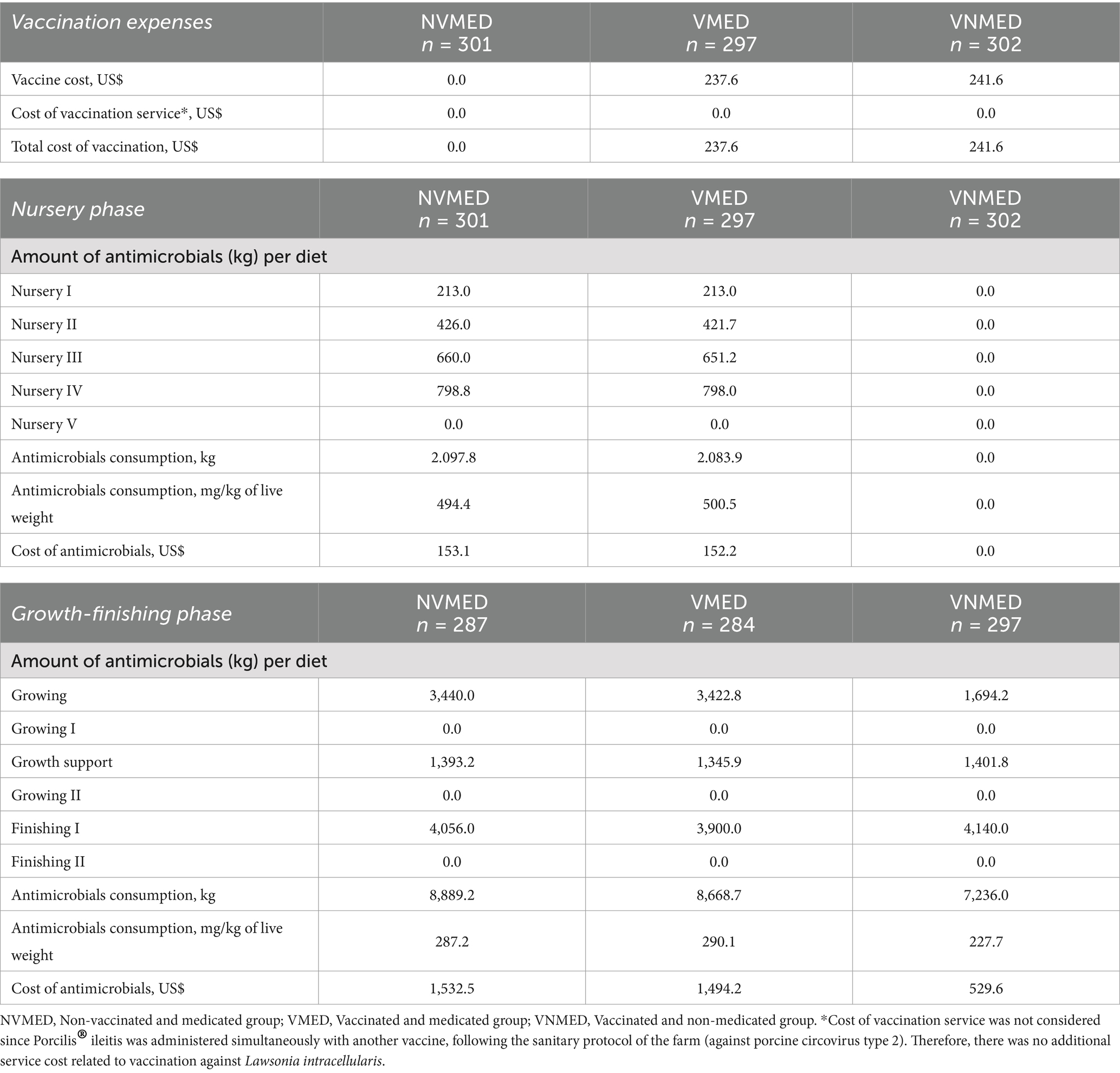
Table 5. Expenses related to the vaccination process against Lawsonia intracellularis and the in-feed antimicrobials consumption during the study period.
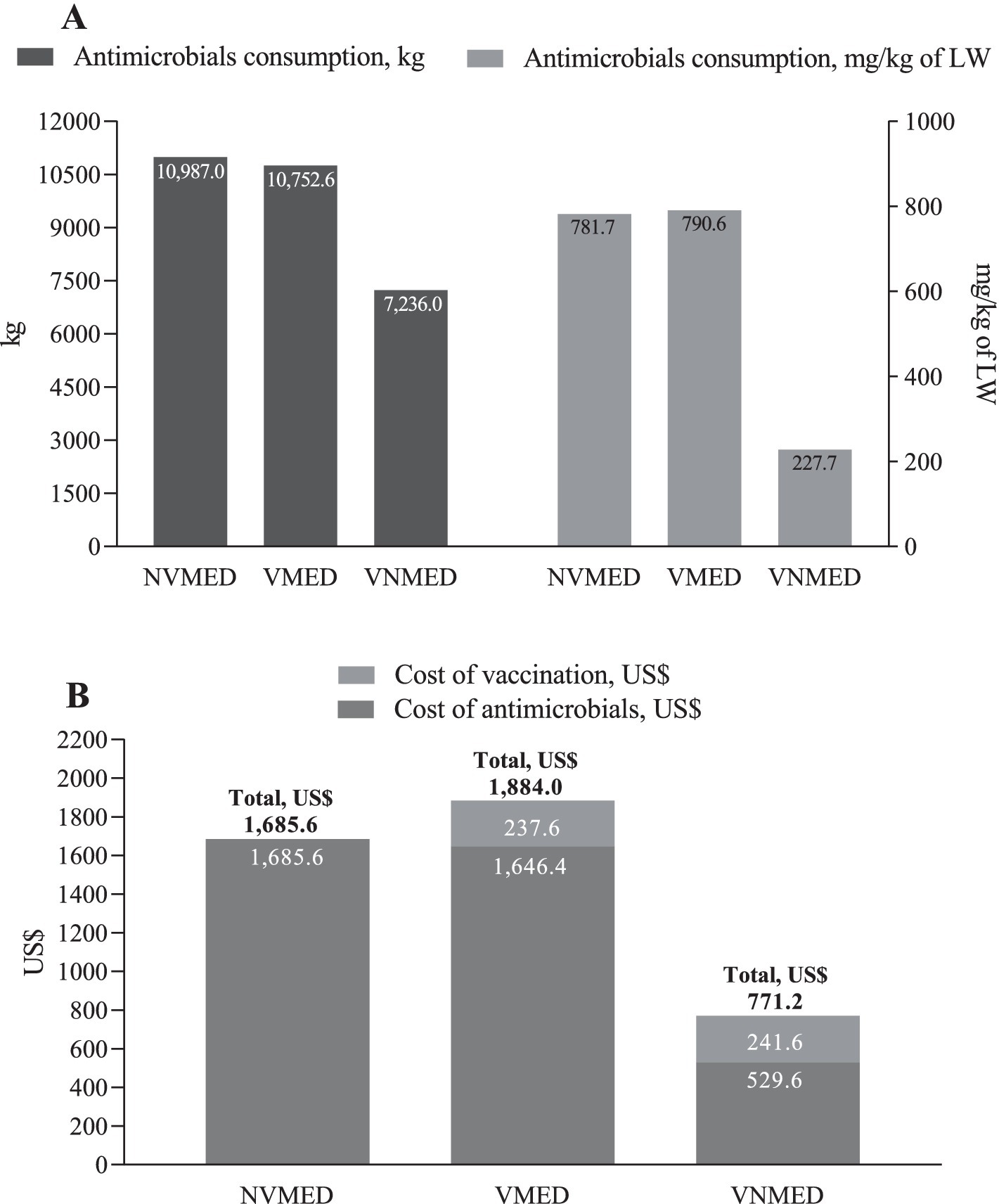
Figure 2. Consumption and costs of in-feed antimicrobials, and cost of vaccination process against Lawsonia intracellularis over the study period. (A) Total consumption of in-feed antimicrobials over the nursery and growth-finishing phases (kg and mg/kg of live weight). (B) Expenses related to the consumption of in-feed antimicrobials and the vaccination process against Lawsonia intracellularis during the nursery and growth-finishing phases. NVMED, Non-vaccinated and medicated group; VMED, Vaccinated and medicated group; VNMED, Vaccinated and non-medicated group. LW, live weight. Cost of vaccination service was not considered since Porcilis® ileitis was administered simultaneously to another vaccine, following the sanitary protocol of the farm (against porcine circovirus type 2). Therefore, there was no additional service cost related to vaccination against Lawsonia intracellularis.
4 Discussion
Lawsonia intracellularis is a highly prevalent microorganism in Brazil (36) that causes porcine proliferative enteropathy. The diagnosis of this disease can be easily performed when the animals present typical clinical signs of the acute and chronic forms of the disease. On the other hand, when the infection presents subclinically, which occurs most of the time, the microorganism is only detected through laboratory diagnosis. In this study, we conducted a field experiment to evaluate the clinical, zootechnical performance and immune profile of a group of piglets vaccinated against L. intracellularis (VNMED) compared to a group of piglets vaccinated and medicated with antibiotics (VMED) and/or non-vaccinated and medicated with antibiotics (NVMED).
As expected, piglets vaccinated at 29 days of age with the Porcilis® Ileitis vaccine developed a clear systemic IgG response after vaccination (Figure 1). This vaccine is formulated with an inactivated strain of L. intracellularis and potentiated with an oil-based adjuvant (37). The immunogenicity of this vaccine has already been demonstrated in controlled immunization and experimental challenge studies (37), and here, in a field experiment. In our study, piglets that received the vaccination showed significantly more antibodies than those not immunized. Furthermore, using the Flow Cytometry Antibody Test (33), we observed that lincomycin, spectinomycin, tilmicosin, and amoxicillin did not reduce the levels of specific IgG; therefore, they can be used concomitantly to the genesis of the adaptive response. On the other hand, ceftiofur, doxycycline, and tulathromycin may significantly reduce the antibody response if used during the development of the vaccine response (24).
To control L. intracellularis infection, pigs can use humoral (antibodies) and cellular (T helper 1 and cytotoxic T lymphocytes) immune responses. These responses can be modulated during the natural infection process (38) or by vaccination (39). Regarding the antibody response, we observed that vaccinated pigs presented high levels of IgG for more than 20 weeks, confirming the duration of immunity of this vaccine (40). As described in Figure 1A, few animals presented positive levels of IgG at 49 days of age, demonstrating that the duration of maternal immunity was ≤7 weeks in this study. An increase in antibody levels was observed at 65 days of age, demonstrating the occurrence of natural infection throughout the nursery phase. Interestingly, at this moment, it was not possible to detect the excretion of L. intracellularis in feces.
In a recent study conducted in Brazil, it was reported that the peak of L. intracellularis fecal shedding occurs when piglets are 90–120 days old (36). Despite that, in this study, none of the piglets from group NVMED presented fecal samples positive for L. intracellularis. Nevertheless, this low shedding may be related to a low infection pressure, which in this study was a consequence of the vaccination or medication process, since all piglets from VNMED, VMED, and NVMED groups were housed in the same farm and facility. Unfortunately, a group of piglets non-vaccinated and non-medicated was not considered in our methodology, which is a limitation that also contributed to a low agent shedding. Despite that, we must highlight that although this trial was performed in a farm positive for L. intracellularis a low shedding was observed even for the piglets vaccinated but not medicated, showing that vaccination was successfully effective in controlling the agent, regardless of the antimicrobial use. Furthermore, the prophylactic use of antimicrobials was not crucial to avoid respiratory diseases, given that the PPI did not differ amongst the three groups.
Concerning the piglets’ performance, the few studies investigating the effects of vaccination against L. intracellularis on piglets’ growth performance within a scenario of no in-feed antimicrobial use reported a greater average daily weight gain when vaccination was implemented (1, 32). Contrarily, in our study, piglets vaccinated and not medicated (VNMED) had lower average daily weight gain and, consequently, lower final weight at the nursery phase compared to piglets non-vaccinated and medicated (NVMED) and piglets vaccinated and medicated (VMED). Nevertheless, at the growth-finishing phase, the VNMED piglets overcame the lower starting weight – probably due to a compensatory effect – reaching similar average daily weight gain and body weight to other piglets. Regardless, it is important to highlight that savings obtained through the VNMED scenario would probably overcome the expenses related to the lower final weight in VNMED piglets at the nursery phase. Our results showed that the reduction of ~71% in antimicrobials consumption in the VNMED scenario led to an economy of US$ 1,156.0 or 1,116.8 compared to the NVMED or VMED approach, respectively (savings of 68 and 69%, respectively). Even when vaccination expenses were considered, an economy of US$ 914.4 (46% of the total costs for NVMED) or US$ 1,112.8 (59% of the total costs for VMED) was reached. This significant cost saving are attributed to the absence of in-feed antimicrobial use during the nursery phase and the low cost of antimicrobials used during the growth-finishing phase for the VNMED group. Although the VNMED group consumed 7,236.0 kg of antimicrobials in the growth and finishing phase – representing 81 to 83% of the consumption of the NVMED and VMED groups (8,889.2 kg and 8,668.7 kg, respectively) – the majority of this consumption was of a lower-cost antimicrobial (amoxicillin) compared to the one most used in the feed for NVMED and VMED groups (tilmicosin).
The overall results of our study, especially the reduction of ~71% in antimicrobial consumption in the VNMED group, comply with the ‘One Health’ premise of reducing antimicrobial use in animal production and, consequently, antimicrobial resistance, which is one of the global challenges that intensive animal production needs to collaborate on in its confrontation. The swine production presented the largest projected increase in antimicrobial consumption and contributed 45% to the total increase between 2017 and 2030. In 2020, the global antimicrobial use intensity in pigs was estimated at 173.1 mg/PCU (population correction units), while for cattle and chicken, it was 59.6 mg/PCU and 35.4 mg/PCU, respectively (28, 29, 41). Mitigating antimicrobial resistance in animals, humans, and the environment depends primarily on reducing the need for antimicrobial use. Nevertheless, currently, the antimicrobial use in pig production seems to be directly related to the common fear among farmers and veterinarians, who believe that a scenario of reduced use of antimicrobials would not result in lower production outputs; therefore, the high use of antimicrobials would still be necessary to support intensive production (42). Thus, the findings of our study are also essential for decision-makers in the swine industry.
The lack of a group of piglets non-vaccinated and non-medicated is an important limitation of our study. Considering non-vaccinated and non-medicated piglets – besides the non-vaccinated and medicated, vaccinated and medicated, and vaccinated and non-medicated groups – to further evaluate the vaccination as a strategy to reduce the use of antimicrobials in swine production is one of the future perspectives for this topic, as well as considering a different sanitary challenge and comparing the vaccination with other alternative approaches [i.e., use of additives; (43)]. Nevertheless, the overall data found in this study strongly suggest that prophylactic use of antimicrobials in swine production may not be needed to achieve satisfactory health and growth performance when vaccination protocols and adequate biosecurity measures are appropriately applied (44), besides leading to an expressive decrease in antimicrobials use and expenses.
5 Conclusion
The use of inactivated vaccine against L. intracellularis to prevent Porcine Proliferative Enteropathy is an effective strategy to reduce the prophylactic use of antimicrobials. Replacing the drug program with vaccination did not change the zootechnical parameters; however, it significantly reduced the expenses with antimicrobials, increasing the profitability of the operation.
Data availability statement
The raw data supporting the conclusions of this article will be made available by the authors, without undue reservation.
Ethics statement
The animal study was approved by the Institutional Committee for Ethical Use of Animals of the Instituto Federal Catarinense – Campus Araquari (protocol no. 396/2022) and followed the Brazilian College of Animal Experimentation guidelines. The experiment was conducted on commercial pig farms located in the southern region of Brazil. The study was conducted in accordance with the local legislation and institutional requirements.
Author contributions
MG: Conceptualization, Data curation, Formal analysis, Funding acquisition, Investigation, Methodology, Project administration, Resources, Writing – original draft, Writing – review & editing. MQ: Conceptualization, Validation, Writing – original draft, Writing – review & editing. RF: Conceptualization, Formal analysis, Investigation, Methodology, Writing – original draft, Writing – review & editing. YT: Investigation, Methodology, Resources, Writing – original draft. AN: Methodology, Resources, Writing – original draft. AC: Data curation, Formal analysis, Methodology, Validation, Writing – original draft, Writing – review & editing. IB: Conceptualization, Data curation, Formal analysis, Investigation, Methodology, Project administration, Supervision, Validation, Writing – original draft, Writing – review & editing. JK: Conceptualization, Investigation, Methodology, Supervision, Validation, Writing – original draft, Writing – review & editing.
Funding
The author(s) declare that financial support was received for the research, authorship, and/or publication of this article. This study was funded by: CNPq (grant # 305575/2021-0 given to I. Bianchi). FAPESC (grant # 1367/2022 given to M.W. Quirino). Embrapa’s research project (SEG 40.19.03.039.00.00). The authors declare that this study received funding from MSD. The funder was not involved in the study design, collection, analysis, interpretation of data, the writing of this article, or the decision to submit it for publication.
Acknowledgments
Authors are thankful to the Conselho Nacional Desenvolvimento Científico e Tecnológico – Brazil (CNPq), Coordenação de Aperfeiçoamento de Pessoal de Nível Superior – Brazil (CAPES), and Fundação de Amparo à Pesquisa e Inovação do Estado de Santa Catarina – Brazil. The authors state that this study was carried out at the facilities of Pamplona Alimentos and used animals owned by the company.
Conflict of interest
MG was employed by SD Saúde Animal. RF is a partner and CEO of AFK Imunotech. AN and YT were employed by Pamplona Alimentos. JK was employed by Embrapa Suínos e Aves.
The remaining authors declare that the research was conducted in the absence of any commercial or financial relationships that could be construed as a potential conflict of interest.
Generative AI statement
The authors declare that no Generative AI was used in the creation of this manuscript.
Publisher’s note
All claims expressed in this article are solely those of the authors and do not necessarily represent those of their affiliated organizations, or those of the publisher, the editors and the reviewers. Any product that may be evaluated in this article, or claim that may be made by its manufacturer, is not guaranteed or endorsed by the publisher.
References
1. McOrist, S, and Smits, RJ. Field evaluation of an oral attenuated Lawsonia intracellularis vaccine for porcine proliferative enteropathy (ileitis). Vet Rec. (2007) 161:26–8. doi: 10.1136/vr.161.1.26
2. Kroll, JJ, Roof, MB, Hoffman, LJ, Dickson, JS, and Harris, DL. Proliferative enteropathy: a global enteric disease of pigs caused by Lawsonia intracellularis. Anim Health Res Rev. (2005) 6:173–97. doi: 10.1079/ahr2005109
3. Paradis, MA, Gottschalk, M, Rajic, A, Ravel, A, Wilson, JB, Aramini, J, et al. Seroprevalence of Lawsonia intracellularis in different swine populations in 3 provinces in Canada. Can Vet J. (2007) 48:57–64. doi: 10.4141/cjas68-008
4. Wu, Z, Ling, Y, Tian, D, Pan, Q, Heegaard, PM, and He, C. Seroprevalence of Lawsonia intracellularis antibodies in intensive pig farms in China. BMC Vet Res. (2014) 10:100. doi: 10.1186/1746-6148-10-100
5. Arnold, M, Crienen, A, Swam, H, von Berg, S, Jolie, R, and Nathues, H. Prevalence of Lawsonia intracellularis in pig herds in different European countries. Porcine Health Manag. (2019) 5:31. doi: 10.1186/s40813-019-0137-6
6. Vannucci, FA, Gebhart, CJ, and McOrist, S. Proliferative enteropathy In: LA Karriker, JJ Zimmerman, A Ramirez, KJ Schwartz, GW Stevenson, and J Zhang, editors. Diseases of swine. 11th ed. Hoboken, NJ, USA: John Wiley Sons, Inc (2019). 475–91.
7. Love, DN, and Love, RJ. Pathology of proliferative haemorrhagic enteropathy in pigs. Vet Pathol. (1979) 16:41–8. doi: 10.1177/030098587901600104
8. Vannucci, FA, and Gebhart, CJ. Recent advances in understanding the pathogenesis of Lawsonia intracellularis infections. Vet Pathol. (2014) 51:465–77. doi: 10.1177/0300985813520249
9. Hardge, T, Nickoll, E, Grunert, H, Elbers, K, Langbein, U, Keller, C, et al. Prevention of porcine proliferative enteropathy (PPE) by vaccination — efficacy and economics in European farms. Pig J. (2004) 54:17–34.
10. Almond, PK, and Bilkei, G. Effects of oral vaccination against Lawsonia intracellularis on growing-finishing pig’s performance in a pig production unit with endemic porcine proliferative enteropathy (PPE). Dtsch Tierarztl Wochenschr. (2006) 113:232–5.
11. Caspari, K, Kummerlen, D, Voets, H, Eichin, E, Zeeh, H, and Zimmermann, W. Field study about the use of Enterisol ileitis in a swine herd in Switzerland. Schweiz Arch Tierheilkd. (2009) 151:31–2. doi: 10.1024/0036-7281.151.1.31
12. Weibel, H, Sydler, T, Brugnera, E, Voets, H, Grosse Liesner, B, and Sidler, X. Efficacy of simultaneous vaccination with Enterisol® ileitis and Ingelvac® CircoFLEX™ in a Swiss breeding farm. Schweiz Arch Tierheilkd. (2012) 154:445–50. doi: 10.1024/0036-7281/a000381
13. Park, S, Lee, JB, Kim, KJ, Oh, YS, Kim, MO, Oh, YR, et al. Efficacy of a commercial live attenuated Lawsonia intracellularis vaccine in a large-scale field trial in Korea. Clin Exp Vaccine Res. (2013) 2:135. doi: 10.7774/cevr.2013.2.2.135
14. Peiponen, KS, Tirkkonen, BT, Junnila, JJT, and Heinonen, ML. Effect of a live attenuated vaccine against Lawsonia intracellularis in weaned and finishing pig settings in Finland. Acta Vet Scand. (2018) 60:18. doi: 10.1186/S13028-018-0374-8
15. Jacobs, AAC, Harks, F, Hazenberg, L, Hoeijmakers, MJH, Nell, T, Pel, S, et al. Efficacy of a novel inactivated Lawsonia intracellularis vaccine in pigs against experimental infection and under field conditions. Vaccine. (2019) 37:2149–57. doi: 10.1016/j.vaccine.2019.02.067
16. Musse, SL, Nielsen, GB, Stege, H, Weber, NR, and Houe, H. Effect of intramuscular vaccination against Lawsonia intracellularis on production parameters, diarrhea occurrence, antimicrobial treatment, bacterial shedding, and lean meat percentage in two Danish naturally infected finisher pig herds. Prev Vet Med. (2023) 212:105837. doi: 10.1016/j.prevetmed.2023.105837
17. França, SA, and Guedes, RM. Antimicrobial use for the control of porcine proliferative enteropathy. Cienc Rural. (2008) 38:288–96. doi: 10.1590/S0103-84782008000100050
18. WOAH. World organisation for animal health. Terrestrial animal health code. (2024). Available in: https://www.woah.org/en/what-we-do/standards/codes-and-manuals/terrestrial-code-online-access/ (Accessed June 12, 2024).
19. Khalifeh, MS, Amawi, MM, Abu-Basha, EA, and Yonis, IB. Assessment of humoral and cellular-mediated immune response in chickens treated with tilmicosin, florfenicol, or enrofloxacin at the time of Newcastle disease vaccination. Poult Sci. (2009) 88:2118–24. doi: 10.3382/ps.2009-00215
20. Pomorska-Mól, M, Kwit, K, Markowska-Daniel, I, and Pejsak, Z. The effect of doxycycline treatment on the postvaccinal immune response in pigs. Toxicol Appl Pharmacol. (2014) 278:31–8. doi: 10.1016/j.taap.2014.04.006
21. Pomorska-Mól, M, Czyżewska-Dors, E, Kwit, K, Wierzchosławski, K, and Pejsak, Z. Ceftiofur hydrochloride affects the humoral and cellular immune response in pigs after vaccination against swine influenza and pseudorabies. Vet Res. (2015) 11:268. doi: 10.1186/s12917-015-0586-3
22. Pomorska-Mól, M, Kwit, K, Czyżewska-Dors, E, and Pejsak, Z. Tulathromycin enhances humoral but not cellular immune response in pigs vaccinated against swine influenza. J Vet Pharmacol Ther. (2019) 42:318–23. doi: 10.1111/jvp.12742
23. Pomorska-Mól, M, Kwit, K, Wierzchosławski, K, Dors, A, and Pejsak, Z. Effects of amoxicillin, ceftiofur, doxycycline, tiamulin and tulathromycin on pig humoral immune responses induced by erysipelas vaccination. Vet Rec. (2016) 178:559. doi: 10.1136/vr.103533
24. Fonseca, EM, Donin, DG, Jesus, AN, Daniel, AG, Gomes, BC, Feronato, C, et al. Effect of amoxicillin, ceftiofur, doxycycline, tiamulin, and tulathromycin on the antibody response of piglets vaccinated against Lawsonia intracellularis. Proceedings of the 27th international pig veterinary society congress – IPVS and 15th European symposium of porcine health management, Leipzig, Germany. (2024).
25. Won, G, Chi, NK, and Park, Y. The effectiveness of commercial vaccination against Lawsonia intracellularis in mitigating the reduction in ADWG, the increased mortality, and fecal shedding of the vaccinated pigs: a systematic review and meta-analysis. Vet Sci. (2022) 9:536. doi: 10.3390/vetsci9100536
26. Maron, DF, Smith, TJS, and Nachman, KE. Restrictions on antimicrobial use in food animal production: an international regulatory and economic survey. Glob Health. (2013) 9:1–11. doi: 10.1186/1744-8603-9-48
27. Burow, E, and Käsbohrer, A. Risk factors for antimicrobial resistance in Escherichia coli in pigs receiving oral antimicrobial treatment: a systematic review. Microb Drug Resist. (2017) 23:194–205. doi: 10.1089/mdr.2015.0318
28. Boeckel, TP, Glennon, EE, Chen, D, Gilbert, M, Robinson, TP, Grenfell, BT, et al. Reducing antimicrobial use in food animals. Science. (2017) 357:1350–2. doi: 10.1126/science.aao1495
29. Mulchandani, R, Wang, Y, Gilbert, M, and Van Boeckel, TP. Global trends in antimicrobial use in food-producing animals: 2020 to 2030. PLOS Glob Public Health. (2023) 3:e0001305. doi: 10.1371/journal.pgph.0001305
30. Cardelle-Cobas, A, Coy-Girón, L, Cepeda, A, and Nebot, C. Swine production: Probiotics as an alternative to the use of antibiotics. In: Antibiotics and Probiotics in Animal Food—Impact and Regulation; Veterinary medicine and science. London, UK: IntechOpen. (2023). doi: 10.5772/intechopen.108308
31. Dutra, MC, Moreno, LZ, Dias, RA, and Moreno, AM. Antimicrobial use in Brazilian swine herds: assessment of use and reduction examples. Microorganisms. (2021) 9:881. doi: 10.3390/microorganisms9040881
32. Bak, H, and Rathkjen, PH. Reduced use of antimicrobials after vaccination of pigs against porcine proliferative enteropathy in a Danish SPF herd. Acta Vet Scand. (2009) 51:1. doi: 10.1186/1751-0147-51-1
33. Baldasso, DZ, Guizzo, JA, Dazzi, CC, Paraboni Frandoloso, GC, Feronato, C, von Berg, S, et al. Development and validation of a flow cytometry antibody test for Lawsonia intracellularis. Front Immunol. (2023) 14:1145072. doi: 10.3389/fimmu.2023.1145072
34. Stahl, M, Kokotovic, B, Hjulsager, CK, Breum, SO, and Ange, O. The use of quantitative PCR for identification and quantification of Brachyspira pilosicoli, Lawsonia intracellularis and Escherichia coli fimbrial types F4 and F18 in pig feces. Vet Microbiol. (2011) 151:307–14. doi: 10.1016/j.vetmic.2011.03.013
35. Sobestiansky, J, Barcellos, D, and Driemeier, D. Matos M P C. Monitoramento de abate In: J Sobestiansky and D Barcellos, editors. Doenças dos suínos (Diseases of Swine). Brazil: Cânone Editorial (2007). 915–21.
36. Dazzi, CC, Baldasso, D, Frandoloso, GP, Kreutz, LC, and Frandoloso, R. Brazilian growing and finishing pig farms are endemic for Lawsonia intracellularis. Proceedings of the 26th international pig veterinary society congress – IPVS, Rio de Janeiro, Brazil. (2022).
37. Roerink, F, Morgan, CL, Knetter, SM, Passat, MH, Archibald, AL, Ait-Ali, T, et al. A novel inactivated vaccine against Lawsonia intracellularis induces rapid induction of humoral immunity, reduction of bacterial shedding and provides robust gut barrier function. Vaccine. (2018) 36:1500–8. doi: 10.1016/j.vaccine.2017.12.049
38. Riber, U, Heegaard, PM, Cordes, H, Stahl, M, Jensen, TK, and Jungersen, G. Vaccination of pigs with attenuated Lawsonia intracellularis induced acute phase protein responses and primed cell-mediated immunity without reduction in bacterial shedding after challenge. Vaccine. (2015) 33:156–62. doi: 10.1016/j.vaccine.2014.10.084
39. Guedes, RM, and Gebhart, CJ. Onset and duration of fecal shedding, cell-mediated and humoral immune responses in pigs after challenge with a pathogenic isolate or attenuated vaccine strain of Lawsonia intracellularis. Vet Microbiol. (2003) 91:135–45. doi: 10.1016/s0378-1135(02)00301-2
40. USDA. Lawsonia intracellularis Bacterin, Intervet Inc In: Summary of studies supporting USDA product licensure USA: USDA (2015)
41. Tiseo, K, Huber, L, Gilbert, M, Robinson, TP, and Van Boeckel, TP. Global trends in antimicrobial use in food animals from 2017 to 2030. Antibiotics. (2020) 9:918. doi: 10.3390/antibiotics9120918
42. Dewulf, J, Joosten, P, Chantziaras, I, Bernaerdt, E, Vanderhaeghen, W, Postma, M, et al. Antibiotic use in European pig production: less is more. Antibiotics. (2022) 11:1–11. doi: 10.3390/antibiotics11111493
43. Güths, MF, Siqueira, HA, Montes, JH, Moreira, F, Rizzoto, G, Peripolli, V, et al. Removal or substitution of in-feed antimicrobials in swine production. Prev Vet Med. (2022) 205:105696. doi: 10.1016/j.prevetmed.2022.105696
Keywords: pig production, Lawsonia intracellularis , vaccination, antibiotics, antimicrobial resistance, antimicrobial prudent use, one health
Citation: Gallina MA, Quirino MW, Frandoloso R, Tutida YH, Norenberg A, Coldebella A, Bianchi I and Kich JD (2025) Vaccination versus antimicrobials to prevent Porcine Proliferative Enteropathy: associated costs and effects on piglets’ growth, health, and serological performance. Front. Vet. Sci. 12:1538206. doi: 10.3389/fvets.2025.1538206
Edited by:
Dalia Hamza, Cairo University, EgyptCopyright © 2025 Gallina, Quirino, Frandoloso, Tutida, Norenberg, Coldebella, Bianchi and Kich. This is an open-access article distributed under the terms of the Creative Commons Attribution License (CC BY). The use, distribution or reproduction in other forums is permitted, provided the original author(s) and the copyright owner(s) are credited and that the original publication in this journal is cited, in accordance with accepted academic practice. No use, distribution or reproduction is permitted which does not comply with these terms.
*Correspondence: Jalusa Deon Kich, amFsdXNhLmtpY2hAZW1icmFwYS5icg==