 Ehab Kotb Elmahallawy1,2*
 Nady Khairy Elbarbary3
 David Cano-Terriza1,4
 Tomás Fajardo1
 Nada Oudah Albalawi5Débora Jiménez-Martín1Marwa M. I. Ghallab6
 Ahmed Gareh7
 Refaat Ras8,9
 Isabelle Villena10
 Sabry A. S. Sadek11
 Hajar AlQadeeb12Hind Alzaylaee13
 Sonia Almería14
 Ignacio García-Bocanegra1,4*
- 1Departamento de Sanidad Animal, Grupo de Investigación en Sanidad Animal y Zoonosis (GISAZ), Universidad de Córdoba, Córdoba, Spain
- 2Department of Zoonoses, Faculty of Veterinary Medicine, Sohag University, Sohag, Egypt
- 3Department of Food Hygiene, Faculty of Veterinary Medicine, Aswan University, Aswan, Egypt
- 4CIBERINFEC, ISCIII CIBER de Enfermedades Infecciosas, Instituto de Salud Carlos III, Madrid, Spain
- 5Department of Biology, Faculty of Science, Taibah University, Alula, Saudi Arabia
- 6Department of Medical Parasitology, Faculty of Medicine, Kafrelsheikh University, Kafr El Sheikh, Egypt
- 7Department of Parasitology, Faculty of Veterinary Medicine, Aswan University, Aswan, Egypt
- 8Department of Parasitology, Faculty of Veterinary Medicine, Zagazig University, Zagazig, Egypt
- 9Department of Microbiology and Parasitology, Faculty of Veterinary Medicine, Badr University in Cairo (BUC), Badr City, Egypt
- 10University of Reims Champagne-Ardenne, UR 7510, National Reference Centre for Toxoplasmosis, Laboratory of Parasitology, Reims Hospital, Reims, France
- 11Department of Zoonotic Diseases, National Research Centre, Giza, Egypt
- 12Department of Medical Laboratory, College of Applied Medical Sciences, Prince Sattam Bin Abdulaziz University, AlKharj, Saudi Arabia
- 13Department of Biology, College of Science, Princess Nourah bint Abdulrahman University, Riyadh, Saudi Arabia
- 14Virology and Parasitology Branch, Division of Food and Environmental Safety, Office of Applied Microbiology and Technology (OAMT), Office of Laboratory Operations and Applied Sciences (OLOAS), Food and Drug Administration, Department of Health and Human Services, Laurel, MD, United States
Toxoplasmosis remains a prevalent parasitic zoonosis worldwide, raising public health concerns. The global information available regarding the role of camels in the epidemiology of Toxoplasma gondii is still limited. This study aimed to assess the seroprevalence of T. gondii in dromedary camels (Camelus dromedarius) from northern and southern Egypt. A total of 513 serum samples were obtained from camels across Cairo (Lower Egypt) and Aswan (Upper Egypt) governorates. The Modified Agglutination Test (MAT) was performed to screen for anti-T. gondii antibodies. The overall seroprevalence was 13.84% (71/513; 95CI%:10.85–16.83). The bivariate analysis showed that animals aged 4–8 years (13.84%, 36/260) and older than 8 years (18.45%, 31/168) showed significantly higher seropositivity compared to those young individuals (≤ 4 years old) (p = 0.011). Additionally, the multiple logistic regression analysis highlighted the geographic region as a potential risk factor for T. gondii exposure. Thus, camels from Lower Egypt had significantly higher seroprevalence of T. gondii (19.92%, 51/256) compared to those from Upper Egypt (7.78%; 20/257; p < 0.001; odds ratio [OR] = 2.94; 95% CI: 1.70–5.10). Our results provide evidence of moderate, widespread, and heterogeneous spatial distribution of T. gondii among camel populations in Egypt, which might have important implications for animal and public health in that country. Surveillance and control programs should be implemented to reduce the risk of exposure of T. gondii in camels.
1 Introduction
Toxoplasmosis is recognized as one of the most significant and globally widespread parasitic zoonoses (1). The disease is caused by the apicomplexan intracellular protozoan, Toxoplasma gondii, which infects all warm-blooded species (2). The distribution of this parasite varies widely across different regions worldwide, reliant on ecological, climatic and environmental factors (3). Toxoplasma gondii is an opportunistic parasite which relies on both definitive and intermediate hosts to complete its cycle. The sexual stage occurs in the intestine of the definitive hosts, which are members of the Felidae family (4). This protozoan undergoes an asexual stage in various tissues of a wide range of warm-blooded animals, including humans, which serve as intermediate hosts (4). Both definitive and intermediate hosts may get infected via one of the three main stages: sporulated oocysts, tachyzoites, or bradyzoites over three major routes: (a) horizontally through oral ingestion of sporulated oocysts from contaminated food and water, (b) horizontally over ingestion of bradyzoite tissue cysts in undercooked meat and offal of intermediate host, and (c) vertically through transplacental transmission of tachyzoites and milk of infected hosts (5).
Dromedary camels (Camelus dromedarius) are versatile animals, serving multiple purposes for humans, such as transportation and providing milk, meat, and hair. They hold significant value in nomadic or pastoralist communities residing in arid or semi-arid regions. The production and management systems of camels in Egypt are predominantly small-scale, with about 120,000 camels censed (6). Egypt also relies heavily on imports from neighboring African countries, particularly Sudan, to meet its demand for camels. However, camel production in Egypt is challenged by unsuitable management systems and a wide range of transmissible diseases affecting this species (7–9). Among them, toxoplasmosis has shown to have a notable impact on this species (10–12). Toxoplasma gondii infection in camels encompasses a wide range of clinical signs, including general signs like fever, lethargy and weight loss, neurological and respiratory symptoms and reproductive disorders such as abortion and stillbirth (10). Also, food products from camels are frequently being consumed by humans, and therefore might represent a major source of zoonotic infection (13).
Previous studies conducted in Egypt have shown varying T. gondii exposure in camel populations across the country with seroprevalence values ranging between 0.81 and 96.42% [Table 1; (3, 8, 14–31)]. However, survey studies comparing seroepidemiological data in Upper and Lower Egypt are still very limited. Therefore, the aims of the present study were to provide an update about the serological occurrence of T. gondii in dromedary camels in Egypt and to determine the seroprevalence in different populations from Lower and Upper Egypt.
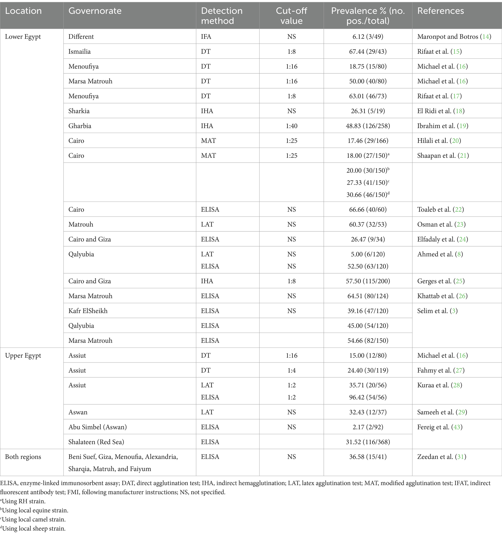
Table 1. Seroprevalence of Toxoplasma gondii reported in dromedary camels (Camelus dromedaries) in Egypt.
2 Materials and methods
2.1 Study area
A cross-sectional study was conducted in the two major governorates of Egypt, namely Cairo and Aswan, representing Lower and Upper Egypt, respectively (Figure 1). Cairo governorate serves as the capital and largest city of the country. The total area is 3,048.676 km2, the inhabited space is 188.982 km2, located near the Nile Delta extends 25 km on the western bank of the Nile River in Egypt at 30° 02′ 30″ N, 031° 14′ 07″ E. Egypt’s capital has a mild-to-hot climate for most of the year, with maximum temperatures around 34°C in summer and 18°C in winter. Aswan governorate covers a total area of 62,726 km2, with an inhabited area of 12,203 km2. It is located in southern Egypt at coordinates 24° 5′ 20.1768″ N and 32° 53′ 59.3880″ E, just north of the Aswan Dam on the east bank of the Nile near the first cataract. The climate of Aswan is very hot and dry in summer and might exceed 41°C, while winter is relatively mild with an average of 26°C and dry and rainfall is non-existent except sometimes in the month of August. Rainfall in Egypt is very low across the country, with an average annual precipitation of less than 80 mm, occurring primarily during the winter months.
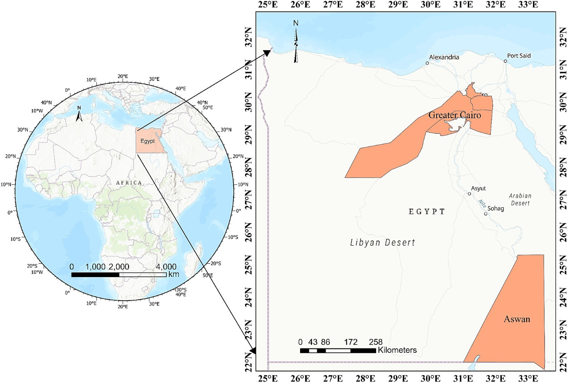
Figure 1. A map of Egypt delineating the geographical locations where camel samples were collected in Upper and Lower Egypt.
2.2 Animals and samples
Between January and May 2023, 513 blood samples were randomly collected from apparently healthy dromedary camels from different regions of Lower and Upper Egypt. Samples were collected from local markets and veterinary campaigns. A total of 513 samples were collected from two regions: 256 from Cairo and 257 from the Aswan governorate. The age of animals ranged from 3 to 15 years, with a median age of 7 years, and were primarily used for fieldwork and meat production. A total of 10 mL blood samples were collected from each animal by puncturing the jugular vein. Plain tubes without anticoagulant were used, along with sterile syringe needles, for the collection process. All samples were kept in portable coolers containing polyethylene ice packs then sent to the laboratories of Zoonotic diseases at National Research Centre (Egypt), for samples collected from Cairo governorate, and Department of Food Hygiene, Aswan University for samples from Aswan governorate. Sera were obtained by centrifugation at 1,107 rcf for 15 min then the supernatant was transferred to a new Eppendorf tube and kept at −20°C until analysis. Data regarding the age, sex, and region of each animal were collected, whenever possible. The ages of the examined camels were estimated and documented based on information provided by their owners and an assessment of the animals’ dentition (32, 33). Camels were classified into three age categories: young (≤4 years old), adults (between 4 and 8 years old), and elders (>8 years old), as described in previous studies (3, 13).
2.3 Serological assessment
Serum samples were serologically examined for the presence of anti-T. gondii antibodies, specifically IgG, using modified agglutination test (MAT), which employs formalin-fixed tachyzoites as described elsewhere (34, 35). Antigen of T. gondii was supplied by NRC on toxoplasmosis (Reims). Sera with titers ≥1:25, were considered positive, which serves as the established cut-off for T. gondii seropositivity, as previously considered for these animal species (36, 37). Sera that initially tested positive at a dilution of ≥1:25 were subsequently retested at dilutions of 1:25, 1:50, 1:100, and ≥1:500.
2.4 Statistical analysis
To assess the seroprevalence of T. gondii, we computed the proportion of seropositive samples relative to the total number of camels examined, accompanied by a 95% confidence interval (95%CI). Associations between explanatory variables [age (categorized as ≤4 years, 4–8 years, and >8 years), sex (male and female), and region (Upper and Lower Egypt)] and serological results were analyzed with the use of a Pearson’s chi-square test and by Fisher’s exact test when observations/category were < 6. Variables with a p-value below 0.10 were selected to be included in the multivariate analysis. Collinearity between pairs of variables was tested using the Cramer’s V, selecting the variable with the highest biological plausibility. Finally, a multiple logistic regression was carried out to study the effect of potential explanatory variables previously selected in the bivariate analysis (38). All analyses were conducted using SPSS software, version 25.0® (Statistical Package for the Social Sciences, Inc., Chicago, IL, United States), and statistical significance was set at p-value < 0.05.
3 Results
Anti-T. gondii antibodies were detected in 71 (13.84%; 95CI%:10.85–16.83) of the 513 camels analyzed. Seropositive animals exhibited varying anti-T. gondii antibodies titers: 1:25 in 14 animals (2.73%), 1:50 in 11 (2.14%), 1:100 in 28 (5.46%), and ≥1:500 in 18 (3.51%). The distribution of T. gondii seropositivity according to region, age and sex is shown in Table 2. The prevalence of anti-T. gondii antibodies was significantly higher in camels from Lower Egypt (19.92%; 51/256; 95 CI%: 15.03–24.81) compared to those from Upper Egypt (7.78%; 20/257; 95CI%: 4.51–11.06) (p < 0.001). Significantly higher seropositivity was also found in older (18.45%; 31/168) and adult (13.84%, 36/260) animals compared to young individuals (4.70%, 4/85) (p = 0.010). The multivariate analysis identified geographic region as a potential risk factor for T. gondii exposure, with significantly higher seropositivity observed in camels from northern Egypt compared to those from southern Egypt (p < 0.001, OR = 2.94; 95% CI: 1.70–5.10).
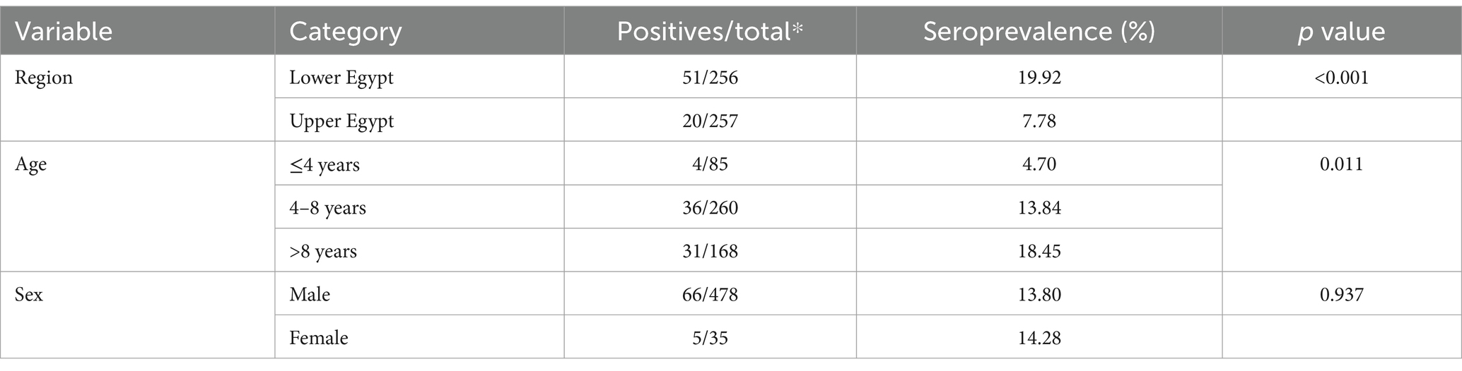
Table 2. Univariable analysis of risk factors associated to T. gondii infection in camels.
4 Discussion
Toxoplasmosis presents significant clinical and economic challenges in both human and animal health. In humans, it typically manifests with flu-like symptoms, including fever, lymphadenopathy, and ocular issues. The infection can also lead to multiple neurological and reproductive disorders (39). While in livestock, it is a leading cause of abortion, stillbirth, and weak offspring, all of which critically impact productivity and economic sustainability (40). Given this significant medical and veterinary implications, regular and periodic updates of seroepidemiological data on T. gondii among different reservoirs are pivotal for establishing a baseline for its control. To the best of the authors knowledge, the current study represents the most extensive serological investigation conducted among dromedary camel populations in Egypt. The overall seroprevalence obtained (13.84%) falls within the values previously reported in this species in Egypt (Table 1). Nevertheless, comparisons among studies should be made with caution given the differences in number of animals examined, serological methods employed and management and environmental factors.
In the present work, we determine the seroprevalence of T. gondii in the northern and southern part of Egypt, evidencing a heterogeneous spatial distribution and exposure to this parasite across the country. The risk factor analysis revealed that camels from Lower Egypt had a 2.9-fold higher risk of T. gondii exposure compared to those in Upper Egypt, suggesting greater circulation of this parasite in the northern region of the country. As depicted, significantly higher seroprevalence was found in camels from Lower Egypt (19.92%) compared to the populations from Upper Egypt (7.78%). It is worth emphasizing that a greater number of studies have been conducted in Lower Egypt, and the prevalence of anti-T. gondii antibodies identified in our study aligns with the rates previously reported in this region (Table 1), although levels varied widely among studies. Using the same diagnostic method as the present study (MAT), similar seroprevalence value (17.46%; 29/166) was observed in a previous study (20) carried out in Lower Egypt (20). Another study assessed the potential efficacy of four antigen strains in the serodiagnosis of toxoplasmosis in 150 camels from Cairo using MAT and observed that the use of the T. gondii RH strain, and a local strain isolated from an equine provided seroprevalence rates of 18.00 and 20.66%, respectively, while a local camel and sheep strains provided seroprevalence rates of 27.33% (41/150) and 30.66% (46/150), respectively (21). On the other hand, several studies performed on a significant number of samples (more than 100 camel samples) using different serological methods, showed high seroprevalence levels (up to 64.51%) in camels in Lower Egypt (3, 21, 25, 26) (Table 1). In this respect, previous research using ELISA on camel blood samples in Lower Egypt revealed an overall seroprevalence rate of 46.9%, with specific rates of 39.16% (47/120) in Kafr El Sheikh, 45.00% (54/120) in Qalyubia, and 54.66% (82/150) in Marsa Matrouh (3). Another previous study conducted in Qalyubia governorate in Lower Egypt reported a high seroprevalence of 52.50% (63/120) using ELISA (8). In contrast, the same study (8) reported lower seroprevalence rates of 5.00% (6/120) for T. gondii in the same governorate using latex agglutination test (LAT) and indirect fluorescent antibody test (IFAT). The previously mentioned results showed important variations related to the method of serological test used. Among other techniques, MAT stands out as one of the most recommended for detection of T. gondii infection in both animals and humans (41). The test offers several advantages over other serological methods and is widely utilized due to its reliability and efficacy in detecting antibodies against the parasite. Additionally, MAT is recognized for its simplicity, cost-effectiveness, relatively high accuracy, and high sensitivity, making it a commonly used technique for serological detection of the parasite in different species, including camels (21).
It should be stressed that few studies have been conducted in Upper Egypt compared to Lower Egypt so far. Furthermore, none of the studies carried out in Upper Egypt used the MAT to detect antibodies against the parasite. The lowest T. gondii seroprevalence (0.81% 368 samples) in Upper Egypt was reported in the Red Sea governorate using ELISA (42). However, some studies, conducted in Aswan governorate, in Upper Egypt, on a smaller number of animals reported higher seroprevalence values (16, 26–28). In contrast to our findings, two previous studies (29, 30) on camels in Aswan governorate investigated the seroprevalence of T. gondii among a total of 37 and 92 animals, reporting seroprevalence rates of 16.30 and 32.43%, respectively, using LAT and ELISA (29, 30). Moreover, a serosurvey conducted on imported camels from Sudan, kept under two regions of Upper Egypt (Red Sea and Aswan), being distributed to other cities throughout the country, revealed significant differences in the seroprevalence between the two study sites (43). In this former study (43), a significantly higher seropositivity to T. gondii was recorded in the Red Sea camels (31.5%, 116/368) than in those sampled in Aswan (2.2%, 2/92). Two additional studies conducted among camels from Assiut governorate (Upper Egypt) documented higher prevalence rates of 15.00% (12/80) and 24.40% (30/119) for anti-T. gondii antibodies, using DT and LAT, respectively (16, 27). Another previous research (28) conducted on 56 camels in Assiut governorate (Upper Egypt) reported higher seroprevalence rates of 35.71 and 96.42% for T. gondii using LAT and ELISA, respectively (28).
Taken into consideration, various contributing factors could involve discrepancies in the spatial distribution of the parasite observed in the present work compared to previous studies at national or international levels. These factors include variations in environmental and climatic conditions, livestock management practices, and biosecurity measures. In certain governorates, camel management is based on traditional, extensive grazing with limited biosecurity measures. Meanwhile, others employ more commercial, urban-based systems with stricter biosecurity protocols, supported by better access to veterinary care and closer proximity to markets. Additionally, differences in the density of definitive hosts, sample sizes, serological methods, and the thresholds and sensitivities of tests and various stress factors that include grazing restrictions, limited movement, temperature changes, water availability, and nutrition further influence these variations (13, 40, 44–46). Meanwhile, discrepancies in seroprevalence values between the two study areas analyzed in the present study may be influenced by several factors including climatic variations encompassing temperature and humidity. In this regard, Lower Egypt typically experiences relatively lower temperatures and higher humidity compared to Upper Egypt (47). These ecological factors may favor the persistence of viable oocysts in the environment, thereby increasing the likelihood of exposure to the parasite (48, 49). Moreover, the higher density of definitive hosts, particularly cats, in the densely populated regions of Lower Egypt, especially Cairo, likely increases the risk of T. gondii exposure to other sympatric species including camels. In this respect, a serological study of stray cats in Cairo found an extremely high seroprevalence 95.55% of 180 cats analyzed by MAT (50). In contrast, there are no prior reports on the seroprevalence of T. gondii in cats in Upper Egypt, highlighting a significant gap in the research.
It is important to note that, although the age was not retained in the multivariate analysis, the bivariate analysis showed a significantly higher prevalence of T. gondii antibodies in camels over 4 years of age (Table 2), indicating a strong trend of increased seropositivity with advancing age. This finding aligns with various previous reports from Egypt (3, 26) and Ethiopia (51), reported higher seroprevalence among camels aged 8 years or older compared to younger ones. In contrary, other previous reports displayed no significant relation between the age of the camels and the seropositivity of T. gondii as (42). Considering that this trend has been observed across all mammals, this finding aligns with those previously observed in this species that reflects a cumulative likelihood for exposure to T. gondii and lifelong persistence of antibodies (3, 26, 51). It should be noted that adult animals, having lived longer, are exposed to a wider array of infection sources compared to younger animals, accounting for the higher seroprevalence rates observed in older animals (52, 53). Collectively, the above-mentioned findings suggest that horizontal transmission might be considered the main route of T. gondii infection in camels.
5 Conclusion
The current study offers updated insights into the exposure of dromedary camels to T. gondii across Upper and Lower Egypt. Our findings revealed a widespread circulation and heterogenous spatial distribution of this protozoan in the camel populations in this country which might have important health implication for this species. While the presence of antibodies against T. gondii does not confirm whether the host harbors viable parasites, the findings of this study highlight the potential risk of zoonotic transmission from camels, particularly through the consumption of raw or undercooked camel milk and meat. Further large-scale serosurvey and molecular investigations are warranted to assess the role of this species in the epidemiology of T. gondii in Egypt. Control measures should be implemented to minimize the risk of camel exposure to this zoonotic parasite. Along with raising public health awareness, these measures should encompass the application of proper farm management practices, preventing contamination of feed and water by T. gondii-infected cats, the control of stray cat populations and their access to animals premisses, and conducting regular testing of animals at large scale level.
Data availability statement
The original contributions presented in the study are included in the article/supplementary material, further inquiries can be directed to the corresponding authors.
Ethics statement
The animal studies were approved by the Institutional Review Board (Ethics Committee) of Faculty of Veterinary Medicine at Aswan University (Egypt) granted approval for this study on [Approval number 11/2022/012]. The studies were conducted in accordance with the local legislation and institutional requirements. Written informed consent was obtained from the owners for the participation of their animals in this study.
Author contributions
EE: Conceptualization, Data curation, Formal analysis, Funding acquisition, Investigation, Methodology, Project administration, Resources, Software, Supervision, Validation, Visualization, Writing – original draft, Writing – review & editing. NE: Data curation, Formal analysis, Methodology, Software, Writing – review & editing. DC-T: Conceptualization, Data curation, Formal analysis, Software, Validation, Visualization, Writing – review & editing. TF: Data curation, Formal analysis, Methodology, Software, Validation, Writing – review & editing. NA: Data curation, Formal analysis, Funding acquisition, Software, Validation, Writing – review & editing. DJ-M: Formal analysis, Software, Validation, Writing – review & editing. MG: Data curation, Formal analysis, Funding acquisition, Methodology, Software, Validation, Visualization, Writing – review & editing. AG: Data curation, Investigation, Methodology, Validation, Visualization, Writing – original draft, Writing – review & editing. RR: Data curation, Formal analysis, Software, Validation, Writing – review & editing. IV: Data curation, Formal analysis, Software, Visualization, Writing – review & editing. SS: Data curation, Methodology, Writing – review & editing. HaA: Data curation, Funding acquisition, Resources, Software, Validation, Writing – review & editing. HiA: Data curation, Formal analysis, Funding acquisition, Resources, Software, Validation, Visualization, Writing – review & editing. SA: Data curation, Formal analysis, Software, Validation, Visualization, Writing – review & editing. IG-B: Conceptualization, Data curation, Formal analysis, Funding acquisition, Investigation, Project administration, Resources, Software, Supervision, Validation, Visualization, Writing – original draft, Writing – review & editing.
Funding
The author(s) declare financial support was received for the research, authorship, and/or publication of this article. This work was partially financed by CIBER -Consorcio Centro de Investigación Biomédica en Red- (CB 2021), Instituto de Salud Carlos III, Ministerio de Ciencia e Innovación and Unión Europea -NextGenerationEU. EE was supported by a postdoctoral contract María Zambrano (University of Córdoba) from the Program of Requalification of the Spanish University System (Spanish Ministry of Universities) financed by the European Union-NextGenerationEU. DJ-M was supported by an FPU grant from the Spanish Ministry of Universities (FPU22/03649). This study was supported by Princess Nourah bint Abdulrahman University Researchers Supporting Project No. (PNURSP2025R401), Princess Nourah bint Abdulrahman University, Riyadh, Saudi Arabia.
Acknowledgments
We would like to acknowledge the Princess Nourah bint Abdulrahman University Researchers Supporting Project no. PNURSP2025R401, Princess Nourah bint Abdulrahman University, Riyadh, Saudi Arabia.
Conflict of interest
The authors declare that the research was conducted in the absence of any commercial or financial relationships that could be construed as a potential conflict of interest.
The reviewer FD declared a past co-authorship with the author IV to the handling editor.
The author(s) declared that they were an editorial board member of Frontiers, at the time of submission. This had no impact on the peer review process and the final decision.
Generative AI statement
The authors declare that no Generative AI was used in the creation of this manuscript.
Publisher’s note
All claims expressed in this article are solely those of the authors and do not necessarily represent those of their affiliated organizations, or those of the publisher, the editors and the reviewers. Any product that may be evaluated in this article, or claim that may be made by its manufacturer, is not guaranteed or endorsed by the publisher.
References
1. de Barros, RAM, Torrecilhas, AC, Marciano, MAM, Mazuz, ML, Pereira-Chioccola, VL, and Fux, B. Toxoplasmosis in human and animals around the world. Diagnosis and perspectives in the one health approach. Acta Trop. (2022) 231:106432. doi: 10.1016/j.actatropica.2022.106432
2. Skariah, S, McIntyre, MK, and Mordue, DG. Toxoplasma gondii: determinants of tachyzoite to bradyzoite conversion. Parasitol Res. (2010) 107:253–60. doi: 10.1007/s00436-010-1899-6
3. Selim, A, Marawan, MA, Abdelhady, A, and Wakid, MH. Seroprevalence and potential risk factors of toxoplasma gondii in dromedary camels. Agriculture. (2023) 13:129. doi: 10.3390/AGRICULTURE13010129
4. Tenter, AM, Heckeroth, AR, and Weiss, LM. Toxoplasma gondii: from animals to humans. Int J Parasitol. (2000) 30:1217–58. doi: 10.1016/s0020-7519(00)00124-7
5. Fatima, T, Mehnaz, S, Wang, M, Yang, J, Sajid, MS, Shen, B, et al. Seroprevalence of toxoplasma gondii in one-humped camels (Camelus dromedarius) of Thal and Cholistan deserts, Punjab, Pakistan. Parasitol Res. (2019) 118:307–16. doi: 10.1007/s00436-018-6124-z
6. Abdel-Aziem, SH, Mabrouk, DM, Abd El-Kader, HA, Alam, SS, and Othman, OE. Genetic similarity and diversity among three camel populations reared in Egypt. J Genet Eng Biotechnol. (2022) 20:154. doi: 10.1186/s43141-022-00435-z
7. Khalafalla, AI. Zoonotic diseases transmitted from the camels. Front Vet Sci. (2023) 10:1244833. doi: 10.3389/FVETS.2023.1244833
8. Ahmed, NE, al–Akabway, L, Ramadan, M, Abd el-Gawad, S, and Moustafa, M. Serological and PCR-sequencing assays for diagnosis of toxoplasma gondii and Neospora caninum infecting camels in Egypt. Benha Vet Med J. (2017) 33:200–10. doi: 10.21608/BVMJ.2017.30466
9. Ahmed, MA, Elmahallawy, EK, Gareh, A, Abdelbaset, AE, El-Gohary, FA, Elhawary, NM, et al. Epidemiological and histopathological investigation of Sarcoptic mange in camels in Egypt. Animals (Basel). (2020) 10:1–11. doi: 10.3390/ANI10091485
10. Hagemoser, WA, Dubey, JP, and Thompson, JR. Acute toxoplasmosis in a camel. J Am Vet Med Assoc. (1990) 196:347. doi: 10.2460/javma.1990.196.02.347
11. Riley, J, Garner, MM, Kiupel, M, and Hammond, EE. Disseminated toxoplasmosis in a captive adult dromedary camel (Camelus dromedarius). J Zoo Wildl Med. (2017) 48:937–40. doi: 10.1638/2016-0057.1
12. Manal, YI, and Maijid, AM. Association of diarrhea with congenital toxoplasmosis in calf-camel (Camelus dromedarius). Int J Trop Med. (2008) 3:10–1.
13. Gebremedhin, EZ, Yunus, HA, Tesfamaryam, G, Tessema, TS, Dawo, F, Terefe, G, et al. First report of toxoplasma gondii in camels (Camelus dromedarius) in Ethiopia: bioassay and seroepidemiological investigation. BMC Vet Res. (2014) 10:222. doi: 10.1186/S12917-014-0222-7
14. Maronpot, RR, and Botros, BA. Toxoplasma serologic survey in man and domestic animals in Egypt. J Egypt Public Health Assoc. (1972) 47:58–67.
15. Rifaat, MA, Morsy, TA, Sadek, MSM, Khalid, MLM, Azab, ME, Makled, MK, et al. Incidence of toxoplasmosis among farm animals in Suez Canal governorates. J Egypt Soc Parasitol. (1977) 7:135–40.
16. Michael, SA, El Refaii, AH, and Morsy, TA. Incidence of toxoplasma antibodies among camels in Egypt. J Egypt Soc Parasitol. (1977) 7:129–32.
17. Rifaat, MA, Morsy, TA, Sadek, MSM, Khalid, MLM, Azab, ME, and Safar, EH. Prevalence of toxoplasma antibodies among slaughtered animals in lower Egypt. J Egypt Soc Parasitol. (1978) 8:339–45.
18. El Ridi, AM, Nada, SM, Aly, AS, Habeeb, SM, and Aboul-Fattah, MM. Serological studies on toxoplasmosis in Zagazig slaughterhouse. J Egypt Soc Parasitol. (1990) 20:677–81.
19. Ibrahim, BB, Salama, MM, Gawish, NI, and Haridy, FM. Serological and histopathological studies on toxoplasma gondii among the workers and the slaughtered animals in Tanta abattoir, Gharbia Governorate. J Egypt Soc Parasitol. (1997) 27:273–8.
20. Hilali, M, Romand, S, Thulliez, P, Kwok, OC, and Dubey, JP. Prevalence of Neospora caninum and toxoplasma gondii antibodies in sera from camels from Egypt. Vet Parasitol. (1998) 75:269–71. doi: 10.1016/s0304-4017(97)00181-7
21. Shaapan, RM, and Khalil, FAM. Evaluation of different toxoplasma gondii isolates as antigens used in the modified agglutination test for the detection of toxoplasmosis in camels and donkeys. J Agric Environ Sci. (2008) 3:837–41.
22. Toaleb, NI, Shaapan, RM, Hassan, SE, and El-Moghazy, FM. High diagnostic efficiency of affinity isolated fraction in camel and cattle toxoplasmosis. World Med Sci J. (2013) 8:61–6. doi: 10.5829/idosi.wjms.2013.8.1.72161
23. Osman, AO, El-Metwaly, HA, Wahba, AA, and Hefny, SF. Studies on causes of abortion in Maghrabian camels. Egypt J Agric Res. (2016) 94:955–67. doi: 10.21608/ejar.2016.153236
24. Elfadaly, HA, Hassanain, NA, Shaapan, RM, Hassanain, MA, Barakat, AM, and Abdelrahma, KA. Molecular detection and genotyping of toxoplasma gondii from Egyptian isolates. Asian J Epidemiol. (2016) 10:37–44. doi: 10.3923/aje.2017.37.44
25. Gerges, AA, Hassanien, TK, and Abdel-Malak, MG. Pathological studies on the effect of toxoplasma gondii on the genitalia of dromedary camels in some governorates of Egypt. J Egypt Vet Med Assoc. (2018) 78:345–67.
26. Khattab, RAH, Barghash, SM, Mostafa, OMS, Allam, SA, Taha, HAH, and Ashour, AAEB. Seroprevalence and molecular characterization of toxoplasma gondii infecting ruminants in the north-west of Egypt. Acta Trop. (2022) 225:106139. doi: 10.1016/J.ACTATROPICA.2021.106139
27. Fahmy, MA, Mandour, AM, Arafa, MS, and Abdel Rahman, BM. Toxoplasmosis of camels in Assiu governorate. J Egypt Vet Med Assoc. (1979) 39:27–31.
28. Kuraa, HM, and Malek, SS. Seroprevalence of toxoplasma gondii in ruminants by using latex agglutination test (LAT) and enzyme-linked immunosorbent assay (ELISA) in Assiut governorate. Trop Biomed. (2016) 33:711–25.
29. Sameeh, S, Mahmoud, AE, El-Salahy, M, Monib, MM, and Eldeek, HEM. Latex agglutination test and PCR assays for diagnosis of toxoplasma gondii infection in red meat producing animals in Aswan governorate, southern Egypt. Slov Vet Res. (2021) 58:281–8. doi: 10.26873/SVR-1447-2021
30. Fereig, RM, Wareth, G, Abdelbaky, HH, Mazeed, AM, El-Diasty, M, Abdelkhalek, A, et al. Seroprevalence of specific antibodies to toxoplasma gondii, Neospora caninum, and Brucella spp. in sheep and goats in Egypt. Animals (Basel). (2022) 12:3327. doi: 10.3390/ani12233327
31. Zeedan, GSG, Abdalhamed, AM, Shaapan, RM, and El-Namaky, AH. Rapid diagnosis of toxoplasma gondii using loop-mediated isothermal amplification assay in camels and small ruminants. Beni Suef Univ J Basic Appl Sci. (2022) 11:1–10. doi: 10.1186/s43088-021-00184-x
32. Food and Agriculture Organization of the United Nations (FAO). Camels; llamas and alpacas In: A manual for the primary animal health care workers. Rome: FAO (1994)
33. Misk, NA, Youssef, HA, Semeika, MM, and El-Khabery, AH. History of dentition in camels (Camelus Dromedarius) In: Proceedings of the XXXVII international congress of the world Association for the History of veterinary medicine & XII Spanish National Congress on the veterinary history. León: (2006). 535–40.
34. Dubey, JP, and Desmonts, G. Serological responses of equids fed toxoplasma gondii oocysts. Equine Vet J. (1987) 19:337–9. doi: 10.1111/j.2042-3306.1987.tb01426.x
35. Dubey, JP. Sarcocystis neurona, Neospora spp. and toxoplasma gondii infections in horses and equine protozoal myeloencephalitis (EPM): five decades of personal experience, perspectives, and update. Parasitology. (2022) 149:1–44. doi: 10.1017/S0031182021002055
36. Dubey, JP, Murata, FHA, Cerqueira-Cézar, CK, and Kwok, OCH. Toxoplasma gondii infections in horses, donkeys, and other equids: the last decade. Res Vet Sci. (2020) 132:492–9. doi: 10.1016/j.rvsc.2020.07.005
37. Cano-Terriza, D, Almería, S, Caballero-Gómez, J, Jiménez-Martín, D, Castro-Scholten, S, Dubey, JP, et al. Exposure to toxoplasma gondii in zoo animals in Spain. Prev Vet Med. (2020) 176:104930. doi: 10.1016/j.prevetmed.2020.104930
38. Hosmer, DWLS. Assessing the fit of the model In: Applied logistic regression. Hoboken, NJ: Wiley (2000). 143–202.
39. Dubey, JP. Outbreaks of clinical toxoplasmosis in humans: five decades of personal experience, perspectives and lessons learned. Parasit Vectors. (2021) 14:263. doi: 10.1186/s13071-021-04769-4
40. Stelzer, S, Basso, W, Benavides Silván, J, Ortega-Mora, LM, Maksimov, P, Gethmann, J, et al. Toxoplasma gondii infection and toxoplasmosis in farm animals: risk factors and economic impact. Food Waterborne Parasitol. (2019) 15:e00037. doi: 10.1016/J.FAWPAR.2019.E00037
41. Fernandes, S, Brilhante-Simões, P, Coutinho, T, Cardoso, L, Dubey, JP, and Lopes, AP. Comparison of indirect and modified agglutination tests for detection of antibodies to toxoplasma gondii in domestic cats. J Vet Diagn Invest. (2019) 31:774–7. doi: 10.1177/1040638719868753
42. Yektaseresht, A, Ghane, M, Atashbar, F, and Aliabadi, J. Serological survey of toxoplasma gondii infection in the one-humped camels (Camelus dromedarius) in the south of Iran. Vet Med Sci. (2023) 9:2386–9. doi: 10.1002/VMS3.1240
43. Fereig, RM, Abdelbaky, HH, El-Alfy, ES, El-Diasty, M, Elsayed, A, Mahmoud, HYAH, et al. Seroprevalence of toxoplasma gondii and Neospora caninum in camels recently imported to Egypt from Sudan and a global systematic review. Front Cell Infect Microbiol. (2022) 12:1042279. doi: 10.3389/FCIMB.2022.1042279
44. Dubey, JP, and Jones, JL. Toxoplasma gondii infection in humans and animals in the United States. Int J Parasitol. (2008) 38:1257–78. doi: 10.1016/J.IJPARA.2008.03.007
45. Robertson, LJ. Parasites in food: occurrence and detection In: Encyclopedia of Food and Health (2016). 219–24.
46. Huertas-López, A, Cantos-Barreda, A, Sánchez-Sánchez, R, Martínez-Carrasco, C, Ibáñez-López, FJ, Martínez-Subiela, S, et al. A systematic review and meta-analysis of the validation of serological methods for detecting anti-toxoplasma gondii antibodies in humans and animals. Vet Parasitol. (2024) 328:110173. doi: 10.1016/J.VETPAR.2024.110173
47. El Kenawy, AM, Lopez-Moreno, JI, McCabe, MF, Robaa, SM, Domínguez-Castro, F, Peña-Gallardo, M, et al. Daily temperature extremes over Egypt: spatial patterns, temporal trends, and driving forces. Atmos Res. (2019) 226:219–39. doi: 10.1016/J.ATMOSRES.2019.04.030
48. Yan, C, Liang, L-J, Zheng, K-Y, and Zhu, X-Q. Impact of environmental factors on the emergence, transmission and distribution of toxoplasma gondii. Parasit Vectors. (2016) 9:137. doi: 10.1186/s13071-016-1432-6
49. Patz, JA, Graczyk, TK, Geller, N, and Vittor, AY. Effects of environmental change on emerging parasitic diseases. Int J Parasitol. (2000) 30:1395–405. doi: 10.1016/S0020-7519(00)00141-7
50. Al-Kappany, YM, Lappin, MR, Kwok, OCH, Abu-Elwafa, SA, Hilali, M, and Dubey, JP. Seroprevalence of toxoplasma gondii and concurrent Bartonella spp., feline immunodeficiency virus, feline leukemia virus, and Dirofilaria immitis infections in Egyptian cats. J Parasitol. (2011) 97:256–8. doi: 10.1645/GE-2654.1
51. Gebremedhin, EZ, Dima, N, Beyi, AF, Dawo, F, Feyissa, N, Jorga, E, et al. Toxoplasmosis in camels (Camelus dromedarius) of Borana zone, Oromia region, Ethiopia: seroprevalence and risk factors. Trop Anim Health Prod. (2016) 48:1599–606. doi: 10.1007/S11250-016-1133-3
52. Katzer, F, Brülisauer, F, Collantes-Fernández, E, Bartley, PM, Burrells, A, Gunn, G, et al. Increased toxoplasma gondii positivity relative to age in 125 Scottish sheep flocks; evidence of frequent acquired infection. Vet Res. (2011) 42:1–9. doi: 10.1186/1297-9716-42-121/FIGURES/4
Keywords: Toxoplasma gondii , camels, serosurvey, modified agglutination test, Egypt
Citation: Elmahallawy EK, Elbarbary NK, Cano-Terriza D, Fajardo T, Albalawi NO, Jiménez-Martín D, Ghallab MMI, Gareh A, Ras R, Villena I, Sadek SAS, AlQadeeb H, Alzaylaee H, Almería S and García-Bocanegra I (2025) Toxoplasma gondii in dromedary camels (Camelus dromedarius) in Egypt: a comparative seroepidemiological study in Upper and Lower Egypt. Front. Vet. Sci. 11:1508496. doi: 10.3389/fvets.2024.1508496
Edited by:
Calin Mircea Gherman, University of Agricultural Sciences and Veterinary Medicine of Cluj-Napoca, RomaniaReviewed by:
Adriana Gyorke, University of Agricultural Sciences and Veterinary Medicine of Cluj-Napoca, RomaniaFilip Dámek, Helmholtz Association of German Research Centres (HZ), Germany
Copyright © 2025 Elmahallawy, Elbarbary, Cano-Terriza, Fajardo, Albalawi, Jiménez-Martín, Ghallab, Gareh, Ras, Villena, Sadek, AlQadeeb, Alzaylaee, Almería and García-Bocanegra. This is an open-access article distributed under the terms of the Creative Commons Attribution License (CC BY). The use, distribution or reproduction in other forums is permitted, provided the original author(s) and the copyright owner(s) are credited and that the original publication in this journal is cited, in accordance with accepted academic practice. No use, distribution or reproduction is permitted which does not comply with these terms.
*Correspondence: Ehab Kotb Elmahallawy, c2EyZWxlbGVAdWNvLmVz; Ignacio García-Bocanegra, djYyZ2FyYm9AdWNvLmVz