 Tamara Ricardo
 Lucía Isabel Azócar-Aedo
 María Andrea Previtali1,2
 Gustavo Monti
- 1Consejo Nacional de Investigaciones Científicas y Técnicas (CONICET), Santa Fe, Argentina
- 2Dpto. de Ciencias Naturales, Facultad de Humanidades y Ciencias (FHUC), Universidad Nacional del Litoral (UNL), Santa Fe, Argentina
- 3Escuela de Medicina Veterinaria, Facultad de Ciencias de la Naturaleza, Universidad San Sebastián, Sede de la Patagonia, Puerto Montt, Chile
- 4Quantitative Veterinary Epidemiology Group, Wageningen University and Research, Wageningen, Netherlands
Leptospirosis is a neglected zoonotic disease transmitted by contact with the urine of animals infected with pathogenic species of the bacteria Leptospira or by contact with environments contaminated with the bacteria. Domestic dogs and cats may act as reservoirs or as sentinels of environmental contamination with leptospires, posing a public health concern. There is a great diversity of leptospires, and one common way to classify them is into serogroups that provide some information on the host species they are associated with. The aims of this study were: (1) to quantitatively summarize the overall prevalence and serogroup-specific prevalence of antibodies against pathogenic leptospires in asymptomatic dogs and cats and (2) to identify environmental and host characteristics that may affect the prevalence. Three electronic databases and the reference lists of eligible articles were screened, for epidemiological studies conducted between the years 2012–2022. We estimated overall and serogroup-specific prevalence using three-level meta-analysis models and assessed potential sources of heterogeneity by moderator analysis and meta-regression. Eighty-four studies met the inclusion criteria (dog studies 66.7%, cat studies 26.2%, and both species 7.1%). There were significant differences between dogs and cats in the overall prevalence model (P < 0.001), but not in the serogroup-specific model (P>0.05). In dogs, the prevalence of Leptospira interrogans serogroup Canicola was significantly higher than the other pathogenic serogroups (P < 0.001), while in cats there were no significant differences among serogroups (P = 0.373). Moderator analysis showed that the prevalence of L. kirschneri serogroup Grippotyphosa was significantly higher in stray/sheltered dogs than in domiciled dogs (P = 0.028). These results suggest that pathogenic serogroups associated with small mammals are circulating among asymptomatic pets and should be taken into account in the transmission cycle of leptospires, as well as in the standard MAT panel for diagnosis in dogs and cats. It also highlights the importance of including both dogs and cats as potential reservoirs when conducting eco-epidemiological studies in different geographical and ecological areas.
1 Introduction
Emerging and re-emerging infectious diseases are mainly of zoonotic origin and have a major impact on public health and the global economy (1, 2). Leptospirosis is a neglected zoonosis caused by spirochetes of the genus Leptospira, which have a complex transmission cycle at the ecosystem interface between animals, humans, and the environment (2). Human infections occur through direct contact with urine from infectious animals or indirectly by exposure to soil or water contaminated by the urine of infected animals (3, 4). Pathogenic Leptospira spp. are generally endemic to certain regions and maintained by specific mammalian hosts, but can infect almost any animal species (3, 5, 6). These incidental infections are usually associated with acute clinical disease and limited renal excretion, whereas maintenance hosts often have subclinical infections and may shed leptospires in their urine for prolonged periods (3, 5). Given the complex transmission process, an effective and sustainable prevention and control strategy requires the application of the “One Health” approach (1, 2). This approach is particularly relevant to epidemiological studies in companion animals because of the close relationship between dogs, cats, and humans, the main route of transmission being urine-contaminated soil or water, and the influence of environmental conditions on bacterial survival, increasing the risk of shared exposure to pathogenic Leptospira spp. (3, 7).
Domestic dogs (Canis lupus familiaris) have traditionally been considered a maintenance host for Leptospira interrogans serovar Canicola, but there is evidence of severe disease in dogs infected with this serovar and reports of chronic infection and possible renal shedding of other commonly detected serovars (3, 5). Although the main clinical signs of leptospirosis in dogs reflect acute tubulointerstitial nephritis and liver dysfunction, the disease is multisystemic, with clinical signs of respiratory, intestinal, muscular, ocular, and reproductive problems, as well as coagulopathies (3). The risk of exposure to pathogenic leptospires in dogs is increased by unsupervised access to the outdoors, poor hygiene, and behaviors such as scavenging for litter or sniffing and licking urine from other dogs (3, 8). Despite this, further research on the serovars associated with both clinical and subclinical infections is still necessary.
Domestic cats (Felis silvestris catus) are also environmentally exposed to pathogenic leptospires due to their rodent hunting habits and, in many areas of the world, by their free-roaming lifestyle (9–12). Because cats are less likely to present clinical signs than dogs (3, 13), fewer studies have been conducted to investigate the clinical manifestations associated with all the serovars reported in this species or to understand the epidemiology of infection. However, some studies reported the presence of anorexia, dyspnea, chronic diarrhea, interstitial nephritis, and hepatitis in cats infected with pathogenic Leptospira spp. (14–18). The International Society of Companion Animal Infectious Diseases (ISCAID) statement indicated that the main serovars detected in this species are L. interrogans serovars Icterohaemorrhagiae, Canicola, Pomona, Bratislava, and Autumnalis; L. kirschneri serovar Grippotyphosa; and L. borgpetersenii serovars Hardjo and Ballum (6). However, there are still controversies on whether they are maintenance hosts or incidental hosts (3, 12, 19). In addition, recent epidemiological studies detected the DNA of pathogenic leptospires in the kidney and urine of domestic cats, indicating a potential role in environmental transmission (10, 20–25).
The care of companion animals and the closeness of human–pet relationships can increase the risk of zoonotic disease transmission (7, 26). For this reason, epidemiologic studies aimed at determining the frequency of presentation and prevalence of zoonoses in animal populations are of great relevance for detecting human and animal populations at risk (27) or differentiating risk areas for control purposes (28–30). Direct transmission of leptospires from dogs or cats to humans is a subject of controversy (3, 6, 12, 19). However, both species may act by zoonotic spillover from rodents or livestock to humans, as sentinels of environmental contamination with pathogenic leptospires, or contribute to contamination of the surroundings of human dwellings (3, 4); thus, there is a need to generate more evidence on this potential risk.
As the immunity induced by vaccination with current Leptospira bacterins is serogroup specific, it is important to recognize the serogroups that commonly cause disease in a given geographical region when designing a new vaccine or updating an existing one (3, 31, 32). Commercial leptospirosis vaccines for companion animals are currently available for dogs but not for cats (3, 6, 19). These vaccines generally contain a combination of L. interrogans serovars Canicola and Icterohaemorrhagiae, but the emergence of canine leptospirosis caused by other serovars has led to the development of multivalent vaccines containing L. interrogans serovars Pomona, Bratislava or Australis, and/or L. kirschneri serovar Grippotyphosa (3, 32). Furthermore, they are thought to provide protection for < 12 months (12, 33) and it is not clear whether commercial vaccines provide cross-protection against other serogroups not included in the formulation of inactivated bacterins (32). It is worth noting that animals vaccinated can present a reaction to serological tests even if they are not infected, which would interfere in serological surveys, for example, by overestimating the prevalence rate, or when validating serological tests affecting sensitivity and specificity estimations.
The microagglutination test (MAT) is the most widely used diagnostic technique for leptospirosis in veterinary practice. However, it has limitations in terms of sensitivity and specificity (3, 31). Several studies have used MAT to investigate asymptomatic infections in domestic dogs and cats, but there is no consensus on the most appropriate MAT titer, the minimum number of serovars and which serovars should be included in the panel, and how to interpret samples that react to more than one serovar. As a result, comparisons of apparent prevalence between studies should be made with caution. This study was motivated by trying to shed light on the numerous gaps in the epidemiology of feline and canine leptospirosis, despite the publication of numerous studies. Therefore, the aims of this study were: (a) to quantitatively summarize the overall prevalence and serogroup-specific prevalence of antibodies against pathogenic Leptospira in asymptomatic dogs and cats and (b) to identify environmental and host characteristics that may affect the seroprevalence.
2 Materials and methods
This meta-analysis was conducted following the Preferred Reporting Items for Systematic Reviews and Meta-Analyses (PRISMA) standards (34). The study protocol was registered into the PROSPERO International Prospective Register of Systematic Reviews with the code CRD4202230129.
2.1 Inclusion and exclusion criteria
Eligible reports were cross-sectional and cohort studies that reported the presence of antibodies against pathogenic Leptospira in domestic dogs (Canis lupus familiaris) or cats (Felis silvestris catus) and were published in peer-reviewed journals between January 2012 and December 2022. Publications written in English, Spanish, Portuguese, French, Italian, and German language were considered eligible. The studies had to use MAT to determine the presence of leptospiral antibodies in apparently healthy dogs and cats of both sexes and of all ages, whether they were house pets or stray animals, regardless of their vaccination status. Publications with null seroprevalence, articles where collection of samples was performed before 2010, and those that included animals with comorbidities or clinical suspicion of leptospirosis were excluded. In addition, conference abstracts, systematic reviews, gray literature, non-peer-reviewed publications, experimental research, case–control studies, ecological studies, case reports, and case series were also excluded. Later in the evaluation process, eligible studies were excluded if the complete text or pertinent data, such as the number of samples reacting to more than one serovar and their titers, were unavailable, and not made available after contacting the authors.
2.2 Search strategy
The electronic databases PubMed, Scopus, and Dimensions were used to search for eligible studies. The search strategy was based on the components: “Leptospira,” “leptospirosis,” “dogs,” “canine,” “cats,” “feline,” “antibodies,” “prevalence,” “seropositivity,” and “infection” and was customized according to the characteristics of each database (Table 1). A “snowball search” was carried out to identify additional studies from the reference lists of eligible publications and systematic reviews on leptospiral infection in domestic dogs or cats.
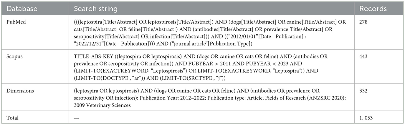
Table 1. Search strings and date limits used in the database literature search.
2.3 Study selection
Two review authors (TR and LAA) independently screened the titles and abstracts of the studies for eligibility, resolving any disagreement by consensus. Search results were screened and cleaned of duplicates using Mendeley Desktop® software; duplicates that were not automatically detected were removed manually.
2.4 Data extraction
Data were collected collaboratively using a Google Sheets® spreadsheet by the same two review authors who screened for eligible articles (TR and LAA). The outcomes considered as response variables were as follows: (1) the overall seroprevalence of leptospiral antibodies, estimated as the number of seropositive samples over the total number of samples and (2) the serogroup-specific seroprevalence, estimated as the number of seropositive samples for a pathogenic serogroup (including one or more frequent serovars) over the total number of samples. The data extraction spreadsheet included identifying variables (first author, title, journal, volume issue, ISSN, DOI, PMID), year of publication, the language of publication, year(s) of sampling, country of study, environmental setting (urban/peri-urban and rural), animals sampled (dogs and cats), the origin of animals (domiciled, sheltered, stray, and working dogs), MAT cut-off titer, the number of sampled animals, and the number of positive individuals for the overall population and per each detected serovar. Data on vaccination against Leptospira and serovars included in the vaccines were also collected for dog studies. Each row of the spreadsheet represented an effect estimate (k), and when a study presented results for different countries, sampling years, environmental settings, origins, vaccination status, or serovars, they were considered as separate effect estimates.
Free-roaming, unowned, dogs and cats were considered strays, regardless of their level of socialization with humans (20). Sheltered animals are stray dogs/cats housed in municipal or private shelters, which usually present overcrowding, poor hygienic conditions, and lack of veterinary care, increasing susceptibility to infections (35–37). Working dogs included those used by hunters to track feral pigs (Sus scrofa) and other wildlife, and dogs trained for herding, protecting properties, rescue operations, detecting drugs or explosives, or assisting people with disabilities (38–40). Each country was assigned to a geographic region and subregion based on the United Nations (UN) classification (https://unstats.un.org/unsd/methodology/m49/overview/). Information on the serovars used as antigens in the MAT panel, the number of serovars tested, the presence of coagglutinations or cross-reactions, and the individual risk-of-bias assessments were collected in a secondary spreadsheet.
2.5 Risk-of-bias assessment
Risk of bias (RoB) was assessed based on certain components of the STROBE-Vet statement (41). Each report was assessed independently by two reviewers (TR and LAA), with discrepancies resolved by consensus. Detailed information on the considered components can be found in Table 2. The overall RoB of each evaluation was calculated as the mean of the seven components and expressed as a percentage. The summary RoB was calculated as the mean of the overall RoB estimations from each reviewer. Scores ≤ 25% were considered high RoB, between 26% and 74% were considered medium RoB, and ≥75% were considered low RoB.
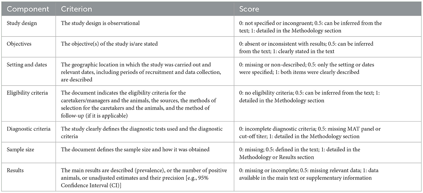
Table 2. Risk-of-bias (RoB) assessment tool for full-text articles based on seven components of the STROBE-Vet criteria (29).
2.6 Statistical analysis
The individual estimations of seroprevalence were log-transformed, and a continuity correction of 0.5 was used for events with probabilities of 0 or 1. When a study reported that serum samples agglutinated for more than one serovar, the serovar with the highest titer was considered responsible for the infection (28, 42). If MAT titers were not available or the sample reacted with equal titers to more than one serovar, they were considered coagglutinations and excluded from the serogroup-level analysis. Effect estimates reporting the prevalence of vaccine serovars in dogs with up-to-date vaccination were excluded, regardless of the cut-off titer used and the percentage of vaccinated dogs in the sample.
The frequencies of categorical variables were compared using either Pearson's chi-square test or Fisher's exact test, and the frequencies of numerical variables were compared using either ANOVA or Kruskal–Wallis ANOVA tests (43, 44). To account for the lack of independence among effect estimates from the same study, we fitted three-level random-effect meta-analysis models. These models allow the estimation of three sources of variance: variance from effect estimates, within-study heterogeneity (), and between-study heterogeneity (I2 level3) (45). All the model parameters were calculated using restricted maximum likelihood (REML). Results of the meta-analyses were back transformed as proportions and their 95% CI.
Potential sources of heterogeneity in the overall seroprevalence model were evaluated by moderator analyses. The categorical variables considered were environmental setting, the origin of the animals, vaccination status, and MAT cut-off titer. The sampling year was also considered as a continuous moderator for a meta-regression model. For the serogroup-specific seroprevalence models, moderator analysis was conducted by type of animal tested (dogs and cats) and serogroup. In addition, moderator analysis by origin of the animals and environmental setting was performed for serogroups detected in at least 20 dog studies. In the case of infrequent factor levels, similar categories were grouped and those with at least five observations were included in the subgroup analyses. Geographic regions and subregions were not considered for the moderator analysis due to the over-representation of studies from Latin America and the Caribbean. The presence of publication bias was assessed using Begg's test and Egger's test (46, 47). We performed a sensitivity analysis using a leave-one-out influence analysis, excluding the effect estimates with higher RoB (48). For the estimation of publication bias and sensitivity analysis, the models were refitted using two-level random-effects meta-analysis models. All the meta-analyses were performed in R software (49). Statistical significance was set at p-values below 0.05.
3 Results
3.1 Overview of the selected studies
A literature search retrieved 1, 053 records, of which 162 were related to the presence of leptospiral antibodies in dogs or cats. Of these, we included 76 studies that met the inclusion criteria and 8 additional studies identified from the “snowball search” (Figure 1). Another 30 potentially eligible articles were excluded because the full text was not available (n = 3) or presented incomplete data (n = 27) and were not made available after contacting the correspondence authors. Based on the 84 selected studies, 66.7% sampled only dogs, 26.2% only cats, and 7.1% both species. The median estimation of risk of bias was 0.89 (IQR: 0.86; 0.96) and ranged between 0.61 and 1, which indicates that RoB was low in most of the studies (92.9%, Supplementary Table S1). The median for publication year was 2018 (IQR: 2016; 2020). The sampling year was missing in 14 studies (16.7%). No significant differences were observed regarding average RoB, publication year, or sampling year among studies that sampled dogs, cats, or both species (P > 0.05). The selected studies were performed in 28 different countries and territories, 11 geographic subregions, and 5 geographic regions. More than half of the studies (56.0%) were conducted in Latin America and the Caribbean, 20.2% in Southern/Southeastern Asia, 8.3% in Southern Europe, 4.8% in Northern America, 3.6% in Sub-Saharan Africa, and 2.4% each in Eastern Asia, Northern/Eastern Europe, and Oceania. The countries with the highest number of publications were Brazil (92.9%), Malaysia (9.5%), and Mexico (7.1%).

Figure 1. PRISMA flowchart for identification and selection of articles. Flow diagram generated using PRISMA2020: R package and ShinyApp for producing PRISMA 2020 compliant flow diagrams (Version 0.0.1) (https://www.eshackathon.org/software/PRISMA2020.html).
3.2 Prevalence of antibodies against Leptospira in asymptomatic dogs and cats
We identified 77 effect estimates of seroprevalence in dogs (k dogs) and 36 effect estimates of seroprevalence in cats (k cats). The overall estimation of seroprevalence of Leptospira was 19.5% in dogs (95% CI: 16.0%; 23.5%, Figure 2) and 9.7% in cats (95% CI: 6.9%; 13.5%, Figure 2). Differences in the seroprevalence between dog studies and cat studies were statistically significant (P < 0.001). The model presented high statistical heterogeneity (I2: 94.2 %), and most of the variations corresponded to between-study heterogeneity ( = 60.8%, = 33.4%). The authors did not detect evidence of publication bias in the dog model (PBegg = 0.291, PEgger = 0.29), but Egger's test showed evidence of publication bias in the cat model (PBegg = 0.108, PEgger < 0.001). Sensitivity analysis did not find significant differences after removing the studies with higher RoB from the dog model (21.2%, 95% CI: 16.8%; 26.5%) or the cat model (9.1%, 95% CI: 6.9%; 12.0%).
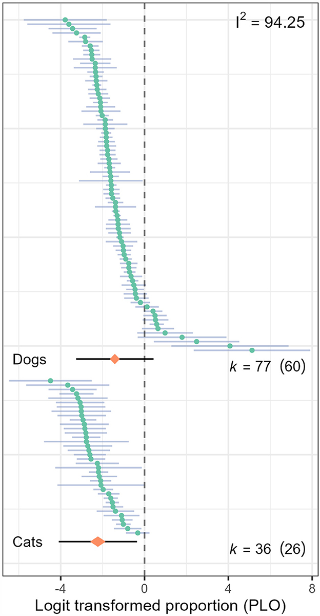
Figure 2. Caterpillar plot of the effect estimates (k) included in the overall seroprevalence model for leptospiral infection in dogs and cats. The orange diamond represents the overall estimator and its 95% confidence interval (CI); the black line represents the prediction interval, and the branches represent the individual effect sizes and their 95% CI.
Half of the effect estimates (50.4%) corresponded to domiciled animals (kdogs = 41, kcats = 16), 22.1% to strays (kdogs = 12, kcats = 13), 18.6% to sheltered (kdogs = 14, kcats = 7), and 8.8% to working dogs (kdogs = 10). Most of the effect estimates (56.6%) were from urban areas (kdogs = 43, kcats = 21), 19.5% from rural areas (kdogs = 16, kcats = 6), while 23.9% did not specify the living environment of the sampled animals. The MAT cut-off titer of 1:100 was used in 77.9% of effect estimates (kdogs = 16, kcats = 6), 17.7% used titers below 1:100 (kdogs = 43, kcats = 21), and 4.4% used titers above 1:100 (kdogs = 18, kcats = 9). The vaccination status of dogs was uncertain in 41.6% of the effect estimates. Among the known cases, 23.4% involved dogs with up-to-date leptospirosis vaccination, while 35.1% were unvaccinated dogs. Among the 18 effect estimates from vaccinated dogs, 50.0% used a bivalent vaccine containing L. interrogans serovars Icterohaemorrhagiae and Canicola, 16.7% used a tetravalent vaccine containing L. interrogans serovars Icterohaemorrhagiae, Canicola, and Pomona and L. kirschneri serovar Grippotyphosa; 11.1% used a tetravalent vaccine containing L. interrogans serovars Icterohaemorrhagiae, Canicola, and Australis and L. kirschneri serovar Grippotyphosa; and the remaining 23.5% used other combinations of serovars. The results of the moderator analysis showed that there was not a significant moderating effect of the origin of the animals (Pdogs = 0.277, Pcats = 0.414, Figure 3), their living environment (Pdogs = 0.56, and Pcats = 0.73, Figure 3), MAT cut-off titer used (Pdogs = 0.693, and Pcats = 0.751, Figure 3), and vaccination status of dogs (Pdogs = 0.392, Figure 3). The moderator analysis also did not detect a significant time trend in either dog studies or cat studies (Pdogs = 0.189, and Pcats = 0.689, Figure 3).
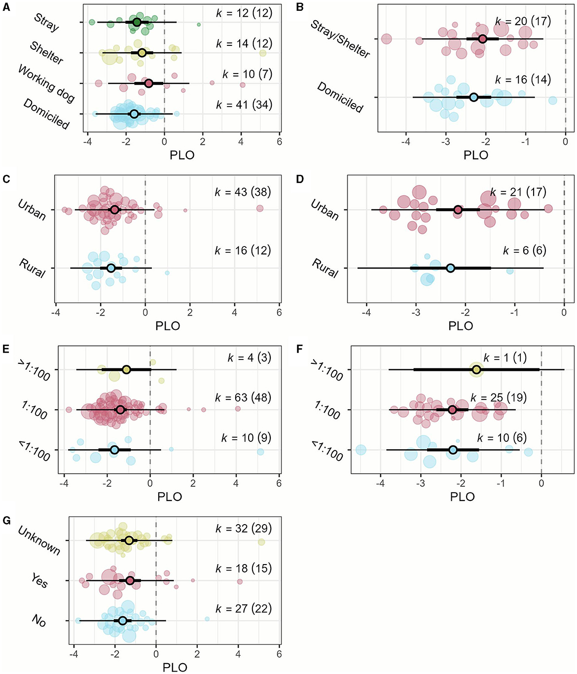
Figure 3. Orchard plots of the overall seroprevalence moderator analyses. (A) Origin of the animal (dogs); (B) origin of the animal (cats); (C) environmental setting (dogs); (D) environmental setting (cats); (E) MAT cut-off titer (dogs); (F) MAT cut-off titer (cats); (G) vaccination status (dogs). The central circles represent the mean effect size estimates, the bold branch represents its 95% confidence interval (CI), and the thin branches represent the prediction interval. Individual effect sizes, weighed by the study sample size, are represented as bubbles. PLO: logit transformed proportion.
3.3 Serogroup and serovar-specific seroprevalence
The selected studies tested a total of 74 serovars belonging to 26 known serogroups, plus two serovars of undetermined serogroups (Cantagalo and Khorat, Supplementary Tables S1, S2). The median number of serovars tested per study was 14 (IQR: 9–22), with a minimum of two and a maximum of 27 (Supplementary Tables S1, S2). Four studies had incomplete information on the serovars used in the MAT panel (Supplementary Table S1). Among the 76 tested serovars, 50 (67.6%) were detected in the sampled animals, of which 22 (44%) were detected in both species, 25 (50%) only in dogs, and L. biflexa serovar Andamana, L. borgpetersenii serovar Arborea, and L. interrogans serovar Rachmati only in cats (Supplementary Table S2).
We identified 17 pathogenic serovars within 13 serogroups that appeared in at least 10 effect estimates from dogs and/or five effect estimates from cats. The overall estimation of seroprevalence for any of these pathogenic serogroups was 2.8% (95% CI: 2.3%; 3.4%), with no significant differences between dog studies and cat studies (P = 0.516) and high statistical heterogeneity (I2 = 85.8%). Based on 290 effect estimates from dog studies, there were significant differences in the seroprevalence of the 12 most frequent pathogenic serogroups (P < 0.001), with L. interrogans serogroup Canicola presenting the highest seroprevalence (5.4%, 95% CI: 3.8%; 7.5%), and L. interrogans serovar Pyrogenes (1.7%, 95% CI: 1.0%; 2.7%), L. kirschneri serogroup Grippotyphosa (1.6%, 95% CI: 1.1%; 2.4%), and L. borgpetersenii serovar Tarassovi (1.4%, 95% CI: 0.7%; 2.8%) the lowest (Figure 4). No significant differences were detected in the seroprevalence of the eight most frequent pathogenic serogroups in cat studies (P = 0.373, Figure 4).

Figure 4. Orchard plots of the serogroup-specific seroprevalence analyses. (A) Dog studies; (B) cat studies. The central circles represent the mean effect size estimates, the bold branch represents its 95% confidence interval (CI), and the thin branches represent the prediction interval. Individual effect sizes are represented as bubbles. PLO: logit transformed proportion.
For dog studies, we fitted individual meta-analysis models to evaluate variables that could affect the seroprevalence in serogroups with at least 20 effect estimates. Seroprevalence of L. kirschneri serogroup Grippotyphosa was significantly higher (P = 0.028) in stray and sheltered dogs (2.8%, 95% CI: 1.4%;5.5%) than domiciled dogs (1.1%, 95% CI: 0.7%;1.8%). No other significant differences were observed regarding the origin or environmental setting of the dogs (P > 0.05). Subgroup analyses for cat studies were not performed due to the small number of effect estimates.
4 Discussion
In this systematic review and meta-analysis, we estimated the seroprevalence of the most frequently detected pathogenic serogroups in asymptomatic dogs and cats in the period 2012–2021, as well as those with greater prevalence in dogs (L. interrogans serogroups Canicola and Autumnalis, L. borgpetersenii serovar Ballum), cats (L. borgpetersenii serovar Javanica), and both species (L. interrogans serogroups Icterohaemorrhagiae, Bataviae, and Pomona). Previous systematic reviews had reported that L. interrogans serogroups Australis, Canicola, Icterohaemorrhagiae, and Pomona; L. kirschneri serogroup Grippotyphosa; and L. borgpetersenii serogroup Sejroe were the most prevalent in dogs (5, 31), and L. interrogans serogroups Autumnalis, Canicola, Icterohaemorrhagiae, and Pomona; L. kirschneri serogroup Grippotyphosa; and L. borgpetersenii serogroup Ballum were the most prevalent in cats (6, 19). Most of these reviews, however, covered different time frames and were limited to Northern Hemisphere countries, and the prevalence of a pathogenic serogroup may present spatiotemporal variations (4, 50). It should also be noted that in our study, the seroprevalence of a particular serogroup was estimated by dividing the number of seropositive individuals by the sample size, and in case of agglutination to multiple serovars, only the one with the highest titer was accounted, which may have lowered the pooled estimates.
In our study, the largest estimation of seroprevalence for domestic dogs corresponded to L. interrogans serogroup Canicola. Dogs are the only known maintenance hosts of this serogroup, and exposure is thought to occur mainly through contact with the urine of infected dogs (5, 31). Moreover, the estimated seroprevalence of L. interrogans serogroup Canicola in domestic cats was low (1.9%), which is consistent with published studies that reported that the risk of transmission from dogs to cats seems to be low (6, 51, 52). However, recent evidence suggests that rodents, horses, and pigs may also play a significant role in the environmental transmission cycle of this serovar (3).
In our study, L. interrogans serogroup Icterohaemorrhagiae was a serogroup frequently found in both species. Rats (Rattus spp.) are the major maintenance hosts for this serogroup (12, 31). Infection in dogs is thought to occur mainly through contact with rat urine and it is usually associated with severe forms of leptospirosis (6, 33), whereas in cats, infections with this serogroup may occur by rodent hunting and there are no reports of clinical illness (6, 12). However, there is evidence of renal excretion in asymptomatic dogs and cats seropositive to L. interrogans serogroup Icterohaemorrhagiae (15, 20, 21, 53, 54). The detection of this serogroup in asymptomatic pets should be of public health concern, as infections with Icterohaemorrhagiae in humans are associated with severe forms of leptospirosis, and domestic dogs and cats may be acting as epidemiological links between infected rats and pet caretakers (55, 56). These results can be related to the One Health concept as they illustrate the links between environmental, animal, and human health. From this perspective, a disturbed ecosystem harboring a large rat population could be contaminated with pathogenic leptospires, which could consequently affect human and animal health. Solutions to such a complex scenario can only be found through the joint efforts of all relevant disciplines and sectors (1, 3, 4).
The seroprevalence of L. interrogans serovars Canicola and Icterohaemorrhagiae in domestic dogs has shown a downward trend in the northern hemisphere over the past few decades, thought to be related to the widespread use of bivalent vaccines (5). This trend contrasts with the relatively high overall pooled frequencies of these serogroups estimated in our meta-analysis and Esteves et al. (31) and may be linked to the different epidemiological contexts found in countries from Latin America and the Caribbean, South and Southeast Asia, and the relatively lower adoption coverage of where canine vaccination is not mandatory. In these areas, the prices of the vaccines are not quite affordable for a large proportion of pet caretakers, and many of them are still unaware of the importance of vaccinating their pets regularly, especially with vaccines other than rabies (31, 57).
L. interrogans serogroup Pomona is maintained by pigs and cattle and was found in the kidneys of skunks and opossums (6, 12). Our results show that this serogroup is quite common in both species, dogs and cats, and its prevalence did not differ significantly between animals living in urban or rural environments, which is in contrast to the findings of Esteves et al. (31). It should be noted that many studies from urban areas have been conducted in slums or peri-urban settlements, where residents tend to keep subsistence livestock animals in backyards without veterinary care and exposed to garbage and wildlife, increasing the likelihood of transmission of leptospires to companion animals (58, 59). It should also be noted that most of the studies that included working dogs, involved hunting dogs, which are in close contact with feral pigs and small mammals and may act as epidemiological links of infections with serogroup Pomona as well (60, 61).
L. interrogans serogroups Autumnalis and Bataviae and L. borgpetersenii serogroup Javanica are also associated with wild rodents, rats (Rattus spp.), and small mammals (62–65), whereas the main reservoirs of L. borgpetersenii serogroup Ballum are house mice (Mus musculus) (6, 66). These serogroups were detected in several dog and cat studies, which may be indicative of direct exposure by rodent hunting, or indirectly by contact with garbage, chicken huts, and other areas contaminated with rodent urine (58, 67–69). However, L. interrogans serogroup Autumnalis presents cross-reactivity with L. interrogans serogroups Pomona, Canicola, and Icterohaemorrhagiae, and its large prevalence should be interpreted with caution (70, 71).
The results of this meta-analysis showed that there were no significant differences in the seroprevalence to a single pathogenic serogroup between dogs and cats (P = 0.648). This may be explained considering that it is rather frequent that the same animal reacts to more than one serovar, sometimes with the same titer, making it impossible to determine whether the animal is infected with multiple serovars or there were cross-reactions between antigenically related serovars (6, 71). To improve knowledge of the species of Leptospira infecting domestic dogs and cats and their maintenance hosts, future epidemiological studies should combine serological and molecular characterization (3, 8, 9, 31).
This study has some limitations worth noting. First, a large proportion of the studies included in the analysis were conducted in Brazil and may not be representative of the worldwide distribution and seroprevalence of pathogenic Leptospira in domestic dogs and cats, and it might influence the pooled estimates. However, publication bias assessment did not suggest any significant effect on that. Second, the number of cat studies found was relatively low, which prevented us from performing subgroup analyses and meta-regression for cats. Third, statistical heterogeneity was high, which can be attributed to the presence of confounding factors that were not considered in the base studies, as well as the over-representation of studies from Brazil. Finally, it should be noted that even though the MAT is specific to serogroup levels, cross-reactions still may occur among antigenically similar serogroups. It should be noted that vaccinated animals may react to serological tests even if they are not infected, which may affect prevalence estimates in serological surveys, especially for the serovars included in the vaccines used in those populations. Despite the lack of specificity of the MAT and other limitations related to the technique mentioned before, we consider that the results of this meta-analysis are of relevance for both veterinary and human health as they show a large burden of infection in a group of animal species with important bonds with humans.
We believe that the results of our study will be useful to health professionals and researchers not only to better understand the pathogenic serogroups of Leptospira currently circulating in asymptomatic companion animals, their potential maintenance hosts, and the risk of environmental contamination to other animal species and humans but also to promote adoption of biosecurity measures and appropriate handling precautions when humans handle or are exposed to companion animals. Our results would also be useful in the development of public health strategies aimed at reducing the transmission of pathogenic leptospires from the environment to domestic and stray animals and the potential risk of transmission to humans.
Author contributions
TR: Data curation, Formal analysis, Investigation, Methodology, Visualization, Writing—original draft, Writing—review & editing. LA-A: Data curation, Investigation, Methodology, Writing—original draft, Writing—review & editing. MP: Resources, Supervision, Writing—original draft, Writing—review & editing. GM: Conceptualization, Investigation, Resources, Supervision, Writing—original draft, Writing—review & editing.
Funding
The author(s) declare that no financial support was received for the research, authorship, and/or publication of this article.
Acknowledgments
We would like to thank Dr. Marcelo Signorini and Dr. Federico Costa for their advice on improving the methodological quality of the manuscript. We also would like to thank BSc. Mayla Lovera and BSc. Ericka Browne for their collaboration in the early stages of literature search and selection.
Conflict of interest
The authors declare that the research was conducted in the absence of any commercial or financial relationships that could be construed as a potential conflict of interest.
Publisher's note
All claims expressed in this article are solely those of the authors and do not necessarily represent those of their affiliated organizations, or those of the publisher, the editors and the reviewers. Any product that may be evaluated in this article, or claim that may be made by its manufacturer, is not guaranteed or endorsed by the publisher.
Supplementary material
The Supplementary Material for this article can be found online at: https://www.frontiersin.org/articles/10.3389/fvets.2024.1301959/full#supplementary-material
References
1. Chen KT. Emerging infectious diseases and one health: implication for public health. Int J Environ Res Public Health. (2022) 19:9081. doi: 10.3390/ijerph19159081
2. Pal M, Roba Bulcha M, Mitiku Bune W. Leptospirosis and one health perspective. Am J Public Health Res. (2021) 9:180–3. doi: 10.12691/ajphr-9-4-9
3. Sykes JE, Francey T, Schuller S, Stoddard RA, Cowgill LD, Moore GE. Updated ACVIM consensus statement on leptospirosis in dogs. J Vet Intern Med. (2023) 2023:jvim.16903. doi: 10.1111/jvim.16903
4. Mwachui MA, Crump L, Hartskeerl R, Zinsstag J, Hattendorf J. Environmental and behavioural determinants of leptospirosis transmission: a systematic review. PLoS Negl Trop Dis. (2015) 9:e0003843. doi: 10.1371/journal.pntd.0003843
5. Ellis WA. Animal Leptospirosis. In:Adler B, , editor. Current Topics in Microbiology and Immunology. Cham: Springer (2015), p. 99–137.
6. Schuller S, Francey T, Hartmann K, Hugonnard M, Kohn B, Nally JE, et al. European consensus statement on leptospirosis in dogs and cats. J Small Anim Pract. (2015) 56:159–79. doi: 10.1111/jsap.12328
7. Overgaauw PAM, Vinke CM, Hagen MAE, van Lipman LJA. A one health perspective on the human-companion animal relationship with emphasis on zoonotic aspects. Int J Environ Res Public Health. (2020) 17:3789. doi: 10.3390/ijerph17113789
8. Ricardo T, Previtali MAA, Signorini M. Meta-analysis of risk factors for canine leptospirosis. Prev Vet Med. (2020) 181:105037. doi: 10.1016/j.prevetmed.2020.105037
9. Ricardo T, Azócar-Aedo L, Signorini M, Previtali M. Leptospiral infection in domestic cats: Systematic review with meta-analysis. Prev Vet Med. (2023) 212:105851. doi: 10.1016/j.prevetmed.2023.105851
10. Murillo A, Cuenca R, Serrano E, Marga G, Ahmed A, Cervantes S, et al. Leptospira detection in cats in spain by serology and molecular techniques. Int J Environ Res Public Health. (2020) 17:1–5. doi: 10.3390/ijerph17051600
11. Ribeiro TMP, Santos HD, Sousa SAP, Galvão SR, Reis TS, Jayme V de S. Infection by Leptospira spp. in domestic cats (Felis silvestris catus) a review. Rev Bras Hig E Sanidade Anim. (2018) 12:1–14. doi: 10.5935/1981-2965.20180011
12. Azócar-Aedo L, Smits HL, Monti G. Leptospirosis in dogs and cats: epidemiology, clinical disease, zoonotic implications and prevention. Arch Med Vet. (2014) 46:337–48. doi: 10.4067/S0301-732X2014000300002
13. Murillo A, Goris M, Ahmed A, Cuenca R, Pastor J. Leptospirosis in cats: current literature review to guide diagnosis and management. J Feline Med Surg. (2020) 22:216–28. doi: 10.1177/1098612X20903601
14. Millán J, Candela MG, López-Bao JV, Pereira M, Jiménez MA, León-Vizcaíno L. Leptospirosis in wild and domestic carnivores in natural areas in Andalusia, Spain. Vector Borne Zoonotic Dis Larchmt N. (2009) 9:549–54. doi: 10.1089/vbz.2008.0081
15. Weis S, Rettinger A, Bergmann M, Llewellyn JR, Pantchev N, Straubinger RK, et al. Detection of Leptospira DNA in urine and presence of specific antibodies in outdoor cats in Germany. J Feline Med Surg. (2017) 19:470–6. doi: 10.1177/1098612X16634389
16. Yaafar NE, Prado A, Favot NA, Poli Gl, Sarradell JE, Anthony LM, et al. Possible clinical leptospirosis in two cats (Felis silvestris catus) from the south of the Santa Fe province. Cienc Vet. (2019) 21:85–98. doi: 10.19137/cienvet-201921206
17. Rose L, Hapke H, Luge E, Mayer-Scholl A, Merle R, Nöckler K, et al. [Prevalence of antibodies and clinical suspected cases of leptospirosis in cats in the Berlin/ Brandenburg area]. Prakt Tierarzt. (2019) 100:324–35. doi: 10.2376/0005-9366-17096
18. Francois S, Poli G, Yaafar N, Prado N, Adrien-Rüeger M, Gorordo M, et al. Estudio serológico de la infección por Leptospira spp. en gatos (Felis silvestris catus) en el sur de la provincia de Santa Fe, Argentina. Clin Infecto Vet. (2020) 6:2–9.
19. Hartmann K, Egberink H, Pennisi MG, Lloret A, Addie D, Belák S, et al. Leptospira species infection in cats: ABCD guidelines on prevention and management. J Feline Med Surg. (2013) 15:576–81. doi: 10.1177/1098612X13489217
20. Bourassi E, Savidge C, Foley P, Hartwig S. Serologic and urinary survey of exposure to Leptospira species in a feral cat population of Prince Edward Island, Canada. J Feline Med Surg. (2021) 23:1155–61. doi: 10.1177/1098612X211001042
21. Holzapfel M, Taraveau F, Djelouadji Z. Serological and molecular detection of pathogenic Leptospira in domestic and stray cats on Reunion Island, French Indies. Epidemiol Infect. (2021) 149:e229. doi: 10.1017/S095026882100176X
22. Kakita T, Kuba Y, Kyan H, Okano S, Morita M, Koizumi N. Molecular and serological epidemiology of Leptospira infection in cats in Okinawa Island, Japan. Sci Rep. (2021) 11:1–8. doi: 10.1038/s41598-021-89872-3
23. Dorsch R, Ojeda J, Salgado M, Monti G, Collado B, Tomckowiack C, et al. Cats shedding pathogenic Leptospira spp. -An underestimated zoonotic risk? PLoS ONE. (2020) 15:e0239991. doi: 10.1371/journal.pone.0239991
24. Donato G, Masucci M, Hartmann K, Goris MGA, Ahmed AA, Archer J, et al. Leptospira spp. Prevalence in cats from southern Italy with evaluation of risk factors for exposure and clinical findings in infected cats. Pathogens. (2022) 11:10. doi: 10.3390/pathogens11101129
25. Mazzotta E, De Zan G, Cocchi M, Boniotti MB, Bertasio C, Furlanello T, et al. Feline Susceptibility to Leptospirosis and Presence of Immunosuppressive Co-Morbidities: First European Report of L. interrogans serogroup Australis sequence type 24 in a cat and survey of leptospira exposure in outdoor cats. Trop Med Infect Dis. (2023) 8:54. doi: 10.3390/tropicalmed8010054
26. Bhat AH. Bacterial zoonoses transmitted by household pets and as reservoirs of antimicrobial resistant bacteria. Microb Pathog. (2021) 155:104891. doi: 10.1016/j.micpath.2021.104891
27. Caldart ET, Constantino C, Pasquali AKS, Benitez ADN, Hamada FN, Dias RCF, et al. Zoonosis in dogs and cats attended by the birth control project: Toxoplasma gondii, Leishmania spp. and Leptospira spp, serodiagnosis and epidemiology. Semin Agrar. (2015) 36:253–66. doi: 10.5433/1679-0359.2015v36n1p253
28. do Nascimento Benitez A, Monica TC, Miura AC, Romanelli MS, Giordano LGP, Freire RL, et al. Spatial and simultaneous seroprevalence of anti-Leptospira antibodies in owners and their domiciled dogs in a major city of southern Brazil. Front Vet Sci. (2021) 7:580400. doi: 10.3389/fvets.2020.580400
29. Fonzar UJV, Langoni H. Geographic analysis on the occurrence of human and canine leptospirosis in the city of Maringá, state of Paraná, Brazil. Rev Soc Bras Med Trop. (2012) 45:100–5. doi: 10.1590/S0037-86822012000100019
30. De Morais EGF, Rodrigues Magalhães FJ, De Lima Filho CDF, Brandespim DF, De Oliveira PRF, Da Costa DF, et al. Geo-epidemiological study of Leptospira spp. infection in cattle, feral cats and rodents of the Fernando de Noronha Island, Brazil. Acta Sci Vet. (2018) 46:1–9. doi: 10.22456/1679-9216.89373
31. Esteves SB, Santos CM, Silva BCS, Salgado FF, Guilloux AGA, Cortez A, et al. Time for change? A systematic review with meta-analysis of Leptospires infecting dogs to assess vaccine compatibility in Brazil. Prev Vet Med. (2023) 213:105869. doi: 10.1016/j.prevetmed.2023.105869
32. Bergmann Esteves S, Moreira Santos C, Ferreira Salgado F, Paldês Gonçales A, Gil Alves Guilloux A, Marinelli Martins C, et al. Efficacy of commercially available vaccines against canine leptospirosis: a systematic review and meta-analysis. Vaccine. (2022) 40:1722–40. doi: 10.1016/j.vaccine.2022.02.021
33. Goldstein RE. Canine leptospirosis. Vet Clin NA Small Anim Pract. (2010) 40:1091–101. doi: 10.1016/j.cvsm.2010.07.008
34. Page MJ, McKenzie JE, Bossuyt PM, Boutron I, Hoffmann TC, Mulrow CD, et al. The PRISMA 2020 statement: An updated guideline for reporting systematic reviews. The BMJ. (2021) 372:n71. doi: 10.1136/bmj.n71
35. Alashraf AR, Khairani-Bejo S, Khor KH, Radzi R, Rani PAMA, Goh SH, et al. Serological detection of anti-Leptospira antibodies among animal caretakers, dogs and cats housed in animal shelters in Peninsular Malaysia. Sains Malays. (2020) 49:1121–8. doi: 10.17576/jsm-2020-4905-17
36. Spangler D, Kish D, Beigel B, Morgan J, Gruszynski K, Naikare H, et al. Leptospiral shedding and seropositivity in shelter dogs in the Cumberland Gap region of Southeastern Appalachia. PLoS ONE. (2020) 15:e0228038. doi: 10.1371/journal.pone.0228038
37. Miotto BA, Guilloux AGA, Tozzi BF, Moreno LZ, da Hora AS, Dias RA, et al. Prospective study of canine leptospirosis in shelter and stray dog populations: Identification of chronic carriers and different Leptospira species infecting dogs. Chang YF, editor PloS One. (2018) 13:e0200384. doi: 10.1371/journal.pone.0200384
38. Orr B, Westman ME, Malik R, Purdie A, Craig SB, Norris JM. Leptospirosis is an emerging infectious disease of pig-hunting dogs and humans in North Queensland. PLoS Negl Trop Dis. (2022) 16:e0010100. doi: 10.1371/journal.pntd.0010100
39. Cilia G, Fratini F, Turchi B, Ebani VV, Turini L, Bilei S, et al. Presence and characterization of zoonotic bacterial pathogens in wild boar hunting dogs (Canis lupus familiaris) in Tuscany (Italy). Animals. (2021) 11:1139. doi: 10.3390/ani11041139
40. Lau SF, Wong JY, Khor KH, Roslan MA, Abdul Rahman MS, Bejo SK, et al. Seroprevalence of leptospirosis in working dogs. Top Companion Anim Med. (2017) 32:121–5. doi: 10.1053/j.tcam.2017.12.001
41. O'Connor AM, Sargeant JM, Dohoo IR, Erb HN, Cevallos M, Egger M, et al. Explanation and elaboration document for the STROBE-vet statement: strengthening the reporting of observational studies in epidemiology—veterinary extension. J Vet Intern Med. (2016) 30:1896–928. doi: 10.1111/jvim.14592
42. Athapattu T, Fernando R, Abayawansha R, Fernando P, Fuward M, Samarakoon N, et al. Carrier status of Leptospira spp. in healthy companion dogs in Sri Lanka vector-borne. Zoonotic Dis. (2022) 22:93–100. doi: 10.1089/vbz.2021.0065
43. Hazra A, Gogtay N. Biostatistics series module 4: comparing groups - categorical variables. Indian J Dermatol. (2016) 61:385–92. doi: 10.4103/0019-5154.185700
44. Hazra A, Gogtay N. Biostatistics series module 3: comparing groups: numerical variables. Indian J Dermatol. (2016) 61:251–60. doi: 10.4103/0019-5154.182416
45. Assink M, Wibbelink C. Fitting three-level meta-analytic models in R : a step-by-step tutorial mark Assink and Carlijn J. M Wibbelink Quant Methods Psychol. (2016) 12:154–74. doi: 10.20982/tqmp.12.3.p154
46. Begg CB, Mazumdar M. Operating characteristics of a rank correlation test for publication bias. Biometrics. (1994) 50:1088–101. doi: 10.2307/2533446
47. Egger M, Smith GD, Schneider M, Minder C. Bias in meta-analysis detected by a simple, graphical test. Br Med J. (1997) 315:629–34. doi: 10.1136/bmj.315.7109.629
48. Bown MJ, Sutton AJ. Quality control in systematic reviews and meta-analyses. Eur J Vasc Endovasc Surg. (2010) 40:669–77. doi: 10.1016/j.ejvs.2010.07.011
49. R Core Team. R: A Language and Environment for Statistical Computing. Vienna: R Foundation for Statistical Computing (2023).
50. Costa F, Hagan JEJE, Calcagno J, Kane M, Torgerson P, Martinez-Silveira MSMS, et al. Global morbidity and mortality of leptospirosis: a systematic review. PLoS Negl Trop Dis. (2015) 9:e0003898. doi: 10.1371/journal.pntd.0003898
51. Murcia CA, Astudillo M, Romero MH. Prevalence of leptospirosis in vaccinated working dogs and humans with occupational risk. Biomed Rev Inst Nac Salud. (2020) 40:62–75. doi: 10.7705/biomedica.5009
52. Mai LTP, Dung LP, Than PD, Dinh TV, Quyet NT, Hai H, et al. Leptospir a infection among human-close-contact animals in different geographical areas in Vietnam. Sci Prog. (2021) 104:1–12. doi: 10.1177/00368504211031747
53. Sant'Anna R, Vieira AS, Grapiglia J, Lilenbaum W. High number of asymptomatic dogs as leptospiral carriers in an endemic area indicates a serious public health concern. Epidemiol Infect. (2017) 145:1852–4. doi: 10.1017/S0950268817000632
54. Chan KW, Hsu YH, Hu WL, Pan MJ, Lai JM, Huang KC, et al. Serological and PCR detection of feline Leptospira in southern Taiwan. Vector Borne Zoonotic Dis Larchmt N. (2014) 14:118–23. doi: 10.1089/vbz.2013.1324
55. Vieira AS, Di Azevedo MIN, D'Andrea PS, do Val Vilela R, Lilenbaum W. Neotropical wild rodents Akodon and Oligoryzomys (Cricetidae: Sigmodontinae) as important carriers of pathogenic renal Leptospira in the Atlantic forest, in Brazil. Res Vet Sci. (2019) 124:280–3. doi: 10.1016/j.rvsc.2019.04.001
56. Browne ES, Callefe JLR, Jesus ERSDE, Zeppelini CG, Cremonese C, Costa F, et al. Systematic review of the geographic distribution of pathogenic Leptospira serovars in the Americas, 1930-2017. An Acad Bras Cienc. (2022) 94:e20201026. doi: 10.1590/0001-3765202220201026
57. Maciel EAP, de Carvalho ALF, Nascimento SF, de Matos RB, Gouveia EL, Reis MG, et al. Household transmission of Leptospira infection in urban slum communities. PLoS Negl Trop Dis. (2008) 2:e154. doi: 10.1371/journal.pntd.0000154
58. Reis RB, Ribeiro GS, Felzemburgh RDM, Santana FS, Mohr S, Melendez AXTO, et al. Impact of environment and social gradient on Leptospira infection in urban slums. PLoS Negl Trop Dis. (2008) 2:e228. doi: 10.1371/journal.pntd.0000228
59. Ricardo T, Bergero LC, Bulgarella EP, Previtali MA. Knowledge, attitudes and practices (KAP) regarding leptospirosis among residents of riverside settlements of Santa Fe, Argentina. PLoS Negl Trop Dis. (2018) 12:e0006470. doi: 10.1371/journal.pntd.0006470
60. Adesiyun AA, Hull-Jackson C, Mootoo N, Halsall S, Bennett R, Clarke NR, et al. Sero-epidemiology of canine leptospirosis in Trinidad: Serovars, implications for vaccination and public health. J Vet Med. (2006) 53:91–9. doi: 10.1111/j.1439-0450.2006.00922.x
61. Machado FP, Kmetiuk LB, Pellizzaro M, Yamakawa AC, Martins CM, Morikawa VM, et al. Leptospira spp. Antibody in wild boars (Sus scrofa), hunting dogs (Canis lupus familiaris), and hunters of Brazil. J Wildl Dis. (2021) 57:184–8. doi: 10.7589/JWD-D-20-00002
62. Villanueva SYAM, Ezoe H, Baterna RA, Yanagihara Y, Muto M, Koizumi N, et al. Serologic and molecular studies of Leptospira and leptospirosis among rats in the Philippines. Am J Trop Med Hyg. (2010) 82:889–98. doi: 10.4269/ajtmh.2010.09-0711
63. Benacer D, Zain SNM, Amran F, Galloway RL, Thong KL. Isolation and molecular characterization of Leptospira interrogans and Leptospira borgpetersenii Isolates from the urban rat populations of Kuala Lumpur, Malaysia. Am J Trop Med Hyg. (2013) 88:704–9. doi: 10.4269/ajtmh.12-0662
64. Boey K, Shiokawa K, Rajeev S. Leptospira infection in rats: a literature review of global prevalence and distribution. PLoS Negl Trop Dis. (2019) 13:e0007499. doi: 10.1371/journal.pntd.0007499
65. Benacer D, Mohd Zain SN, Sim SZ, Mohd Khalid MKN, Galloway RL, Souris M, et al. Determination of Leptospira borgpetersenii serovar Javanica and Leptospira interrogans serovar Bataviae as the persistent Leptospira serovars circulating in the urban rat populations in Peninsular Malaysia. Parasit Vectors. (2016) 9:117. doi: 10.1186/s13071-016-1400-1
66. Millán J, Cevidanes A, Chirife AD, Candela MG, León-Vizcaíno L. Risk factors of Leptospira infection in Mediterranean periurban micromammals. Zoonoses Public Health. (2018) 65:e79–85. doi: 10.1111/zph.12411
67. Azócar-Aedo L, Monti G, Jara R. Leptospira spp. in domestic cats from different environments: prevalence of antibodies and risk factors associated with the seropositivity. Anim open access J MDPI. (2014) 4:612–26. doi: 10.3390/ani4040612
68. Moreira da, Silva J, Prata S, Domingues TD, Leal RO, Nunes T, Tavares L, et al. Detection and modeling of anti-Leptospira IgG prevalence in cats from Lisbon area and its correlation to retroviral infections, lifestyle, clinical and hematologic changes. Vet Anim Sci. (2020) 10:100144. doi: 10.1016/j.vas.2020.100144
69. Azócar-Aedo L, Monti G. Seroprevalence of pathogenic Leptospira spp. in domestic dogs from southern Chile and risk factors associated with different environments. Prev Vet Med. (2022) 206:105707. doi: 10.1016/j.prevetmed.2022.105707
70. Davis MA, Evermann JF, Petersen CR, VanderSchalie J, Besser TE, Huckabee J, et al. Serological survey for antibodies to Leptospira in dogs and raccoons in Washington State. Zoonoses Public Health. (2008) 55:436–42. doi: 10.1111/j.1863-2378.2008.01137.x
Keywords: leptospirosis, Canis lupus familiaris, Felis silvestris catus, pathogenic serotypes, meta-analysis
Citation: Ricardo T, Azócar-Aedo LI, Previtali MA and Monti G (2024) Seroprevalence of pathogenic Leptospira serogroups in asymptomatic domestic dogs and cats: systematic review and meta-analysis. Front. Vet. Sci. 11:1301959. doi: 10.3389/fvets.2024.1301959
Received: 25 September 2023; Accepted: 23 January 2024;
Published: 16 February 2024.
Edited by:
Veasna Duong, Institut Pasteur du Cambodge, CambodiaReviewed by:
Sara Savic, Scientific Veterinary Institute Novi Sad, SerbiaArthur Willian De Lima Brasil, Federal University of Paraíba, Brazil
Copyright © 2024 Ricardo, Azócar-Aedo, Previtali and Monti. This is an open-access article distributed under the terms of the Creative Commons Attribution License (CC BY). The use, distribution or reproduction in other forums is permitted, provided the original author(s) and the copyright owner(s) are credited and that the original publication in this journal is cited, in accordance with accepted academic practice. No use, distribution or reproduction is permitted which does not comply with these terms.
*Correspondence: Gustavo Monti, Z3VzdGF2by5tb250aUB3dXIubmw=
†Present address: Tamara Ricardo, Dpto. de Investigación Epidemiológica, Instituto Nacional de Epidemiología “Dr. Juan H. Jara” (INE), ANLIS Malbrán, Mar del Plata, Buenos Aires, Argentina