
94% of researchers rate our articles as excellent or good
Learn more about the work of our research integrity team to safeguard the quality of each article we publish.
Find out more
BRIEF RESEARCH REPORT article
Front. Vet. Sci., 24 January 2024
Sec. Animal Reproduction - Theriogenology
Volume 11 - 2024 | https://doi.org/10.3389/fvets.2024.1287872
 Fernanda Barthelson Carvalho de Moura1Zara Alves Lacerda1
 José Luiz Catão-Dias2Pedro Enrique Navas-Suárez2,3Karin Werther4Sarah Raquel Jesus Santos Simões4
 Renato de Lima Santos5
 Daniel Felipe Barrantes Murillo6
 Tatiane Terumi Negrão Watanabe7
 Carlos Eduardo Fonseca-Alves8*†Noeme Sousa Rocha1†
The giant anteater (Myrmecophaga tridactyla) is a vulnerable species in South America and is considered endangered or near extinction in Central America. Therefore, studies describing the reproductive characteristics of this species are pivotal for its conservation. Thus, this study aimed to provide a morphological description of the female reproductive tissues of this species. We collected tissue samples from six female giant anteaters and performed gross, morphological, and histochemical analyses. Five adult subjects and one juvenile were included in the study. In the ovary, classifications were made according to the follicle and oocyte sizes: primordial, primary, secondary, early antral, or antral. Typical follicles with a single oocyte surrounded by a simple or stratified layer of cubic epithelium, atretic follicles, corpora lutea, corpora albicans, and ovarian cysts were also observed. No ovarian lesions were observed. By contrast, endometritis, metritis, mucometra, and endometrial cysts were identified in the uterus. Uterine alterations in these subjects were frequent and could affect reproduction.
Xenarthra is a superorder of placental mammals endemic to the American Continent, including the orders Pilosa (anteaters and sloths) and Cingulata (armadillos). The giant anteater (Myrmecophaga tridactyla) is a member of the Myrmecophagidae family. According to the International Union for Conservation of Nature (IUCN) Red List of Threatened Species, giant anteaters are considered to be a vulnerable or extinct species in different Latin American countries (1). Several factors, such as predatory hunting, forest burning, and blunt trauma secondary to wildlife–vehicle collisions, contribute to the reduction in the giant anteater population (1–7). The lack of governmental programs for conservation (8), slow reproductive cycles in nature (long gestation periods, birth of only one cub per year, and extended parental care) (2), and difficulties with captive breeding programs are factors that influence species preservation (9). The population of giant anteaters has been increasingly affected by vehicle collisions. Studies have shown that death rates are higher in males than in females among giant anteaters living near roads (within <2 km) (10, 11).
The scarcity of studies enhancing reproductive understanding and of available reproductive protocols and biotechnologies also contributes to challenges in reproduction (9). Morphological and histopathological descriptions of the reproductive tracts of this species are highly relevant in determining the typical features or recognizing clinically significant lesions in these organs, which may interfere with individual fertility. The lack of knowledge in this area can hinder conservation work if normal and abnormal findings in the sexual organs are poorly defined. A recent study has identified the structural and ultrastructural morphology of the prostate gland in giant anteaters and revealed its histological and immunohistochemical features (12). Another study has described the morphology and histology of the reproductive tracts (13).
Different lesions and female reproductive disorders have been described in several domestic and wild species, including lesser anteaters (Tamandua tetradactyla) (14–17). To our knowledge, there have been few studies that have investigated and analyzed the histochemical features and common lesion alterations in giant female anteaters (18). Histopathological characterization of these alterations is important for an understanding of individual fertility. Thus, this study aimed to generate new knowledge regarding pathologies affecting the ovaries and uterus of giant anteaters to improve the diagnosis of reproductive disorders in this charismatic species.
This study was approved by three Brazilian committees responsible for conducting wildlife research: the System for Genetic Heritage and Associated Traditional Knowledge (#C1018E9), the Chico Mendes Institute for Biodiversity Conservation (#7685–1), and São Paulo State University Committee on the Use of Animals in Research (177/2020).
Six female giant anteaters were included after being received at our institutional wildlife center within a maximum interval of 12-h after death due to blunt trauma force (Table 1). Their ages were determined based on their body weights according to the existing literature (13). Representative sections of both the ovaries and uterus were systematically collected and fixed in neutral-buffered 10% formalin during necropsy. Samples were collected at São Paulo State University (UNESP, Brazil), the University of São Paulo (USP, Brazil), and the Universidade Federal de Minas Gerais (UFMG, Brazil) between January 2000 and December 2021.
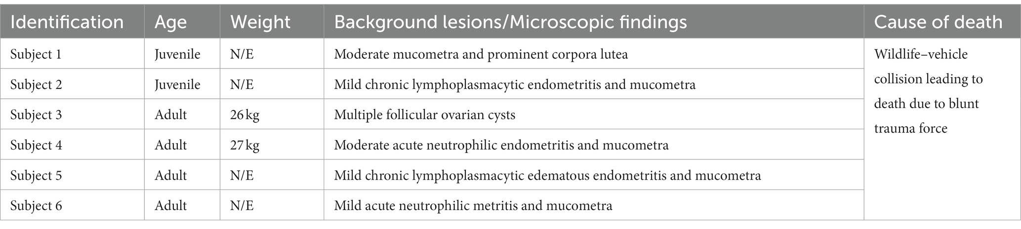
Table 1. Giant anteater autopsied information.
For the histochemical analysis, tissue samples were fixed in 10% buffered formalin and embedded in paraffin. Afterward, 4-μM-thick tissue sections were stained with hematoxylin and eosin for morphological evaluation. Periodic acid–Schiff (PAS) and Masson’s trichrome staining were performed to evaluate structural findings.
Ovarian and uterine morphological analyses were performed according to the methods established by Rossi et al. (17) and Fromme et al. (13). The evaluation was performed in a double-blind fashion: one at São Paulo State University (N.S.R.) and the other at North Carolina State University (T.T.N.W.). A qualitative analysis was conducted instead of a descriptive analysis due to the small number of animals included. Lesions were described to determine the frequency of each type of lesion.
The reproductive tracts of six free-living female giant anteaters were collected during routine postmortem examination. After the postmortem examination, ovary and uterine samples from five adult subjects (> 2 years) and one juvenile (< 2 years) were subjected to gross and microscopic assessment. There were no significant differences observed between tissues from subjects in these two different age categories. The ovaries showed an ovoid morphology and measured approximately 2 cm in length, 0.5 cm in width, and 0.5 in height. The simple uterine body was approximately 3 cm in length, 2.5 cm in width, and 2 cm in height. The ovaries and uterus were located dorsal to the urinary bladder and ventral to the rectum (Figures 1, 2).
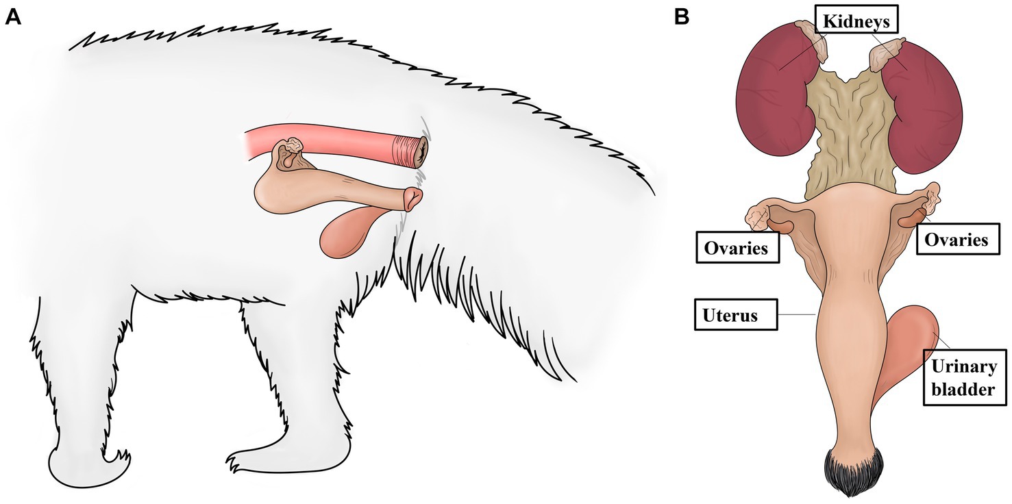
Figure 1. Schematic representation of the anatomic localization of the ovaries and uterus of the giant anteater. (A) Localization of the ovaries and uterus dorsal to the urinary bladder (and ventral to the rectum). (B) The urogenital tract of a female giant anteater, showing the position of the ovaries and uterus cranial to the urinary bladder and caudal to the kidneys.
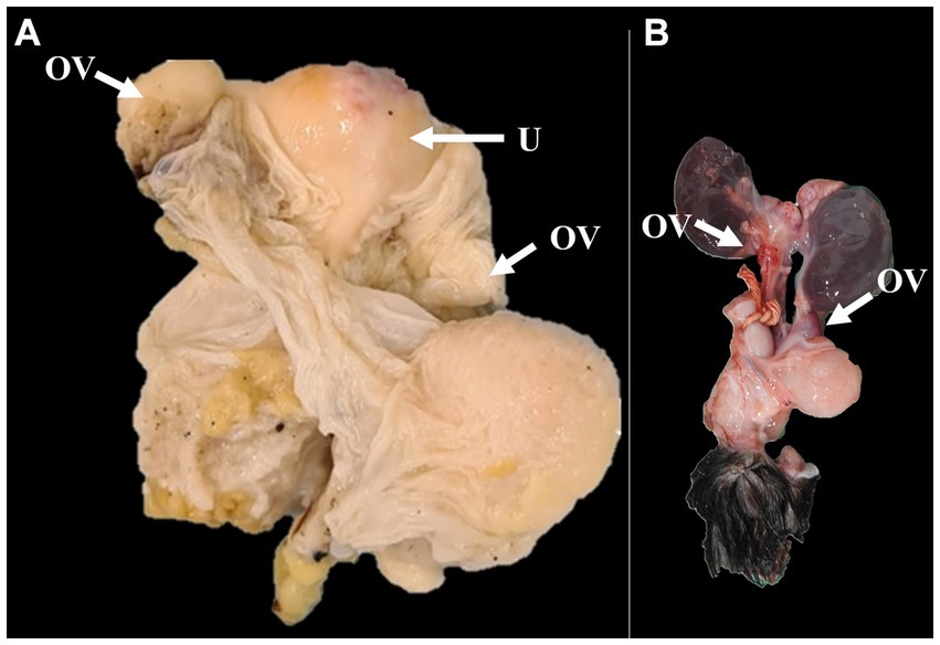
Figure 2. Gross images of the ovaries and uterus of adult (A) and juvenile (B) giant anteaters. (A) The formalin-fixed ovaries and the simple uteri are observed. (B) Macroscopic appearance of the ovaries of the giant anteater, ovoid in shape, located dorsal to the urinary bladder. OV: ovary; U: uterus.
Microscopically, the ovaries showed an architecture consisting of a simple cuboidal epithelium, ovaries, serosa, and dense connective tissue in the tunica albuginea. The ovarian parenchyma consisted of two zones. The cortical zone was located under the tunica albuginea and contained ovarian follicles at different stages of development (Figure 2). These stages were classified according to the follicle and oocyte sizes as primordial (mean diameter [follicle 88.24 μm; oocyte 47.2 μm; SD: 33.37 μm]; mean follicle number: 58), primary (mean diameter [follicle 125.02 μm; oocyte 38.72 μm; SD: 27.38 μm]; mean follicle number: 8), secondary (mean diameter [follicle 313.87 μm; oocyte 64.64 μm; SD: 176.23 μm]; mean follicle number: 4), early antral (mean diameter [follicle 953.68 μm; oocyte 152.27 μm; SD: 566.69 μm]; mean follicle number: 2), or antral (mean diameter [follicle 1806.89 μm; oocyte 374.42 μm; SD: 1012.28 μm]; mean follicle number: 1). The ovarian cortex surrounded the medullary zone, where the major vessels and nerves entered the ovary centrally and ramified to the periphery proximal to the follicles. We observed that the collagen fibers from the cortical and medullary zones were colored blue with Masson’s trichrome staining, showing rich collagen bunds in the uterine parenchyma, and that the zona pellucida and theca ovarian cells were positive for PAS staining.
Common lesions, such as typical atretic follicles containing degenerative granulosa cells and oocytes, were observed. Additionally, corpora lutea were visualized as centrally filled with the remains of the blood clot that formed after ovulation, surrounded by granulosa lutein cells with lutein cells on the outside. Some dense, round connective tissue organization, consisting of corpora albicans, was also identified in the cortical zone. The most typical background lesions were thin-walled ovarian cysts filled with pale acidophilic residual degenerative oocytes often surrounded by cell debris (Figure 3).
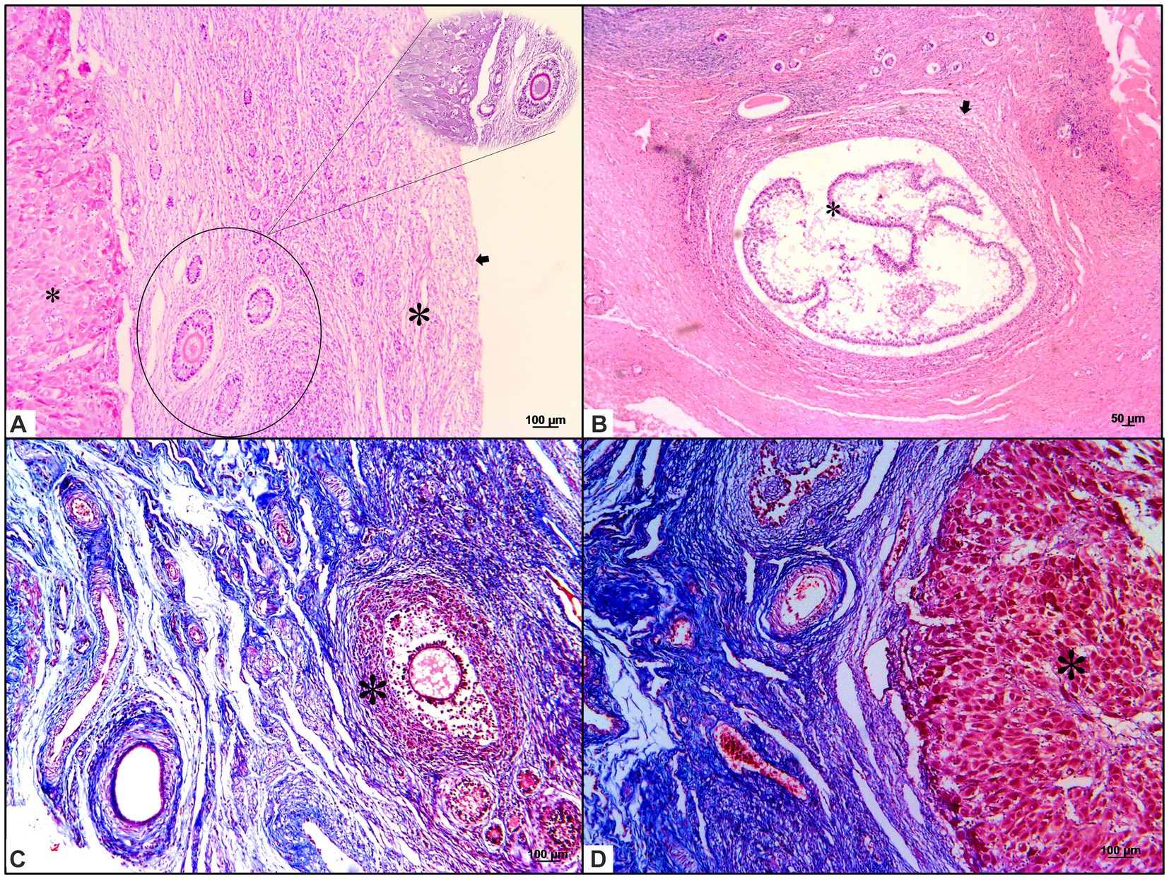
Figure 3. Histochemical characterization of background and common lesions in the ovaries of giant anteaters. (A) Ovaries with a simple cuboidal epithelium (small asterisk) on the serosa and dense connective tissue; the tunica albuginea (arrows), which is the ovarian parenchyma, is composed of two zones. The cortical zone (inside lined and non-lined circles, stained with hematoxylin–eosin and periodic acid–Schiff staining, respectively) is located under the tunica albuginea. Ovarian follicles in different stages of development are presented. The medullar zone (small asterisk), which is surrounded by the ovarian cortex (large asterisk), is where the major vessels and nerves enter the ovary centrally and ramify to the periphery proximal to the follicles. The ovarian serosa and parenchyma, stained with hematoxylin and eosin. (B,C) Typical follicles (arrows) with a single oocyte surrounded by a simple or stratified layer of cubical epithelium, atretic follicles, and ovarian cysts (large and small asterisks in B,C, respectively) were observed. Hematoxylin and eosin and Masson’s trichrome staining. (D) Medullary zone filled with a fibrous stroma; the presence of vessels and a corpora lutea (large asterisk) can be observed centrally. Masson’s trichrome staining.
Three different layers were observed in the uterine samples. First, the perimetrium was composed of loose connective tissues located peripherally. Second, the myometrium was found to consist of a thin external longitudinal layer and a thick internal circular muscular layer. The simple cuboidal epithelium with a folded endometrium near the uterine lumen presented with simple tubular glands surrounded by dense fibrous connective tissue. We observed that the collagen fibers from the perimetrium and muscle fibers from the myometrium were blue and red, respectively, after Masson’s trichrome staining (Figure 4C).
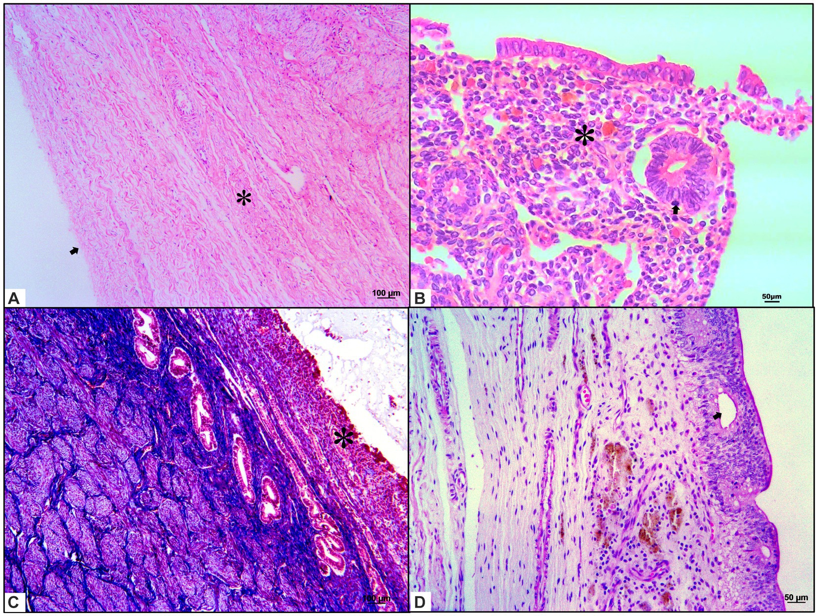
Figure 4. Histochemical characterization of background and common lesions in the uterus of giant anteaters. (A) The anteater uterus presents with three different layers. The outer layer, a perimetrium, is composed of loose connective tissues located peripherally (arrow). Second, the myometrium consists of one thin external longitudinal muscular layer and a second thick internal circular muscular layer (asterisk). Hematoxylin and eosin staining. (B) The simple cuboidal epithelium with folded endometrium near the uterine lumen presents as simple tubular glands surrounded by dense fibrous connective tissue (asterisk) with discrete leukocytic infiltration in uterine mucus (arrow). Hematoxylin and eosin staining. (C,D) Luminal secretion (asterisk, Masson’s trichrome staining in C) can be observed, and endometrial cysts (arrow, periodic acid–Schiff staining in D) disposed in the endometrial mucosa are accentuated.
The most common lesion was a mucometra, which was characterized by a discrete uterine lumen dilated with fluid. The most commonly identified background lesions were endometritis and metritis, consisting of a uterine lumen moderately dilated with fluid and discrete leukocytic infiltration composed of mononuclear cells with the presence of lymphocytes and plasma cells in the endometrium and myometrium, respectively. One case showed endometrial cysts comprising thin-walled, round epithelial structures in the endometrium. Luminal secretions and epithelial cyst cells disposed of in the endometrial mucosa were positive for PAS staining (Figure 4).
Studies investigating and analyzing the histochemical features of background and common lesions and alterations in female giant anteaters are rare (18). Histopathological characterization of reproductive abnormalities is important for an understanding of individual fertility. Our results contribute to the information available on ovarian and uterine lesions in giant anteaters through “ba/b = @histological analyses.
Previous studies have characterized the morphology and histology of the reproductive organs in giant anteaters (11, 12). Two studies involving female lesser anteaters (Tamandua tetradactyla) were performed. One other piece of research was conducted with the three-banded armadillo (Tolypeutes matacus), describing a case of uterine adenomyosis. A number of background and common ovarian lesions have been reported, including atretic follicles, corpora lutea, and corpora albicans (5, 17, 19).
We consider our results to be similar to the structural findings in female giant anteaters made in a previous study (13). Furthermore, the background and common reproductive lesions visualized have previously been described in humans and in domestic and wildlife species (14–16).
We observed that the giant anteater’s ovarian appearance is homologous with that observed in dogs. It is essential to mention that zone distribution occurs in both of these two species. Ovarian lesions have been studied to a greater extent in dogs, this being therefore a good model to better understand their pathogenesis in giant anteaters, which have been found to present features in common with dogs; this could allow us to infer that both species may develop similar lesions in the ovarian tissue. The follicles of giant anteaters were generally found to be larger than those of dogs. Nevertheless, oocyte size, granulosa cell number, and follicle diameter compared to other developmental follicular stages were homologs of those observed in dogs (20).
In giant anteaters and dogs, the atretic follicles have an irregular format, and granulosa cells have been observed in apoptosis; the follicular cysts present with thick layers of luteinizing granulosa cells, amid loose stroma; the luminal space is filled with eosinophilic material, and the corpora lutea have several vacuolated cells (20, 21).
The fallopian tubes could not be evaluated in this study because samples were not obtained during the research period. However, their structure in this species is already known. According to Fromme et al. (13), it is possible to histologically identify the infundibulum, ampulla, and isthmus, which are composed of mucosal, muscular, and serosal layers in giant anteaters. These three portions show variations in the lumen extension and width of the muscular layer, which become smaller and more significant as they become more distant from the ovaries and closer to the uterus, respectively (13).
We noted that the simple uterus in giant anteaters presented with extensive, well-marked fibrous tissue in the endometrium surrounding the tubular uterine glands. This feature is considered abnormal in domestic species, such as equines, interfering with an individual’s fertility (22, 23). Additionally, we observed that the myometrial layer was more prominent in this species than in domestic species or lesser anteaters (17, 24).
In giant anteaters and dogs, uterine background lesions have been found to consist of inflammatory infiltrates in different uterine layers, resulting in endometritis and metritis; moreover, luminal uterine secretion/edema is present, which may be associated with varying phases of the estrus cycle (19–21, 25–27).
The critical conservation status of giant anteaters significantly influences their ecological and evolutionary importance in the environment. As a species facing extinction in numerous countries, they are categorized as “vulnerable” by the IUCN, highlighting the urgency of conservation efforts (1, 28, 29). Thus, research involving the reproductive tract and fertility is fundamental to improving species conservation and protection and guaranteeing the maintenance of research programs involving these individuals. Another study has been conducted to evaluate the anatomical disposition of organs in the abdominal cavity in this species through computed tomography. All these complementary methods help to improve the detection of reproductive system lesions, contributing substantially to reproductive programs (30).
The results of the present study provide the first histochemical characterization of background lesions and common reproductive lesions in female giant anteaters. Overall, our results suggest that there is a degree of similarity between the ovarian and uterine pathophysiology of giant anteaters and that of other domestic species and humans. This information may help veterinary pathologists to report on reproductive alterations in female giant anteaters and allow them to characterize their impact on fertility.
The raw data supporting the conclusions of this article will be made available by the authors, without undue reservation.
This study was approved by three Brazilian committees responsible for conducting wildlife research: system for Genetic Heritage and Associated Traditional Knowledge (#C1018E9), Chico Mendes Institute for Biodiversity Conservation (#7685-1), and São Paulo State University Committee on the Use of Animals in Research (177/2020). The study was conducted in accordance with the local legislation and institutional requirements.
FM: Conceptualization, Visualization, Writing – original draft, Writing – review & editing. ZL: Writing – review & editing. JC-D: Writing – review & editing. PN-S: Writing – review & editing. KW: Writing – review & editing. SS: Writing – review & editing. RS: Writing – review & editing. DM: Writing – review & editing. TW: Conceptualization, Visualization, Writing – review & editing. CF-A: Conceptualization, Visualization, Writing – original draft, Writing – review & editing. NR: Conceptualization, Visualization, Writing – original draft, Writing – review & editing.
The author(s) declare financial support was received for the research, authorship, and/or publication of this article. This research was funded by Morris Animal Foundation, grant number D21ZO-601.
We want to thank the Morris Animal Foundation for their support and encouragement for this study.
The authors declare that the research was conducted in the absence of any commercial or financial relationships that could be construed as a potential conflict of interest.
The author(s) declared that they were an editorial board member of Frontiers, at the time of submission. This had no impact on the peer review process and the final decision.
All claims expressed in this article are solely those of the authors and do not necessarily represent those of their affiliated organizations, or those of the publisher, the editors and the reviewers. Any product that may be evaluated in this article, or claim that may be made by its manufacturer, is not guaranteed or endorsed by the publisher.
1. Miranda, F , Bertassoni, A , and Abba, AM . Myrmecophaga tridactyla The IUCN Red List of Threatened Species (2014). doi: 10.2305/IUCN.UK.2014-1.RLTS.T14224A47441961.en (Accessed January 13, 2024).
2. Medri, ÍM , Mourão, GM , and Harada, AY . Edentata: Dieta de Tamanduá-bandeira (Myrmecophaga tridactyla) no Pantanal da Nhecolândia, Brasil (ed. Fonseca, G. A. B. et al.); (2002) pp. 1413–4411.
3. Vidolin, GP . In: ed. IAP/Projeto Paraná Biodiversidade , editor. Planos de Conservação para Espécies de Mamíferos Ameaçados (2009). 01–316.
4. Brazil’s new president adds to global threat to science. Nature. (2018) 563:5–6. doi: 10.1038/d41586-018-07236-w
5. Arenales, A , Gardiner, CH , Miranda, FR , Dutra, KS , Oliveira, AR , Mol, JPS, et al. Pathology of free-ranging and captive Brazilian anteaters. J Comp Pathol. (2020) 180:55–68. doi: 10.1016/j.jcpa.2020.08.007
6. Pinto, FA , Bager, A , Clevenger, AP , and Grilo, C . Giant anteater (Myrmecophaga tridactyla) conservation in Brazil: Analysing the relative effects of fragmentation and mortality due to roads. Biol Conserv. (2018) 228:148–57. doi: 10.1016/j.biocon.2018.10.023
7. de Freitas, CH , Justino, CS , and Setz, EZ . Road-kills of the giant anteater in South-Eastern Brazil: 10 years monitoring spatial and temporal determinants. Wildl Res. (2015) 41:673–80.
9. Miranda, FR , Chiarello, AG , Röhe, F , Braga, F , Mourão, G , Braga, G, et al. Avaliação do Risco de Extinção de Myrmecophaga tridactyla Linnaeus, 1758 no Brasil, Processo de ava-liação do risco de extinção da fauna brasileira, 2015; Preprint at ICMBio: Available at: http://www.icmbio.gov.br/portal/biodiversidade/fauna-brasileira/lista-de-especies/7049-mamiferos-myrmecophaga-tridactyla-tamandua-bandeira.html.
10. Navas-Suárez, PE , Diaz-Delgado, J , Caiaffa, MG , da Silva, MC , Yogui, DR , Alves, MH, et al. Characterization of traumatic injuries due to motor vehicle col-lisions in Neotropical wild mammals. J Comp Pathol. (2022) 197:1–18. doi: 10.1016/j.jcpa.2022.06.003
11. Noonan, MJ , Ascensão, F , Yogui, DR , and Desbiez, AL . Roads as ecological traps for giant anteaters. Anim Conserv. (2021) 25:182–94. doi: 10.1111/acv.12728
12. Moura, F , Sampaio, L , Kobayashi, P , Laufer-Amorim, R , Ferreira, JC , Watanabe, TTN, et al. Structural and ultrastructural morphological evaluation of Giant anteater (Myrmecophaga tridactyla) prostate gland. Biology (Basel). (2021) 10:231. doi: 10.3390/biology10030231
13. Fromme, L , Yogui, DR , Alves, MH , Desbiez, ALJ , Langeheine, M , Quagliatto, A, et al. Morphology of the genital organs of male and female giant anteaters (Myrmecophaga tridactyla). PeerJ. (2021) 9:e11945. doi: 10.7717/peerj.11945
14. Fromme, L , Yogui, DR , Alves, MH , Díaz-Delgado, J , Desbiez, ALJ , Santos, ALQ, et al. Ovarian Filariasis in a wild southern Tamandua (Tamanduatetradactyla; Mammalia: Myrmecophagidae). Pathogens. (2022) 11:918. doi: 10.3390/pathogens11080918
15. Pavone, ME , Hirshfeld-Cytron, J , Tingen, C , Thomas, C , Thomas, J , Lowe, MP, et al. Human ovarian tissue cortex surrounding benign and malignant lesions. Reprod Sci. (2014) 21:582–9. doi: 10.1177/1933719113506498
16. McInnes, E.F. Background lesions in laboratory animals: A color atlas. Saunders/Elsevier: Edinburgh, (2012); pp. 101–122.
17. Rossi, LF , Luaces, JP , Marcos, HJ , Cetica, PD , Gachen, G , Jimeno, GP, et al. Female reproductive tract of the lesser anteater (Tamandua tetradactyla, myrmecophagidae, Xenarthra). Anatomy and histology. J Morphol. (2011) 272:1307–13. doi: 10.1002/jmor.10983
18. Macêdo, AA , Silva, APC , Pessanha, ÂT , Soave, SA , Paixão, TA , and Santos, RL . Endometrite purulenta em tamanduá-mirim (Tamandua tetradactyla) e tamanduá-bandeira (Myrmecophaga tridactyla). Arch Vet Sci. (2013) 1:18.
19. Marrow, J , Viner, T , Thompson, R , and Boedeker, N . Uterine adenomyosis in southern three-banded armadillos (Tolypeutes matacus). J Zoo Wildl Med 2013. (2013) 44:1018–26. doi: 10.1638/2013-0008R.1
20. Songsasen, N , Fickes, A , Pukazhenthi, BS , and Wildt, DE . Follicular morphology, oocyte diameter and localisation of fibroblast growth factors in the domestic dog ovary. Reprod Domest Anim. (2009) 44:65–70. doi: 10.1111/j.1439-0531.2009.01424.x
21. Chandra, SA , and Adler, RR . Frequency of different estrous stages in purpose-bred beagles: a retrospective study. Toxicol Pathol. (2008) 36:944–9. doi: 10.1177/0192623308326150
22. Rehm, S , Stanislaus, DJ , and Williams, AM . Estrous cycle-dependent histology and review of sex steroid receptor expression in dog reproductive tissues and mammary gland and associated hormone levels. Birth Defects Res B Dev Reprod Toxicol. (2007) 80:233–45. doi: 10.1002/bdrb.20121
23. Blanchard, TL , Garcia, MC , Kintner, LD , and Kenney, RM . Investigation of the representativeness of a single endometrial sample and the use of trichrome staining to aid in the detection of endometrial fibrosis in the mare. Theriogenology. (1987) 28:445–50. doi: 10.1016/0093-691X(87)90249-4
24. Snider, TA , Sepoy, C , and Holyoak, GR . Equine endometrial biopsy reviewed: observation, interpretation, and application of histopathologic data. Theriogenology. (2011) 75:1567–81. doi: 10.1016/j.theriogenology.2010.12.013
26. Schulman, ML , and Bolton, LA . Uterine horn aplasia with complications in two mixed-breed bitches. J S Afr Vet. (1997) 68:150–3.
27. Darko, M , Milan, A , Slobodanka, V , Svetlana, N , and Vladimir, M . Morphological characteristics and expression of estrogen and progesterone receptors in the canine endometrium during the estrus cycle, cystic endometrial hyperplasia and Pyometra. Acta Vet Brno. (2018) 68:239–50. doi: 10.4102/jsava.v68i4.899
28. Gaudin, TJ , and Croft, DA . Paleogene Xenarthra and the evolution of south American mammals. J Mammal. (2015) 96:622–34. doi: 10.1093/jmammal/gyv073
29. Patzl, M , Schwarzenberger, F , Osmann, C , Bamberg, E , and Bartmann, W . Monitoring ovarian cycle and pregnancy in the giant anteater (Myrmecophaga tridactyla) by faecal progestagen and oestrogen analysis. Anim Reprod Sci. (1998) 53:209–19. doi: 10.1016/S0378-4320(98)00114-6
Keywords: female, reproduction, morphology, histopathology, xenarthra
Citation: de Moura FBC, Lacerda ZA, Catão-Dias JL, Navas-Suárez PE, Werther K, Simões SRJS, Santos RL, Murillo DFB, Watanabe TTN, Fonseca-Alves CE and Rocha NS (2024) Background and common lesions in the female reproductive organs of giant anteaters (Myrmecophaga tridactyla). Front. Vet. Sci. 11:1287872. doi: 10.3389/fvets.2024.1287872
Edited by:
Regiane R. Santos, Schothorst Feed Research, NetherlandsReviewed by:
Ezequiel Andres Vanderhoeven, National Scientific and Technical Research Council (CONICET), ArgentinaCopyright © 2024 de Moura, Lacerda, Catão-Dias, Navas-Suárez, Werther, Simões, Santos, Murillo, Watanabe, Fonseca-Alves and Rocha. This is an open-access article distributed under the terms of the Creative Commons Attribution License (CC BY). The use, distribution or reproduction in other forums is permitted, provided the original author(s) and the copyright owner(s) are credited and that the original publication in this journal is cited, in accordance with accepted academic practice. No use, distribution or reproduction is permitted which does not comply with these terms.
*Correspondence: Carlos Eduardo Fonseca-Alves, Y2FybG9zLmUuYWx2ZXNAdW5lc3AuYnI=
†These authors have contributed equally to this work
Disclaimer: All claims expressed in this article are solely those of the authors and do not necessarily represent those of their affiliated organizations, or those of the publisher, the editors and the reviewers. Any product that may be evaluated in this article or claim that may be made by its manufacturer is not guaranteed or endorsed by the publisher.
Research integrity at Frontiers
Learn more about the work of our research integrity team to safeguard the quality of each article we publish.