 Ramon A. Juste1*Leire Fernández-Veiga1Miguel Fuertes1Ignacio Fernández-Ortiz de Murua2Guillermo Cardona2Maria V. Geijo1Joseba M. Garrido1Iker A. Sevilla1
- 1Animal Health Department, NEIKER – Basque Institute for Agricultural Research and Development, Basque Research and Technology Alliance (BRTA), Derio, Spain
- 2Servicio de Ganadería, Diputacion Foral de Alava, Vitoria-Gasteiz, Spain
Tuberculosis (TB) is a disease caused by members of the M. tuberculosis complex (MTC) that affects numerous species. M. caprae, a member of the complex which is close to M. bovis, is emerging and affects several different hosts that include goats, cattle, sheep, pigs, rabbits, wild boar, red deer, foxes and also humans. A new M. caprae spoligotype (SB2737) was isolated from an outbreak of sheep tuberculosis affecting a mixed sheep (323)-goat (29) farm in 2021. The index case was detected by the La Rioja slaughterhouse veterinary inspection. Tracing back to the farm of origin, both species were submitted to Comparative Intradermal Tuberculin Test (CITT) and M. bovis-specific antibody ELISA tests. A subsample was also examined by IFN-γ release assay (IGRA) and all positives were slaughtered and pathologically and microbiologically investigated. Only 1.2% of sheep and no goat were positive in the CITT, and 11.4% in the IGRA sheep subsample, while up to 36.8% were positive in two consecutive M. bovis-specific antibody ELISA tests. Goats had always tested negative in annual intradermal follow-up since 2013. Upon confirmation of the immunologically positive sheep at slaughter, all the remaining negative animals were killed and 29.2% of sheep were still found infected. This raised the final overall prevalence to 37.5%. Antibody ELISA was the most sensitive (81.4%) in vivo detection method still showing a 85.0% specificity relative to pathological and microbiological tuberculosis status. It was nearly 10 times more sensitive than skin test and had an 86.8% positive predictive value. Notwithstanding a possible singular pathogenesis of the new spoligotype, this outbreak adds up to previous reports suggesting that sheep tuberculosis could be huge reservoir of infection worldwide overlooked by skin test low sensitivity or simply lack of investigation. This makes it urgent to extend the use antibody tests to address the Trojan horse of hidden M. tuberculosis complex infections on bovine TB control programs.
Introduction
Tuberculosis (TB) is a disease caused by members of the M. tuberculosis complex (MTC) that affects numerous species. The two more common agents are M. tuberculosis itself, which affects mainly humans, and M. bovis, which affects many different ruminant and non-ruminant species. However, infection with M. caprae, another member of the complex that is close to M. bovis, is emerging and affects several different hosts including goats, cattle, sheep, pigs, rabbits, wild boars, red deer, foxes, and also humans (1–4). Spoligotyping has proven very useful to characterize the different variants for epidemiological studies and to link outbreaks. Given its impact on human health and the connection of bovine TB with human cases, especially digestive forms in children caused by transmission through milk, eradication of the infection from cattle was soon undertaken in many countries. The success of the programs designed to identify and cull infected cattle through subclinical infection detection made them compulsory in many countries and have defined the standards for international animal health policies (5). Since the main focus of these programs was to prevent milk transmission of MTC, their objective has been almost exclusively the bovine species, which is the main source of milk for human consumption. The lower risk associated with meat transmission, the smaller individual value, and the restricted market of small ruminant milk has attracted much less attention to TB control in goats and sheep, and therefore, these species have not been included in TB eradication programs until very recently, and even so, only in relation to cattle TB outbreaks (6, 7). Although the role of goats was more widely acknowledged, sheep had long been considered resistant to tuberculous infections (8). Therefore, the disease was considered extremely rare in this species (9, 10) and sheep is not even mentioned by the WOAH Terrestrial Code with regard to tuberculosis (11). However, recent studies have shown that the species is fully susceptible and is often infected with MTC mycobacteria in bovine and caprine mixed farms where TB has been detected (7, 12–17). Here, we report a case of MTC infection in a sheep-goat mixed farm whose index case was detected by passive slaughterhouse surveillance and where standard cellular tests failed to detect the vast majority of infected individuals, but a humoral multispecies test showed a high sensitivity. We think that these case features, sheep-only outbreak, new spoligotype, and humoral test outperformance of cellular ones, are novel features that, contributing to the depiction of a more complex model of bovine TB (18), must be taken into account to make animal TB control a reachable objective in the not-too far future.
Materials and methods
Lesions compatible with TB were detected in the routine slaughterhouse inspection (Slaughterhouse of Logroño, La Rioja, Spain) of a culled ewe coming from Laguardia (Basque Country, Spain) on 18 April, 2021 (see the timeline presented in Figure 1). Samples from lesions were sent to the European Union Reference Laboratory (EU-RL) for Bovine TB (VISAVET-Universidad Complutense de Madrid) for MTC culture, and M. caprae was isolated. Spoligotyping yielded a new pattern that was registered at the Spanish MycoDB database with code SB2737. Regional animal health authorities immediately checked that the goats in the flock had been annually skin test (ST) negative to TB since the caprine TB eradication program began in 2013 (the last control on 31 August 2020). There was no history of paratuberculosis in the flock, and paratuberculosis vaccination had never been used. Then, a prevalence investigation that included a comparative intradermal tuberculin test (CITT) and an antibody ELISA (INgezim Tuberculosis DR; INGENASA-Eurofins, Gold Standard Diagnostics) (19) on the whole flock (321 sheep and 29 goats) was ordered and performed on 28 June 2021. The ST was performed and interpreted following the EU-RL guidelines published by the Spanish Ministry of Agriculture, Fisheries and Food (MAPA), which are in agreement with the regulations of the European Union (Commission Delegated Regulation 2020/689 of 17 December 2019). Goats and sheep were administered 0.1 mL PPD-B (2,500 IU) and PPD-A (2,500 IU) (CZ VACCINES, Pontevedra, Spain) through separate intradermal injections in the neck using a Dermojet (Akra Dermojet, Pau, France). The skin thickness was measured before and 72 h after inoculation. Interpretation was done for both single intradermal tuberculin test (SITT) and comparative intradermal tuberculin test (CITT). An animal was considered SITT positive, inconclusive, or negative when the increase of the PPD-B site was 4 mm or greater, between 2 mm and 4 mm, or less than 2 mm, respectively. For CITT, animals were considered positive, inconclusive, or negative if the increase of the PPD-B injection site exceeded that of the PPD-A site by greater than 4 mm, 1–4 mm, and less than 1 mm, respectively. The antibody ELISA was run and interpreted according to the supplier instructions. At 72 h ST reading, only three ewes and one ram were positive to the ST (according to both SITT and CITT interpretation) and were sent to NEIKER-BRTA laboratories for necropsy on 6 July 2021. Antibodies against Mycobacterium bovis MPB83 protein (conserved also in M. caprae) were detected by ELISA in 41 sheep (37 positive and 4 doubtful), including three out of the four ST-positive individuals. TB-characteristic lesions were confirmed, and SB2737 M. caprae was isolated from all four necropsied sheep. Subsequently, the remaining sheep (except three) with positive or doubtful ELISA result (n = 35) were slaughtered (15 July 2021), and blood was collected for interferon-γ release assay testing (IGRA). For IGRA, sodium heparin whole blood was distributed into cell culture plates (1.5 mL/well) and stimulated with PPD-B, PPD-A (both PPDs at 20 μg/mL) and phosphate-buffered saline (PBS; no stimulation) in individual wells (no mitogen was included), starting within 4 h of blood collection. After overnight incubation at 37°C in 5% CO2 atmosphere, samples were centrifuged and supernatants analyzed using the MAPA validated IDScreen® Ruminant interferon-gamma ELISA kit (IDvet, Grabels, France) following manufacturer’s instructions. Results were interpreted according to the indications of the kit (positive cutoff: s/p ≥ 16). During necropsy, all tissues were macroscopically inspected, and samples were collected for histopathological (lung, liver, spleen, intestine, pancreas and mediastinal, tracheobronchial, tracheal, hepatic, mesenteric, ileocecal, and ruminal lymph nodes) and microbiological (mediastinal lymph node) analysis. M. caprae infection was confirmed in all of them except for three (one of the ELISA doubtful and two ELISA positive). Given the high seroprevalence detected with the antibody ELISA and the high infection confirmation rate among positives, an anamnestic antibody ELISA was performed (23 July). In view of the increased seroprevalence detected with the anamnestic ELISA, the whole flock including goats was sent to the slaughterhouse between 13 and 19 August 2021. This time 24 out of the 200 sheep displaying a negative result in both ELISA tests, and all goats (n = 29) were submitted to necropsy and MTC isolation. Gross inspection of all tissues and microscopic examination of lung and mediastinal and tracheobronchial lymph nodes were carried out. Samples from tissues with lesions or a pool of the tissues specified above if lesions were not present were submitted to MTC isolation culture. TB infection was confirmed if TB lesions were observed and/or M. caprae was isolated. Thus, a total of 93 animals (64 sheep and 29 goats) were necropsied and had pathological and microbiological results in addition to in vivo tests results.
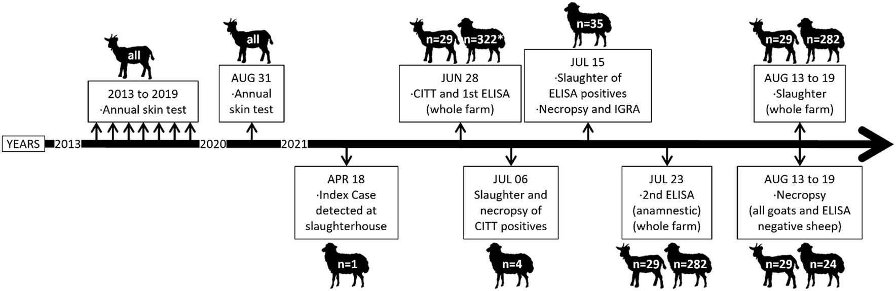
Figure 1. Chronological overview of the events and actions undertaken in relation to the tuberculosis outbreak detected in the goat-sheep mixed farm. *One ewe had only a record for CITT on 28 June control.
The epidemiological investigation included collection of information to determine management, replacement rates, origin of purchased animals, possible contacts with other farms, and recent TB outbreaks in the municipality. Drinking points in the grazing area were investigated for MTC by isolation culture.
Fisher’s exact probability test (FET) was used for comparison of frequencies (20). A complementary sensitivity was calculated as the percent of positive to one test and negative to the reference over the total number of positives in the reference test. This figure gives a measure of how much a test outperforms the reference to which it is compared (21). A generalized linear model and a correlation analysis were carried out to assess the association of age with optical density (OD) of immune tests using the jamovi statistical software (20, 22).
Results
Table 1 shows a summary of the animals according to different grouping criteria and test results. A total of 351 animals (322 sheep and 29 goats) were processed at different times and with different methods (see Figure 1). Four breeds of sheep were present in the farm (70.9% Navarrese breed). About two-thirds were breeders 24 months or older. Only 1.2% reacted positive in the intradermal test. In contrast, there were about 10 times more positive animals (12.8%) in the first ELISA testing and twice as many (28.4%) in the anamnestic ELISA. Jointly, the ELISA identified 37.27% antibody-positive sheep. The infection was confirmed in as much as the 94.6% (35/37) of positive/doubtful individuals of the first ELISA. The IGRA was applied only to 35 of the 41 antibody ELISA-positive/doubtful sheep and was positive only in 4 of them. In total, TB was pathologically or microbiologically confirmed in 44 out of the 64 sheep investigated (68.75%), including the index case. However, of these, 24 had been randomly selected at slaughter among test negative ones. Since seven of them (29.2%) were also TB confirmed, it can be estimated that there were about 77 additional infected sheep that had been missed. This would make the overall prevalence in the sheep flock reach 37.5% (121 out of 323). The age of these randomly selected animals was significantly lower than the age of the rest (p = 0.0187). Age was positively correlated with OD readings in the anamnestic ELISA (r = 0.2235, p = 0.0002) and its increase (r = 0.1292, p = 0.0455), but not with OD readings in the first antibody ELISA (r = −0.0880, p = 0.1418), or bovine antigen in IGRA (r = −0.0483, p = 0.7830).
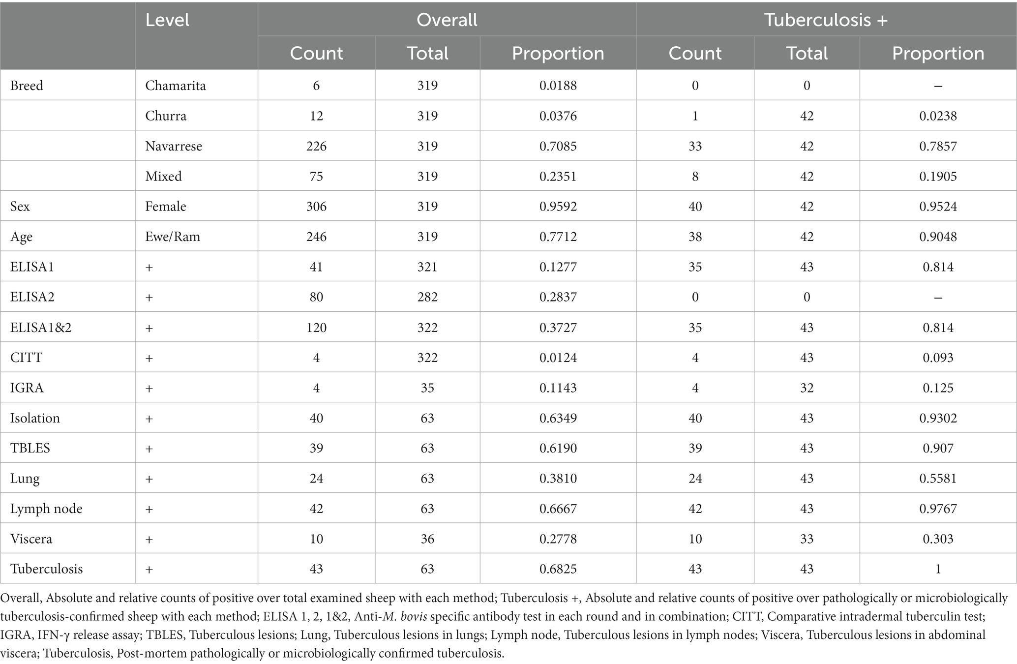
Table 1. General results in sheep.
The diagnostic method performance showed large differences regarding sensitivity (Table 2). While both standard cellular tests had a low sensitivity at around 10%, the ELISA had over 80% sensitivity. These figures led to a high outperformance of the ELISA relative to the cellular tests (over 700%), which, however, only had a 7% complementary sensitivity relative to the post-mortem tuberculosis status.
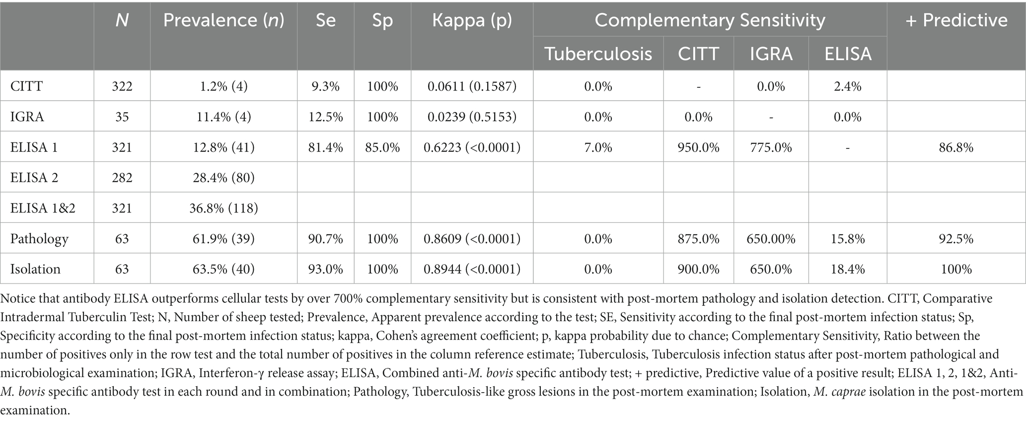
Table 2. Performance of the tests.
Interestingly, all sheep had very good body condition and 24 out of 44 sheep with confirmed infection had TB lung lesions (54.5%). C. pseudotuberculosis was isolated from one animal that was negative in the ELISA, and whose gross lesions in lung were histologically compatible with pseudotuberculosis, but that yielded M. caprae from the tracheobronchial lymph node. Most of the lesions were observed in lymph nodes (61.9%), while only 36.5 and 7.9% from lung and viscera, respectively. Lesions were very similar to those seen in typical bovine TB: multiple coalescing necrotic foci of a pale-yellow color with intense mineralization and fibrosis both in lymph nodes and in lungs. No significant differences were found according to breed (FET p = 0.3023) or sex in the first ELISA, but adults had a significantly higher seroprevalence (16.9% vs. 5.9%; p = 0.0051). In the anamnestic ELISA, breed and age differences were highly significant (Table 2), but not sex. Taken jointly, only the difference between adults (52.0%) and lambs (11.9%) was statistically significant (p < 0.0001).
All goats were negative to all the diagnostic tests performed (ST, IGRA, ELISA, and pathological and microbiological methods).
The flock was the last one in the village to use the traditional management system of daily walking from the farm to the surrounding fields with crop residues and spontaneous grass between harvests. Feed was purchased from registered producers and straw from the local producers. Sheep and goats spent the days together en route and grazing but were housed in separate barns for the nights. Average replacement rate during the last 4 years was 38.1% for sheep and 18.7% for goats, with similar culling rates. Sheep breeders were introduced in the flock from the same or a neighboring province in 2018 (10 from same province, nine from neighboring province), 2019 (38 from neighboring province), and 2020 (15 from same province, one from the same neighboring province). A total of six goats were introduced from same province in 2021. A TB outbreak occurred in a goat farm in the same municipality in 2014 caused by M. caprae spoligotype SB0416 and caused its end. Drinking points were negative for MTC.
Discussion
Routine slaughterhouse inspection allowed detection of unapparent MTC infection in a sheep-goat mixed flock. This underlines the importance of this type of surveillance (23), which largely depends on the good performance of public health veterinarians and stresses the importance of collaboration between them and animal health services (24–26). Our results further reinforce the need to allocate the resources that secure an efficient performance of this surveillance method (27, 28). It must be noticed that the index case was a routinely culled ewe because of production, not clinical reasons, from a mixed flock with a goat population that had undergone annual TB skin testing in the past 8 years before (last test on 31 August 2020), always with negative results. The commercial antibody ELISA used in this case allowed detection of 82.5% of culture-positive sheep out of the 63 that were necropsied (index case was not tested through ELISA). The figure was slightly lower if positives to either culture or pathology were used as the reference (81.4%). ST only detected 9.3% of positives to either of these two reference methods and IGRA only 12.5%. This may look surprising since humoral immunity tests have been considered to have low sensitivity in cattle (29, 30). In spite of scientific evidence pointing out that it could very specifically complement the sensitivity of cellular immunity-based tests (detection of up to an additional 33% infected animals not identified by cellular tests) (28, 30), its use has not been incorporated into cattle control programs yet (29, 31). The only other large study of TB in sheep showed similar high prevalence rates between isolation (50.4%), histopathology (83.2%), and ELISA (59.0%), but much lower for IGRA (4.9%) and ST (24.9%) with highly concordant rates of sensitivity (93.5 to 100%) for the former three. Although the sensitivity and specificity calculations were based on a small number of cases (23 and 31, respectively), the low specificity rates (37.5–50.0%) found suggest that many truly infected cases might have failed to be detected rather than being false positives and, therefore, likely indicating that the real prevalence was substantially higher (6). Qualitative and quantitative analyses indicate that infection increases with age. That might explain why the antibody ELISA missing in a few cases could have been due to low antibody levels in recent infections. In general, this pattern of immune diagnostic response must be seriously taken into account because it means that the official cellular diagnostic techniques for TB diagnosis in cattle and goats might be dramatically failing to identify a potential hidden MTC reservoir in an important and well managed domestic species but not submitted to periodic controls for the detection of infection. If we add the recent TB findings in free-ranging swine (32–36), it becomes evident that it is urgent to include non-cattle domestic species in eradication programs taking advantage of new improved humoral TB diagnostic protocols. While these insights do cover all angles in such a costly and advanced eradication program focused on a single species, it cannot be overlooked that this outbreak was caused by a totally new spoligotype that might have specific pathogenic and epidemiological particularities such as low virulence and high sheep adaptation. Actually, the failure of the epidemiological investigation to point out an origin suggests that the bacteria might have been evolving in this sheep flock acquiring new clinicopathological and epidemiological features that could eventually require a higher taxonomical status. However, lesions did not show any pathological trait that could be attributed to a differential particularity of this specific strain. Further characterization of the strain (e.g., whole genome sequencing) might shed light in this sense.
In general terms, cellular tests have proven to be much more sensitive than humoral ones for the diagnosis of bovine TB in cattle submitted to periodical TB eradication programs. However, these results confirm our own unpublished experience in cattle that antibody tests could complement cellular ones for the detection of residual/anergic individuals, because the former detect fewer cases but still have a high complementary sensitivity for identifying residual infections that the cellular tests are missing. These cases might explain why in Spain, up to 25.3% of bovine TB-infected herds are attributed to residual internal or undetected external infection according to the last report of the Spanish Ministry of Agriculture on bovine TB (37) that also confirms an earlier observation reporting a 22.3% (28). The skin test has been in service for over one century and has allowed resonant success, but current evidence that a few infected individuals still go undetected in spite of the recurrent and huge administrative and economic investments should show us the way to take a new approach. Knowing that humoral tests provide an additional sensitivity that is close to the observed rates of residual and imported new TB outbreaks, it seems clear that a breakthrough is necessary. Contrary to other studies, in this case cellular tests, including IGRA, have much lower sensitivity (about one third) than the ELISA that, actually, had a complementary sensitivity of 7.0% relative to combined pathology and isolation. In a recently published Irish study (38) reporting 30.7% of cattle negative to the official comparative test although positive to a highly specific commercial antibody test with a WOAH homologated specificity of 99.7% (39), authors suggest that the serological results might relate to intensive skin testing. However, it could also be related to a limited sensitivity of the ST when residual/anergic infection (not detected by ST) is more significant than expected.
There is another striking finding of this study that goes against generally accepted knowledge on TB. It is that infection was highly prevalent in sheep, but not in goats. Goats have long been recognized as a host of MTC infections and have even led to name the bacterial species M. caprae (40–42). The fact that in this mixed herd all goats had been negative to repeated testing and still were negative to all tests when sheep had such a high prevalence is indeed an extraordinary finding that challenges previous understanding of TB epidemiology in domestic ruminants and must be considered in the future planning of bovine TB control programs. Sheep cannot be any longer ignored in the epidemiology of TB, which should not be defined as bovine anymore (11).
Regarding post-mortem inspection, 54.5% of tuberculous sheep had TB lesions in lungs. That is, about half of the infected animals had lesions only in lymph nodes that required an exhaustive post-mortem examination. The rough similarity of TB lesions with caseous lymphadenitis, together with the assumption that ovine TB is rare, is likely to have heavily contributed to overlooking sheep TB.
This silent pattern of TB infection in sheep reported here, building on previous studies indicating an unsuspected high prevalence in mixed species farms (6), raises fears that a huge reservoir of infection in a domestic species might have been worldwide overlooked by a technical issue. Addressing it by extending the use of humoral tests is urgent to help neutralize the Trojan horse-like impact of hidden MTC infections on bovine TB control programs.
Failure to use the cheaper and more complementarily sensitive antibody tests for residual infection detection could make impossible to effectively eradicate bovine TB in many countries once arrived at its last stages. Given the effectiveness of slaughterhouse passive surveillance, complementing visual inspection with systematic (all animals, a percent, or only those from suspect or unknown TB status farms or counties depending on the epidemiological status and resources availability) application of the relatively unexpensive serological tests could become a powerful tool for tracing undetected TB-infected farms. We think that in a moment in which the European TB eradication programs have reached substantial regional eradication, the recognition of ELISA as an official test both for cattle and for other TB-susceptible species and generalizing its use in active TB infection detection should be very effective in reducing exposure risks to humans and cattle. In summary, after acknowledgment of the complex epidemiology determined by cattle sharing environments with other TB-susceptible species like wild and domestic ruminants, rabbits (43) wild boars, and even badgers (41, 42, 44–50), adapting diagnostic methods to specific species epidemiology and pathogenesis could more efficiently lead to faster TB control.
Data availability statement
The datasets presented in this study can be found in online repositories. The names of the repository/repositories and accession number(s) can be found at: https://www.visavet.es/mycodb/index-en.php, SB2737.
Ethics statement
Ethical approval was not required for the studies involving animals in accordance with the local legislation and institutional requirements because this study was carried out as a part of an EU-Spain approved Animal Health control program carried out by the local Animal Health and Welfare authority. Written informed consent was obtained from the owners for the participation of their animals in this study.
Author contributions
RJ: Conceptualization, Formal analysis, Investigation, Methodology, Writing – original draft, Writing – review & editing. LF-V: Investigation, Methodology, Writing – review & editing. MF: Investigation, Methodology, Writing – review & editing. IF: Data curation, Investigation, Methodology, Writing – review & editing. GC: Investigation, Methodology, Writing – review & editing. MG: Investigation, Writing – review & editing. JG: Conceptualization, Data curation, Funding acquisition, Investigation, Methodology, Project administration, Writing – review & editing. IS: Conceptualization, Data curation, Formal analysis, Funding acquisition, Investigation, Methodology, Project administration, Writing – review & editing.
Funding
The author(s) declare financial support was received for the research, authorship, and/or publication of this article. This study has been carried out partly with funds from Grant PID2019-105155RB-C33 and Grant PID2022-142939OR-C21 funded by MCIN/AEI/10.13039/501100011033 and by “ERDF A way of making Europe.”
Acknowledgments
The authors are indebted to the owner of the farm, who provided all kinds of facilities for the study of the case. We thank the staff of the Slaughterhouse of Logroño and the Public Health Services of La Rioja Government for carcass inspection, sample collection, and submission to NEIKER. We also want to thank ABERE ZERBITZU TEKNIKOAK KOOP for their excellent collaboration in the collection of information and the field diagnostic and sample collection. LF-V holds a pre-doctoral fellowship from the Department of Economic Development, Sustainability and Environment of the Basque Government.
Conflict of interest
The authors declare that the research was conducted in the absence of any commercial or financial relationships that could be construed as a potential conflict of interest.
The author(s) declared that they were an editorial board member of Frontiers, at the time of submission. This had no impact on the peer review process and the final decision.
Publisher’s note
All claims expressed in this article are solely those of the authors and do not necessarily represent those of their affiliated organizations, or those of the publisher, the editors and the reviewers. Any product that may be evaluated in this article, or claim that may be made by its manufacturer, is not guaranteed or endorsed by the publisher.
Supplementary material
The Supplementary material for this article can be found online at: https://www.frontiersin.org/articles/10.3389/fvets.2023.1310205/full#supplementary-material
References
1. Rodríguez, S, Bezos, J, Romero, B, de Juan, L, Álvarez, J, Castellanos, E, et al. Mycobacterium caprae infection in livestock and wildlife, Spain. Emerg Infect Dis. (2011) 17:532. doi: 10.3201/EID1703.100618
2. Varela-Castro, L, Gerrikagoitia, X, Alvarez, V, Geijo, MV, Barral, M, and Sevilla, IA. A long-term survey on Mycobacterium tuberculosis complex in wild mammals from a bovine tuberculosis low prevalence area. Eur J Wildl Res. (2021) 67:43. doi: 10.1007/s10344-021-01489-z
3. Pérez de Val, B, Perea, C, Estruch, J, Solano-Manrique, C, Riera, C, Sanz, A, et al. Generalized tuberculosis due to Mycobacterium caprae in a red fox phylogenetically related to livestock breakdowns. BMC Vet Res. (2022) 18:1–6. doi: 10.1186/S12917-022-03454-7/FIGURES/2
4. Martínez-Lirola, M, Herranz, M, Serrano, SB, Rodríguez-Grande, C, Inarra, ED, Garrido-Cárdenas, JA, et al. A one health approach revealed the long-term role of Mycobacterium caprae as the hidden cause of human tuberculosis in a region of Spain, 2003 to 2022. Eur Secur. (2023) 28:2200852. doi: 10.2807/1560-7917.ES.2023.28.12.2200852/CITE/PLAINTEXT
5. WOAH. (2023). Mammalian tuberculosis (infection with Mycobacterium tuberculosis complex), manual of diagnostic tests and vaccines for terrestrial animals. Available at: https://www.woah.org/en/what-we-do/standards/codes-and-manuals/terrestrial-manual-online-access/ (Accessed August 29, 2023).
6. Muñoz-Mendoza, M, Romero, B, del Cerro, A, Gortázar, C, García-Marín, JF, Menéndez, S, et al. Sheep as a potential source of bovine TB: epidemiology, pathology and evaluation of diagnostic techniques. Transbound Emerg Dis. (2016) 63:635–46. doi: 10.1111/tbed.12325
7. Muñoz Mendoza, M, de Juan, L, Menéndez, S, Ocampo, A, Mourelo, J, Sáez, JL, et al. Tuberculosis due to Mycobacterium bovis and Mycobacterium caprae in sheep. Vet J. (2012) 191:267–9. doi: 10.1016/j.tvjl.2011.05.006
8. Radostits, OM, Gay, CC, Blood, DC, and Hinchcliff, KW. Veterinary medicine. London: W.B. Saunders (2000).
9. Behrens, H. Tuberkulose In: Lehrbuch Der Schafkrankheiten. 3rd ed. Berlin and Hamburg: Paul Parey (1987). 73–4.
10. Davidson, RM, Alley, MR, and Beatson, NS. Tuberculosis in a flock of sheep. N Z Vet J. (1981) 29:1–2. doi: 10.1080/00480169.1981.34775
11. World Organization for Animal Health. (2023). Terrestrial code online access – WOAH – world organization for animal health. Available at: https://www.woah.org/en/what-we-do/standards/codes-and-manuals/terrestrial-code-online-access/?id=169&L=1&htmfile=chapitre_bovine_tuberculosis.htm (Accessed November 22, 2023)
12. Infantes-Lorenzo, JA, Gortázar, C, Domínguez, L, Muñoz-Mendoza, M, Domínguez, M, and Balseiro, A. Serological technique for detecting tuberculosis prevalence in sheep in Atlantic Spain. Res Vet Sci. (2020) 129:96–8. doi: 10.1016/J.RVSC.2020.01.013
13. Malone, FE, Wilson, EC, Pollock, JM, and Skuce, RA. Investigations into an outbreak of tuberculosis in a flock of sheep in contact with tuberculous cattle, journal of veterinary medicine series B: infectious diseases and veterinary. Public Health. (2003) 50:500–4. doi: 10.1046/J.1439-0450.2003.00714.X
14. Van Der Burgt, GM, Drummond, F, Crawshaw, T, and Morris, S. An outbreak of tuberculosis in Lleyn sheep in the UK associated with clinical signs. Vet Rec. (2013) 172:69. doi: 10.1136/VR.101048
15. Cordes, DO, Bullians, JA, Lake, DE, and Carter, ME. Observations on tuberculosis caused by Mycobacterium bovis in sheep. N Z Vet J. (1981) 29:60–2. doi: 10.1080/00480169.1981.34798
16. Marianelli, C, Cifani, N, Capucchio, MT, Fiasconaro, M, Russo, M, la Mancusa, F, et al. A case of generalized bovine tuberculosis in a sheep. J Vet Diagn Investig. (2010) 22:445–8. doi: 10.1177/104063871002200319
17. Gelalcha, BD, Zewude, A, and Ameni, G. Tuberculosis caused by Mycobacterium bovis in a sheep flock Colocated with a tuberculous dairy cattle herd in Central Ethiopia. J Vet Med. (2019) 2019:1–6. doi: 10.1155/2019/8315137
18. Gortázar, C, de la Fuente, J, Perelló, A, and Domínguez, L. Will we ever eradicate animal tuberculosis? Ir Vet J. (2023, 2023) 76:1–11. doi: 10.1186/S13620-023-00254-9
19. Bezos, J, Roy, Á, Infantes-Lorenzo, A, González, I, Venteo, Á, Romero, B, et al. The use of serological tests in combination with the intradermal tuberculin test maximizes the detection of tuberculosis infected goats. Vet Immunol Immunopathol. (2018) 199:43–52. doi: 10.1016/j.vetimm.2018.03.006
20. JAMOVI. (2022). The jamovi project, jamovi (version 2.3) [computer software]. Available at: https://www.jamovi.org/ (Accessed March 24, 2022)
21. Juste, RA, Garrido, JM, Geijo, M, Elguezabal, N, Aduriz, G, Atxaerandio, R, et al. Comparison of blood polymerase chain reaction and enzyme-linked immunosorbent assay for detection of Mycobacterium avium subsp. paratuberculosis infection in cattle and sheep. J Vet Diagn Investig. (2005) 17:354–9. doi: 10.1177/104063870501700409
22. Gallucci, M.. (2019). GAMLj: General analyses for the linear model in Jamovi. Available at: https://gamlj.github.io/ (Accessed July 9, 2023)
23. García-Díez, J, Saraiva, S, Moura, D, Grispoldi, L, Cenci-Goga, BT, and Saraiva, C. The importance of the slaughterhouse in surveilling animal and public health: a systematic review. Vet Sci. (2023) 10:167. doi: 10.3390/vetsci10020167
24. Napp, S, Ciaravino, G, Pérez de Val, B, Casal, J, Saéz, JL, and Alba, A. Evaluation of the effectiveness of the surveillance system for tuberculosis in cattle in Spain. Prev Vet Med. (2019) 173:104805. doi: 10.1016/J.PREVETMED.2019.104805
25. Vidal, E, Tolosa, E, Espinar, S, de Val, BP, Nofrarías, M, Alba, A, et al. Six-year follow-up of slaughterhouse surveillance (2008-2013): the Catalan slaughterhouse support network (SESC). Vet Pathol. (2016) 53:532–44. doi: 10.1177/0300985815593125
26. Pozo, P, Cardenas, NC, Bezos, J, Romero, B, Grau, A, Nacar, J, et al. Evaluation of the performance of slaughterhouse surveillance for bovine tuberculosis detection in Castilla y Leon, Spain. Prev Vet Med. (2021) 189:105307. doi: 10.1016/J.PREVETMED.2021.105307
27. Vidal, E, Balseiro, A, Marco, A, and Domingo, M. Diagnóstico anatomopatológico y vigilancia en mataderos In: A Balseiro, C Gortazar, and JL Saez, editors. Tuberculosis Animal: Una Aproximación Desde La Perspectiva de La Ciencia y La Administración, Ministerio de Agricultura. Madrid: Pesca y Alimentacion. Gobierno de España (2020). 141–8.
28. Guta, S, Casal, J, Napp, S, Saez, JL, Garcia-Saenz, A, Perez de Val, B, et al. Epidemiological investigation of bovine tuberculosis herd breakdowns in Spain 2009/2011. PLoS One. (2014) 9:e104383. doi: 10.1371/journal.pone.0104383
29. Casal, C, Infantes, JA, Risalde, MA, Díez-Guerrier, A, Domínguez, M, Moreno, I, et al. Antibody detection tests improve the sensitivity of tuberculosis diagnosis in cattle. Res Vet Sci. (2017) 112:214–21. doi: 10.1016/j.rvsc.2017.05.012
30. Casal, C, Díez-Guerrier, A, Álvarez, J, Rodriguez-Campos, S, Mateos, A, Linscott, R, et al. Strategic use of serology for the diagnosis of bovine tuberculosis after intradermal skin testing. Vet Microbiol. (2014) 170:342–51. doi: 10.1016/j.vetmic.2014.02.036
31. Infantes-Lorenzo, JA, Moreno, I, Thomas, J, and Risalde, MA. Serología en especies domésticas y silvestres In: A Balseiro, C Gortazar, and JL Saez, editors. Tuberculosis Animal: Una Aproximación Desde La Perspectiva de La Ciencia y La Administración, Ministerio de Agricultura. Madrid: Ministerio de Agricultura, Pesca y Alimentación (2020). 106–19.
32. Parra, A, Fernández-Llario, P, Tato, A, Larrasa, J, García, A, Alonso, JM, et al. 401 Seroepidemiology of tuberculosis in sheep in southern Spain. Prev Vet Med. (2023) 215:402–105920. doi: 10.1016/j.prevetmed.2023.105920
33. Parra, A, Fernández-Llario, P, Tato, A, Larrasa, J, García, A, Alonso, JM, et al. Epidemiology of Mycobacterium bovis infections of pigs and wild boars using a molecular approach. Vet Microbiol. (2003) 97:123–33. doi: 10.1016/j.vetmic.2003.08.007
34. Cardoso-Toset, F, Gómez-Laguna, J, Amarilla, SP, Vela, AI, Carrasco, L, Fernández-Garayzábal, JF, et al. Multi-etiological nature of tuberculosis-like lesions in condemned pigs at the slaughterhouse. PLoS One. (2015) 10:e0139130. doi: 10.1371/journal.pone.0139130
35. Cardoso-Toset, F, Luque, I, Carrasco, L, Jurado-Martos, F, Risalde, Á, Venteo, Á, et al. Evaluation of five serologic assays for bovine tuberculosis surveillance in domestic free-range pigs from southern Spain. Prev Vet Med. (2017) 137:101–4. doi: 10.1016/j.prevetmed.2016.12.016
36. Bailey, SS, Crawshaw, TR, Smith, NH, and Palgrave, CJ. Mycobacterium bovis infection in domestic pigs in Great Britain. Vet J. (2013) 198:391–7. doi: 10.1016/j.tvjl.2013.08.035
37. Sanz, C, Grau, A, Fernandez, C, Muñoz, M, Solares, S, Fernandez, M, et al. Informe Final Técnico-Financiero Tuberculosis Bovina Año 2022. Madrid: Ministry of Agriculture, Fisheries and Food (2023).
38. Madden, J, Casey-Bryars, M, More, SJ, Barrett, D, Gormley, E, and Ryan, E. Investigation of the association between the Enferplex bovine tuberculosis antibody test and the future risk of bovine tuberculosis in irish cattle in infected herds: a pilot field study. Vet Res Commun. (2023). doi: 10.1007/s11259-023-10200-3
39. Enfer Scientific ULC. (2019). OIE procedure for registration of diagnostic kits validation studies abstract. Enferplex Bovine TB antibody test. Available at: https://www.woah.org/app/uploads/2021/03/oie20abstract2026-07-19.pdf (Accessed August 26, 2023)
40. Aranaz, A, Cousins, D, Mateos, A, and Domínguez, L. Elevation of Mycobacterium tuberculosis subsp. caprae Aranaz et al. 1999 to species rank as Mycobacterium caprae comb. nov., sp. nov. Int J Syst Evol Microbiol. (2003) 53:1785–9. doi: 10.1099/ijs.0.02532-0
41. Napp, S, Allepuz, A, Mercader, I, Nofrarias, M, Lopez-Soria, S, Domingo, M, et al. Evidence of goats acting as domestic reservoirs of bovine tuberculosis. Vet Rec. (2013) 172:663. doi: 10.1136/vr.101347
42. Aranaz, A, Liebana, E, Gomez-Mampaso, E, Galan, JC, Cousins, D, Ortega, A, et al. Mycobacterium tuberculosis subsp. caprae subsp. nov.: a taxonomic study of a new member of the Mycobacterium tuberculosis complex isolated from goats in Spain. Int J Syst Bacteriol. (1999) 49:1263–73. doi: 10.1099/00207713-49-3-1263
43. Sevilla, IA, Arnal, MC, Fuertes, M, Martín, E, Comenge, J, Elguezabal, N, et al. Tuberculosis outbreak caused by Mycobacterium caprae in a rabbit farm in Spain. Transbound Emerg Dis. (2020) 67:431–41. doi: 10.1111/tbed.13366
44. AECOSAN. Plan de Actuación sobre TUBerculosis en Especies Silvestres. Madrid: Ministerio de Agricultura, Pesca y Alimentación (2023).
45. Infantes-Lorenzo, JA, Moreno, I, Roy, A, Risalde, MA, Balseiro, A, De Juan, L, et al. Specificity of serological test for detection of tuberculosis in cattle, goats, sheep and pigs under different epidemiological situations. BMC Vet Res. (2019) 15:70. doi: 10.1186/s12917-019-1814-z
46. Infantes-Lorenzo, JA, Dave, D, Moreno, I, Anderson, P, Lesellier, S, Gormley, E, et al. New serological platform for detecting antibodies against Mycobacterium tuberculosis complex in European badgers. Vet Med Sci. (2019) 5:61–9. doi: 10.1002/vms3.134
47. Thomas, J, Infantes-Lorenzo, JA, Moreno, I, Romero, B, Garrido, JM, Juste, R, et al. A new test to detect antibodies against Mycobacterium tuberculosis complex in red deer serum. Vet J. (2019) 244:98–103. doi: 10.1016/j.tvjl.2018.12.021
48. Richomme, C, Boadella, M, Courcoul, A, Durand, B, Drapeau, A, Corde, Y, et al. Exposure of wild boar to Mycobacterium tuberculosis complex in France since 2000 is consistent with the distribution of bovine tuberculosis outbreaks in cattle. PLoS One. (2013) 8:e77842. doi: 10.1371/JOURNAL.PONE.0077842
49. Aurtenetxe, O, Barral, M, Vicente, J, De La Fuente, J, Gortázar, C, and Juste, RA. Development and validation of an enzyme-linked immunosorbent assay for antibodies against Mycobacterium bovis in European wild boar. BMC Vet Res. (2008) 4:43. doi: 10.1186/1746-6148-4-43
50. Alvarez, J., Bezos, J., Diez-Guerrier, A., Gonzalez, S., and Saez, J.L. (2018). Manual de detección de animales infectados anérgicos en explotaciones T2, de riesgo o con antecedentes de tuberculosis, Madrid. Available at: https://www.mapa.gob.es/es/ganaderia/temas/sanidad-animal-higiene-ganadera/manualdedetecciondeanimalesinfectadosanergicosenexplotacionest2deriesgooconantecedentesdetuberculosis_3_tcm30-479532.pdf (Accessed September 19, 2023)
Keywords: sheep tuberculosis, goat tuberculosis, SB2737, Mycobacterium caprae , tuberculosis antibody test, bovine tuberculosis, skin test
Citation: Juste RA, Fernández-Veiga L, Fuertes M, Fernández-Ortiz de Murua I, Cardona G, Geijo MV, Garrido JM and Sevilla IA (2024) A humoral diagnostic test outperforms cellular tests in a farm with a latent tuberculosis outbreak caused by a new Mycobacterium tuberculosis complex spoligotype that affected sheep but not goats. Front. Vet. Sci. 10:1310205. doi: 10.3389/fvets.2023.1310205
Edited by:
Michael Kogut, United States Department of Agriculture, United StatesReviewed by:
Álvaro Roy, Complutense University of Madrid, SpainAlicia Aranaz, Complutense University of Madrid, Spain
Copyright © 2024 Juste, Fernández-Veiga, Fuertes, Fernández-Ortiz de Murua, Cardona, Geijo, Garrido and Sevilla. This is an open-access article distributed under the terms of the Creative Commons Attribution License (CC BY). The use, distribution or reproduction in other forums is permitted, provided the original author(s) and the copyright owner(s) are credited and that the original publication in this journal is cited, in accordance with accepted academic practice. No use, distribution or reproduction is permitted which does not comply with these terms.
*Correspondence: Ramon A. Juste, rjuste@neiker.eus