
95% of researchers rate our articles as excellent or good
Learn more about the work of our research integrity team to safeguard the quality of each article we publish.
Find out more
ORIGINAL RESEARCH article
Front. Vet. Sci. , 06 December 2023
Sec. Veterinary Infectious Diseases
Volume 10 - 2023 | https://doi.org/10.3389/fvets.2023.1217135
This article is part of the Research Topic Zoonotic Bacteria: Genomic Evolution, Antimicrobial Resistance, Pathogenicity, and Prevention Strategies View all 6 articles
 Philippe Charron‡
 Ruimin Gao†‡
 John Chmara
 Emily Hoover
 Susan Nadin-Davis
 Danielle Chauvin
 Jennifer Hazelwood
 Kennedy Makondo
 Marc-Olivier Duceppe
 Mingsong Kang*
Glanders is a highly contagious and life-threatening zoonotic disease caused by Burkholderia mallei (B. mallei). Without an effective vaccine or treatment, early diagnosis has been regarded as the most effective method to prevent glanders transmission. Currently, the diagnosis of glanders is heavily reliant on serological tests. However, given that markedly different host immune responses can be elicited by genetically different strains of the same bacterial species, infection by B. mallei, whose genome is unstable and plastic, may result in various immune responses. This variability can make the serodiagnosis of glanders challenging. Therefore, there is a need for a comprehensive understanding and assessment of how B. mallei genomic variations impact the appropriateness of specific target antigens for glanders serodiagnosis. In this study, we investigated how genomic variations in the B. mallei genome affect gene content (gene presence/absence) and expression, with a special focus on antigens used or potentially used in serodiagnosis. In all the genome sequences of B. mallei isolates available in NCBI’s RefSeq database (accessed in July 2023) and in-house sequenced samples, extensive small and large variations were observed when compared to the type strain ATCC 23344. Further pan-genome analysis of those assemblies revealed variations of gene content among all available genomes of B. mallei. Specifically, differences in gene content ranging from 31 to 715 genes with an average of 334 gene presence-absence variations were found in strains with complete or chromosome-level genome assemblies, using the ATCC 23344 strain as a reference. The affected genes included some encoded proteins used as serodiagnostic antigens, which were lost due mainly to structural variations. Additionally, a transcriptomic analysis was performed using the type strain ATCC 23344 and strain Zagreb which has been widely utilized to produce glanders antigens. In total, 388 significant differentially expressed genes were identified between these two strains, including genes related to bacterial pathogenesis and virulence, some of which were associated with genomic variations, particularly structural variations. To our knowledge, this is the first comprehensive study to uncover the impacts of genetic variations of B. mallei on its gene content and expression. These differences would have significant impacts on host innate and adaptive immunity, including antibody production, during infection. This study provides novel insights into B. mallei genetic variants, knowledge which will help to improve glanders serodiagnosis.
Glanders is an infectious and usually fatal zoonosis caused by Burkholderia mallei (B. mallei). Compared to other species in the family Burkholderiaceae, most of which are soil residents, B. mallei is a mammalian intracellular pathogen (1). Horses are highly susceptible to B. mallei infections and have been regarded as the natural reservoir (2). Other solipeds, such as mules and donkeys, are also susceptible to B. mallei infection, as well as humans (2). As B. mallei is highly infectious through aerosols, it was used as a biological weapon during World War I and has since been classified as a category B bioterrorism agent by the Centers for Disease Control and Prevention. Glanders has now been eradicated from most developed countries, however, it remains endemic in a number of Asian, European, African, Middle Eastern, and South American countries where sporadic infections have been detected, including Germany, Russia, Brazil, India, Turkey, and China, and significantly affects regional economic activities and international trade (3, 4). These incidents highlight the potential for importing the glanders agent to glanders-free countries or areas, thus posing a significant risk to equine and public health with huge economic loss and potentially fatal consequences globally.
So far, due to the absence of licensed vaccines or preventive treatment against glanders (5), a fast and accurate early diagnosis remains the most efficient way to prevent B. mallei transmission and infection. Diagnosis of glanders is mainly conducted using serodiagnostic assays for surveillance and international trade of Equidae. The complement fixation test (CFT), a serological test officially recommended by the World Organization for Animal Health (WOAH, founded as OIE) with decades of utilization in the equine industry, represents a practical method and is currently the internationally accepted approach for glanders diagnosis. However, the specificity of CFT may vary and its standardization has been questioned for years (4, 6). In order to improve the accuracy of glanders diagnosis, the immunoblot (IB) assay using Lipopolysaccharides (LPS) antigen (7) has been used to confirm CFT “positive” results. However, such a subjective test is still quite technically cumbersome, especially the production of the LPS antigen. In addition, IB may also generate false-positive results due to similar LPS structures between Burkholderia species, especially those acting as opportunistic pathogens (8, 9). Therefore, it would be beneficial to establish a simpler, more objective, and quantifiable serological test, such as an ELISA. ELISAs using single or double recombinant protein antigens have been developed and evaluated, and the results are promising (6, 10, 11).
Theoretically, B. mallei is believed to have evolved from an ancestral B. pseudomallei environmental isolate following an animal infection (12), by the expansion of insertion sequence (IS) elements, prophage elimination, and genome rearrangements, specifically deletions (13). The B. mallei genome (5.8 Mb) is 20% smaller than the B. pseudomallei genome (7.2 Mb), and many genes needed for environmental survival were lost from B. mallei, while those critical for host survival were maintained (13, 14). It has also been shown that the genome of B. mallei is significantly less stable than that of B. pseudomallei, and large-scale genomic alterations, such as insertions/deletions (INDELs), and structural variants (SVs) related to chromosomal rearrangements, have been observed in many B. mallei genomes (15–17). These dynamic processes may impact gene content (gene presence/absence) and expression through various mechanisms (18).
Recently, the WOAH reference laboratory for glanders in France reported that a genetic variant of B. mallei compromised the molecular diagnosis of glanders (19). Since interspecies differences in genome sequences, especially the accessory genome, also elicit distinctly different host adaptive immune responses (20), the high plasticity of B. mallei genomes may cause more variations in immunological responses that potentially disrupt the reliability of serodiagnosis, a vital tool in detecting glanders. In this study, using integrative genomic and transcriptomic analyses, we characterized the effects of genetic variation on gene content and expression to evaluate currently used and potentially useful serodiagnostic biomarkers to help improve glanders serodiagnosis.
All available Burkholderia mallei genome assemblies (n = 108) and corresponding metadata, including NCBI RefSeq ID, level of assembly, contig count, L50, N50, size, and geographic location (Supplementary File S1), were downloaded from the NCBI Reference Sequence database (as of July 2023) using Bit v1.8.351 and EDirect v15.6.2 All B. mallei genome assemblies and annotation completeness were evaluated using BUSCO with a completeness score cut-off value of 90% (21–23).
B. mallei type strain ATCC 23344 was transferred from The National Centre for Foreign Animal Disease (NCFAD) in Winnipeg (24) and strains Zagreb, Mukteswar and Bogor were kindly provided by the Friedrich Loeffler Institute, Federal Research Institute for Animal Health in Germany (7). Live B. mallei strains were handled in Containment Level 3 (CL3) facilities. Strains were cultured on a blood agar plate with 3% glycerol for 48 h at 37°C. Cultures were harvested for RNA purification or heat-killed at 85°C for 30 min and the sterility was verified by our standard operating procedure prior to further processing.
Genomic DNA was extracted from heat-killed B. mallei cultures using the PureLink Genomic DNA Mini Kit (Invitrogen, United States) or NanoBind CBB Kit (PacBio, United States) following the manufacturer’s instructions. The quality and the quantity of extracted DNA were determined by NanoDrop™ OneC Spectrophotometer and Qubit™ 4 Fluorometer, respectively. The extracted genomic DNA samples have a 260/280 ratio of 1.8-2.0 and a 260/230 ratio of 2.0–2.3 with a concentration above 20 ng/μl. Library preparations and whole genome sequencing were performed as described previously (25). Briefly, Illumina (short-read) libraries were prepared using an Illumina DNA Prep (M) Tagmentation kit (Cat.No: 20060059, Illumina, United States), and sequenced on the Illumina MiSeq platform to generate 300-bp paired-end reads. MinION (long-read) libraries were established using Ligation Sequencing Kits (SQK-LSK108 or SQK-LSK109) with Native Barcoding Expansion (EXP-NBD103 or EXP-NBD104) (Oxford Nanopore Technologies, UK) without shearing and were sequenced using an FLO-MIN106 (R9.4.1) flow cell and a MinION Mk1B device.
Whole genome assembly and evaluation were performed as previously described with modifications (25). Briefly, the raw data (FASTQ files) generated from Illumina MiSeq were filtered and trimmed with BBTools v38.87 or Fastp v0.23.2 (26). Raw data (FAST5 files) generated from Nanopore MinION were base-called using Guppy v5.0.11 with the super accuracy mode, trimmed using Porechop v0.2.3,3 and filtered using Filtlong v0.2.1.4 Hybrid assembly of strain Zagreb using Illumina paired-end and MinION reads was performed by Unicycler v0.4.5 (27). Long read assembly of strains ATCC 23344, Bogor and Mukteswar was performed using Flye v2.9 (28), corrected using Medaka v1.4.45 and polished with Illumina MiSeq reads using a combination of NextPolish v1.4.0 (29) and Polypolish v0.5.0 (30). Nanopore long-read and Illumina short-read data were mapped to the assembled genome sequences using minimap2 (31), and the generated mapping bam files were quality checked. Qualimap (v2.2.1) (32) determined the sequencing coverage depth. The assembled genome was then annotated using the NCBI prokaryotic genome annotation pipeline (PGAP v6.2) (33).
Small variants, including SNPs and INDELs, were determined using Snippy v4.6.0 with default parameters,6 and SVs were determined using MUM&Co v3.8.07 (34). The core genome SNP alignment obtained from Snippy was used for maximum-likelihood phylogenetic tree construction using IQtree v2.1.28 (35) with 1,000 ultrafast bootstraps. The phylogeny was partitioned into lineages using FastBaps v1.0.89 (36). The phylogenetic tree was visualized using iTOL v6.6.10 The distribution of SNPs, INDELs, and SVs was analyzed as described previously (37).
Pan-genomes of 112 available B. mallei genome sequences were analyzed using PPanGGOLiN v1.2.7411 (38), with PGAP–annotated Genbank files as input. In the clustering step, the default parameters were used. Further visualization and analysis of pan-genome results were performed using R v4.2.2, packages ggplot2 v3.4.0,12 ggtree v3.2.1 (39), pheatmap v1.0.12,13 UpSetR v1.4.0 (40) and vegan v2.6-4.14
Total RNA was extracted from live B. mallei strains ATCC 23344 and Zagreb with three biological replicates for each strain in the Containment Level 3 (CL3) laboratory using the PureLink RNA mini kit with the On-column PureLink DNase Treatment (Invitrogen, United States) according to the manufacturer’s instructions. The RNA samples were sent to Centre d’expertise et de service Génome Québec (Québec, Canada) for sequencing. Briefly, total RNA was quantified using a NanoDrop Spectrophotometer ND-1000 (Thermo Scientific, United States), and its integrity was assessed on a 2,100 Bioanalyzer (Agilent Technologies, United States). rRNA were depleted using QIAseq FastSelect -5S/16S/23S kits (QIAGEN, United States). cDNA synthesis was obtained using the NEBNext RNA First Strand Synthesis and NEBNext Ultra Directional RNA Second Strand Synthesis Modules (New England BioLabs, United States). The library preparation was performed using the NEBNext Ultra II DNA Library Prep Kit for Illumina (New England BioLabs, United States), followed by quantification using the Kapa Illumina GA with Revised Primers-SYBR Fast Universal kit (Kapa Biosystems, Switzerland). The libraries were normalized and pooled for sequencing. After ExAMP was added, the pooled library was loaded on an Illumina cBot, and the flowcell was run on a HiSeq 4000 for 2 × 100 cycles (paired-end mode) to generate over 10 million reads for each sample (Supplementary Table S2). A PhiX library was used as a control and mixed with libraries at a 1% level. The Illumina control software was HCS HD 3.4.0.38, and the real-time analysis program was RTA v2.7.7. The bcl2fastq2 v2.20 tool was used to demultiplex samples and generate FASTQ reads.
The file (.faa file) containing coding sequences (CDS) of strain ATCC 23344 was used as an input of PathoFact v1.0 (ORF version) for the prediction of virulence factors (VFs) using default parameters (41). Only CDSs with predicted virulence confidence levels 1 (secreted virulence factor) and 2 (non-secreted virulence factor) were used for further analysis.
Raw reads from the sequenced libraries were quality checked with fastp v0.23.2 (26). The reference cDNA sequences (CDS from genomic FASTA file) of B. mallei type strain ATCC 23344 were downloaded from the NCBI RefSeq database. Transcript abundance was quantified using kallisto v0.46.1 (42) with 100 bootstraps. Differential expression analysis was performed using the R package, sleuth v0.30.0 (43). Differential gene expressions (DEGs) were identified based on a fold change >2 and a q-value <10−3 for further filtering using the Wald test in sleuth. The transcript abundance data were imported into R v4.2.2, and principal coordinates analysis (PCoA) was performed using vegan v2.6-415 and visualized using ggplot2 v3.4.0 (https://cran.r-project.org/web/packages/ggplot2/index.html).
The identified differentially expressed genes (DEGs) were applied to explore the potentially affected Kyoto encyclopedia of genes and genomes (KEGG) pathways. The corresponding KO (KEGG ORTHOLOGY) table was obtained by GhostKOALA v2.2 (44) using the genus_prokaryotes database. The obtained KO terms corresponding to the DEGs were fed to the KEGG Mapper reconstruction tool (last updated: July 1, 2021)16 (45), and the affected pathways were evaluated.
Based on the simple model that the destruction of operon structure or disruption of the non-coding region upstream of operons can interfere with gene expression (46), variant calls (SNPs, INDELs, and SVs) were compared with RNA-seq data to link DEGs to specific genomic variation. Specifically, we identified the operons using operonSEQer v1.0 (47) and investigated changes in gene expression in relation to an alteration in the operon structure mediated by SVs, as well as changes in the non-coding region upstream of the operon, including promoter and regulators, mediated by SNPs or INDELs. This was achieved using custom scripts modified from a previously described method (48).
Due to genetic variability after laboratory or host passages, four B. mallei isolates, ATCC 23344, Bogor, Mukteswar, and Zagreb, were re-sequenced using both Nanopore long-read and Illumina short-read sequencing technologies to obtain complete or chromosome-level whole genome assemblies (Supplementary Table S1). In combination with 108 published genomes available on RefSeq (Supplementary File S1), we identified a total of 3,806 core SNPs by using strain ATCC 23344 (RefSeq accession number: GCF_000011705.1) as a reference. The density and distribution of SNPs are similar among chromosome I (CH1) and chromosome II (CH2) (Figure 1A). In addition to SNPs, a large number of INDELs and SVs were identified. Several dense spots of variants indicating common SNPs, INDELs, and SVs are noticeable in multiple sites on both CH1 and CH2 (Figure 1A). To further explore this observation, the distribution and number of SNPs, INDELs, and SVs for 38 strains with complete and chromosome-level genome assembly were examined, respectively (Figures 1B,C). Multiple typical “hotspots” of SNPs and INDELs exist in all genomes, including derivatives of ATCC 23344 as shown in the five inner circles (Figure 1B). Around 70% of small variants, including SNPs, INDELs, MNPs (multiple nucleotide polymorphisms), and complex variants (combination of SNPs and MNPs), were located within coding sequences (CDS) (Figure 2A), some of which introduce early stop codons for several genes, including a gene encoding the serodiagnostic antigen ImpJ (TssK) shown in Table 1. The majority of small variants are still SNPs and INDELs (Figure 2A). Similarly, multiple types of SVs, especially large deletions, in B. mallei isolates were identified using strain ATCC 23344 as a reference, except for some ATCC 23344 derivatives (Figures 1C, 2B). These SVs were scattered in both chromosomes, and multiple hotspot regions were observed in most isolates on both chromosomes (Figure 1C).
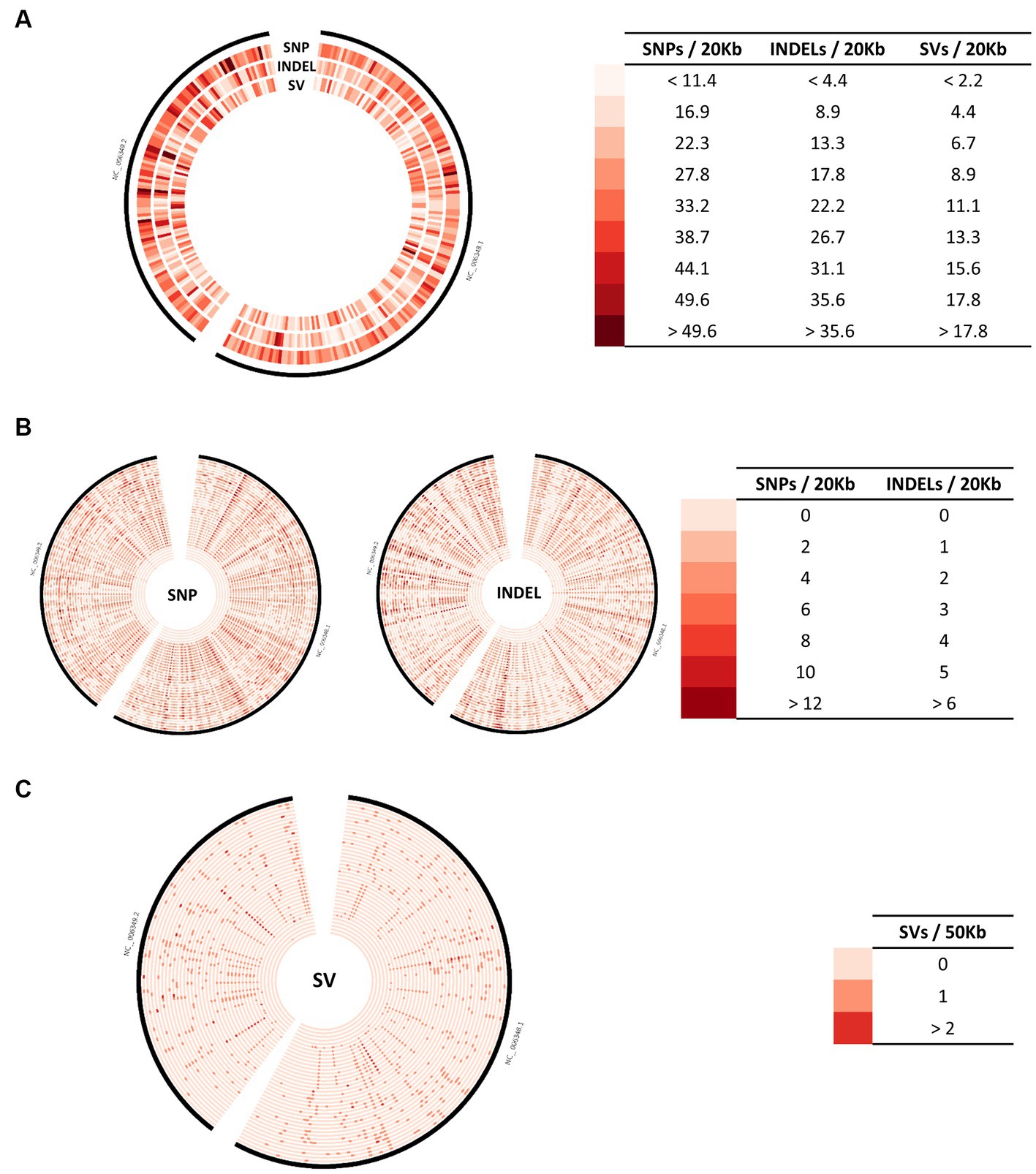
Figure 1. Distribution of genomic variations across the genome of B. mallei. (A) Distribution of SNPs, INDELs, and SVs across the genome for 112 B. mallei strains. (B) Distribution of SNPs and INDELS for 38 isolates with complete or chromosome-level genome assembly. (C) Distribution of SVs for 38 isolates with complete or chromosome-level genome assembly. The red gradient represents the density of each type of genomic variation.
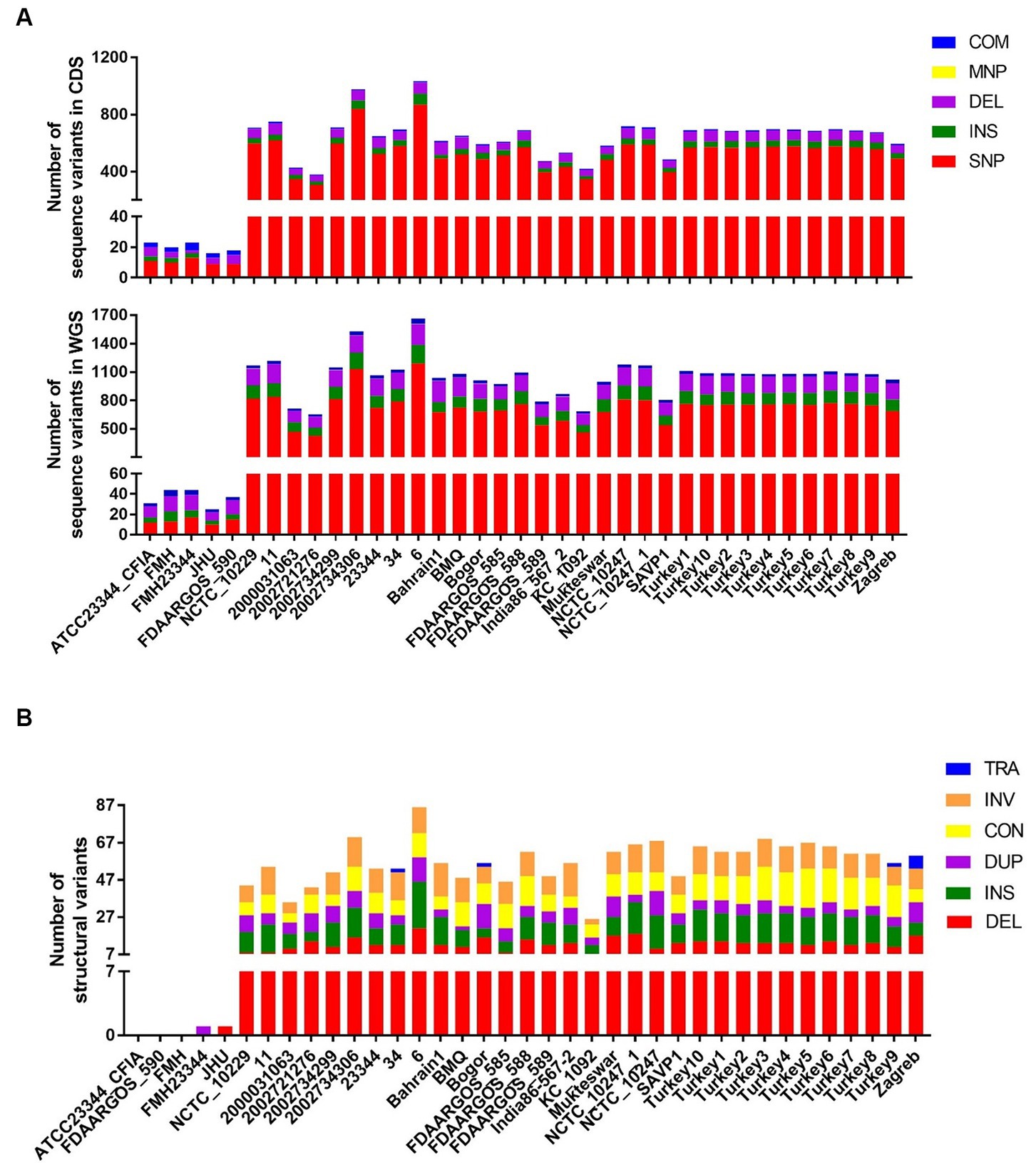
Figure 2. Various types of genomic variations identified in each B. mallei strain with complete or chromosome-level genome assembly. (A) The number of SNP, INDEL, MNP, and complex variants identified in the coding regions and whole genome sequences. (B) The number of multiple types of SVs identified in WGS. SNP, Single Nucleotide Polymorphism; MNP, Multiple Nucleotide Polymorphism; INS, Insertion; DEL, Deletion; COM, Complex (combination of SNP/MNP); TRA, Translocation; INV, Inversion; CON, Contraction; DUP, Duplication.
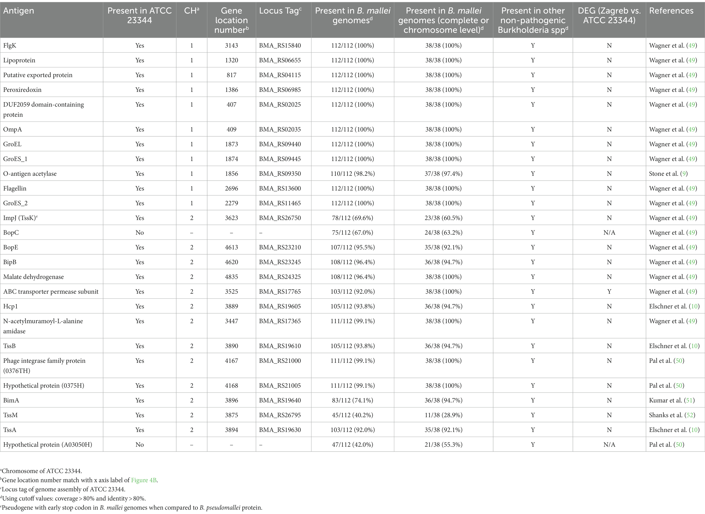
Table 1. Gene-encoded purified proteins used for ELISA development or as potential serodiagnostic markers.
All 112 available genome assemblies of B. mallei were used to identify the core and accessory genomes. Upon successive addition of each genome, the number of gene families in the pan-genome increased to 5,332 and seemed to reach a plateau with a very low γ-value (0.03) of a Heaps’ law fitting, which reflects that B. mallei’s pan-genome is nearly closed (38) (Supplementary Figures S1A,B). Overall, 5,332 gene families were identified (Supplementary Figure S1B), in which there were 3,464 persistent genes present in over 95% of the B. mallei genomes, 1,386 shell genes that were present at intermediate frequencies, and 482 cloud genes that were found in one, or very few individuals. The gene frequencies tend to show a strongly asymmetric U-shaped distribution with a larger proportion of persistent genes (Supplementary Figure S1B), indicating a lower level of gene gain achieved through horizontal gene transfer (HGT) within B. mallei (53). The pan-genome matrix shown in Figure 3A demonstrated substantial variations of gene content among all the genomes of B. mallei available in this study. These data indicate gene loss is a significant consequence of B. mallei’s genetic variation as limited gene gain was observed.
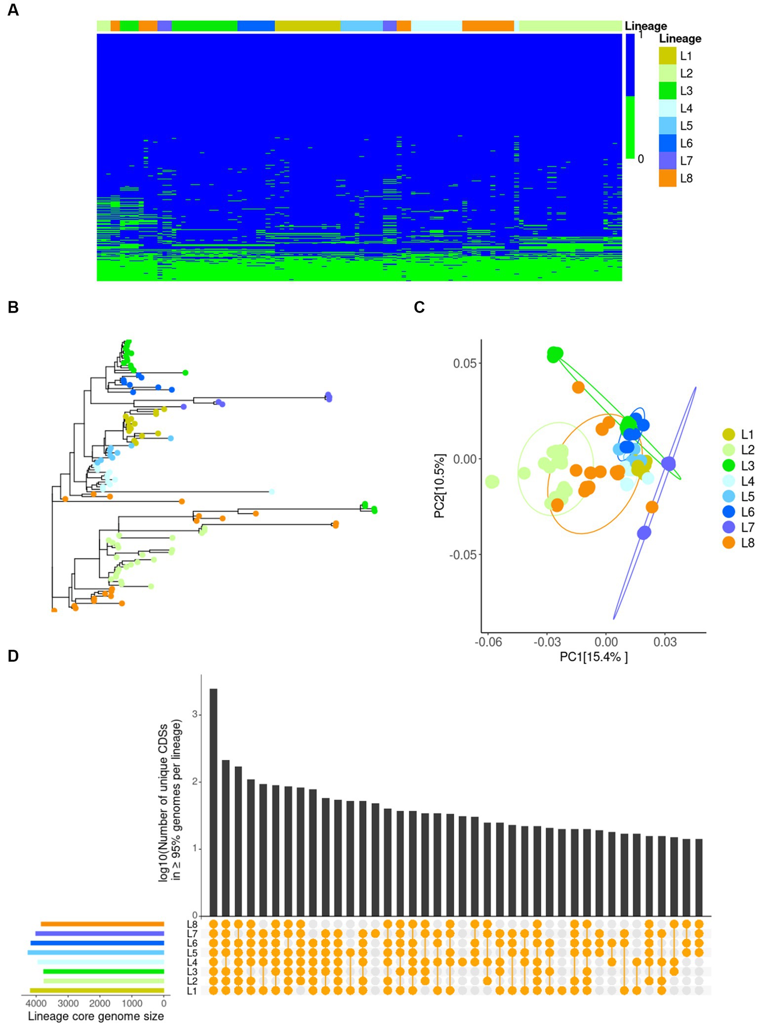
Figure 3. Variations in gene content of B. mallei isolates as identified by Pan-genome analysis. (A) A presence/absence matrix of 5,332 genes in the B. mallei pan-genome. Each row represents a gene cluster. Each column corresponds to a strain’s gene content. The color bar at the top represents different lineages. (B) Phylogenetic tree of 112 B. mallei isolates using the binary data of presence/absence gene generated by IQtree. (C) Principal coordinates analysis (PCoA) plot of gene content between each lineage based on Bray–Curtis dissimilarity. The color represents different lineages. (D) UpSetR plot showing the large intersection of lineage-specific core genomes. Intersections between lineages as shown by the presence of a yellow dot in the presence/absence matrix underneath the bar plot. The bar plot represents a logarithm of the number of genes in each intersection.
Gene content variations of a bacterial pathogen have been demonstrated to contribute to various acute and adaptive immune responses elicited by different strains of the same bacterial species (20). Due to the highly plastic genome of B. mallei (14), it is highly possible that long term evolution of this species may magnify this phenomenon thereby increasing the complexity of glanders serodiagnosis. To achieve greater accuracy in analyzing gene content variations and avoid unreliable or unavailable metadata mentioned previously (54), we implemented hierarchical Bayesian clustering via FastBaps, which further divided the worldwide phylogeny based on core SNPs into strain-level clusters (Supplementary Figure S2). In total, we identified eight clusters corresponding to different lineages (L1-8) (Supplementary Figure S2). L1 was built up from genomes of the type strain ATCC 23344 and derivatives of ATCC 23344 (US as geographic origin). Other Chinese isolates, such as strain China 5 and ATCC 10399, were found in L5. Interestingly, strains FDAARGOS_587, 2000031063, and KC_1092 were also clustered in L5, an observation which is consistent with previous publications that have pointed out inaccurate metadata for these isolates (15, 54, 55). The majority of isolates from Turkey were grouped into L2. Within the Indian strains in L3, the Budapest strain from Hungary was surprisingly close to strain SAVP1, as previously reported (15). Five genome sequences of possible Russian isolates with SCPM Identifier comprised L4. An exception was the strain SCPM-O-B-7093, which was most closely related to the strains Bahrain1, Bogor, Zagreb, and Mukteswar isolated in the 1990s or 2010s within L6. The genomes of UK strains 2002734306, FDAARGOS_586, and NCTC 120 were grouped with the Turkey strain 6 in L7. The L8 group consisted of strains isolated from different geographical locations and hosts.
Comparing gene contents between different lineages, isolates from the same lineage or close geographic location tended to have rather similar gene contents (Figures 3A,B), except for those of L3 and L8 which overlapped with other lineages. To investigate this finding further, using binary data of the presence/absence of genes, we performed Principal Coordinates Analysis (PCoA) to explore the dissimilarity of gene content between lineages and further confirmed the distinct gene presence-absence variations within L3 and L8 (Figure 3C). Additionally, there were clear differences in gene content between L2 or L7 and the rest of the lineages (Figure 3C), demonstrating that the gene content of the Turkey or UK strains is quite different from those of strains isolated from other geographic locations. Variations in each lineage-core genome (genes present in over 95 percent of the genomes in each lineage) were evident (Figure 3D). All these data demonstrate that B. mallei strains from different lineages or isolated from various geographic locations have different gene contents, suggesting various adaptive immune responses can possibly be elicited by strains from different lineage or geographic locations.
Moreover, we explored gene content diversity in the complete and chromosome-level genome assemblies to uncover gene presence-absence variations at the individual strain level using strain ATCC 23344 as a reference. Gene loss for each isolate and missing genes in the ATCC 23344 genome (gene gain in other genomes) were identified (Figure 4A). Interestingly, some strains, such as strains 6, 2002734306, and Turkey 6, even lost many persistent genes, while strain ATCC 23344 also lost four persistent genes (Figure 4A). The absence of genes in each isolate was mapped to the chromosomes of ATCC 23344, and multiple hotspots on CH1 and CH2 were observed, especially on CH2 (Figure 4B). Notably, most of the genes encoding antigens, such as BimA, HCP1, TssA, TssB, TssM, A03050H, and 0376H, used in established or developing serodiagnostic tests for glanders or melioidosis are located within those CH2 hotspots and are thus missing from some B. mallei isolates (Figure 4B and Table 1). All these data strongly demonstrate that B. mallei genome plasticity affects gene content, including some genes which encode proteins used for glanders serodiagnosis.
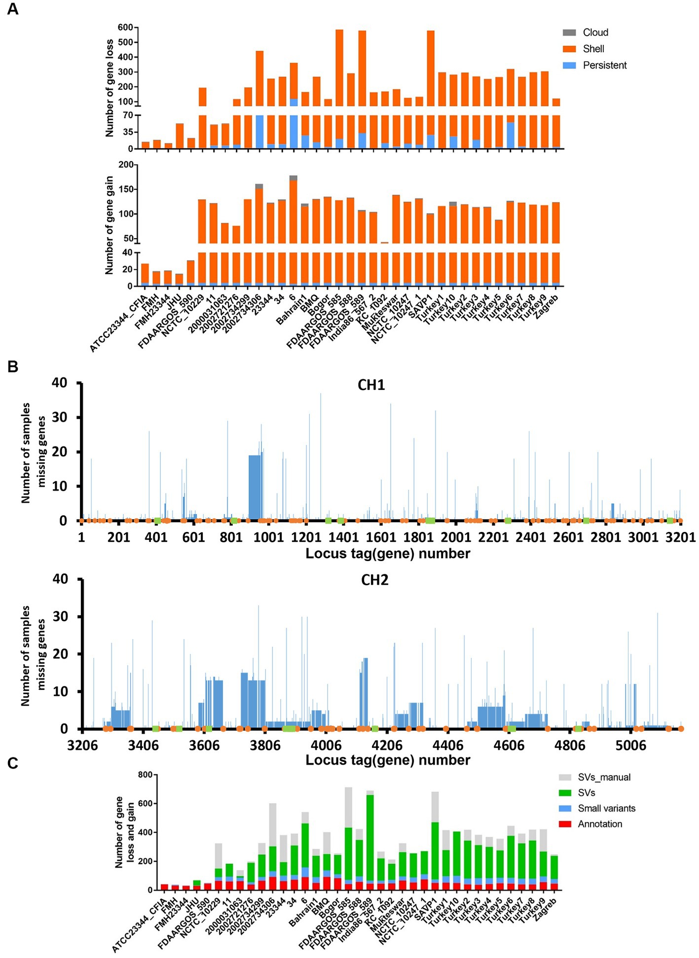
Figure 4. Effects of genomic variations on gene content. (A) Number of genes lost and gained (loss in strain ATCC 23344) in each B. mallei strain with complete or chromosome-level genome assembly. (B) Distribution of gene losses in B. mallei strains with complete or chromosome-level genome assembly on the genome of ATCC 23344. The orange dot represents transposase, and the green square shows gene coding antigens shown in Table 1. (C) Number of gene losses and gains in each B. mallei strain with complete or chromosome level genome assembly, compared to strain ATCC 23344. The color represents the cause of the gene loss or gain. Annotation: genes were not predicted by PGAP; Small variants: gene losses are due to SNPs, INDELs, MNPs, and complex variants. SVs: gene loss due to SVs. SV_manual: gene loss due to SVs that were not identified by MUM&Co.
To determine how genomic variations correlated with gene loss, we focused on the cross-comparison of genetic variation with gene content. Although a large number of small variants have been detected in CDS (Figure 2A), resulting in early stop codons and frameshifts, most of the gene loss events were associated with SVs (Figure 4C), which further confirms the key role of SVs in genome plasticity and its role in the loss of genes encoding antigens related to serodiagnosis such as Hcp1, TssB, BimA, and TssA (Figure 4B and Table 1). Additionally, some gene losses are related to the annotation performed by PGAP, which are mainly labeled as pseudogenes due to frameshifts or genes not being annotated (Figure 4C).
To understand potential effects of sequence variations on gene expression, we compared the transcriptome profiles of strain Zagreb with ATCC 23344 and found slight, but not significant differences overall (PERMANOVA: R2 = 0.415 Pr(> F) = 0.1) (Supplementary Figure S3A), due to a variation of gene expression of less than 10% (Figure 5A and Supplementary File S2). To be specific, only 388 DEGs were identified, for which a total of 155 available KEGG Orthology (KO) were retrieved from the KEGG mapper reconstruction server. The results showed that a total of 82 pathways were affected. The key differences in affected gene functions involve various bacterial metabolic pathways, pathogenesis, and virulence, including ABC transporters, quorum sensing (QS), two-component system, and biofilm formation (Supplementary Figure S3B). Specifically, the expression of ABC transporter permease subunit, which has been regarded as a potential serodiagnostic marker (56) was significantly different between these two strains (Table 1). Since proteins related to bacterial pathogenesis and virulence are typically used as serodiagnostic biomarkers, we investigated expression differences for virulence factors. A total of 842 virulence factors and 2,294 potential virulence factors were predicted using the PathoFact pipeline (Supplementary File S3). Fifty-eight (58) out of the 842 virulence genes, which code for 24 secreted and 34 non-secreted virulence factors, were among the identified DEGs, including some genes related to the type IV secretion system (Figure 5B and Table 2).
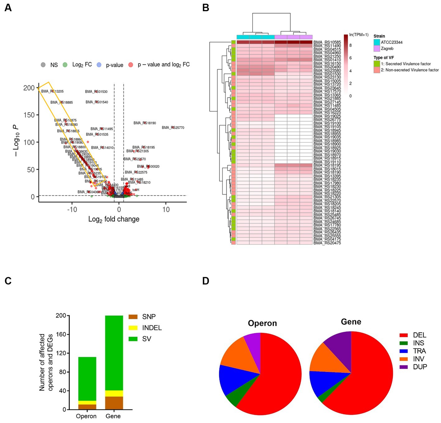
Figure 5. Effects of genomic variants on gene expression. (A) Volcano plot to display differentially expressed genes (DEGs) between type strain ATCC 23344 and strain Zagreb of B. mallei. The x and y axis represent Log2fold change and –log10P, respectively. The genes indicated in red dots represent the DEGs with |log2 (FC)| > 1 and p < 0.001. (B) The expression level of 58 differentially expressed virulence genes coding secreted VF (green) and non-secreted VF (pink) between strain ATCC 23344 and Zagreb. The density of the red color represents the gene expression level of ln(TPM + 1). Three biological replicates for each strain. (C) The number of SNP (brown), INDEL (yellow), and SV (green) related to affected operons and DEGs. (D) The percentage of different SVs related to affected operons and DEGs. The association of genomic variants on DEGs or affected operons was predicted using the simple predictive model that the destruction of the structure of operon and alteration of the non-coding regions, can interfere with gene expression. INS, Insertion; DEL, Deletion; TRA, Translocation; INV, Inversion; DUP, Duplication.
In an attempt to understand the effects of genomic variants on gene expression, we focused on not only changes in transcription related to the alteration of operon structures mediated by SVs, but also changes of non-coding regions upstream of operons, including promoter and regulator regions, mediated by SNPs or INDELs, respectively. Using transcriptomic data of strain ATCC 23344, OperonSEQer predicted a total of 2,455 operons (Supplementary Files S4, S5). One hundred and eighty-five of the DEGs were located within 108 operons disturbed by at least one type of genomic variant, especially SVs, with a large number being caused by deletions (Figures 5C,D). A large deletion from locus_tag BMA_RS18805 to BMS_RS19170 was observed in the genome of strain Zagreb, which was also reflected in transcriptomic data shown within the yellow rectangle in Figure 5A. Besides deletions, translocations also had a big impact on the expression of many genes, including several virulence genes (Figure 5D and Table 2).
Glanders diagnosis mainly relies on serological tests. Apart from different assay platforms, selection of appropriate antigens used in serological assays is critical to achieving adequate diagnostic sensitivity and specificity. Due to its simple standardization and objective analysis, ELISAs using recombinant proteins as antigens have shown very promising results with a potential replacement of CFT for glanders serodiagnosis in the near future (6). However, in the current study, we showed that, due to its high plasticity, the genome of B. mallei contains large-scale variants, resulting in differences in gene content and expression among each isolate. A number of gene-encoded proteins, including antigens used for the development of glanders ELISAs and virulence factors, are deleted, modified, or inactivated, in some isolates thereby compromising the efficacy of serodiagnostic tests using purified recombinant antigens.
Using all the B. mallei genome sequences available online and in-house, we undertook a comprehensive investigation of the nature of genetic variations of B. mallei isolates. SNPs are the most common type of genetic variation. The rate of single nucleotide alterations generated upon passage or infection has been reported as being typically very low (17). This suggests that SNPs may have a minor effect on genome plasticity, which is further supported by the small number of core SNPs identified in our study. Therefore, SNPs may provide a good level of discrimination and avoid genome alteration during passages. Indeed, the whole genome SNP-based phylogenetic analysis presented in this study demonstrates a high resolution of relationships among B. mallei isolates collected from diverse geographical locations (Supplementary Figure S2). Our phylogeny is quite similar to the phylogenetic relationships obtained from previous cgMLST (15, 54) or WGS-based SNP typing analyses (57). Compared to the previous three lineages (57) or 12 clusters (BHR, UAE, IND1/2/3, CHN1/2, TUR1/2/3, HUN, RUS) (15), hierarchical Bayesian clustering (FastBaps) used in this study subdivided the phylogeny into eight lineages (L1-8). The previously defined L1 is the same as our L7 (57), containing strain 6 and 20027344306 (NCTC120 or FDAARGOS_586), while previously reported L2 was further subdivided into current L1 (cluster CHN2), L5 (cluster CHN1), L3 (cluster IND1), and L6 (clusters IND2 and BHR). The previously defined L3 consisted of our L2 (cluster TUR1), L4 (cluster RUS) and L8 (clusters TUR 2, TUR3, HUN, and IND3).
Furthermore, our eight lineages match clusters identified through high-resolution melting PCR (PCR-HRM) (58). Except for L8 containing assemblies from three subtypes: L3B2, L3B3sB1, and L3B3sB3, each lineage is comprised of only genomes from the same PCR-HRM cluster, such as L1 (L2B2sB1Grp1), L2 (L3B3sB2), L3 (L2B2sB2), L4 (L3B1), L5 (L2B2sB1Grp2), L6 (L2B1), and L7 (L1). It is likely that L8 can be further subdivided using FastBap, which may match PCR-HRM cluster analysis. The lineage analysis in this study will provide new clues to enhance future molecular typing.
Besides SNPs, a relatively small number of INDELs were also identified across all B. mallei genomes available in RefSeq (Figure 1B). In contrast to consequences of SNPs, it is believed that there are some links between the accumulation of INDELs and the evolution of B. mallei (17). INDELs in both coding and non-coding regions were observed in the B. mallei genome upon passage in vitro and in vivo, thereby resulting in a few ORF frameshifts and alteration of gene expression, respectively, (17). Similar results can be seen in our data (Figures 2A, 4C, 5C).
It is generally believed that B. mallei evolved from an ancestral B. pseudomallei following serendipitous infection of a living host followed by ongoing evolution in which the genome underwent large-scale chromosome rearrangements and reduction (16, 17). Following this change, B. mallei continues to evolve with additional large genome reductions resulting from chromosomal rearrangements, which represent a significant contributor to genome plasticity (16, 17). Therefore, it is not surprising that a large number of SVs, specifically deletions, were observed in many B. mallei isolates (Figures 1C, 2B). So far, consequences, causes, and underlying mechanisms of SVs in B. mallei have not been well characterized. Our analysis revealed that SVs, in particular large deletions, are a significant cause for gene loss (Figure 4C) and altered operons, resulting in interference with structural gene expression (Figure 5D). Additionally, the strain JHU, obtained from a laboratory-acquired infection of B. mallei ATCC 23344, had evidence of an approximately 97 kb deletion starting from 1,226,811 to 1,324,421 on the CH2, compared to the reference strain ATCC 23344, which had not been reported in the previous study (17), thus suggesting that chromosome rearrangement could also occur in B. mallei during infection.
Consistent with this study, a previous report clearly indicated that the smaller secondary chromosome evolves more rapidly than the larger one, showing greater substitution rates and gene dispensability (59). This secondary chromosome usually serves as an evolutionary testing ground where genes are weakly preserved (18), which may explain the higher frequency of gene loss on the B. mallei CH2, especially in vivo (17). Based on previous transcriptional data, transposases on CH1 were down-regulated, and most of the over-expressed genes were located on CH2 in vivo (60), Over-expression of transposases on CH2 may be associated with instability of CH2 in vivo, which can be investigated and confirmed by future studies.
Genetically different strains of the same pathogenic bacterial species can elicit markedly diverse host immune responses during infection (20). The presence of large scale genetic changes in B. mallei may amplify this phenomenon during long–term evolution, particularly in vivo, as demonstrated in other Burkholderia species (61–64). Many genes in B. mallei CH2 have virulence or virulence-related functions, playing a pivotal role in pathogenesis (60). The extensive gene losses observed in CH2 (Figure 4B) could potentially lead to a reduction in its virulence during infection, thereby contributing to B. mallei’s ability to transmit and colonize between hosts. This serves as an example of optimized interaction between pathogenic bacteria and hosts to maximize their reproductive success, such as transmission, survival, and colonization (65). However, due to gene loss and alteration of gene expression, it is highly possible that antigens may be disrupted at the genomic or transcriptomic levels during infection, a phenomenon demonstrated in previous studies. For example, one spleen isolate of B. mallei obtained post-infection displayed a change in LPS phenotype, from smooth to rough, due to loss of O-polysaccharide during infection (66). Additionally, several B. mallei strains, such as B. mallei ATCC 23344 human and horse isolates, showed several gene deletions, including BMAA0367 (GNAT family N-acetyltransferase), BMAA0623, BMA2996, BMAA0729 (TssM), and BMA0685 (TonB) due to INDELs (17). TssM and TonB are well-known virulence factors of B. mallei (52, 67). Furthermore, TssM in B. pseudomallei has been investigated as a potential serodiagnostic biomarker (68) and used to develop a vaccine for melioidosis (69). BMAA0367, a GNAT family N-acetyltransferase, has not been well characterized in Burkholderia spp., but it was up-regulated during B. mallei infections (60), indicating that it is associated with B. mallei’s pathogenesis. Here, we found that genes encoding several antigens used to develop ELISA or potential serodiagnostic biomarkers, such as TssB, TssA, BimA, A03050H, and Hcp1, are not present in several B. mallei complete genome assemblies, due to genomic variations, but are present in other environmental Burkholderia spp. (Table 1). Most importantly, most of these genes are located within hotspots of gene loss on CH2 (Figure 4B). Loss of these gene-encoded antigens or virulence factors in B. mallei may be more likely to occur during natural infection compared to laboratory culturing since genome evolution in bacterial pathogens can occur faster and at higher frequency during the span of short host infections (13, 70–72). Based on the instability of CH2, it would be optimal to identify stable, unique, and conserved serodiagnostic biomarkers using genes located on CH1, such as GroEL (73). Moreover, by targeting multiple conserved serodiagnostic antigens from different lineages in a single serodiagnostic test, we could mitigate the effects of specific antigen loss and could cover different lineages, leading to a potential improvement in sensitivity and specificity.
Genomic variations, especially structural variations, have a significant impact on overall gene expression. Unfortunately, most SV studies mainly focus on humans and other multicellular eukaryotic organisms. To study the effects of multiple types of variants on gene expression in bacteria, we focused on the B. mallei strain Zagreb. This strain was chosen due to its wide usage for diagnostic antigen production and the presence of a large number of inter-chromosomal translocations observed in its genome when compared to ATCC 23344 (data not shown). Using the simple predictive model that the destruction of the operon structure and alteration of the 5′ flanking non-coding regions, including promoters and transcriptional regulators, can interfere with gene expression, this study has shown that genomic variations, specifically SVs, can alter gene expression, including some virulence genes (Figure 5B and Table 2). Interestingly, deletion of LuxI/LuxR-type QS systems observed in the strain Zagreb may also be indirectly altering the expression of many other genes, including those of virulence factors, toxins, and biofilm components (74).
In this study, we used all available B. mallei genome assemblies in the RefSeq database and assemblies of four additional in-house B. mallei strains. It is important to note that the quality of bacterial genome assemblies in the RefSeq database may vary. To address this issue, we assessed the quality of the 112 genome assemblies and annotations using BUSCO’s completeness score (Supplementary File S1) for quality control. However, it should be acknowledged that the presence of sequencing and assembly errors, in addition to accuracy of aligners and variant callers, may affect the genetic variation identification performed in our study (75). Nevertheless, this limitation must be taken into consideration. Besides that, bacterial gene expression and regulation are complicated and have yet to be fully elucidated, so it is hard to comprehensively investigate the effect of genomic variations on gene expression using the limited sample size in this study. Furthermore, there is a significant difference in gene expression profiling of a B. mallei isolate between in vivo and in vitro, particularly with most of the over-expressed genes on CH2 in vivo (60). This indicates in vitro transcriptional data may not be instrumental in predicting in vivo gene expression and regulation, and the possibility that virulence genes or serodiagnostic markers located on CH2 show variable expression between different isolates during infection will be investigated further. However, it is likely that the disruption of operon structures by SVs, such as deletion and translocation, impacts the expression of genes within or near the operon during infection as well, since these co-regulated genes under a common control system are interrupted (46). The specific impacts of SVs on gene expression needs to be further investigated using more transcriptome data, especially in relation to bacterial infection and virulence factors.
In summary, we have demonstrated that the plasticity of the B. mallei genome interferes with its gene content and expression. Accordingly, methods of glanders serodiagnosis using only purified recombinant antigens could be potentially compromised. Multiple instances have demonstrated that certain targeted antigens, including BimA, HCP1, TssA, TssB, TssM, A03050H, or potentially valuable antigens like virulence factors, may undergo loss or significant changes in expression due to genetic variations. As a result, these alterations can potentially lead to modifications in the immune response, including antibody production, during infection. This study provides novel insights into B. mallei genome plasticity, including details and consequences of genomic variation. These findings will be useful for informing improved glanders serodiagnosis and molecular typing in the future.
The whole-genome assemblies of strains ATCC 23344, Zagreb, Mukteswar and Bogor have been deposited in GenBank under accession number CP124607-CP124608, CP124605-CP124606, CP124602-CP124603, and CP124604, respectively. All the raw sequencing data are available under BioProject number PRJNA954830. Specifically, the RNA-sequencing raw data are available in the NCBI Sequence Read Archive under accession numbers SRR24153017 to SRR24153022 and whole genome sequencing raw data are available in the NCBI Sequence Read Archive under accession numbers SRR24860065 to SRR24860072.
MK designed the experiments. MK, RG, and PC wrote the manuscript. PC, RG, JC, EH, SN-D, DC, JH, KM, M-OD, and MK reviewed and edited the manuscript. JC, EH, and SN-D performed WGS. PC, MK, RG, JC, and M-OD analyzed sequencing data. DC, JH, and KM conducted bacterial culture and RNA purification experiments. All authors contributed to the article and approved the submitted version.
This research was supported by the Canadian Food Inspection Agency.
We would like to thank the Friedrich Loeffler Institute for sharing B. mallei strains Zagreb, Mukteswar, and Bogor with us. We also thank Centre d’expertise et de service Génome Québec for performing the RNA sequencing.
The authors declare that the research was conducted in the absence of any commercial or financial relationships that could be construed as a potential conflict of interest.
All claims expressed in this article are solely those of the authors and do not necessarily represent those of their affiliated organizations, or those of the publisher, the editors and the reviewers. Any product that may be evaluated in this article, or claim that may be made by its manufacturer, is not guaranteed or endorsed by the publisher.
The Supplementary material for this article can be found online at: https://www.frontiersin.org/articles/10.3389/fvets.2023.1217135/full#supplementary-material
SUPPLEMENTARY FIGURE S1 | Pan-genome analysis of 112 B. mallei genomes. (A) Rarefaction curves for the core (persistent) genome size and pan-genome. (B) U-shaped distribution demonstrating gene cluster count among 112 B. mallei genomes.
SUPPLEMENTARY FIGURE S2 | SNP-based Phylogenetic tree of B. mallei isolates and related metadata. The phylogenetic tree was obtained by IQtree with 1000 ultrafast bootstraps. Lineages (L1-8) were determined by Fastbap. Geographic location was collected from BioSample and Assembly database from NCBI: Bahrain (BH), Brazil (BR), China (CN), Hungary (HU), India (IN), Indonesia (ID), Iran (IR), Myanmar (MM), Pakistan (PK), Russia (RU), Turkey (TR), United Kingdom (UK), United States (US), and Yugoslavia (YU).
SUPPLEMENTARY FIGURE S3 | Differences in gene expression and Kyoto Encyclopedia of Genes and Genomes (KEGG) pathways between strain ATCC 23344 and Zagreb. (A) PCoA plot of gene expression between strain ATCC 23344 and Zagreb based on Bray-Curtis dissimilarity. Three biological replicates for each strain. (B) KEGG pathways correlating to virulence DEGs between strain ATCC 23344 and Zagreb. The red color bars represent pathways related to bacterial pathogeny and virulence.
SUPPLEMENTARY Table S1 | Summary of genome assembly information.
SUPPLEMENTARY Table S2 | Number of reads for each sample in RNA-seq experiment.
SUPPLEMENTARY FILE S1 | Genomes and assemblies used in this study.
SUPPLEMENTARY FILE S2 | Summary of DEGs generated from RNA-seq analysis.
SUPPLEMENTARY FILE S3 | Virulence factors predicted by PathoFact.
SUPPLEMENTARY FILE S4 | Operons on CH1 predicted by OperonSEQer.
SUPPLEMENTARY FILE S5 | Operons on CH2 predicted by OperonSEQer.
1. ^https://github.com/AstrobioMike/bit#bioinformatics-tools-bit
2. ^https://astrobiomike.github.io/unix/ncbi_eutils
3. ^https://github.com/rrwick/Porechop
4. ^https://github.com/rrwick/Filtlong
5. ^https://github.com/nanoporetech/medaka
6. ^https://github.com/tseemann/snippy
7. ^https://github.com/SAMtoBAM/MUMandCo
8. ^https://github.com/iqtree/iqtree2
9. ^https://github.com/gtonkinhill/fastbaps
11. ^https://github.com/labgem/PPanGGOLiN
12. ^https://cran.r-project.org/web/packages/ggplot2/index.html
13. ^https://cran.r-project.org/web/packages/pheatmap/
14. ^https://github.com/vegandevs/vegan
1. Whitlock, GC, Valbuena, GA, Popov, VL, Judy, BM, Estes, DM, and Torres, AG. Burkholderia mallei cellular interactions in a respiratory cell model. J Med Microbiol. (2009) 58:554–62. doi: 10.1099/jmm.0.007724-0
2. Van Zandt, KE, Greer, MT, and Gelhaus, HC. Glanders: an overview of infection in humans. Orphanet J Rare Dis. (2013) 8:131. doi: 10.1186/1750-1172-8-131
3. Kettle, AN, and Wernery, U. Glanders and the risk for its introduction through the international movement of horses. Equine Vet J. (2016) 48:654–8. doi: 10.1111/evj.12599
4. Khan, I, Wieler, LH, Melzer, F, Elschner, MC, Muhammad, G, Ali, S, et al. Glanders in animals: a review on epidemiology, clinical presentation, diagnosis and countermeasures. Transbound Emerg Dis. (2013) 60:204–21. doi: 10.1111/j.1865-1682.2012.01342.x
5. Estes, DM, Dow, SW, Schweizer, HP, and Torres, AG. Present and future therapeutic strategies for melioidosis and glanders. Expert Rev Anti-Infect Ther. (2010) 8:325–38. doi: 10.1586/eri.10.4
6. Elschner, MC, Melzer, F, Singha, H, Muhammad, S, Gardner, I, and Neubauer, H. Validation of a commercial glanders ELISA as an Alternative to the CFT in international trade of equidae. Front Vet Sci. (2021) 8:628389. doi: 10.3389/fvets.2021.628389
7. Elschner, MC, Scholz, HC, Melzer, F, Saqib, M, Marten, P, Rassbach, A, et al. Use of a Western blot technique for the serodiagnosis of glanders. BMC Vet Res. (2011) 7:4. doi: 10.1186/1746-6148-7-4
8. Sengyee, S, Yoon, SH, West, TE, Ernst, RK, and Chantratita, N. Lipopolysaccharides from different burkholderia species with different lipid a structures induce toll-like receptor 4 activation and react with melioidosis patient sera. Infect Immun. (2019) 87:e00692-19. doi: 10.1128/IAI.00692-19
9. Stone, JK, Mayo, M, Grasso, SA, Ginther, JL, Warrington, SD, Allender, CJ, et al. Detection of Burkholderia pseudomallei O-antigen serotypes in near-neighbor species. BMC Microbiol. (2012) 12:250. doi: 10.1186/1471-2180-12-250
10. Elschner, MC, Laroucau, K, Singha, H, Tripathi, BN, Saqib, M, Gardner, I, et al. Evaluation of the comparative accuracy of the complement fixation test, Western blot and five enzyme-linked immunosorbent assays for serodiagnosis of glanders. PLoS One. (2019) 14:e0214963. doi: 10.1371/journal.pone.0214963
11. Singha, H, Malik, P, Goyal, SK, Khurana, SK, Mukhopadhyay, C, Eshwara, VK, et al. Optimization and validation of indirect ELISA using truncated TssB protein for the serodiagnosis of glanders amongst equines. ScientificWorldJournal. (2014) 2014:469407:1–6. doi: 10.1155/2014/469407
12. Godoy, D, Randle, G, Simpson, AJ, Aanensen, DM, Pitt, TL, Kinoshita, R, et al. Multilocus sequence typing and evolutionary relationships among the causative agents of melioidosis and glanders, Burkholderia pseudomallei and Burkholderia mallei. J Clin Microbiol. (2003) 41:2068–79. doi: 10.1128/JCM.41.5.2068-2079.2003
13. Losada, L, Ronning, CM, Deshazer, D, Woods, D, Fedorova, N, Kim, HS, et al. Continuing evolution of Burkholderia mallei through genome reduction and large-scale rearrangements. Genome Biol Evol. (2010) 2:102–16. doi: 10.1093/gbe/evq003
14. Nierman, WC, Deshazer, D, Kim, HS, Tettelin, H, Nelson, KE, Feldblyum, T, et al. Structural flexibility in the Burkholderia mallei genome. Proc Natl Acad Sci U S A. (2004) 101:14246–51. doi: 10.1073/pnas.0403306101
15. Appelt, S, Rohleder, AM, Jacob, D, Von Buttlar, H, Georgi, E, Mueller, K, et al. Genetic diversity and spatial distribution of Burkholderia mallei by core genome-based multilocus sequence typing analysis. PLoS One. (2022) 17:e0270499. doi: 10.1371/journal.pone.0270499
16. Bochkareva, OO, Moroz, EV, Davydov, I, and Gelfand, MS. Genome rearrangements and selection in multi-chromosome bacteria Burkholderia spp. BMC Genomics. (2018) 19:965. doi: 10.1186/s12864-018-5245-1
17. Romero, CM, Deshazer, D, Feldblyum, T, Ravel, J, Woods, D, Kim, HS, et al. Genome sequence alterations detected upon passage of Burkholderia mallei ATCC 23344 in culture and in mammalian hosts. BMC Genomics. (2006) 7:228. doi: 10.1186/1471-2164-7-228
18. Cooper, VS, Vohr, SH, Wrocklage, SC, and Hatcher, PJ. Why genes evolve faster on secondary chromosomes in bacteria. PLoS Comput Biol. (2010) 6:e1000732. doi: 10.1371/journal.pcbi.1000732
19. Laroucau, K, Aaziz, R, Vorimore, F, Varghese, K, Deshayes, T, Bertin, C, et al. A genetic variant of Burkholderia mallei detected in Kuwait: Consequences for the PCR diagnosis of glanders. Transbound Emerg Dis. (2021) 68:960–3. doi: 10.1111/tbed.13777
20. Sela, U, Euler, CW, Correa Da Rosa, J, and Fischetti, VA. Strains of bacterial species induce a greatly varied acute adaptive immune response: The contribution of the accessory genome. PLoS Pathog. (2018) 14:e1006726. doi: 10.1371/journal.ppat.1006726
21. Feron, R, and Waterhouse, RM. Assessing species coverage and assembly quality of rapidly accumulating sequenced genomes. Gigascience. (2022) 11:giac006. doi: 10.1093/gigascience/giac006
22. Lawniczak, MKN, Durbin, R, Flicek, P, Lindblad-Toh, K, Wei, X, Archibald, JM, et al. Standards recommendations for the Earth BioGenome Project. Proc Natl Acad Sci U S A. (2022) 119:e2115639118. doi: 10.1073/pnas.2115639118
23. Simao, FA, Waterhouse, RM, Ioannidis, P, Kriventseva, EV, and Zdobnov, EM. BUSCO: assessing genome assembly and annotation completeness with single-copy orthologs. Bioinformatics. (2015) 31:3210–2. doi: 10.1093/bioinformatics/btv351
24. Lopez, J, Copps, J, Wilhelmsen, C, Moore, R, Kubay, J, St-Jacques, M, et al. Characterization of experimental equine glanders. Microbes Infect. (2003) 5:1125–31. doi: 10.1016/j.micinf.2003.07.004
25. Kang, M, Chmara, J, Naushad, S, and Huang, H. Complete genome sequence of a canadian strain of Raoultella planticola with metal and antimicrobial resistance genes. Microbiol Resour Announc. (2021) 10:e0041521. doi: 10.1128/MRA.00415-21
26. Chen, S, Zhou, Y, Chen, Y, and Gu, J. fastp: an ultra-fast all-in-one FASTQ preprocessor. Bioinformatics. (2018) 34:i884–90. doi: 10.1093/bioinformatics/bty560
27. Wick, RR, Judd, LM, Gorrie, CL, and Holt, KE. Unicycler: Resolving bacterial genome assemblies from short and long sequencing reads. PLoS Comput Biol. (2017) 13:e1005595. doi: 10.1371/journal.pcbi.1005595
28. Kolmogorov, M, Yuan, J, Lin, Y, and Pevzner, PA. Assembly of long, error-prone reads using repeat graphs. Nat Biotechnol. (2019) 37:540–6. doi: 10.1038/s41587-019-0072-8
29. Hu, J, Fan, J, Sun, Z, and Liu, S. NextPolish: a fast and efficient genome polishing tool for long-read assembly. Bioinformatics. (2020) 36:2253–5. doi: 10.1093/bioinformatics/btz891
30. Wick, RR, and Holt, KE. Polypolish: Short-read polishing of long-read bacterial genome assemblies. PLoS Comput Biol. (2022) 18:e1009802. doi: 10.1371/journal.pcbi.1009802
31. Li, H. Minimap2: pairwise alignment for nucleotide sequences. Bioinformatics. (2018) 34:3094–100. doi: 10.1093/bioinformatics/bty191
32. Okonechnikov, K, Conesa, A, and Garcia-Alcalde, F. Qualimap 2: advanced multi-sample quality control for high-throughput sequencing data. Bioinformatics. (2016) 32:292–4. doi: 10.1093/bioinformatics/btv566
33. Tatusova, T, Dicuccio, M, Badretdin, A, Chetvernin, V, Nawrocki, EP, Zaslavsky, L, et al. NCBI prokaryotic genome annotation pipeline. Nucleic Acids Res. (2016) 44:6614–24. doi: 10.1093/nar/gkw569
34. O'donnell, S, and Fischer, G. MUM&Co: accurate detection of all SV types through whole-genome alignment. Bioinformatics. (2020) 36:3242–3. doi: 10.1093/bioinformatics/btaa115
35. Minh, BQ, Schmidt, HA, Chernomor, O, Schrempf, D, Woodhams, MD, Von Haeseler, A, et al. IQ-TREE 2: new models and efficient methods for phylogenetic inference in the genomic era. Mol Biol Evol. (2020) 37:1530–4. doi: 10.1093/molbev/msaa015
36. Tonkin-Hill, G, Lees, JA, Bentley, SD, Frost, SDW, and Corander, J. Fast hierarchical Bayesian analysis of population structure. Nucleic Acids Res. (2019) 47:5539–49. doi: 10.1093/nar/gkz361
37. Mortazavi, M, Ren, Y, Saini, S, Antaki, D, St Pierre, CL, Williams, A, et al. SNPs, short tandem repeats, and structural variants are responsible for differential gene expression across C57BL/6 and C57BL/10 substrains. Cell Genom. (2022) 2:100102. doi: 10.1016/j.xgen.2022.100102
38. Gautreau, G, Bazin, A, Gachet, M, Planel, R, Burlot, L, Dubois, M, et al. PPanGGOLiN: Depicting microbial diversity via a partitioned pangenome graph. PLoS Comput Biol. (2020) 16:e1007732. doi: 10.1371/journal.pcbi.1007732
39. Yu, G. Using ggtree to Visualize Data on Tree-Like Structures. Curr Protoc Bioinformatics. (2020) 69:e96. doi: 10.1002/cpbi.96
40. Conway, JR, Lex, A, and Gehlenborg, N. UpSetR: an R package for the visualization of intersecting sets and their properties. Bioinformatics. (2017) 33:2938–40. doi: 10.1093/bioinformatics/btx364
41. De Nies, L, Lopes, S, Busi, SB, Galata, V, Heintz-Buschart, A, Laczny, CC, et al. PathoFact: a pipeline for the prediction of virulence factors and antimicrobial resistance genes in metagenomic data. Microbiome. (2021) 9:49. doi: 10.1186/s40168-020-00993-9
42. Bray, NL, Pimentel, H, Melsted, P, and Pachter, L. Near-optimal probabilistic RNA-seq quantification. Nat Biotechnol. (2016) 34:525–7. doi: 10.1038/nbt.3519
43. Pimentel, H, Bray, NL, Puente, S, Melsted, P, and Pachter, L. Differential analysis of RNA-seq incorporating quantification uncertainty. Nat Methods. (2017) 14:687–90. doi: 10.1038/nmeth.4324
44. Kanehisa, M, Sato, Y, and Morishima, K. BlastKOALA and GhostKOALA: KEGG tools for functional characterization of genome and metagenome sequences. J Mol Biol. (2016) 428:726–31. doi: 10.1016/j.jmb.2015.11.006
45. Kanehisa, M, Sato, Y, and Kawashima, M. KEGG mapping tools for uncovering hidden features in biological data. Protein Sci. (2022) 31:47–53. doi: 10.1002/pro.4172
46. Lawrence, JG. Shared strategies in gene organization among prokaryotes and eukaryotes. Cells. (2002) 110:407–13. doi: 10.1016/S0092-8674(02)00900-5
47. Krishnakumar, R, and Ruffing, AM. OperonSEQer: A set of machine-learning algorithms with threshold voting for detection of operon pairs using short-read RNA-sequencing data. PLoS Comput Biol. (2022) 18:e1009731. doi: 10.1371/journal.pcbi.1009731
48. Hamala, T, Wafula, EK, Guiltinan, MJ, Ralph, PE, Depamphilis, CW, and Tiffin, P. Genomic structural variants constrain and facilitate adaptation in natural populations of Theobroma cacao, the chocolate tree. Proc Natl Acad Sci U S A. (2021) 118:e2102914118. doi: 10.1073/pnas.2102914118
49. Wagner, GE, Berner, A, Lipp, M, Kohler, C, Assig, K, Lichtenegger, S, et al. Protein microarray-guided development of a highly sensitive and specific dipstick assay for glanders serodiagnostics. J Clin Microbiol. (2023) 61:e0123422. doi: 10.1128/jcm.01234-22
50. Pal, V, Kumar, S, Malik, P, and Rai, GP. Evaluation of recombinant proteins of Burkholderia mallei for serodiagnosis of glanders. Clin Vaccine Immunol. (2012) 19:1193–8. doi: 10.1128/CVI.00137-12
51. Kumar, S, Malik, P, Verma, SK, Pal, V, Gautam, V, Mukhopadhyay, C, et al. Use of a recombinant burkholderia intracellular motility a protein for immunodiagnosis of glanders. Clin Vaccine Immunol. (2011) 18:1456–61. doi: 10.1128/CVI.05185-11
52. Shanks, J, Burtnick, MN, Brett, PJ, Waag, DM, Spurgers, KB, Ribot, WJ, et al. Burkholderia mallei tssM encodes a putative deubiquitinase that is secreted and expressed inside infected RAW 264.7 murine macrophages. Infect Immun. (2009) 77:1636–48. doi: 10.1128/IAI.01339-08
53. Brockhurst, MA, Harrison, E, Hall, JPJ, Richards, T, Mcnally, A, and Maclean, C. The ecology and evolution of pangenomes. Curr Biol. (2019) 29:R1094–103. doi: 10.1016/j.cub.2019.08.012
54. Brangsch, H, Saqib, M, Sial, AUR, Melzer, F, Linde, J, and Elschner, MC. Sequencing-based genotyping of Pakistani Burkholderia mallei strains: a useful way for investigating glanders outbreaks. Pathogens. (2022) 11:614. doi: 10.3390/pathogens11060614
55. Brangsch, H, Singha, H, Laroucau, K, and Elschner, M. Sequence-based detection and typing procedures for Burkholderia mallei: Assessment and prospects. Front Vet Sci. (2022) 9:1056996. doi: 10.3389/fvets.2022.1056996
56. Wagner, JK, Setayeshgar, S, Sharon, LA, Reilly, JP, and Brun, YV. A nutrient uptake role for bacterial cell envelope extensions. Proc Natl Acad Sci U S A. (2006) 103:11772–7. doi: 10.1073/pnas.0602047103
57. Laroucau, K, De Assis, L, Santana, V, Girault, G, Martin, B, Miranda Da Silveira, PP, et al. First molecular characterisation of a Brazilian Burkholderia mallei strain isolated from a mule in 2016. Infect Genet Evol. (2018) 57:117–20. doi: 10.1016/j.meegid.2017.11.014
58. Girault, G, Wattiau, P, Saqib, M, Martin, B, Vorimore, F, Singha, H, et al. High-resolution melting PCR analysis for rapid genotyping of Burkholderia mallei. Infect Genet Evol. (2018) 63:1–4. doi: 10.1016/j.meegid.2018.05.004
59. Morrow, JD, and Cooper, VS. Evolutionary effects of translocations in bacterial genomes. Genome Biol Evol. (2012) 4:1256–62. doi: 10.1093/gbe/evs099
60. Kim, HS, Schell, MA, Yu, Y, Ulrich, RL, Sarria, SH, Nierman, WC, et al. Bacterial genome adaptation to niches: divergence of the potential virulence genes in three Burkholderia species of different survival strategies. BMC Genomics. (2005) 6:174. doi: 10.1186/1471-2164-6-174
61. Diaz Caballero, J, Clark, ST, Wang, PW, Donaldson, SL, Coburn, B, Tullis, DE, et al. A genome-wide association analysis reveals a potential role for recombination in the evolution of antimicrobial resistance in Burkholderia multivorans. PLoS Pathog. (2018) 14:e1007453. doi: 10.1371/journal.ppat.1007453
62. Hassan, AA, Dos Santos, SC, Cooper, VS, and Sa-Correia, I. Comparative evolutionary patterns of Burkholderia cenocepacia and B. multivorans during chronic co-infection of a cystic fibrosis patient lung. Front Microbiol. (2020) 11:574626. doi: 10.3389/fmicb.2020.574626
63. Lee, AH, Flibotte, S, Sinha, S, Paiero, A, Ehrlich, RL, Balashov, S, et al. Phenotypic diversity and genotypic flexibility of Burkholderia cenocepacia during long-term chronic infection of cystic fibrosis lungs. Genome Res. (2017) 27:650–62. doi: 10.1101/gr.213363.116
64. Lieberman, TD, Michel, JB, Aingaran, M, Potter-Bynoe, G, Roux, D, Davis, MR Jr, et al. Parallel bacterial evolution within multiple patients identifies candidate pathogenicity genes. Nat Genet. (2011) 43:1275–80. doi: 10.1038/ng.997
65. Diard, M, and Hardt, WD. Evolution of bacterial virulence. FEMS Microbiol Rev. (2017) 41:679–97. doi: 10.1093/femsre/fux023
66. Bernhards, RC, Cote, CK, Amemiya, K, Waag, DM, Klimko, CP, Worsham, PL, et al. Characterization of in vitro phenotypes of Burkholderia pseudomallei and Burkholderia mallei strains potentially associated with persistent infection in mice. Arch Microbiol. (2017) 199:277–301. doi: 10.1007/s00203-016-1303-8
67. Mott, TM, Vijayakumar, S, Sbrana, E, Endsley, JJ, and Torres, AG. Characterization of the Burkholderia mallei tonb mutant and its potential as a backbone strain for vaccine development. PLoS Negl Trop Dis. (2015) 9:e0003863. doi: 10.1371/journal.pntd.0003863
68. Tan, KS, Chen, Y, Lim, YC, Tan, GY, Liu, Y, Lim, YT, et al. Suppression of host innate immune response by Burkholderia pseudomallei through the virulence factor TssM. J Immunol. (2010) 184:5160–71. doi: 10.4049/jimmunol.0902663
69. Burtnick, MN, Shaffer, TL, Ross, BN, Muruato, LA, Sbrana, E, Deshazer, D, et al. Development of subunit vaccines that provide high-level protection and sterilizing immunity against acute inhalational melioidosis. Infect Immun. (2018) 86:e00724-17. doi: 10.1128/IAI.00724-17
70. Grote, A, and Earl, AM. Within-host evolution of bacterial pathogens during persistent infection of humans. Curr Opin Microbiol. (2022) 70:102197. doi: 10.1016/j.mib.2022.102197
71. Kraft, C, Stack, A, Josenhans, C, Niehus, E, Dietrich, G, Correa, P, et al. Genomic changes during chronic Helicobacter pylori infection. J Bacteriol. (2006) 188:249–54. doi: 10.1128/JB.188.1.249-254.2006
72. Mikonranta, L, Mappes, J, Laakso, J, and Ketola, T. Within-host evolution decreases virulence in an opportunistic bacterial pathogen. BMC Evol Biol. (2015) 15:165. doi: 10.1186/s12862-015-0447-5
73. Dohre, SK, Kamthan, A, Singh, S, Alam, SI, and Kumar, S. Identification of a new diagnostic antigen for glanders using immunoproteome analysis. Comp Immunol Microbiol Infect Dis. (2017) 53:26–32. doi: 10.1016/j.cimid.2017.06.007
74. Kai, K. Bacterial quorum sensing in symbiotic and pathogenic relationships with hosts. Biosci Biotechnol Biochem. (2018) 82:363–71. doi: 10.1080/09168451.2018.1433992
Keywords: Burkholderia mallei , genomic variation, serodiagnostic antigen, transcriptome, virulence factors
Citation: Charron P, Gao R, Chmara J, Hoover E, Nadin-Davis S, Chauvin D, Hazelwood J, Makondo K, Duceppe M-O and Kang M (2023) Influence of genomic variations on glanders serodiagnostic antigens using integrative genomic and transcriptomic approaches. Front. Vet. Sci. 10:1217135. doi: 10.3389/fvets.2023.1217135
Received: 04 May 2023; Accepted: 20 November 2023;
Published: 06 December 2023.
Edited by:
Min Yue, Zhejiang University, ChinaReviewed by:
Eunice M. Machuka, International Livestock Research Institute (ILRI), KenyaCopyright © 2023 Charron, Gao, Chmara, Hoover, Nadin-Davis, Chauvin, Hazelwood, Makondo, Duceppe and Kang. This is an open-access article distributed under the terms of the Creative Commons Attribution License (CC BY). The use, distribution or reproduction in other forums is permitted, provided the original author(s) and the copyright owner(s) are credited and that the original publication in this journal is cited, in accordance with accepted academic practice. No use, distribution or reproduction is permitted which does not comply with these terms.
*Correspondence: Mingsong Kang, bWluZ3Nvbmcua2FuZ0BpbnNwZWN0aW9uLmdjLmNh
†Present address: Ruimin Gao, National Microbiology Laboratory, Public Health Agency of Canada, Winnipeg, MB, Canada
‡These authors have contributed equally to this work
Disclaimer: All claims expressed in this article are solely those of the authors and do not necessarily represent those of their affiliated organizations, or those of the publisher, the editors and the reviewers. Any product that may be evaluated in this article or claim that may be made by its manufacturer is not guaranteed or endorsed by the publisher.
Research integrity at Frontiers

Learn more about the work of our research integrity team to safeguard the quality of each article we publish.