- 1Dhirubhai Ambani International School, Mumbai, India
- 2Private Veterinary Practice, Posh Vets Clinic, Mumbai, India
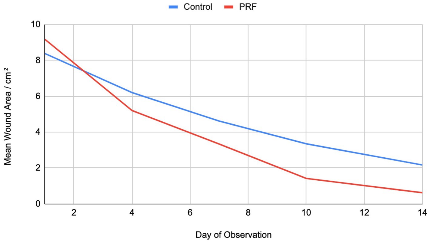
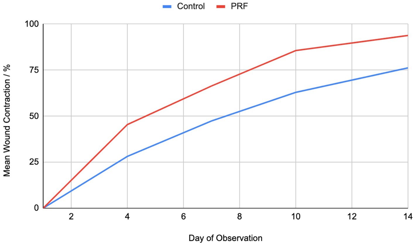
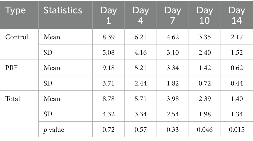
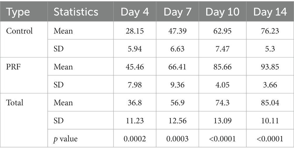
Street cats commonly present large skin wounds that pose significant challenges in veterinary practice. Platelet-rich fibrin (PRF) is a second-generation platelet concentrate increasingly used in humans to promote wound healing. Ease of use and clinical success in humans has prompted interest in using PRF in veterinary practice. However, until now, there is no reported study on the use of autologous PRF in feline wound management. This study evaluated the effect of application of autologous PRF in cats with naturally occurring cutaneous wounds. 16 cats with full-thickness cutaneous acute/subacute wounds were randomly allocated to PRF or Control (standard care) groups. Each cat was enrolled for 2 weeks. PRF was prepared according to previously described procedures. PRF was applied on Days 1 and 4 in addition to standard wound care. Wound size was measured using tracing planimetry. Wound surface area was calculated using SketchAndCalc™ software on scanned tracing images. Average wound sizes at enrolment were 8.39 cm2 (Control) (standard deviation (SD) 5.08 cm2) and 9.18 cm2 (PRF) (SD 3.71 cm2) (range 2.42–15.97 cm2). By Day 14, the mean wound size for the Control group was 2.17 cm2 (SD 1.52 cm2) and for the PRF was 0.62 cm2 (SD 0.44 cm2) (p = 0.015). At Day 14, the PRF group showed mean 93.85% wound contraction with SD 3.66, while the control group showed mean 76.23% wound contraction with SD 5.30 (p = <0.0001). Based on the results, PRF could be further investigated to promote wound healing in cats as a low-risk and convenient adjunctive therapy.
1. Introduction
Street cats commonly suffer from large, infected wounds. Dog bites, territorial cat fight injuries, and even minor cuts may turn into open infected wounds with environmental contamination and oral bacteria (1). Cutaneous wounds in cats are generally slow to heal, even compared with dogs (2), and present considerable challenges in veterinary medicine as well as a significant economic burden (3). There is a need to develop cost-effective solutions to enhance wound healing in cats, particularly in countries with large stray cat populations.
Platelets are central to the healing of skin wounds. Growth factors provided by platelets play a key role in the proliferation phase of healing, including fibroplasia, reepithelialisation and neovascularization (4–8). Platelet-related products have been developed for use in modern regenerative medicine. Platelet-rich fibrin (PRF) is a second-generation platelet derivative that was first used in 2001 by Choukron et al. (9). PRF is a natural bioscaffold, providing a biophysical and biochemical milieu to promote tissue healing and regeneration. It contains platelets, leukocytes, cytokines and adhesive proteins such as fibrinogen, fibronectin, vitronectin, and thrombospondin-1 (10). PRF has increasingly been used in human medicine in the treatment of wounds from varied etiologies such as burns and diabetes (11–13). The evidence supporting its effectiveness is growing, with success in vitro and in vivo studies (14–18). This clinical success, in addition to other appealing properties of being autologous, and simple and inexpensive to produce, has led to an interest in using PRF in veterinary medicine (19, 20). Early, limited studies have examined autologous PRF use in donkeys (21) and dogs (22), with one preliminary study using heterologous canine-origin PRF in four cats with naturally occurring wounds (23).
To our knowledge, no studies have examined the use of autologous PRF in cutaneous wound management in felines. The purpose of our study was to evaluate the effectiveness of PRF as an adjunctive treatment in feline cutaneous wound management. Our hypothesis was that the use of autologous PRF will reduce the time for second intention cutaneous wound healing in cats. To test our hypothesis, we compared wound healing parameters between wounds treated with PRF and those undergoing a standard wound management protocol on full-thickness cutaneous wounds in cats.
2. Materials and methods
Cats were recruited at three locations in Mumbai, including a low-cost spay-neuter centre, a private veterinary clinic and a non-governmental animal shelter. Cats with one or more cutaneous wounds were screened to determine eligibility. Select photographs of cats taken by the rescuers are included in the Supplementary Material. The wounds had to be either acute (less than 1 week old) or subacute (1–2 weeks old), at least 2 cm2 in size, and of full thickness; the location and cause of the wound were not considered. A general physical examination was done by the attending veterinary doctor. If a wound was deemed suitable for surgical closure, it was excluded. For others, complete blood counts and serum biochemistry analyses were done. Cats with comorbidities and other medical conditions that could affect wound healing such as anaemia, hepatitis, kidney disease, malnutrition, and diabetes mellitus were excluded. Cats with white blood cell (WBC) counts up to 25,000/µL were considered as eligible if all other blood values were normal. For cats who fulfilled the inclusion criteria, rescuers provided informed consent. All eligible cats were enrolled with no rescuer refusing participation. The study was conducted at a private veterinary clinic, staffed with one veterinary surgeon and two veterinary doctors, all three of whom participated in the study. Each cat was enrolled in the study for 2 weeks with five clinical visits on Days 1, 4, 7, 10, and 14 at the study location. The cats were housed in a paid foster service for 2 weeks. Following the two-week study period, the rescuers were provided the option of continuing care at the vet and foster if needed. The enrolled cats were randomly allocated into PRF or Control groups using Microsoft Excel (RAND function).
2.1. Wound treatment protocol: PRF and control
All wounds were determined to be contaminated, based on gross examination noting edema and/or purulent exudate, and needed debridement. Debridement was done on Day 1 for all wounds using a surgical blade to remove necrotic tissue to create a fresh wound bed. At subsequent visits, no debridement was necessary. For the Control group, standard wound care dressing was done at each visit on Days 1, 4, 7, 10, and 14. Standard wound care included using non-adhesive Bactigras® dressing and Triple Antibiotic Ointment, with soft gauze dressing to collect excess discharge as needed. For the PRF group, the PRF application was done on Days 1 and 4 in addition to standard wound care on Days 1, 4, 7, 10, and 14. All cats received antibiotics (Clindamycin 15 mg/kg once a day orally for 7 days) and anti-inflammatory medication and pain relief treatment (Meloxicam at a starting dosage of 0.1 mg/kg once on the first day of treatment followed by a dosage of 0.05 mg/kg once a day for 2 additional days). Following the end of the study period of 2 weeks, wound care was done as needed according to the healing progression and considering the convenience of the rescuers for making clinic visits until wound closure was reached.
2.2. Platelet-rich fibrin preparation and application
PRF clots were prepared as described in previous studies (9). Five mL of whole blood was collected from the jugular vein in a sterile 10 ml glass tube without any clot activators. Immediately after blood collection, it was centrifuged using Remi Clinical Centrifuge C-854/8 at 3500 rpm for 10 min at room temperature, which activated coagulation. The PRF clot formed between the red blood cell layer at the bottom and the plasma on the top. Using sterile tweezers, the PRF clot was removed and compressed between two slides. Using sterile surgical scissors, the PRF was separated from the blood clot. If the wound was large in size ~10cm2 or more, the PRF was cut into smaller pieces. The PRF or pieces of PRF were distributed across the wounds to ensure adequate coverage. Bandaging was done using sterile paraffin gauze to hold the PRF in place (Figures 1, 2).
2.3. Assessment of wound area
Tracing planimetry was done to document the wound size for all cats on Days 1, 4, 7, 10, and 14. This involved placing a double-layer tracing film on the wound and manually tracing the margin of the wound using a fine-point indelible marker (24). Tracings included the outer border of the wounds or the epithelial edge as the healing process progressed. The layer which had contact with the wound was disposed. Wound surface area was calculated by scanning the tracings and using SketchAndCalc™ software to assess the area of the scanned images (25). Wound depth was not considered. The wound area on the initial day of treatment was taken to be 100%. The change of wound size on Days 4, 7, 10, and 14 was compared with that of Day 1 and was reported as the percentage of wound contraction. The percentage of wound contraction (%WC) was calculated using the previously published formula: [(Wound Area on Initial Day − Wound Area on Day n)/Initial Wound Area] × 100 (26–29).
2.4. Statistical analysis
Means and standard deviations (SDs) were presented for linear variables. Means across groups were tested using the unpaired t-test. Statistically significant differences were assessed. Statistical analysis was done using STATA version 17 (StataCorp, United States).
3. Results
A total of 16 cats were enrolled in the study. All cats were male, ages unknown. The wounds were all infected with mean size at time of enrolment of 8.39 cm2 (Control) and 9.18 cm2 (PRF) (range 2.42–15.97 cm2, p = 0.72). At the end of the study period, a significant difference in the percentage of wound area reduction was noted between the two groups. By Day 14, the mean wound size for the Control group was 2.17 cm2 and for the PRF was 0.62 cm2 (p = 0.015). At Day 14, the PRF group showed mean 93.85% wound contraction with standard deviation of 3.66, while the control group showed mean 76.23% wound contraction with standard deviation of 5.30 (p = <0.0001) (Tables 1, 2).
4. Discussion
PRF is a second-generation platelet concentrate with several advantages over the first-generation concentrate of Platelet-Rich Plasma (PRP). PRF is a three-dimensional fibrin matrix with supportive mechanical properties as well as platelets, growth factors and cells trapped in it that are released after a certain time. Unlike PRP, no additives are used, thereby avoiding any adverse reactions, and there is a higher concentration of immune cells with prolonged release of growth factors (GFs) (14, 30). The therapeutic effect of PRF and reduction in healing time may be explained by the aforementioned increase in concentrations of GFs, stem-like cells with high regenerative potential, hemostatic and antibiotic peptides, chemokines, in addition to the scaffolding provided by the fibrin matrix (31).
In felines in particular, PRF presents important advantages over PRP. Standard commercial centrifuge systems have not shown consistency in achieving ideal platelet concentrations in preparing feline PRP (32). Significant obstacles are presented by platelet aggregation. One study showed up to 40% of feline blood samples collected resulted in platelet clumping (32). Furthermore, felines are small blood volume animals, posing a challenge for PRP preparation which requires 12.5–15 mL of blood to produce 2–4 mL of PRP, whereas PRF preparation requires 4–5 ml of blood (33).
Additional benefits of PRF are the ease of preparation without the need for expensive equipment or advanced technical operator skills (9, 34–36). The methodology of PRF preparation is shown to be easily reproduced and consistent in the platelet concentration along with GFs, cytokines and hormones (37).
The limitations of the study are related to the intrinsic variability of PRF. PRF properties depend on a multitude of factors based on the characteristics of the patients and would thereby impact the responses to the treatment. A further limitation of employing PRF in wound management of cats is that autologous PRF is not possible when there are pre-existing hematological, metabolic or immune disorders with abnormal blood values. In our own prior experience, all cats with cutaneous wounds at our study sites routinely present with grossly infected, contaminated wounds. As such, we extended the allowable WBC count to 25,000/µL as the upper limit to be eligible for the present study (normal 20,000/µL).
Given that all the wounds were traumatic and contaminated, they were not uniform. In a study that artificially creates surgical wounds, identically sized control and treatment arms could exist in the same animal on different sides. However, given the large numbers of stray cats with naturally occurring traumatic cutaneous wounds at our study location, and the results observed in human use of PRF on wounds, it was considered ethically and scientifically sound to have a control group receiving standard care while studying the effects of PRF on naturally occurring wounds.
The PRF preparation in our study followed procedures as described in previous human and canine studies (9). We used 5 ml of autologous venous blood, considering the low-blood-volume nature of cats. PRF preparation from 4 to 5 mL of blood has been previously reported in human and canine studies (33, 38–40). Additionally, we used a glass tube in the PRF preparation. The use of a glass tube has been shown to activate coagulation during centrifugation, leading to the formation of a solid PRF matrix with entrapped platelets, leukocytes, proteins and GFs (41, 42). Further the PRF clot forms quicker and retracts easier in a glass tube as compared with a plastic tube (43).
Single use of platelet concentrates may not provide satisfactory clinical results, particular in infected wounds with high demands on healing processes (44). Repeated application of PRF has been employed in a previous study with heterologous canine PRF to manage wounds in compromised felines, with the intent to increase in-site concentration of cytokines and GFs (23). As such, we decided to have two applications of PRF per cat for our treatment protocol.
Wound area assessment was done using the tracing method, which is a reliable planimetry method (24, 45–47). This method was particularly suitable in our study with several wounds located on contoured surfaces, such as spanning the base of the ear to the back of the neck rendering single plane digital imaging unfeasible.
As this study was the first to examine the use of autologous PRF on wounded cats, there are several other aspects of PRF that merit investigation. For instance, this study did not consider street cats with comorbidities. However, since comorbidities are common among street cats, could PRF be used effectively even in such cases? Does the use of PRF lead to shorter duration for which medication is needed, including antibiotics and anti-inflammatory medicines? Which wound dressing will be most effective to use with PRF? Would a single application of PRF also promote healing? Such studies will contribute to the development of a reliable, reproducible and effective protocol to promote wound healing in cats using PRF.
5. Conclusion
This study demonstrated that autologous PRF can be used to significantly shorten the healing time in infected wounds in cats. Furthermore, this holds true for infected wounds with elevated WBC counts. Based on the results, this work supports further investigation into using PRF to promote healing in cats as a low-cost, low-risk and convenient adjunctive therapy.
Data availability statement
The raw data supporting the conclusions of this article will be made available by the authors, without undue reservation.
Ethics statement
Ethical review and approval was not required for the animal study because Platelet Rich Fibrin (PRF)’s safety has been established for over a decade for its wound healing properties. PRF has been effectively used in multiple medical specialties both in Human and Veterinary Medicine, including orthopaedics, dental and maxillofacial surgery, dermatology, ophthalmology, and cosmetic surgery. PRF is safe because it is autologous plasma with no additives. The current clinical study was carried out with cats brought in by their rescuers. All rescuers included in the study signed a written consent after having been explained all the relevant treatment and project information. The informed consent was discussed during the consultation and contained information about the treatment. All cats that participated in the study were directly overseen by a veterinarian to ensure no harm was incurred during study participation. Written informed consent was obtained from the owners for the participation of their animals in this study.
Author contributions
AC-R and NB: conception and design of study, acquisition of data, and interpretation of data. AC-R: drafting the manuscript and revising. MB and KS: acquisition of data. All authors contributed to the article and approved the submitted version.
Acknowledgments
The authors are grateful to Kiran Shekar of Save Our Strays, Mumbai for her support in recruiting cats and Sangeeta Jadhav for fostering the cats during the study period.
Conflict of interest
The authors declare that the research was conducted in the absence of any commercial or financial relationships that could be construed as a potential conflict of interest.
Publisher’s note
All claims expressed in this article are solely those of the authors and do not necessarily represent those of their affiliated organizations, or those of the publisher, the editors and the reviewers. Any product that may be evaluated in this article, or claim that may be made by its manufacturer, is not guaranteed or endorsed by the publisher.
Supplementary material
The Supplementary material for this article can be found online at: https://www.frontiersin.org/articles/10.3389/fvets.2023.1180447/full#supplementary-material
References
2. Bohling, MW, Henderson, RA, Swaim, SF, Kincaid, SA, and Wright, JC. Cutaneous wound healing in the cat: a macroscopic description and comparison with cutaneous wound healing in the dog. Vet Surg. (2004) 33:579–87. doi: 10.1111/j.1532-950X.2004.04081.x
3. Iacopetti, I, Patruno, M, Melotti, L, Martinello, T, Bedin, S, Badon, T, et al. Autologous platelet-rich plasma enhances the healing of large cutaneous wounds in dogs. Front Vet Sci. (2020) 7:575449. doi: 10.3389/fvets.2020.575449
4. Stadelmann, WK, Digenis, AG, and Tobin, GR. Physiology and healing dynamics of chronic cutaneous wounds. Am J Surg. (1998) 176:26S–38S. doi: 10.1016/S0002-9610(98)00183-4
5. Rozman, P, and Bolta, Z. Use of platelet growth factors in treating wounds and soft-tissue injuries. Acta Dermatovenerol Alp Pannonica Adriat. (2007) 16:156–65.
6. Werner, S, and Grose, R. Regulation of wound healing by growth factors and cytokines. Physiol Rev. (2003) 83:835–70. doi: 10.1152/physrev.2003.83.3.835
7. Robson, MC. The role of growth factors in the healing of chronic wounds. Wound Repair Regen. (1997) 5:12–7. doi: 10.1046/j.1524-475X.1997.50106.x
8. Crovetti, G, Martinelli, G, Issi, M, Barone, M, Guizzardi, M, Campanati, B, et al. Platelet gel for healing cutaneous chronic wounds. Transfus Apher Sci. (2004) 30:145–51. doi: 10.1016/j.transci.2004.01.004
9. Choukroun, J, Adda, F, Schoffler, C, and Vervelle, A. Une opprtumnité en paroimplantologie: Le PRF. Implantodontie. (2001) 42:55–62.
10. Choukroun, J, Diss, A, Simonpieri, A, Girard, MO, Schoeffler, C, Dohan, SL, et al. Platelet-rich fibrin (PRF): a second-generation platelet concentrate. Part V: histologic evaluations of PRF effects on bone allograft maturation in sinus lift. Oral Surg Oral Med Oral Pathol Oral Radiol Endod. (2006) 101:299–303. doi: 10.1016/j.tripleo.2005.07.012
11. Pinto, NR, Ubilla, M, Zamora, Y, Del Rio, V, Dohan Ehrenfest, DM, and Quirynen, M. Leucocyte- and platelet-rich fibrin (L-PRF) as a regenerative medicine strategy for the treatment of refractory leg ulcers: a prospective cohort study. Platelets. (2018) 29:468–75. doi: 10.1080/09537104.2017.1327654
12. Shreyas, NS, Agrwal, S, Agrwal, C, Karki, S, and Khare, S. Efficacy of autologous platelet-rich fibrin in chronic cutaneous ulcer: a randomized controlled trial. IP Indian J Clin Exp Dermatol. (2017) 3:172–81.
13. Somani, A, and Rai, R. Comparison of efficacy of autologous platelet-rich fibrin versus saline dressing in chronic venous leg ulcers: a randomised controlled trial. J Cutan Aesthet Surg. (2017) 10:8–12. doi: 10.4103/JCAS.JCAS_137_16
14. Ghanaati, S, Booms, P, Orlowska, A, Kubesch, A, Lorenz, J, Rutkowski, J, et al. Advanced platelet-rich fibrin: a new concept for cell-based tissue engineering by means of inflammatory cells. J Oral Implantol. (2014) 40:679–89. doi: 10.1563/aaid-joi-D-14-00138
15. Schär, MO, Diaz-Romero, J, Kohl, S, Zumstein, MA, and Nesic, D. Platelet-rich concentrates differentially release growth factors and induce cell migration in vitro. Clin Orthop Relat Res. (2015) 473:1635–43. doi: 10.1007/s11999-015-4192-2
16. Preeja, C, and Arun, S. Platelet-rich fibrin: its role in periodontal regeneration. Saudi J Dent Res. (2014) 5:117–22. doi: 10.1016/j.ksujds.2013.09.001
17. Masuki, H, Okudera, T, Watanebe, T, Suzuki, M, Nishiyama, K, Okudera, H, et al. Growth factor and pro-inflammatory cytokine contents in platelet-rich plasma (PRP), plasma rich in growth factors (PRGF), advanced platelet-rich fibrin (A-PRF), and concentrated growth factors (CGF). Int J Implant Dent. (2016) 2:19. doi: 10.1186/s40729-016-0052-4
18. Naik, B, Karunakar, P, Jayadev, M, and Marshal, VR. Role of platelet rich fibrin in wound healing: a critical review. J Conserv Dent. (2013) 16:284–93. doi: 10.4103/0972-0707.114344
19. Soares, CS, Barros, LC, Saraiva, V, Gomez-Florit, M, Babo, PS, Dias, IR, et al. Bioengineered surgical repair of a chronic oronasal fistula in a cat using autologous platelet-rich fibrin and bone marrow with a tailored 3D printed implant. J Feline Med Surg. (2018) 20:835–43. doi: 10.1177/1098612X18789549
20. Soares, CS, Babo, PS, Reis, RL, Carvalho, PP, and Gomes, ME. Platelet-derived products in veterinary medicine: a new trend or an effective therapy? Trends Biotechnol. (2021) 39:225–43. doi: 10.1016/j.tibtech.2020.07.011
21. Hamed, MA, Abouelnasr, KS, El-Adl, M, Elfadl, EAA, Farag, A, and Lashen, S. Effectiveness of allogeneic platelet-rich fibrin on second-intention wound healing of experimental skin defect in distal limb in donkeys (Equus asinus). J Equine Vet Sci. (2019) 73:131–8. doi: 10.1016/j.jevs.2018.12.014
22. Soares, CS, Dias, IR, Pires, MA, and Carvalho, PP. Effects of autologous platelet-rich fibrin (PRF) therapy on wound healing in dogs. Res Sq. (2022) 73. doi: 10.21203/rs.3.rs-1409109/v1
23. Soares, CS, Dias, IR, Pires, MA, and Carvalho, PP. Canine-origin platelet-rich fibrin as an effective biomaterial for wound healing in domestic cats: a preliminary study. Vet Sci. (2021) 8:213. doi: 10.3390/vetsci8100213
24. Wunderlich, RP, Peters, EJ, Armstrong, DG, and Lavery, LA. Reliability of digital videometry and acetate tracing in measuring the surface area of cutaneous wounds. Diabetes Res Clin Pract. (2000) 49:87–92. doi: 10.1016/S0168-8227(00)00145-5
25. Dobbs, EM. Sketch and Calc (2011). Available at: www.SketchAndCalc.com (Accessed May, 2022)
26. Demaria, M, Stanley, BJ, Hauptman, JG, Steficek, BA, Fritz, MC, Ryan, JM, et al. Effects of negative pressure wound therapy on healing of open wounds in dogs. Vet Surg. (2011) 40:658–69. doi: 10.1111/j.1532-950X.2011.00849.x
27. Kaufman, T, Levin, M, and Hurwitz, DJ. The effect of topical hyperalimentation on wound healing rate and granulation tissue formation of experimental deep second degree burns in guinea-pigs. Burns Incl Therm Inj. (1984) 10:252–6. doi: 10.1016/0305-4179(84)90003-2
28. Sardari, K, Kakhki, EG, and Mohri, M. Evaluation of wound contraction and epithelialization after subcutaneous administration of Theranekron ® in cows. Comp Clin Path. (2007) 16:197–200. doi: 10.1007/s00580-006-0657-8
29. Xu, R, Xia, H, He, W, Li, Z, Zhao, J, Liu, B, et al. Controlled water vapor transmission rate promotes wound-healing via wound re-epithelialization and contraction enhancement. Sci Rep. (2016) 6:1–12. doi: 10.1038/srep24596
30. Miron, R, Choukroun, J, and Ghanaati, S. Controversies related to scientific report describing g-forces from studies on platelet-rich fibrin: necessity for standardization of relative centrifugal force values. Int J Growth Factors Stem Cells Dent. (2018) 1:80–9. doi: 10.4103/GFSC.GFSC_23_18
31. Jansen, EE, Braun, A, Jansen, P, and Hartmann, M. Platelet-therapeutics to improve tissue regeneration and wound healing-physiological background and methods of preparation. Biomedicine. (2021) 9:869. doi: 10.3390/biomedicines9080869
32. Ferrari, JT, and Schwartz, P. Prospective evaluation of feline sourced platelet-rich plasma using centrifuge-based systems. Front Vet Sci. (2020) 7:322. doi: 10.3389/fvets.2020.00322
33. Kornsuthisopon, C, Pirarat, N, Osathanon, T, and Kalpravidh, C. Autologous platelet-rich fibrin stimulates canine periodontal regeneration. Sci Rep. (2020) 10:1850. doi: 10.1038/s41598-020-58732-x
34. Toffler, M, Toscano, N, Holtzclaw, D, Corso, MD, and Dohan Ehrenfest, DM. Introducing Choukroun’s platelet rich fibrin (PRF) to the reconstructive surgery milieu. J Implant Clin Adv Dent. (2009) 1:21–30.
35. Cortese, A, Pantaleo, G, Borri, A, Caggiano, M, and Amato, M. Platelet-rich fibrin (PRF) in implant dentistry in combination with new bone regenerative technique in elderly patients. Int J Surg Case Rep. (2016) 28:52–6. doi: 10.1016/j.ijscr.2016.09.022
36. Al-Ashmawy, MMM, Ali, HEM, and Baiomy, AABA. Effect of platelet-rich fibrin on the regeneration capacity of bone marrow aspirate in alveolar cleft grafting (clinical and radiographic study). Dentistry. (2017) 7:428. doi: 10.4172/2161-1122.1000428
37. Bottegoni, C, Dei Giudici, L, Salvemini, S, Chiurazzi, E, Bencivenga, R, and Gigante, A. Homologous platelet-rich plasma for the treatment of knee osteoarthritis in selected elderly patients: an open-label, uncontrolled, pilot study. Ther Adv Musculoskelet Dis. (2016) 8:35–41. doi: 10.1177/1759720X16631188
38. Keswani, D, and Pandey, RK. Revascularization of an immature tooth with a necrotic pulp using platelet-rich fibrin: a case report. Int Endod J. (2013) 46:1096–104. doi: 10.1111/iej.12107
39. Suzuki, S, Morimoto, N, and Ikada, Y. Gelatin gel as a carrier of platelet-derived growth factors. J Biomater Appl. (2013) 28:595–606. doi: 10.1177/0885328212468183
40. Ji, B, Sheng, L, Chen, G, Guo, S, Xie, L, Yang, B, et al. The combination use of platelet-rich fibrin and treated dentin matrix for tooth root regeneration by cell homing. Tissue Eng Part A. (2015) 21:26–34. doi: 10.1089/ten.tea.2014.0043
41. Miron, RJ, Pinto, NR, Quirynen, M, and Ghanaati, S. Standardization of relative centrifugal forces in studies related to platelet-rich fibrin. J Periodontol. (2019) 90:817–20. doi: 10.1002/JPER.18-0553
42. Ghanaati, S, Al-Maawi, S, Schaffner, Y, Sader, R, Choukroun, J, and Nacopoulos, C. Application of liquid platelet-rich fibrin for treating hyaluronic acid-related complications: a case report with 2 years of follow-up. Int J Growth Factors Stem Cells Dent. (2018) 1:74–7. doi: 10.4103/GFSC.GFSC_11_18
43. Jianpeampoolpol, B, Phuminart, S, and Subbalekha, K. Platelet-rich fibrin formation was delayed in plastic tubes. Br J Med Med Res. (2016) 14:1–9. doi: 10.9734/BJMMR/2016/25169
44. Ding, ZY, Tan, Y, Peng, Q, Zuo, J, and Li, N. Novel applications of platelet concentrates in tissue regeneration (review). Exp Ther Med. (2021) 21:226. doi: 10.3892/etm.2021.9657
45. Foltynski, P, Ciechanowska, A, and Ladyzynski, P. Wound surface area measurement methods. Biocybernetics and Biomedical Engineering. (2021) 41:1454–65. doi: 10.1016/j.bbe.2021.04.011
46. Bhedi, A, Saxena, AK, Gadani, R, and Patel, R. Digital photography and transparency-based methods for measuring wound surface area. Indian J Surg. (2013) 75:111–4. doi: 10.1007/s12262-012-0422-y
Keywords: cat, platelet therapy, autologous platelet-rich fibrin, cutaneous wound healing, regenerative medicine
Citation: Changrani-Rastogi A, Swadi K, Barve M and Bajekal N (2023) Autologous platelet-rich fibrin promotes wound healing in cats. Front. Vet. Sci. 10:1180447. doi: 10.3389/fvets.2023.1180447
Edited by:
Eleonora Iacono, University of Bologna, ItalyReviewed by:
Ana Ivanovska, University of Galway, IrelandDario Drudi, DOCVET Clinica Veterinaria Nervianese, Italy
Copyright © 2023 Changrani-Rastogi, Swadi, Barve and Bajekal. This is an open-access article distributed under the terms of the Creative Commons Attribution License (CC BY). The use, distribution or reproduction in other forums is permitted, provided the original author(s) and the copyright owner(s) are credited and that the original publication in this journal is cited, in accordance with accepted academic practice. No use, distribution or reproduction is permitted which does not comply with these terms.
*Correspondence: Anamika Changrani-Rastogi, YW5hbWlrYWNyQGdtYWlsLmNvbQ==
 Anamika Changrani-Rastogi
 Krutika Swadi2


