
95% of researchers rate our articles as excellent or good
Learn more about the work of our research integrity team to safeguard the quality of each article we publish.
Find out more
ORIGINAL RESEARCH article
Front. Vet. Sci. , 24 March 2022
Sec. Comparative and Clinical Medicine
Volume 9 - 2022 | https://doi.org/10.3389/fvets.2022.849500
This article is part of the Research Topic Rising Stars in Comparative and Clinical Medicine: 2021 View all 21 articles
 Xing Gao1†Luosong Xire2†Zhao Zhang1Chuxian Quan1Shimeng Zhou1Kewei Li1Rende Song3Suonan Zhao4Xiangying Kong4Cairang Naori5
 Muhammad Fakhar-e-Alam Kulyar1Yuhua Bao2*
 Jiakui Li1,6*
Cystic echinococcosis (CE) is a livestock disease caused by a parasite known as Echinococcus granulosus. It is one of the primary cause for illness and poverty especially for herders on the Qinghai–Tibet plateau, China. Meanwhile, the Qinghai–Tibet plateau has been a key area for echinococcosis control in China. Here in current study, we determined the seroprevalence of E. granulosus in ruminants on this region. A total of 2,730 serum samples (1,638 samples from yaks and 1,092 samples from sheep) were collected on the plateau during the period of 2017. The samples were assayed for E. granulosus antibodies by commercial enzyme-linked immunosorbent assay kits. Our results exhibited a prevalence percentage of 52.2% in Tibetan yaks and 38.2% in Tibetan sheep. Moreover, there was more chance of being infected with E. granulosus infection in old animals due to more exposure to contaminated sources of infection. However, no significant difference was observed. Furthermore, we observed that the rainfall and presence of several lakes has increased the risk of CE infection in yaks and sheep in the Qinghai, Qinglong, and Baingoin areas. Hence, with this investigation, it was possible to determine the frequency and distribution of CE in yaks and Tibetan sheep on the Qinghai-Tibet plateau, that laying the groundwork for its prevention and management.
Cystic echinococcosis (CE) is a livestock disease caused by a parasite called Echinococcus granulosus. It is transmitted by dogs, wolves, and foxes, causing different symptoms in different viscera or brain (1–3). Approximately 30 million livestock are infected with this globally distributed disease every year, causing more than 1.92 billion US dollars loss to the global animal husbandry (4, 5). Moreover, the health of livestock and herders is seriously endangered with the low development of breeding industry under the affect of CE. It is one of the main factors causing illness and making herders poor on the Qinghai–Tibet plateau. The Qinghai–Tibet plateau has been a key area for echinococcosis control in China.
E. granulosus is mainly found in low-lying moist areas and swamps (6, 7). The strategies to control the risk of E. granulosus are more important particularly in such areas, where humans and domestic livestock are in the same environment (8–12).
A number of diagnostic tests are available for the detection of E. granulosus, such as polymerase chain reaction, enzyme-linked immunosorbent assay (ELISA), indirect ELISA, and colloidal gold method (13–18). The ELISA approach is notable for its inexpensive cost, increased sensitivity, and specificity as compared to other methods, which often overlook infections with low parasitemia (14, 15).
In current research, we determined the seroprevalence of E. granulosus in ruminants using ELISA. With the investigation, the prevalence and distribution of CE were basically clarified in yaks and Tibetan sheep on the Qinghai–Tibet plateau, which provided a basis for the prevention and control of the disease.
The Qinghai–Tibet plateau is located on the southwestern border of China and south-central Eurasia. It is the largest and highest plateau in China (latitude and longitude, 20°00'−39°47'N and 73°19'−104°47'E, respectively). The average altitude is above 4,000 m with a complex climate, low temperature, and a sufficient sunshine. There were more than 300 lakes within 10 km2 on the plateau. Also, it is one of the important pastoral areas in China with abundant grassland (19).
Yak is a unique bovine species on the Qinghai–Tibet plateau. More than 14 million yaks are mainly distributed on the Qinghai–Tibet plateau in China, while there is a small distribution in Afghanistan, India, and Pakistan. The yaks are necessary for herders because of the milk, wool, and meat (20, 21). Tibetan sheep is one of the three original varieties in China and the biggest proportion in livestock, with more than 30,000,000 sheep on the plateau (22).
A total of 2,370 blood samples (1,638 samples from yaks, Figure 1; 1,092 samples from sheep, Figure 2) were collected during 2017 on Qinghai, Gansu, and Tibet, respectively. Age, sex, and region was the information that obtained for each animal, involving this study. Then serum of each animal was separated by centrifugation and stored at −20°C till analysis.
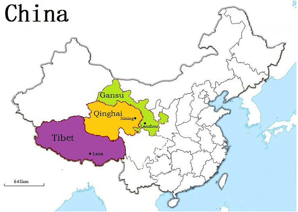
Figure 1. Geographic distribution of yaks enrolled in study.
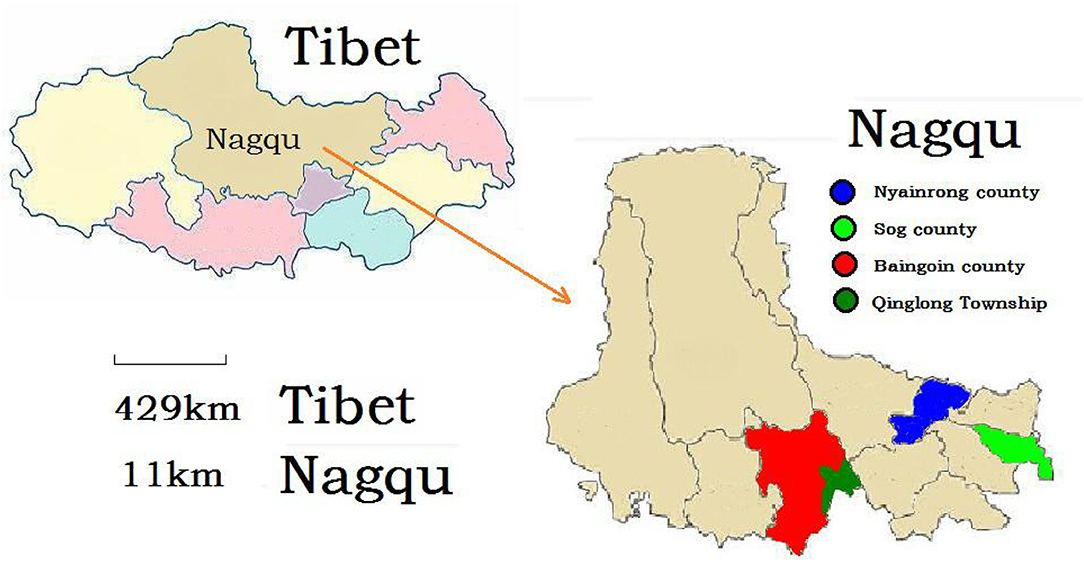
Figure 2. Geographic distribution of Tibetan sheep enrolled in study.
All serum had been determined for anti-E. granulosus antibodies by using two commercial enzyme-linked immunosorbent kits (Jianlun Biological Pharmaceuticals Co., Ltd., Guangzhou China; Duoyu Biological Pharmaceuticals Co., Ltd., Shanghai, China) according to the manufacturer's instructions. The detailed method was consistent with the previous research (23).
A total of 855 of the 1,638 (52.2%) yaks were detected to have a CE infection, of which 328 (50.2%) were males and 527 (53.6%) were females. The prevalence values were 45.1, 64.3, and 49.5% on Tibet, Qinghai, and Gansu, respectively. While the prevalence ranged from 17.4 to 57.5% in different ages (Table 1).
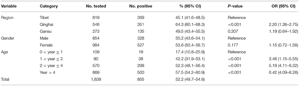
Table 1. Prevalence and risk factors of Echinococcus granulosus infection in yaks on Qinghai–Tibet plateau.
In the current research, the more influencing risk factors were region and age according to logistic regression models. Qinghai yaks were considered to be 2.20 times of higher risk of being positive compared with Tibet yaks, whereas Gansu yaks were considered to be 1.19 times at higher risk of CE infection compared with Tibet yaks (Table 1). In different ages, yaks <1 year ≤ 2 years had 3.46 times higher risk of CE infection compared with yaks <0 years ≤ 1 year; both yaks of <2 years ≤ 4 years and >4 years (57.54%) had 5.19 times and 6.42 times higher risk of being positive, respectively, when compared with yaks <0 years ≤ 1 year (Table 1). Also, there was no significant difference between males and females for yaks (Table 1).
In our research, 1,092 serum samples of Tibetan sheep were tested, 38.2% were detected to be positive for E. granulosus, with the distribution of 25.3% (Sog county), 15.4% (Nyainrong), 54.4% (Qinglong), and 56.8% (Baingoin) (Table 2). Tibetan sheep from both Nyainrong and Sog county had a significantly lower risk of CE infection compared with that from Qinglong and Baingoin (Table 2). With regard to sex, there was a non-significant difference. However, the seroprevalence values were 25.7% (juveniles), 42.8% (sub-adults), and 50.7% (adults) (Table 2). The Tibetan sheep in sub-adults and adults had two times higher risk of CE infection in juveniles (Table 2).
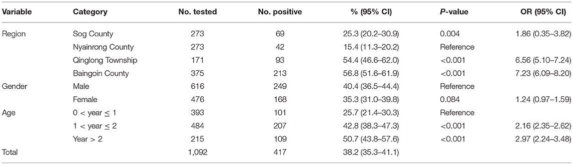
Table 2. Prevalence and risk factors of Echinococcus granulosus infection in Tibetan sheep on Qinghai–Tibet plateau.
Parasites have lived on Earth for as long as life has existed, and no species, whether animal or human, is exempt to parasites (24). E. granulosus had caused a tremendous economic loss and a severe public health risk in China as a foodborne neglected parasitic disease (25). More than seven million livestock are infected by CE yearly (8).
The seroprevalence of CE infection in yaks was 52.2% in our study, which was higher than the prevalence in the previous research in Turkey (41.1%), Greece (42%), and Ethiopia (27.6%), and significantly higher than the prevalence in Southern Brazil (13.7%) and Pakistan (13.46%) (3). Meanwhile, the previous investigation showed that the average infection rates of sheep decreased from 8.17% in 2012 to 3.68% by 2018 in the Western Sichuan Plateau (26). It was also significantly lower than the seroprevalence of CE infection in Tibetan sheep in our research (38.2%).
The previous study identified that the prevalence of CE was bound with environment culture, exerting complicated and combined effects (27). Meanwhile, the low temperatures could be a possible reason for the seroprevalence of CE. The Qinghai–Tibet plateau had a low surface temperature for years and has been a suitable place for CE due to its high altitude (28, 29). On the other hand, the stagnant economy is one of the main reason for CE. In rural areas, people had no awareness to undergo a personal medical checkup before the illness. People could be infected easily by echinococcosis due to the lack of education, information, and low sanitation conditions (30–33). A recent study showed that the plateau is the key area of CE in China (3).
In our research, the analysis showed that E. granulosus infection is closely related to region and age of animals. Due to the rainfall and many lakes, the ruminants had a higher risk of CE infection in Qinghai province, Qinglong, and Baingoin (23). In addition, the results suggested that old animals had more chances to acquire E. granulosus infection due to more exposure to the source of infection. However, no significant difference was observed in sex. Therefore, persons had a much higher risk of E. granulosus infection by frequent exposure to infected animals.
Our research showed the high seroprevalence rate of E. granulosus infection in ruminants on the Qinghai–Tibet plateau in China. It was indicated that E. granulosus could cross-transmit between the environment and host, including human beings. Therefore, effective measures must be taken to control the spread of E. granulosus by considering the role of various factors. Hence, our study might be useful to wiping off such transmissible disease on Qinghai–Tibet plateau of China.
The original contributions presented in the study are included in the article/supplementary material, further inquiries can be directed to the corresponding author/s.
Blood samples were collected under the permission of the relevant institutions. All procedures were approved and performed by Laboratory Animals Research Centre of Hubei, Qinghai, Gansu and Tibet in China, and the Ethics Committee of Huazhong Agricultural University, China (Permit number: 4200695757). All animal experiments and procedures were conducted under the relevant procedures of Proclamation of the Standing Committee of Hubei People's Congress (PSCH No.5), China.
XG, YB, and JL conceived and designed the study. LX, RS, SZha, XK, and CN collected the sample. XG, ZZ, and SZho executed the experiment and analyzed the samples. XG, CQ, and KL analyzed the data. XG and LX finished the first draft. MK revised the manuscript. All authors interpreted the data, critically revised the manuscript for important intellectual contents, and approved the final version.
This study was supported by the Chinese Agricultural Research Systems (CARS-37) and the Key Research and Development Program of Tibet Autonomous Region (XZ202001ZY0044N).
The authors declare that the research was conducted in the absence of any commercial or financial relationships that could be construed as a potential conflict of interest.
All claims expressed in this article are solely those of the authors and do not necessarily represent those of their affiliated organizations, or those of the publisher, the editors and the reviewers. Any product that may be evaluated in this article, or claim that may be made by its manufacturer, is not guaranteed or endorsed by the publisher.
1. Gavidia CM, Gonzalez AE, Barron EA, Ninaquispe B, Llamosas M, Verastegui MR, et al. Evaluation of oxfendazole, praziquantel and albendazole against Cystic Echinococcosis: a randomized clinical trial in naturally infected sheep. PLoS Negl Trop Dis. (2010) 4:e616. doi: 10.1371/journal.pntd.0000616
2. Schurer JM, Rafferty E, Farag M, Zeng W, Jenkins EJ. Echinococcosis: an economic evaluation of a veterinary public health intervention in rural Canada. PLoS Negl Trop Dis. (2015) 9:e0003883. doi: 10.1371/journal.pntd.0003883
3. Li K, Zhang L, Zhang H, Lei Z, Luo H, Mehmood K, et al. Epidemiological investigation and risk factors of Echinococcus granulosus in yaks (Bos grunniens), Tibetan pigs and Tibetans on Qinghai Tibetan plateau. Acta Trop. (2017) 173:147. doi: 10.1016/j.actatropica.2017.06.019
4. Hélène C, Francisco JBR, José RS, Benner CT, Benito A, Fernández-Crespo JC, et al. Cystic echinococcosis in the province of Álava, North Spain: the monetary burden of a disease no longer under surveillance. PLoS Negl Trop Dis. (2014) 8:e3069. doi: 10.1371/journal.pntd.0003069
5. Qian MB, Abelaridder B, Wu WP, Zhou XN. Combating echinococcosis in China: strengthening the research and development. Infect Dis Poverty. (2017) 6:161. doi: 10.1186/s40249-017-0374-3
7. Maharana BR, Kumar B, Allaie IM. Veterinary Parasitology: A Complete Objective Type Guide Kolkata, West Bengal: Kalyani Publisers (2017).
8. Budke CM, Deplazes P, Torgerson PR. Global socioeconomic impact of cystic echinococcosis. Emerg Infect Dis. (2006) 12:296–303. doi: 10.3201/eid1202.050499
9. Torgerson PR, Krista K, Mellissa M. The global burden of alveolar echinococcosis. PLoS Negl Trop Dis. (2010) 4:e722. doi: 10.1371/journal.pntd.0000722
10. Wang LY, Wu WP, Zhu XH. The endemic status of hydatidosis in China from 2004 to 2008. Chin J Zoonoses. (2010) 26:699–702.
11. Wang Q, Huang Y, Huang L, Yu W, He W, Zhong B, et al. Review of risk factors for human echinococcosis prevalence on the Qinghai-Tibet plateau, China: a prospective for control options. Infect Dis Poverty. (2014) 3:3. doi: 10.1186/2049-9957-3-3
12. Budke CM, Campos-Ponce M, Qian W, Torgerson PR. A canine purgation study and risk factor analysis for echinococcosis in a high endemic region of the Tibetan plateau. Vet Parasitol. (2005) 127:43–9. doi: 10.1016/j.vetpar.2004.08.024
13. Ris DR, Hamel KL, Mackle ZM. Use of two polysaccharide antigens in ELISA for the detection of antibodies to Echinococcus granulosus in sheep sera. Res Vet Sci. (1987) 43:257–63. doi: 10.1016/S0034-5288(18)30784-7
14. Simsek S, Balkaya I, Ciftci AT, Utuk AE. Molecular discrimination of sheep and cattle isolates of Echinococcus granulosus by SSCP and conventional PCR in Turkey. Vet Parasitol. (2011) 178:367–9. doi: 10.1016/j.vetpar.2011.01.033
15. Osman AMA, Aradaib IE, Ashmaig ALK, Gameel AA. Detection and differentiation of Echinococcus granulosus-complex using a simple PCR-based assay. Int J Trop Med. (2009) 4:21–6. Available online at: http://www.medwelljournals.com/abstract/?doi=ijtmed.2009.21.26
16. Hjalmar V. Double-antibody sandwich ELISA using biotinylated antibodies for the detection of Echinococcus granulosus, coproantigens in dogs. Acta Trop. (2005) 95:9–15. doi: 10.1016/j.actatropica.2005.03.005
17. Zhuo X, Yu Y, Chen X, Zhang Z, Yang Y, Du A. Development of a colloidal gold immunochromatographic strip based on HSP70 for the rapid detection of Echinococcus granulosus in sheep. Vet Parasitol. (2017) 240:34. doi: 10.1016/j.vetpar.2017.03.027
18. Gao JS, Zhang XC, Liu Y, Kou JH, Zhang Z, Li JH, et al. Preparation of colloidal gold strip for detection of Echinococcus granulosus infection in dogs. Chin J Biol. (2018) 31:271–5. doi: 10.13200/j.cnki.cjb.002119
19. Zhang XX, Feng SY, Ma JG, Zheng WB, Yin MY, Qin SY, et al. Seroprevalence and risk factors of fascioliasis in yaks, Bos grunniens, from three counties of Gansu province, China. Korean J Parasitol. (2017) 55:89–93. doi: 10.3347/kjp.2017.55.1.89
20. Li K, Gao JF, Shahzad M, Han Z, Nabi F, Liu M, et al. Seroprevalence of Toxoplasma gondii infection in yaks (Bos grunniens) on the Qinghai-Tibetan Plateau of China. Vet Parasitol. (2014) 205:354–6. doi: 10.1016/j.vetpar.2014.07.014
21. Li JK, Li K, Shahzad M, Han Z, Nabi F, Gao J, et al. Seroprevalence of Bluetongue virus in domestic yaks (Bos grunniens) in Tibetan regions of China based on circulating antibodies. Trop Anim Health Prod. (2015) 47:1221–3. doi: 10.1007/s11250-015-0853-0
22. Xin GS, Long RJ, Guo XS, Irvine J, Ding L. Blood mineral status of grazing Tibetan sheep in the Northeast of the Qinghai–Tibetan plateau. Livest Sci. (2011) 136:102–7. doi: 10.1016/j.livsci.2010.08.007
23. Gao X, Zhang L, Tong X, Zhang H, Mehmood K, Jiang X, et al. Epidemiological survey of fasciolosis in yaks and sheep living on the Qinghai-Tibet plateau, China. Acta Trop. (2020) 201:105212. doi: 10.1016/j.actatropica.2019.105212
24. Mitchell PD. Human parasites in the Roman World: health consequences of conquering an empire. Parasitology. (2016) 144:48–58. doi: 10.1017/S0031182015001651
25. Da SA. Human echinococcosis: a neglected disease. Gastroenterol Res Pract. (2010) 2010:583297. doi: 10.1155/2010/583297
26. Yuan DB, Hao L, Yin NC, Zhou MZ, Yang AG, Zeng ZX, et al. Epidemiological survey of hydatid disease of livestock in the western Sichuan Plateau from 2012 to 2018. Chin Vet J. (2020) 256:20–21+24.
27. Huang D, Li RD, Qiu J, Sun X, Yuan R, Shi Y, et al. Geographical environment factors and risk mapping of human Cystic Echinococcosis in Western China. Int J Environ Res Public Health. (2018) 15:1729. doi: 10.3390/ijerph15081729
28. Eckert J, Gemmell MA, Meslin FX, Pawlowski ZS. WHO/OIE Manual on Echinococcosis in Humans and Animals: A Public Health Problem of Global Concern. : Paris: World Organisation for Animal Health; Geneva: World Health Organization (2001). p. 265.
29. Hu HH, Wu PW, Guan YY, Wang LY, Wang Q, Cai HX, et al. Village-based multidisciplinary study on factors affecting the intensity of Cystic Echinococcosis in an endemic region of the Tibetan plateau, China. Epidemiol Infect. (2014) 142:1214–20. doi: 10.1017/S0950268813002124
30. Yang YR, Craig PS, Sun T. Echinococcosis in Ningxia Hui Autonomous Region, northwest China. Trans R Soc Trop Med Hyg. (2008) 102:319–28. doi: 10.1016/j.trstmh.2008.01.007
31. Possenti A, Manzanoromán R, Sánchezovejero C, Boufana B, La Torre G, Siles-Lucas M, et al. Potential risk factors associated with human Cystic Echinococcosis: systematic review and meta-analysis. PLoS Negl Trop Dis. (2016) 10:e0005114. doi: 10.1371/journal.pntd.0005114
32. Merino V, Westgard CM, Bayer AM, García PJ. Knowledge, attitudes, and practices regarding Cystic Echinococcosis and sheep herding in Peru: a mixed-methods approach. BMC Vet Res. (2017) 13:213. doi: 10.1186/s12917-017-1130-4
Keywords: seroprevalence, Echinococcus granulosus, risk factors, yaks, Tibetan sheep
Citation: Gao X, Xire L, Zhang Z, Quan C, Zhou S, Li K, Song R, Zhao S, Kong X, Naori C, Kulyar MF-e-A, Bao Y and Li J (2022) Seroprevalence of Cystic Echinococcosis in Yaks and Sheep During 2017 on the Qinghai–Tibet Plateau, China. Front. Vet. Sci. 9:849500. doi: 10.3389/fvets.2022.849500
Received: 06 January 2022; Accepted: 25 January 2022;
Published: 24 March 2022.
Edited by:
Fazul Nabi, Lasbela University of Agriculture, Water and Marine Sciences, PakistanReviewed by:
Yung-Fu Chang, Cornell University, United StatesCopyright © 2022 Gao, Xire, Zhang, Quan, Zhou, Li, Song, Zhao, Kong, Naori, Kulyar, Bao and Li. This is an open-access article distributed under the terms of the Creative Commons Attribution License (CC BY). The use, distribution or reproduction in other forums is permitted, provided the original author(s) and the copyright owner(s) are credited and that the original publication in this journal is cited, in accordance with accepted academic practice. No use, distribution or reproduction is permitted which does not comply with these terms.
*Correspondence: Jiakui Li, bGlqazIxMEBzaW5hLmNvbQ==; Yuhua Bao, NjUxMzIwMzE4QHFxLmNvbQ==
†These authors have contributed equally to this work and share first authorship
Disclaimer: All claims expressed in this article are solely those of the authors and do not necessarily represent those of their affiliated organizations, or those of the publisher, the editors and the reviewers. Any product that may be evaluated in this article or claim that may be made by its manufacturer is not guaranteed or endorsed by the publisher.
Research integrity at Frontiers
Learn more about the work of our research integrity team to safeguard the quality of each article we publish.