
94% of researchers rate our articles as excellent or good
Learn more about the work of our research integrity team to safeguard the quality of each article we publish.
Find out more
ORIGINAL RESEARCH article
Front. Trop. Dis., 29 June 2022
Sec. Antimicrobial Resistance
Volume 3 - 2022 | https://doi.org/10.3389/fitd.2022.890296
This article is part of the Research TopicBiological Alternatives To Combat Drug Resistant Tropical Bacterial InfectionsView all 4 articles
 Brice Armand Fanou1
 Jean Robert Klotoe1,2*
 Victorien Dougnon1
 Phénix Assogba1
 Eric Agbodjento1
 Charles Hornel Koudokpon1
 Lauris Fah1
 Kévin Sintondji1Rodrigue Kpoze1Frédéric Loko1
The search for new bioactive molecules with antifungal properties to combat resistance to classical antifungals represents a great challenge. This study aimed to explore the virulence factors and resistance profile of Candida species isolated from urine samples in Benin and the in vitro efficacy of organic extracts of Cyanthillium cinereum (L.) H.Rob., Lippia multiflora Moldenke and Khaya senegalensis (Desv.) A.Juss. on the growth of these Candida spp. The study focused on Candida strains isolated from urine samples collected from patients admitted to the bacteriological analysis laboratories of hospitals in Southern Benin. The sensitivity of these strains to classical antifungal agents was determined by the simple diffusion method. Their pathogenicity was investigated via several virulence factors (gelatinase, hemolysin, hydrophobicity, adhesin, biofilm and lecithinase). The in vitro efficacy of the aqueous, ethanolic and hydro-ethanolic extracts of the plants on Candida albicans ATCC 90028 and on six clinical strains was evaluated by the method of determination of the inhibition diameters. The results obtained showed that 51 different Candida strains were isolated from the collected urine samples with a respective predominance of Candida albicans (52.94%) and Candida glabrata (17.64%) species. All identified species were sensitive to amphotericin B and nystatin but 20% are resistant to fluconazole and present 15 different resistance profiles. Six different virulence factors were identified with a high frequency of hydrophobicity (96.08%) and adhesin (94.12%). Antifungal tests revealed that at 100 mg/mL the plant extracts were active on the tested strains with better activity for Cyanthilium cinereum and Khaya senegalensis. Cyanthilium cinereum, Khaya senegalensis and Lippia multiflora showed antifungal activity on virulent Candida strains suggesting the possibility to explore them further for the discovery of new antifungal molecules.
Candidiasis is the most frequent fungal infection in human pathology (1). Affecting millions of women each year, candidiasis is considered as a major public health problem (2). This benign affection has a very negative impact on life quality and high health costs (3). Fungal pathologies have various degrees of severity, ranging from superficial and benign infections to invasive and fatal infections (4). According to some epidemiological data, their incidence has increased in the last two decades due to the pathological state of immunocompromised patients, invasive medical procedures and therapy with broad spectrum antibiotics (5).
Candidiasis is caused by different species of Candida with Candida albicans as the main species isolated from infected patients. Its contamination is almost exclusively endogenous and its importance depends on the location of the responsible yeast species (5).
Urinary tract candidiasis is a most common nosocomial fungal infection worldwide (6). Usually asymptomatic, it is frequently seen in hospitalized patients and can be due to cystitis, pyelonephritis, prostatitis, epididymo-orchitis or disseminated candidiasis (7). Major risk factors include diabetes mellitus, use of broad-spectrum antibiotics, urinary tract obstruction, viral infections, admission to an intensive care unit, among others (7, 8). Candida albicans is the main cause of this nosocomial infection. Candida UTIs can be caused by hematogenous spread following candidemia, or by retrograde route via the urethra (9). The presence of Candida species in the urine of asymptomatic patients does not warrant antifungal therapy except for neutropenic patients, very low birth weight infants, and patients undergoing urologic procedures.
The management of urinary candidiasis is effectively done with fluconazole, which is the antifungal agent of choice for this type of nosocomial infection However, amphotericin B or flucytosine can also be used for this purpose in rare cases (10, 11). In recent years, the increase in urinary tract candidiasis has led to the emergence of Candida species that are resistant to conventional antifungals (6). Thus, Candida glabrata, Candida krusei strains are increasingly associated with urinary candidiasis (12). Some authors had even reported cross-resistance of Candida albicans to itraconazole and fluconazole (13, 14). These Candida species have the potential to develop antifungal resistance either intrinsically or during treatment (15). The emergence of fungal infections is favored by several virulence factors responsible for the degree of pathogenicity of Candida species. Nowadays, the emergence of new resistant and virulent Candida species that are virtually untreatable has further highlighted this public health problem (16, 17).
Faced with this resistance observed nowadays to classical antifungal drugs, even those of last resort (fluconazole for example) used for the treatment of candidiasis, it is necessary to resort to safe and effective alternative solutions. The search for new bioactive molecules with antifungal properties to combat resistance to classical antifungals represents a great challenge for researchers in the field of biomedical research. The use of medicinal plants is the first reflex of most populations, especially those in developing countries, for socio-cultural and economic reasons, and is one of the most promising alternative solutions. Indeed, in recent years, scientific studies conducted on a large number of medicinal plants have identified about 7,000 natural bioactive compounds currently used in modern medicine, which increases the global market value of medicinal plant products (18). Over 90% of today’s therapeutic classes are derived from a prototype natural product, the discovery of which has led to significant changes in the practice of modern medicine (19, 20).
The flora of Benin (a West African country), rich in 2807 plant species, offers the possibility of using many medicinal plants in the treatment of various pathologies (21). Several plant species including Cyanthillium cinereum (L.) H.Rob., Khaya senegalensis (Desv.) A.Juss. et Lippia multiflora Moldenke are used in the management of candidiasis in Benin (22). However, very little scientific evidence exists on the efficacy of plants against Candida strains as well as on the resistance and virulence profiles of Candida strains.
This study aimed to explore the virulence factors and resistance profile of Candida species isolated from urine samples in Benin and the in vitro efficacy of organic extracts of Cyanthilium cinereum, Lippia multiflora and Khaya senegalensis on the growth of these Candida spp.
Candida strains were isolated from urine samples collected from patients sent to the laboratory for urine cytobacteriological examinations + antibiotic susceptibility testing in various bacteriological analysis laboratories in hospitals in southern Benin. Each strain was identified according to the methodology described by Khan et al. (23) and Pandey et al. (24). For this purpose, macroscopic examination was performed on the strains obtained on Sabouraud agar supplemented with 0.5% chloramphenicol (SDA+C). The phenotypic identification of the strains was done based simultaneously on the aspects of the colonies obtained on the differential media ChromAgar and Tetrazolium Reduction Medium. The ability of yeast to produce germ tubes was studied by the blast test.
Candida spp. strains were also identified by restriction fragment length polymorphism (RFLP) PCR using the MspI restriction enzyme according to the methods of Dehghan et al. (25) and Hamzehee et al. (26).
DNA extraction was performed using 24 hours young colonies isolated on YEPD medium. The chloroform-phenol technique described by Silva et al. (27) has been used. The PCR was done in two steps: the first step consists in amplifying the ITS1 and ITS4 region named ITS1-ITS4 and the second was digesting the PCR product of the ITS region with restriction enzyme. For the first step, total DNA amplification was performed using the primer pair ITS1 (5’-TCCGTAGGTGAACCTGCGG-3’) and ITS4 (5’-TCCTCCGCTTATTGATATGC-3’). The reaction mixture, with a total volume of 25µL, consisted of 10µL of master mix (Invitrogen), 12µL of water without DNA, 1µL of each primer and 1µL of the DNA of the yeast to be identified. The PCR was performed in a thermal cycler (PeQlab, Germany) with the following amplification conditions: 1 cycle of DNA denaturation of 5 min at 95°C, followed by 35 hybridization cycles of 30s at 94°C, 1 min at 56°C and 1 min at 72°C and a final extension step at 72°C for 10 minutes. A positive and negative control consisting of respectively a reference strain of Candida albicans ATCC 90028 and DNase, RNase free water were used.
Each PCR product was then individually digested in an aliquot with the MspI enzyme (R0106S, England Biolabs). Indeed, 10U of Msp1 enzyme corresponding to approximately 1µL of enzyme was added to 20 μl of the PCR product contained in an aliquot with 9µL of a buffer solution making a final reaction volume of 30 μl. The mixture was incubated at 37°C for 30mn and then the PCR products (ITS) and digestion products were then migrated onto agarose gels prepared at 1.5% in 0.5x TBE buffer stained with ethidium bromide, through an electrophoresis chain for 30 minutes at 100V. The observed band patterns were then observed and the observed amplicon sizes for each strain were noted and used to differentiate Candida species (28). The ITS1-ITS4 region size were presented in Table 1.

Table 1 Size of ITS1-ITS4 region of Candida species before and after digestion with MspI.
The susceptibility of isolates to antifungal agents was determined by the agar diffusion method (23, 29). The inhibition diameters were measured and interpreted to establish the resistance profile of each strain. A reference strain of C. albicans ATCC 90028 was used for quality assurance (Ref. 0264P Microbiologics, Paris, France). The antifungals used were Amphotericin B, Nystatin, Fluconazole, Kétoconazole, Clotrimazole and Itraconazole.
Several virulence factors were investigated from the fungal suspensions prepared at approximately 107UFC/ml (according the method of Noumi et al. (30). Briefly, 2mL of sterile YEPD broth was inoculated with a young colony obtained on SDA + chloramphenicol agar and incubated at 30°C. After 18h of incubation, the culture was then centrifuged and washed twice with sterile PBS at 3000g for 5 minutes. After removing the supernatant from the last wash, 1ml of PBS was added to the pellet and resuspended by vortexing. The resulting fungal suspension was diluted to obtain a solution with an optical density between 0.4 and 0.5 corresponding to a cell concentration of about 107 CFU/ml. Hydrophobicity, stickiness, biofilm, lecithinase, gelatinase and hemolysin were the virulence factors investigated.
Method described by Zanni et al. (31) which consists of measuring the adhesion of yeast to hydrocarbons such as cyclohexane, was used to evaluate the hydrophobicity of the yeast cell surface (CSH). The Optical Densities of the fungal suspensions were read and adjusted between 0.4 and 0.5 (A0). Then, 200µl of each fungal suspension to be tested was transferred into two wells of a sterile microplate to which 60µl of cyclohexane (BDH Laboratries LTD, England) was added. In the two negative control wells, the Candida suspension was replaced by distilled water. The plate was shaken vigorously for 3 min and then allowed to stand for 20 min at room temperature in a hood. Finally, 100µL of the aqueous phases from the reaction between the fungal suspensions and cyclohexane were transferred to a new microplate and the optical densities (A1) read at 620nm with the plate reader. The percentage of cells adhering to the cyclohexane layer (%CSH) was determined by the following formula:
The percentage of CSH was used to assess the importance of cell surface hydrophobicity following the criteria specified by El-Houssaini et al. (32)
The power of adhesion to polystyrene and the ability of isolates to form a biofilm on it were evaluated in this study according to the method of Zanni et al. (31) modified. Thus, we had two sterile microplates (96 wells) (one for adhesion and the other for biofilm). One hundred microliquids of RPMI 1640 medium were transferred to the test and negative control wells of a sterile microplate. 100µL of fungal suspension and 100µL of PBS solution, were added to the test and two negative control wells respectively. The treated plates were incubated at 37°C for 2 hours.
The adhesion plate was removed and the wells were washed twice with PBS solution and then stained for 1 min with 100µL of 1% diluted crystal violet. The stained wells were then washed twice with PBS solution. The treated microplate was left at room temperature for 20 min and the wells were destained for 30 min with 100µL of alcohol-acetone mixture. The supernatants were transferred to a new microplate and their optical densities were read at 560nm with a microplate reader (Infinite F200 Pro, Tecan 504).
For the biofilm plate (the second microplate), the wells were washed every 24h of incubation with sterile PBS solution and RPMI medium renewed at each occasion. The incubation time was 66h. The last wash before staining was performed after the incubation time. The adhesion and biofilm formation powers of the strains were determined from the optical densities read and then interpreted according to the criteria specified by Noumi et al. (30).
The production of phospholipase or lecithinase and gelatinase was investigated according to the methodology of El-Houssaini et al. (32) and that of Elavarashi et al. (33). 10µL of the prepared fungal suspension was seeded onto the agar and incubated at 37°C in the oven for 5-7 days. The presence of an opaque zone around the fungal colony indicates the production of lecithinase. The altering power of this enzyme (Pz) was evaluated according to the formula: Pz =D0/Dt Where, D0 = colony diameter and Dt = colony diameter + halo (large diameter). The different interpretations were made.
Gelatinase production was demonstrated according to the method described by Elavarashi et al. (33). 4000µL of gelatin-enriched nutrient broth in a hemolysis tube, were seeded with a young colony of the strain to be studied previously isolated on YEPD medium. The inoculated tubes were incubated at room temperature (25°C) and the reactions were read every 24 hours for two weeks after 30 minutes of refrigeration at 4°C. A strain of Staphylococcus aureus was used as a positive control. A tube containing only the unseeded nutrient broth was used as a negative control.
- A positive result means that the medium liquefies after cooling in the refrigerator.
- A negative result means that the medium solidifies after cooling
The investigation of hemolysin production by the strains was done by culturing them on 7% fresh blood (GSL) enriched agar medium (24, 32). Indeed, 10µL of the prepared fungal suspension was seeded on GSL medium and then incubated at 37°C for 48h in CO2 enriched atmosphere (5%). The presence of a greenish halo or white halo around the fungal colony shows the presence of hemolysin (alpha or beta respectively).
The medicinal plants used in this study were selected from an ethno pharmacological survey conducted in Benin on medicinal plants used in the traditional treatment of mycoses (22). These plants have been identified in the national herbarium of Benin at the University of Abomey-Calavi. These plants were collected in Houèto in the commune of Abomey-Calavi. Table 2 presents the plant material and the parts used.Three types of extracts (aqueous, hydroethanolic and ethanolic) were obtained following the protocol described by Klotoé et al. (34).

Table 2 Different parts of the plants used and the identification numbers in the national herbarium.
A qualitative screening of the three studied plants was carried out in order to assess the main chemical groups present according to the precipitation and colouring reactions described by Houghton and Raman (35). The main groups investigated were polyphenolic compounds (flavonoids, total phenols, tannins), anthocyanins, saponosides, reducing compounds, mucilages, and alkaloids. In the extracts produced, total polyphenols were quantified using the commercial Folin Ciocalteu reagent. Total polyphenol levels were determined using the equation from the calibration curve of gallic acid (0-200 µg/mL) taken as reference standard. Samples were prepared in triplicate for each analysis. Total polyphenol content was determined in mg gallic acid equivalent/g extract (mg GAE/g) by the formula used by Ahmed et al. (36):
With TPT, the Total Polyphenol Content, X the Gallic Acid concentration in mg/mL; V the volume of extract used in mL and m the mass of the extract in grams.
The antioxidant activity of the extracts was evaluated by the DPPH free radical scavenging assay which is one of the most widely used methods for the evaluation of antioxidant activities of herbal extracts. DPPH (2,2- Diphenyl-1-picrylhydrazyl) is a stable purplish colored free radical that absorbs at 517 nm. In the presence of anti-free radical compounds, the DPPH radical is reduced and changes its color to yellow. The method adopted in this study is that of Klotoé et al. (34). Thus 100 μL of different concentrations of each extract is added to 1900 µL of the ethanolic solution of DPPH (0.4 mg/mL). The blank is prepared by mixing 100 μL of the extraction solvent with 1900 µL of the DPPH solution. After incubation in the dark for 1 hour at room temperature, absorbance readings were taken at 517 nm using a MINDRAY spectrophotometer (BA-88-A). The recorded optical densities were used to calculate the percentage of DPPH radical scavenging which is proportional to the antioxidant power of the sample. Vitamin C and BHT were used as reference standards. Samples were prepared in triplicate for each analysis. The percentage of DPPH radical scavenging was determined by the formula:
P: Percentage of trapping; Ab: Absorbance of blank; Ae: Absorbance of sample
For the sensitivity test, seven Candida strains isolate for urines were used. These were six clinical strains of Candida and a reference strain of Candida albicans ATCC 90028. A stock concentration of 100 mg/ml of each of the nine extracts were prepared and tested separately on the in vitro growth of a reference strain of Candida albicans ATCC 90028 by the diffusion method on Muëller-Hinton agar supplemented with 2% glucose and 0.5% Methylene Blue (MHGB) (37). The fungal suspensions were prepared with Yeast Extract Peptone Dextrose (YPD or YEPD) broth. After inoculating the agar with the fungal suspension, 100 µl of each type of extract from the three plants was sterilely transferred onto Whatman paper cut into small pieces and previously deposited on the agar surface. The same procedure was followed for distilled water and fluconazole (100µg/mL) which served as negative and positive controls respectively. After 24 hours of incubation, the diameters of inhibition observed around Whatman paper were measured and interpreted. The most active plant is the one whose extracts were active on the strain with the largest inhibition diameters. The most active plant extracts were thus identified and tested following the same process on previously characterized clinical Candida strains. The anti-candida activity of the extracts was tested on the majority resistance profiles of the three most isolated Candida species (Table 4).
The Minimum Inhibitory Concentrations (MIC50) of the effective extracts were determined following the methodology of Okou et al. (37) in 96-wells microplates. A stock solution of these extracts was prepared at a concentration of 100 mg/ml in distilled water. Fluconazole at 100mg/ml was used as the reference antifungal agent. 100µl of a fungal suspension made with Yeast Extract Peptone Dextrose (YEPD) (38) broth was placed in the first eight wells. The first two wells of the microplate served as positive control (YEPD broth with yeast) and reference (YEPD broth with yeast mixed with 100µl of fluconazole solution), respectively. The ninth well is the negative control containing only YEPD broth without yeast. Then, 100 µl of the stock extract solution was added to the third well. Five serial two-for-two dilutions were then performed starting from the third well to well 8 where the additional 100 µl was discarded. For each extract the same procedure was followed for six different Candida strains. The microplates were covered with aluminium foil and placed for 24 hours in an oven at 37°C. The MIC50 corresponds to the minimum concentration where 50% of the yeasts are inhibited (39). They were estimated by counting the number of yeast using the Malassez cell under a microscope with an with the X40 lens.
Data were entered using Microsoft Excel 2010. Data analysis was done using Graph Pad Prism version 8. Quantitative variables are presented as mean and standard deviation. Qualitative variables are presented as percentages. Probit analysis was used for the determination of the IC50 and MIC of the most active extract. Student’s t test was used to compare the means. The significance level was set at 5%.
A total of 51 Candida strains were isolated and identified from urine samples collected from patients sent to the laboratory for urine cytobacteriological examination. Figure 1 shows that Candida albicans (52.94%) was the most identified species, followed by Candida glabrata (17.64%). Figures 2, 3 show electrophoresis images of the PCR product of the ITS1-ITS4 region and the PCR product of the digestion of the DNA of Candida isolates, respectively.
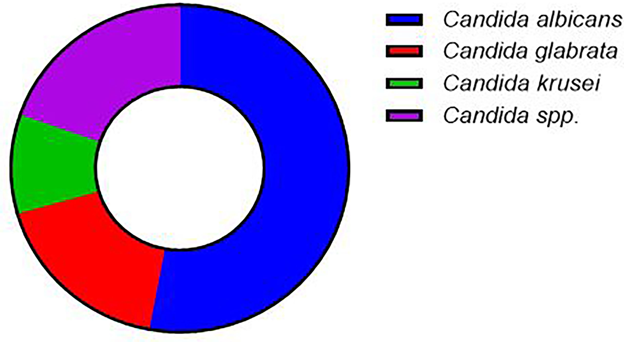
Figure 1 Frequency of identification of different Candida species from urine samples.
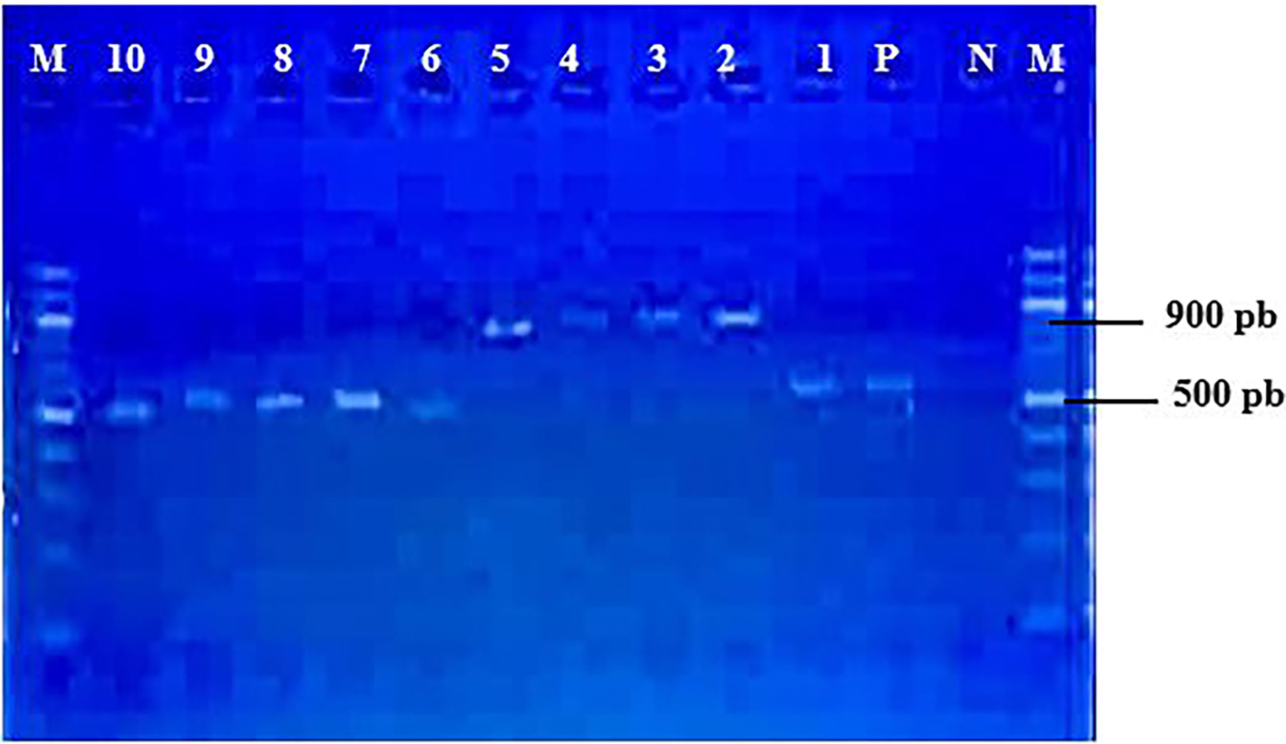
Figure 2 Gel images showing migration band profiles of the ITS1-ITS4 region of Candida strains. (M): Gene ruler 100pb; (N): Negative control; (P): Positive control (Candida albicans ATCC 90028); (1, 6, 7, 8, 9, 10): Candida albicans/Candida krusei; (2, 3, 4, 5): Candida glabrata.

Figure 3 Gel images showing MspI restriction fragment profiles of the ITS1-ITS4 region of Candida strains. (M): Gene ruler 100pb; (N): Negative control; (P): Positive control (Candida albicans ATCC 90028); (1, 7, 8, 9): Candida albicans; (2, 3, 4, 5): Candida glabrata; (6, 10): Candida krusei.
The study on sensitivity of the isolated Candida to antifungal drugs showed that all the isolated species are sensitive to amphotericin B and nystatin, two antifungal drugs of the polyene family and the most used against candidiasis. For the other antibiotics, namely, fluconazole, ketoconazole, clotrimazole and itraconazole, the Candida strains showed low levels of intermediate resistance. Only 3.70% of Candida albicans strains were resistant to clotimazole. No Candida glabrata strains were resistant to any of the antibiotics tested but showed intermediate resistance levels of 22.22% to 55.55% to fluconazole, ketoconazole, clotrimazole and itraconazole. Eighty percent of Candica krusei strains were resistant to fluconazole (Table 3).
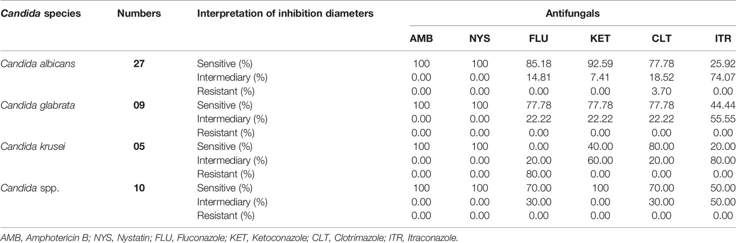
Table 3 Resistance profile of the isolated Candida strains to the tested antifungal agents.
Table 4 show the antibiotic resistance profiles of Candida albicans, Candida glabrata, Candida krusei and Candida spp. strains. Candida albicans strains showed 8 different antibiotic resistance profiles, with the most dominant being AmBS NysS FluS KetS CtrS ItrI. The other strains, notably Candida glabrata and Candida krusei, each showed 5 different antibiotic resistance patterns.
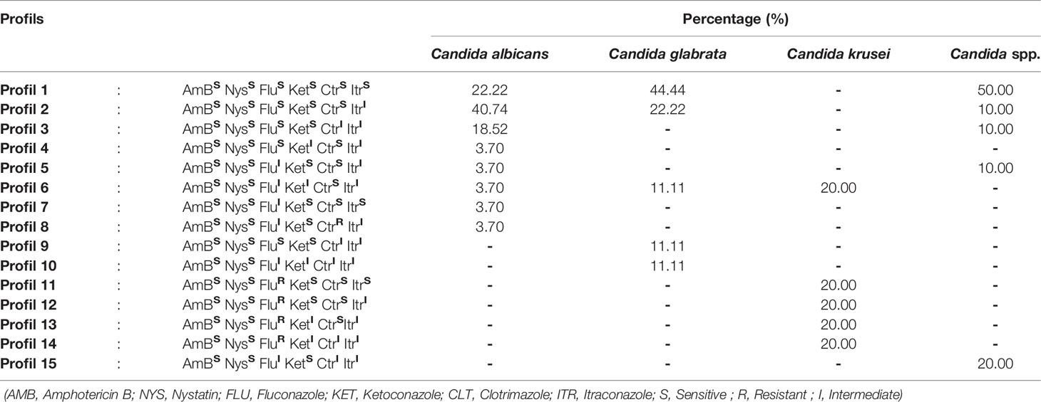
Table 4 Resistance profile of Candida strains to antifungal agents.
The study of virulence factors showed that the majority of the isolated strains showed hydrophobicity (96.08%), hemolysin (90.20%) and adhesin (94.12%) activity. Of all the virulence factors investigated, only gelatinase was absent in Candida krusei strains. Regarding the hemolytic power of Candida isolated from urine, most strains can degrade haemoglobin. Both types of hemolysis (alpha hemolysis and beta hemolysis) were observed with a predominance of beta hemolysis in all strains (Table 5a). In addition, all Candida species showed lecithinase activity. Regarding the powers of adhesion, hydrophobicity and biofilm activity, Candida isolates were found to be highly adherent (94.12%), hydrophobic (96.08%) and biofilm activity (90.20%). All Candida glabrata and Candida krusei strains were 100% hydrophobic and biofilm producer (Table 5b).
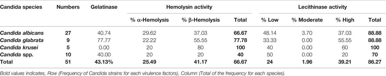
Table 5a Frequency of Candida strains producing the virulence factors.
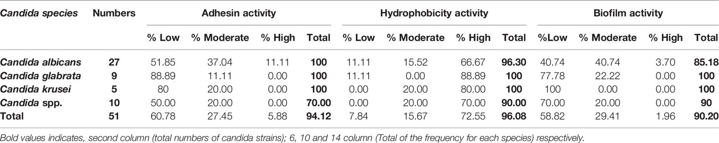
Table 5b Frequency of Candida strains producing the virulence factors (Second part).
The phytochemical analysis of the compounds of the three plants studied reveal the presence of several chemical compounds. These are tannins, flavonoids, anthocyanins, leuco anthocyanins, saponosides, reducing compounds, alkaloids, mucilages and sterols. These different data are presented in the table below. The sign (+) indicates a positive reaction and the sign (-) indicates a negative reaction (Table 6).
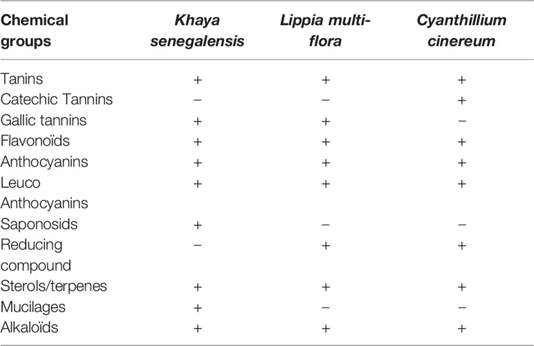
Table 6 Phytochemical composition of the three plants used.
The results of the quantification of total polyphenols in the extracts produced are presented in Table 7. From this table it is to be noticed that the aqueous extracts of the studied plants presented a not significantly high content of total polyphenols compared to the hydro-ethanolic and ethanolic extracts (P<0,05) except in the case of Cyanthillium cinereum for which the ethanolic extract is higher.
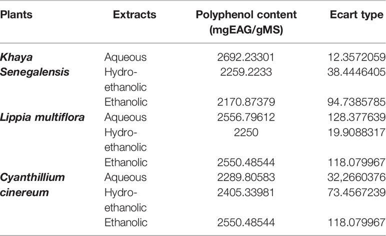
Table 7 Polyphenol content of the different plant extracts.
All the tested extracts reduced the DPPH radical in variable proportions. This inhibition of the DPPH radical translating the antioxidant activity of the extracts is proportional to the increase of the concentration of the extracts. The DPPH radical inhibitory power of the different extracts and reference standards is expressed in IC50 (Table 8). Since the IC50 is inversely proportional to the antioxidant potential of the extract, the lower the IC50 value, the better the antioxidant power of the extract.

Table 8 Antioxidant activity of plant extracts expressed in IC50.
The study of the sensitivity of the aqueous, hydro-ethanolic and ethanolic extracts of Cyantillium cinereum, Khaya senegalensis and Lippia multiflora on the reference strain of Candida albicans ATCC 90028 showed a good sensitivity of the extracts of Cyantillium cinereum and Kaya senegalensis. The largest inhibition diameters were obtained with the extracts of Cyantillium cinereum particularly the hydro-ethanolic (Figure 4).
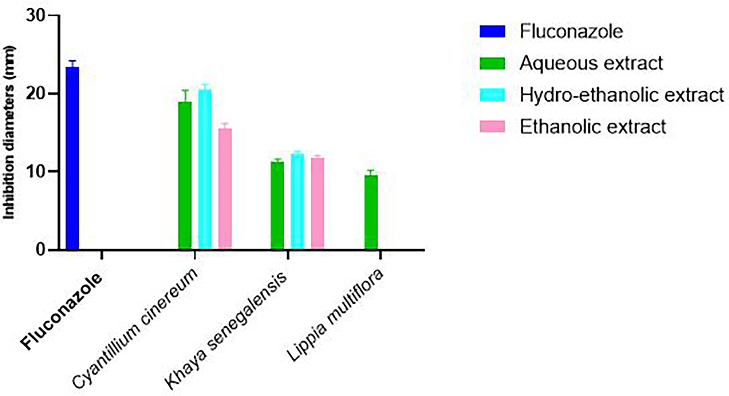
Figure 4 Effect of plant extracts on Candida albicans reference strain ATCC 90028.
Figure 5 shows the effect of aqueous, hydroethanolic and ethanolic extracts of Cyantillium cinereum on the in vitro growth of clinical strains of Candida isolated from urinary tract infections. From this figure, it appears that all the extracts were active on all the strains studied with variable inhibition diameters depending on the extract and the strain. The greatest diameters of inhibition were obtained with the aqueous extract of this plant on most of the strains except for the strains of Candida krusei k1 and Candida glabrata g1. Regarding Khaya senegalensis, these extracts were active on four different clinical strains (Candida krusei k1 and k2, Candida glabrata g1 and Candida albicans a1). The aqueous extract was more active. The hydro-ethanolic extract was not active on any Candida albicans strain and on the Candida glabrata g2 strain (Figure 6). The aqueous extracts of the two plants (Cyantillium cinereum and Khaya senegalensis) were used to determine the Minimum Inhibitory Concentrations (MIC50) on each clinical strain. The MIC50 obtained varied from one strain to another and according to plant. In general, Cyantillium cinereum presented the best anti-Candida activity (MIC50 low) compared to Khaya senegalensis (MIC50 high) (Table 9). The best anti-Candida activity of Cyantillium cinereum was obtained on Candida albicans a1 and Candida glabrata g1.
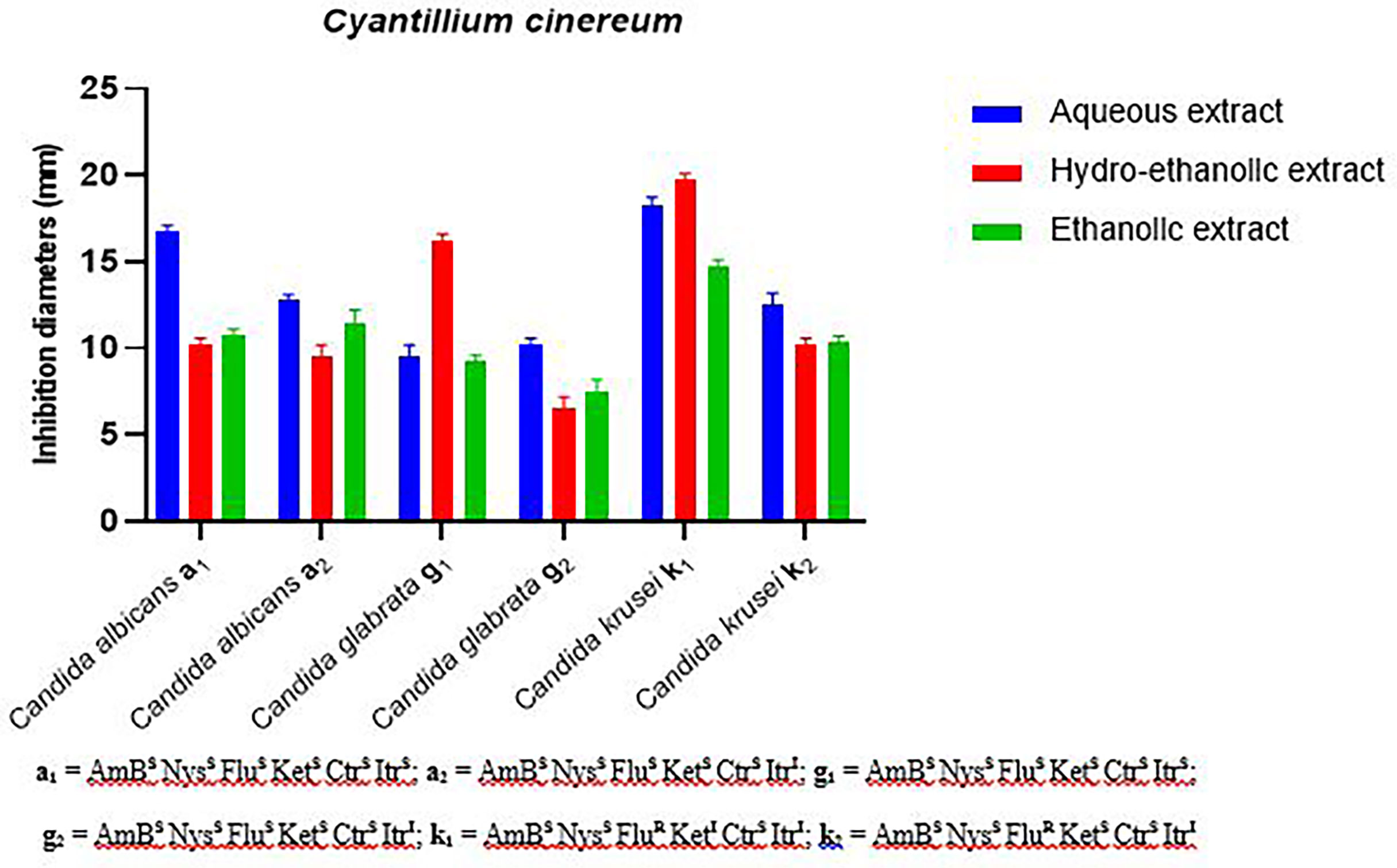
Figure 5 Effect of Cyantillium cinereum extracts on the characterized clinical strains.

Figure 6 Effect of Khaya senegalensis extracts on characterized clinical strains.
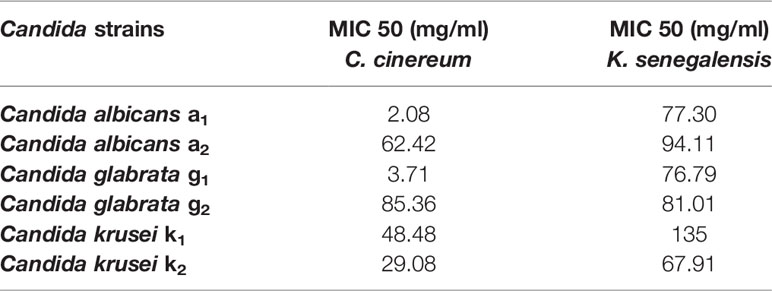
Table 9 Minimum inhibitory concentration (MIC 50) of the aqueous extract Cyantillium cinereum and Khaya senegalensis on the characterized clinical strains.
The present study is part of the search for new sources of bioactive molecules useful to fight resistance to classical antifungal agents. It aimed at exploring the virulence factors and the resistance profile of Candida species isolated from urine samples in Benin and the in vitro efficacy of organic extracts of Cyanthilium cinereum, Lippia multiflora and Khaya senegalensis on the growth of Candida spp. strains.
The search for Candida strains in urine samples from patients in South Benin resulted in the isolation of 51 different Candida species. Candida albicans (52.94%) was the most identified species followed by Candida glabrata (17.65%). The high proportion of Candida albicans species found in this study was also mentioned by several reports in the scientific literature (1, 40; 41) for candidasis in general and urinary candidiasis (6). Indeed, urinary tract candidiasis is known as the most frequent nosocomial fungal infection worldwide (42).
The study of the susceptibility of isolates to antifungal agents showed that all species were susceptible to amphotericin B and nystatin. This sensitivity of the strains to nystatin and amphotericin B obtained in the present study contrasts with the data reported by Ekpo et al. (43) related of Candida strains isolated from urine samples in Cameroon. These differences could be justified by the origin of the strains, the study area, self-medication and the misuse of chemicals against vaginal infections. However, variable levels of resistance have been obtained to other classical antifungal agents (fluconazole, ketoconazole, itraconazole, clotrimazole). Furthermore, the Candida species isolated presented different patterns of resistance to antifungal drugs. Similar data reported a variety of resistance of Candida strains to antifungal drugs (23, 44). Jensen et al. (45) explained that this observed resistance would be due to the induction of drug efflux pumps and sequestration of antifungal agents. The great variability of resistance to antifungal drugs could be linked to the inactivation of proteins essential to ergosterol biosynthesis, leading to ergosterol depletion (target of amphotericin B) and the formation of other sterols (46)
The search for some virulence factors showed that the Candida strains isolated from urine in this study possess several virulence factors such as hemolysin production, biofilm, adhesin, lecithinase, gelatinase and hydrophobicity activity. Candida albicans biofilm formation reflects a powerful resistance structure that is installed by many pathogens such as Candida albicans to resist antimicrobials (47, 48). According to Pandey et al. (24), the production of hydrolytic enzymes by Candida albicans would increase yeast adhesion. Lecithinase being a phospholipase, its activity by Candida albicans could destroy cell lipids, one of the main components of the membrane (49) and promote oxidative stress (50–52).
The antifungal tests carried out on the plants studied show a variable activity depending on the plant, the type of extract and the strain tested. Indeed, on Candida albicans ATCC 90028, Cyanthilium cinereum and Khaya senegalensis were more active in inhibiting the growth of this strain. On clinical strains selected, the extracts of these plants showed anti-Candida activity depending of the plant. Khaya senegalensis, presented a low activity anti-Candida (MIC 50 high) compared to Cyanthilium cinereum (MIC50 low). The best activity of Cyanthilium cinereum was obtained on Candida albicans a1 and Candida glabrata g2. This antifungal activity would be related to the bioactive secondary metabolites such as flavonoids, tannins, coumarins, anthocyanins, leuco anthocyanins identified in the plant (53–56). Belila and Ounis (57) report that alkaloids possess a broad spectrum of biological activities including antibiotic properties. However, the lowest anti-Candida activity of the Khaya senegalensis will be related of the resistance profile and pathogenicity of the tested strains.
The scientific data generated by the present study highlight the possibility of using the Cyanthilium cinereum as probable sources of new bioactive molecules with anti-candida properties for an efficient fight against antifungal drugs resistance.
In conclusion, this study revealed that several Candida species are involved in urinary tract infections. The Candida species isolated from urine are all-sensitive to amphotericin B, nystatin, and show 15 different resistance profiles. These bacteria also possess several virulence factors, the most represented of which are adhesin, hydrophobicity and Biofilm. In vitro antifungal tests of aqueous, ethanolic and hydro-ethanolic extracts of Khaya senegalensis, Cyanthilium cinereum and Lippia multiflora revealed a variation in antifungal power depending on the plant and the type of extract. Cyanthilium cinereum have a good in vitro activity on the growth of clinical and virulent of Candida strains. These results permit new research perspectives on the mechanisms of action of the antifungal effect of the studied plants.
All authors have actively contributed to the manipulation and writing of this article
The raw data supporting the conclusions of this article will be made available by the authors, without undue reservation.
The studies involving human participants were reviewed and approved by Institutional Ethics Committee of Research Unit in Applied Microbiology and Pharmacology of Natural Substances of University of Abomey-Calavi N° 002_CE_URMAPha_UAC. The patients/participants provided their written informed consent to participate in this study
The authors declare that the research was conducted in the absence of any commercial or financial relationships that could be construed as a potential conflict of interest.
All claims expressed in this article are solely those of the authors and do not necessarily represent those of their affiliated organizations, or those of the publisher, the editors and the reviewers. Any product that may be evaluated in this article, or claim that may be made by its manufacturer, is not guaranteed or endorsed by the publisher.
The authors are grateful to the members of Research Unit in Applied Microbiology and Pharmacology of natural substances.
1. Cassone A. Vulvovaginal Candida Albicans Infections: Pathogenesis, Immunity and Vaccine Prospects. BJOG.: Int J Obstet. Gy. (2015) 122:785–94. doi: 10.1111/1471-0528.12994
2. Gonçalves B, Ferreira C, Alves CT, Henriques M, Azeredo J, Silva S. Vulvovaginal Candidiasis: Epidemiology, Microbiology and Risk Factors. Crit Rev Microbiol (2016) 42:905–27. doi: 10.3109/1040841X.2015.1091805
3. Jacob L, John M, Kalder M, Kostev K. Prevalence of Vulvovaginal Candidiasis in Gynecological Practices in Germany: A Retrospective Study of 954,186 Patients. Curr Med Mycol. (2018) 4:6–13. doi: 10.18502/cmm.4.1.27
4. Kemaykin VM, Tabinbaev NB, Khudaibergenova MS, Olifirovich AA, Abdrakhmanova LM, Denning DW, et al. An Estimate of Severe and Chronic Fungal Diseases in the Republic of Kazakhstan. J Fungi. (Basel). (2018) 4:34–42. doi: 10.3390/jof4010034
5. Hashemi SE, Shokohi T, Abastabar M, Aslani N, Ghadamzadeh M, Haghani I. Species Distribution and Susceptibility Profiles of Candida Species Isolated From Vulvovaginal Candidiasis, Emergence of C. Lusitaniae. Curr Med Mycol. (2019) 5:26–31. doi: 10.18502/cmm.5.4.2062
6. Behzadi P, Behzadi E, Ranjbar R. Urinary Tract Infections and Candida Albicans. Cent. Eur J Urol. (2015) 68:96–101. doi: 10.5173/ceju.2015.01.474
7. Odabasi Z, Mert A. Candida Urinary Tract Infections in Adults. World J Urol. (2020) 38:2699–707. doi: 10.1007/s00345-019-02991-5
8. Griffith N, Danziger L. Candida Auris Urinary Tract Infections and Possible Treatment. Antibio. (Basel). (2020) 9:898. doi: 10.3390/antibiotics9120898
9. Poloni JAT, Rotta LN. Urine Sediment Findings and the Immune Response to Pathologies in Fungal Urinary Tract Infections Caused by Candida Spp. J Fungi. (Basel). (2020) 6:E245. doi: 10.3390/jof6040245
10. Malani AN, Kauffman CA. Candida Urinary Tract Infections: Treatment Options. Expert Rev Anti Infect Ther (2007) 5:277–84. doi: 10.1586/14787210.5.2.277
11. Thomas L, Tracy CR. Treatment of Fungal Urinary Tract Infection. Urol. Clin North Am (2015) 42:473–483. doi: 10.1016/j.ucl.2015.05.010
12. Osawa K, Shigemura K, Yoshida H, Fujisawa M, Arakawa S. Candida Urinary Tract Infection and Candida Species Susceptibilities to Antifungal Agents. J Antibio. (2013) 66:651–4. doi: 10.1038/ja.2013.68
13. Corsello S, Spinillo A, Osnengo G, Penna C, Guaschino S, Beltrame A, et al. An Epidemiological Survey of Vulvovaginal Candidiasis in Italy. Eur J Obstet. Gynecol. Reprod Biol (2003) 110:66–72. doi: 10.1016/S0301-2115(03)00096-4
14. Sojakova M, Liptajova D, Borovsky M, Subik J. Fluconazole and Itraconazole Susceptibility of Vaginal Yeast Isolates From Slovakia. Mycopathologia (2004) 157:163–9. doi: 10.1023/B:MYCO.0000020594.35357.b0
15. Khosravi AR, Sharifzadeh A, Nikaein D, Almaie Z, Nasrabadi HG. Chemical Composition, Antioxidant Activity and Antifungal Effects of Five Iranian Essential Oils Against Candida Strains Isolated From Urine Samples. J Mycologie. Med. (2018) 28:355–60. doi: 10.1016/j.mycmed.2018.01.005
16. Dabas PS. An Approach to Etiology, Diagnosis and Management of Different Types of Candidiasis. Journal of Yeast and Fungal Research (2013) 4:63–74.
17. Wang F-J, Dai Zhang Z-HL, Wu W-X, Bai H-H, Dong H-Y. Species Distribution and In Vitro Antifungal Susceptibility of Vulvovaginal Candida Isolates in China. Chin Med J (2016) 129:1161–9. doi: 10.4103/0366-6999.181964
18. Arrey Tarkang P, Franzoi KD, Lee S, Lee E, Vivarelli D, Freitas-Junior L, et al. In Vitro Antiplasmodial Activities and Synergistic Combinations of Differential Solvent Extracts of the Polyherbal Product, Nefang. BioMed Res Int (2014) 2014:e835013. doi: 10.1155/2014/835013
19. McChesney JD, Venkataraman SK, Henri JT. Plant Natural Products: Back to the Future or Into Extinction? Phytochemistry (2007) 68:2015–22. doi: 10.1016/j.phytochem.2007.04.032
20. Petrovska BB. Historical Review of Medicinal Plants’ Usage. Pharmacogn. Rev (2012) 6:1–5. doi: 10.4103%2F0973-7847.95849
21. Akoègninou A, van der Burg WJ, van der Maesen LJG. Flore Analytique Du Bénin. Backhuys: Cotonou and Wageningen (2006).
22. Fanou BA, Klotoe JR, Fah L, Dougnon V, Koudokpon CH, Toko G, et al. Ethnobotanical Survey on Plants Used in the Treatment of Candidiasis in Traditional Markets of Southern Benin. BMC Complement. Med Therap. (2020) 20:1–18. doi: 10.1186/s12906-020-03080-6
23. Khan M, Ahmed J, Gul A, Ikram A, Lalani FK. Antifungal Susceptibility Testing of Vulvovaginal Candida Species Among Women Attending Antenatal Clinic in Tertiary Care Hospitals of Peshawar. IDR (2018) 11:447–56. doi: 10.2147/IDR.S153116
24. Pandey N, Gupta MK, Tilak R. Extracellular Hydrolytic Enzyme Activities of the Different Candida Spp. Isolated From the Blood of the Intensive Care Unit-Admitted Patients. J Lab Physician. (2018) 10:392–6. doi: 10.4103/JLP.JLP_81_18
25. Dehghan P, Mohammadi F, Javaheri MR, Nekoeian S. Identification of Candida Species in the Oral Cavity of Diabetic Patients. mazu-cmm (2016) 2:0–0. doi: 10.18869/acadpub.cmm.2.2.4
26. Hamzehee S, Kalantar-Neyestanaki D, Mohammadi MA, Nasibi S, Mousavi SAA. Identification of Candida Spp. Isolated From Oral Mucosa in Patients With Leukemias and Lymphomas in Iran. Iran J Microbiol (2019) 11:114–9.
27. Silva GA, da, Bernardi TL, Schaker PDC, Menegotto M, Valente P. Rapid Yeast DNA Extraction by Boiling and Freeze-Thawing Without Using Chemical Reagents and DNA Purification. Braz Arch Biol Technol (2012) 55:319–27. doi: 10.1590/S1516-89132012000200020
28. Zahir RA, Himratul-Aznita WH. Distribution of Candida in the Oral Cavity and its Current Patient Perspectives Differentiation Based on the Internally Transcribed Spacer (ITS) Regions of rDNA. Wiley. Online Lib. (2013) 1:13–23. doi: 10.1002/yea.2937
29. ElFeky DS, Gohar NM, El-Seidi EA, Ezzat MM, AboElew SH. Species Identification and Antifungal Susceptibility Pattern of Candida Isolates in Cases of Vulvovaginal Candidiasis. AJM. Elsevier. (2016) 52:269–77. doi: 10.1016/j.ajme.2015.10.001
30. Noumi E, Snoussi M, Noumi I, Saghrouni F, Aouni M, Valentin E. Phenotypic Characterization and Adhesive Properties of Vaginal Candida Spp. Strains Provided by the CHU Farhat Hached (Sousse, Tunisia). Rev Iberoam. Micol. (2015) 32:170–9. doi: 10.1016/j.riam.2014.06.006
31. Zanni PCMD, Bonfim-Mendonça P, de S, Negri M, Nakamura SS, Donatti L, et al. Virulence Factors and Genetic Variability of Vaginal Candida Albicans Isolates From HIV-Infected Women in the Post-Highly Active Antiretroviral Era. Rev Do. Instituto. Medic. Trop São. Paulo. (2017) 59:1–10. doi: 10.1590/s1678-9946201759044
32. El-Houssaini HH, Elnabawy OM, Nasser HA, Elkhatib WF. Correlation Between Antifungal Resistance and Virulence Factors in Candida Albicans Recovered From Vaginal Specimens. Microbial. Pathogen. (2019) 12:13–9.
33. Elavarashi E, Kindo AJ, Rangarajan S. Enzymatic and non-Enzymatic Virulence Activities of Dermatophytes on Solid Media. J Clin Diagn Res (2017) 3:11–23. doi: 10.7860/JCDR/2017/23147.9410
34. Klotoé J, Agbodjento E, Dougnon V, Yovo M, Sacramento T, Deguenon ELM, et al. Exploration of the Chemical Potential and Antioxidant Activity of Some Plants Used in the Treatment of Male Infertility in Southern Benin. J Pharm Res Int (2020) 32:1–12. doi: 10.9734/jpri/2020/v32i430418
35. Houghton PJ, Raman A. Laboratory Handbook for the Fractionation of Natural Extracts. London, Thomson publishing (1998) p. 154–62.
36. Ahmed R, Tariq M, Hussain M, Andleeb A, Masoud MS, Ali I, et al. Phenolic Contents-Based Assessment of Therapeutic Potential of Syzygium Cumini Leaves Extract. PloS One (2019) 14(8):e0221318. doi: 10.1371/journal.pone.0221318
37. Okou OC, Yapo SE-S, Kporou KE, Baibo GL, Monthaut S, Djaman AJ. Évaluation De L’activité Antibactérienne Des Extraits De Feuilles De Solanum Torvum Swartz (Solanaceae) Sur La Croissance In Vitro De 3 Souches D’entérobactéries. J App. Biosci. (2018) 122:12–20. doi: 10.4314/jab.v122i1.8
38. Silva F, dos S, Landell MF, Paulino GVB, Coutinho HDM, Albuquerque UP. Antifungal Activity of Selected Plant Extracts Based on an Ethnodirected Study. Acta Bot Bras (2020) 34:442–8. doi: 10.1590/0102-33062020abb0003
39. Lavaee F, Moshaverinia M, Malekhosseini S, Jamshidzade A, Zarei M, Jafarian H, et al. Antifungal Effect of Sesame Medicinal Herb on Candida Species: Original Study and Mini-Review. Braz J Pharm Sci (2019) 55. doi: 10.1590/s2175-97902019000117479
40. Nakamura-Vasconcelos SS, Fiorini A, Zanni PD, Bonfim-Mendonça P, de S, Godoy JR, et al. Emergence of Candida Glabrata in Vulvovaginal Candidiasis Should be Attributed to Selective Pressure or Virulence Ability? Arch Gynecol. Obstet. (2017) 296:519–26. doi: 10.1007/s00404-017-4465-y
41. Yano J, Sobel JD, Nyirjesy P, Sobel R, Williams VL, Yu Q, et al. (2019). Current Patient Perspectives of Vulvovaginal Candidiasis: Incidence, Symptoms, Management and Post-Treatment Outcomes. BMC Women’s Health 19: 1–8. doi: 10.1186/s12905-019-0748-8.
42. Kauffman CA. Diagnosis and Management of Fungal Urinary Tract Infection. Infect Dis Clin North Am (2014) 28:61–74. doi: 10.1016/j.idc.2013.09.004
43. Ekpo IA, Kechia FA, Iwewe YS, Ngueguim AD, Nangwat C, Dzoyem JP. Species Distribution and Antifungal Susceptibility Profile of Candida Spp Isolated From Urine of Hospitalized Patients in Dschang District Hospital, Cameroon. Int J Biol Chem Sci (2017) 11:1212–21. doi: 10.4314/ijbcs.v11i3.23
44. Maraki S, Mavromanolaki VE, Stafylaki D, Nioti E, Hamilos G, Kasimati A. Epidemiology and Antifungal Susceptibility Patterns of Candida Isolates From Greek Women With Vulvovaginal Candidiasis. Mycoses (2019) 62:692–7. doi: 10.1111/myc.12946
45. Jensen RH, Astvad KMT, Silva LV, Sanglard D, Jørgensen R, Nielsen KF, et al. Stepwise Emergence of Azole, Echinocandin and Amphotericin B Multidrug Resistance In Vivo in Candida Albicans Orchestrated by Multiple Genetic Alterations. J Antimicrob Chemother (2015) 70:2551–5. doi: 10.1093/jac/dkv140
46. Nouraei S, Amir Ali Akbari S, Jorjani M, Alavi Majd H, Afrakhteh M, Ghafoorian A, et al. Comparison Between Fluconazole With Oral Protexin Combination and Fluconazole in the Treatment of Vulvovaginal Candidiasis. Int Scholarly. Res Not. (2012) 2012:1–8. doi: 10.5402/2012/375806
47. Kanwar K, Pandey R, Gezici S, Azmi W. Enzymes as Competent Tool for Efficient Management of Pathogen’s Biofilms. AP (2019) 8:70–81. doi: 10.21276/ap.2019.8.1.8
48. Koo H, Allan RN, Howlin RP, Stoodley P, Hall-Stoodley L. Targeting Microbial Biofilms: Current and Prospective Therapeutic Strategies. Nat Rev Microbiol (2017) 15:740–55. doi: 10.1038/nrmicro.2017.99
49. Nicolson GL, Ash ME. Membrane Lipid Replacement for Chronic Illnesses, Aging and Cancer Using Oral Glycerolphospholipid Formulations With Fructooligosaccharides to Restore Phospholipid Function in Cellular Membranes, Organelles, Cells and Tissues. Biochim Biophys Acta Biomem. (2017) 1859:1704–24. doi: 10.1016/j.bbamem.2017.04.013
50. Camini FC, da Silva Caetano CC, Almeida LT, de Brito Magalhães CL. Implications of Oxidative Stress on Viral Pathogenesis. Arch Virol (2017) 162:907–17. doi: 10.1007/s00705-016-3187-y
51. Ivanov AV, Bartosch B, Isaguliants MG. Oxidative Stress in Infection and Consequent Disease. Oxid Med Cell Longevity (2017) 2017:1–3. doi: 10.1155/2017/3496043
52. Nykiel-Szymańska J, Różalska S, Bernat P, Słaba M. Assessment of Oxidative Stress and Phospholipids Alterations in Chloroacetanilides-Degrading Trichoderma Spp. Ecotoxicol. Environ Saf (2019) 184:1–9. doi: 10.1016/j.ecoenv.2019.109629
53. Fagbohun ED, Lawal OU, Ore ME. The Proximate, Mineral and Phytochemical Analysis of the Leaves of Ocimum Gratissimum L., Melanthera Scandens A. And Leea Guineensis L. And Their Medicinal Value. Int J Appl Biol Pharm Technol (2012) 3:15–22.
54. Kouchadé SA, Adjatin AR, Adomou AC, Dassou HG, Akoègninou A. Phytochimiques Des Plantes Médicinales Utilisées Dans La Prise En Charge Des Maladies Infantiles Au Sud-Bénin. Eur Sci J (2017) 13:471–88. doi: 10.19044/esj.2017.v13n3p471
55. Rahmouni M. Extraction Et Activité Biologique De Quelques Principes Actifs (Flavonoïdes) (PhD Thesis). Université Ahmed Draïa-Adrar (2019).
56. Soré H, Sanon S, Hilou A. Antiplasmodial Properties of Plants Isolated Flavonoids and Their Derivatives. Int J Herbal. Med (2018) 6:43–56.
Keywords: Cyanthillium cinereum, Khaya senegalensis, Lippia multiflora, antifungal susceptibility, virulence factors, Candida spp
Citation: Fanou BA, Klotoe JR, Dougnon V, Assogba P, Agbodjento E, Koudokpon CH, Fah L, Sintondji K, Kpoze R and Loko F (2022) Efficacy of Extracts of Cyanthillium Cinereum, Khaya senegalensis and Lippia multiflora on Candida Strains Isolated From Urine Samples in Benin (West Africa). Front. Trop. Dis 3:890296. doi: 10.3389/fitd.2022.890296
Received: 05 March 2022; Accepted: 27 April 2022;
Published: 29 June 2022.
Edited by:
Yaovi Mahuton Gildas Hounmanou, University of Copenhagen, DenmarkReviewed by:
Neda Kiasat, Ahvaz Jundishapur University of Medical Sciences, IranCopyright © 2022 Fanou, Klotoe, Dougnon, Assogba, Agbodjento, Koudokpon, Fah, Sintondji, Kpoze and Loko. This is an open-access article distributed under the terms of the Creative Commons Attribution License (CC BY). The use, distribution or reproduction in other forums is permitted, provided the original author(s) and the copyright owner(s) are credited and that the original publication in this journal is cited, in accordance with accepted academic practice. No use, distribution or reproduction is permitted which does not comply with these terms.
*Correspondence: Jean Robert Klotoe, anJrbG90b2VAeWFob28uZnI=
Disclaimer: All claims expressed in this article are solely those of the authors and do not necessarily represent those of their affiliated organizations, or those of the publisher, the editors and the reviewers. Any product that may be evaluated in this article or claim that may be made by its manufacturer is not guaranteed or endorsed by the publisher.
Research integrity at Frontiers
Learn more about the work of our research integrity team to safeguard the quality of each article we publish.