 Zhi Yuan Zhang
 Huan Deng2,3
 Qi Chun Liang
 Yi He Wang
94% of researchers rate our articles as excellent or good
Learn more about the work of our research integrity team to safeguard the quality of each article we publish.
Find out more
CASE REPORT article
Front. Surg., 28 March 2025
Sec. Genitourinary Surgery and Interventions
Volume 12 - 2025 | https://doi.org/10.3389/fsurg.2025.1528819
This article is part of the Research TopicInnovations and Challenges in Surgical EducationView all 10 articles
Background: Foreign bodies retained in the urethra or bladder present a rare but complex challenge in adult urological practice. Magnetic beads, in particular, are difficult to manage due to their mutual attraction and the large quantities often involved. The presence of such beads complicates removal procedures, especially in male patients with a long urethra. We describe a novel and simple method for retrieving magnetic beads from the bladder.
Case description: A 23-year-old man presented with painful urination after inserting approximately 40 small magnetic beads into his urethra for sexual stimulation. Pelvic computed tomography confirmed the presence of multiple metallic bodies in the bladder. Given his preference for a minimally invasive approach and opposition to open surgery, we devised a novel retrieval method. To remove the foreign bodies in a minimally invasive manner, we used orthopedic wire to create a spoon-shaped extractor, which was inserted through a resectoscopic sheath. Using direct cystoscopic visualization, the extractor successfully removed up to six beads at a time. A total of 48 beads were retrieved from the bladder, and the patient was discharged on the second postoperative day, with no complications or residual symptoms.
Conclusions: The self-made extractor reduced the risks associated with removing spherical foreign bodies and shortened the overall surgical time. This new device offers valuable insights into the efficient removal of spherical objects from the bladder, making it suitable for primary care settings where conventional instruments may be limited.
Foreign bodies retained in the urethra or bladder present a rare but challenging issue in adult urological practice. A wide variety of foreign objects have been documented in the lower urinary tract, with a significant number being self-inserted by patients (1, 2). In adult patients, the most common reason for self-insertion of urethral foreign bodies is the pursuit of sexual gratification. Since both the urethra and glans penis share innervation from the dorsal nerve of the penis, patients can experience significant sexual gratification during urethral insertion of foreign objects (3). Among these foreign bodies, magnetic beads are particularly difficult to manage due to their large quantity and the strong magnetic attraction of magnets. This issue is further complicated in male patients, as the longer male urethra makes the surgical procedure more challenging. The successful removal of magnetic beads requires both surgical expertise and specialized instruments. In their study, Mahadevappa et al. (4) reported a total of 10 cases of intravesical foreign bodies, among which 3 cases were managed by open surgical procedures. Urologists have used a variety of tools, such as forceps, pneumovesicoscopy, and laparoscopy, to safely extract different types of foreign bodies (5, 6).
Given that our hospital is a primary care center, it is equipped solely with conventional medical instruments. Herein, we describe an innovative method devised by us for the efficient and convenient removal of such magnetic beads. In this report, we present an unusual case involving the self-insertion of a large number of magnetic beads into the urethra, which had settled in the bladder.
A healthy 23-year-old man was admitted to our hospital with a history of inserting magnetic balls into his urethra 1 day ago. He reported inserting approximately 40 magnetic beads into his urethra, each measuring 5 mm in diameter. The patient did not exhibit symptoms of frequent or urgent urination, nor did he experience gross hematuria; however, he did complain of painful urination. A thorough physical examination revealed no abnormalities, and no foreign objects were palpable in the urethra. Routine urinalysis showed a white blood cell count of 47 /µl, while blood tests, liver function, renal function, and coagulation function were all within normal limits. Pelvic computed tomography (CT) confirmed the presence of multiple metallic bodies in the pelvic region (Figure 1). Given his desire to return to work, the patient expressed a strong preference for a minimally invasive approach and declined open bladder surgery.
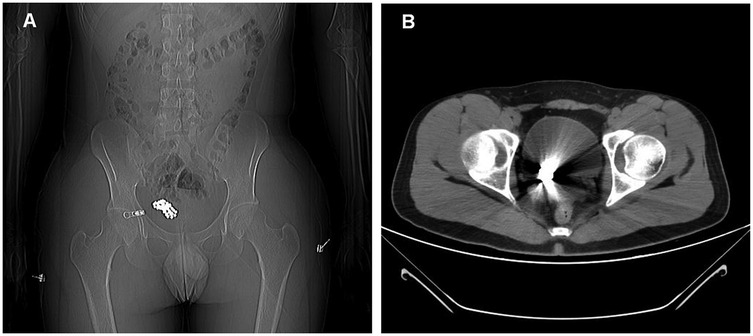
Figure 1. Pelvic plain computed tomography (CT) image. (A) Pelvic plain CT localizer radiograph showing numerous metal beads. (B) Plain pelvic CT scan being affected by metal artifacts.
Under general anesthesia, a 26 Fr sheath was carefully inserted into the urethra, revealing a cluster of metallic beads within the bladder. The primary challenge in retrieving these beads arose from their strong magnetic properties. Initially, we attempted to remove them using a resection electrode, but this proved ineffective due to the beads’ strong mutual attraction, which also caused significant deformation of the electrode. To overcome this, we devised a spoon-shaped extractor using an orthopedic wire made of non-magnetic stainless steel (Figure 2), with a diameter of 0.6 mm, which was not affected by the magnetic force of the beads.
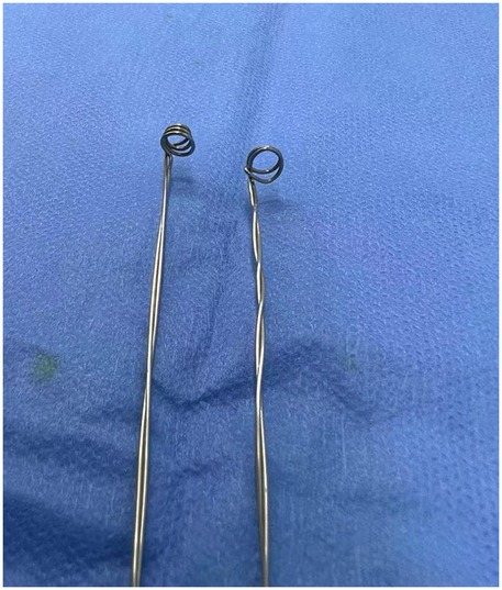
Figure 2. Two spoon-shaped extractors fabricated for bead removal.
After sterilization, the extractor was inserted into the resectoscope sheath and guided under direct visualization with a cystoscope. Initially, we were able to retrieve one to three beads at a time. As the number of beads in the bladder decreased, we successfully removed up to six beads at once (Figure 3). The entire procedure lasted approximately 50 min. Following surgery, a 16 Fr urinary catheter was retained for 24 h. The patient was discharged on postoperative day 2, without painful urination or symptoms of frequent urination, urgency, or gross hematuria.
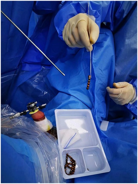
Figure 3. Demonstration of the tool in action, extracting several magnetic beads.
Foreign bodies within the urinary tract, including the bladder and the urethra can occur in both pediatric and adult populations. In this case, our patient sought medical attention after realizing he was unable to expel the magnetic beads through urination. The diagnosis was confirmed via CT imaging. Current methods for removing magnetic beads include open cystostomy, laparoscopic intervention, and cystoscopic extraction. According to literature reports, Graziottin et al. (7) successfully retrieved magnetic spheres from a 22-year-old male patient using endoscopic forceps, while Brooks et al. (8) described the extraction of magnetic beads via transurethral cystoscopy with a basket device and a three-pronged grasper in a 26-year-old male. Most patients prefer cystoscopic procedures due to their minimally invasive nature and shorter recovery times.
We purchased similar beads to simulate the surgical procedure and observed that the magnetic force between them was exceptionally strong. Additionally, instruments commonly used in laparoscopy were easily attracted by the magnetic pull. When attempting to remove the magnetic beads using laparoscopic separation forceps through a 22 Fr percutaneous nephroscope sheath, the beads were drawn toward the base of the forceps. This caused the forceps to open excessively, making it difficult to pass them through the sheath. These findings align with previous report findings of difficulties in using stone baskets or foreign body forceps in similar cases (9). Due to the strong magnetic force, the surgeon was unable to remove the magnetic beads using a stone removal basket or foreign body forceps, ultimately requiring a switch to open surgery. We initially attempted to use a loop electrode, commonly used for transurethral resection of the prostate, to separate the beads between the gaps. However, this approach was unsuccessful on the first attempt. Although the loop electrode was not attracted to the beads, it failed to extract them and became significantly deformed in the process. This made it clear that a new tool would be needed for the procedure.
The use of self-made tools for the removal of foreign bodies from the bladder has also been reported. Zeng et al. (10) reported successfully extracting magnetic beads using a self-made sheath with magnetic properties. We reached out to local factories for small magnetic bars, but they were unable to supply them, and the magnetism of the bars diminished after high-temperature sterilization. As a result, we decided to develop a non-magnetic device to extract the beads. We fashioned a tool from orthopedic wire made of non-magnetic stainless steel to prevent magnetic interference (Figure 2). The diameters of the top and bottom of the extractor were approximately 6 and 4 mm, respectively. After sterilization, this device was effectively used to smoothly remove all the magnetic beads.
Previous studies have reported that the treatment approaches adopted for female patients with bladder foreign bodies are similar to those for male patients, both involving the removal of the foreign body through cystoscopy or cystotomy (11). Very few works of literature have reported on the use of self-made tools to treat patients with bladder foreign bodies in females. Moreover, female patients are more likely to insert strip-shaped or rod-shaped foreign bodies into the bladder via the urethra (11, 12). If the foreign body possesses such a configuration, our tool may exhibit reduced efficacy in achieving its retrieve. The simplicity of the tool's design and the easy availability of materials make it particularly suitable for use in primary care hospitals. However, this tool is more appropriate for the removal of spherical foreign bodies than elongated, cord-like objects.
For additional requirements for specific article types and further information please refer to “article types” on every Frontiers journal page. This study presents a novel method for retrieving magnetic beads from the bladder using a self-made tool. This approach not only reduces the operative duration and complexity but also minimizes surgical trauma. The self-made device employed in this study proved to be relatively simple yet effective in successfully removing the magnetic beads from the bladder.
The original contributions presented in the study are included in the article/Supplementary Material, further inquiries can be directed to the corresponding author.
This study involving humans was approved by the ethics committee at Shenzhen Hengsheng Hospital. This study was conducted in accordance with the local legislation and institutional requirements. The participants provided their written informed consent to participate in this study. Written informed consent was obtained from the individual(s) for the publication of any potentially identifiable images or data included in this article.
ZZ: Conceptualization, Methodology, Writing – original draft. HD: Writing – original draft. QL: Writing – review & editing. YW: Conceptualization, Methodology, Supervision, Writing – review & editing.
The author(s) declare that no financial support was received for the research and/or publication of this article.
The authors declare that the research was conducted in the absence of any commercial or financial relationships that could be construed as a potential conflict of interest.
The author(s) declare that no Generative AI was used in the creation of this manuscript.
All claims expressed in this article are solely those of the authors and do not necessarily represent those of their affiliated organizations, or those of the publisher, the editors and the reviewers. Any product that may be evaluated in this article, or claim that may be made by its manufacturer, is not guaranteed or endorsed by the publisher.
1. Fotovat A, Yavari S, Ayati M, Nowroozi MR, Sharifi L. A case report of a self-inserted foreign body in the urethra/bladder causing urinary calculus formation, and a review of the literature. Heliyon. (2023) 9(3):e14038. doi: 10.1016/j.heliyon.2023.e14038
2. Venkataramani S, Ghazi NM, Kazmi FH, Khan I. Foreign body in the male urinary bladder: a case report. Cureus. (2024) 16(2):e54592. doi: 10.7759/cureus.54592
3. Yang CC, Bradley WE. Innervation of the human anterior urethra by the dorsal nerve of the penis. Muscle Nerve. (1998) 21(4):514–8. doi: 10.1002/(SICI)1097-4598(199804)21:4%3C514::AID-MUS10%3E3.0.CO;2-X
4. Mahadevappa N, Kochhar G, Vilvapathy KS, Dharwadkar S, Kumar S. Self-inflicted foreign bodies in lower genitourinary tract in males: our experience and review of literature. Urol Ann. (2016) 8(3):338–42. doi: 10.4103/0974-7796.184904
5. Zhang K, Zhang Y, Zhang Y, Chao M. Pneumovesicoscopy: an available technique for the retrieval of a rare foreign body in the urinary bladder. Asian J Surg. (2022) 45(5):1180–1. doi: 10.1016/j.asjsur.2022.01.095
6. Liu ZH, Zhu XF, Zhou N. Retrieval of 159 magnetic balls from urinary bladder: a case report and literature review. Urol Case Rep. (2019) 26:100975. doi: 10.1016/j.eucr.2019.100975
7. Graziottin TM, de Freitas GSD, Da Ros CT, Sogari PR, Telöken C, Laste PR. Magnetic spheres as foreign body into the bladder. J Sex Med. (2013) 10(10):2590–2. doi: 10.1111/j.1743-6109.2012.02772.x
8. Brooks T, Zreick J, Iocca A. Urinary obstruction from sexual practice involving magnetized beads inserted in the male urethra. CMAJ. (2013) 185(18):1597–8. doi: 10.1503/cmaj.130397
9. Levine MA, Evans H. Open removal as a first-line treatment of magnetic intravesical foreign bodies. Can Urol Assoc J. (2013) 7(1-2):E25–8. doi: 10.5489/cuaj.190
10. Zeng SX, Li HZ, Zhang ZS, Lu X, Yu XW, Yang QS, et al. Removal of numerous vesical magnetic beads with a self-made magnetic sheath. J Sex Med. (2015) 12(2):567–71. doi: 10.1111/jsm.12762
11. Bansal A, Yadav P, Kumar M, Sankhwar S, Purkait B, Jhanwar A, et al. Foreign bodies in the urinary bladder and their management: a single-centre experience from North India. Int Neurourol J. (2016) 20(3):260–9. doi: 10.5213/inj.1632524.262
Keywords: magnetic beads, foreign body, minimally invasive technique, cystoscope, surgery
Citation: Zhang ZY, Deng H, Liang QC and Wang YH (2025) Self-made spoon-shaped tool for removal of magnetic foreign bodies from the bladder: a case report. Front. Surg. 12:1528819. doi: 10.3389/fsurg.2025.1528819
Received: 15 November 2024; Accepted: 10 March 2025;
Published: 28 March 2025.
Edited by:
Andee Dzulkarnaen Zakaria, Universiti Sains Malaysia, MalaysiaReviewed by:
Nikolay Grigoryev, European Medical Center (EMC), RussiaCopyright: © 2025 Zhang, Deng, Liang and Wang. This is an open-access article distributed under the terms of the Creative Commons Attribution License (CC BY). The use, distribution or reproduction in other forums is permitted, provided the original author(s) and the copyright owner(s) are credited and that the original publication in this journal is cited, in accordance with accepted academic practice. No use, distribution or reproduction is permitted which does not comply with these terms.
*Correspondence: Yi He Wang, d2FuZ3lpaGV6enVAMTI2LmNvbQ==
Disclaimer: All claims expressed in this article are solely those of the authors and do not necessarily represent those of their affiliated organizations, or those of the publisher, the editors and the reviewers. Any product that may be evaluated in this article or claim that may be made by its manufacturer is not guaranteed or endorsed by the publisher.
Research integrity at Frontiers
Learn more about the work of our research integrity team to safeguard the quality of each article we publish.