 Zhen-jiang Liu1
 Shu-chao Wang
94% of researchers rate our articles as excellent or good
Learn more about the work of our research integrity team to safeguard the quality of each article we publish.
Find out more
ORIGINAL RESEARCH article
Front. Surg., 21 April 2023
Sec. Reconstructive and Plastic Surgery
Volume 10 - 2023 | https://doi.org/10.3389/fsurg.2023.1180624
This article is part of the Research TopicThe Role of Skin Surgery and Artificial Intelligence in Mechanisms of Skin Disorders, Lesions, and Skin Disease DiagnosisView all 4 articles
Background: Skin regeneration is a challenging issue worldwide. Increasing research has highlighted the role of immune cells in healing and the underlying regulatory mechanism. The purpose of this study was to identify the hotspots and trends in skin regeneration and inflammation research through bibliometrics and to provide insights into the future development of fundamental research and disease treatment.
Methods: Publications were collected from the Web of Science Core Collection on March 1, 2022. Articles and reviews published in English from January 1, 1999, to December 31, 2022, were selected, and statistical analyses of countries, institutions, authors, references, and keywords were performed using VOSviewer 1.6.18 and CiteSpace 5.8.
Results: A total of 3,894 articles and reviews were selected. The number of publications on skin inflammation and regeneration showed an increasing trend over time. Additionally, authors and institutions in the United States, United Kingdom, Canada, and China appeared to be at the forefront of research in the field of skin inflammation and regeneration. Werner Sabine published some of the most cited papers. Wound Repair and Regeneration was the most productive journal, while Journal of Investigative Dermatology was the most cited journal. Angiogenesis, diamonds, collagen, cytokine, and keratinocytes were the five most commonly used keywords.
Conclusion: The number of publications on skin inflammation and regeneration show an increasing trend. Moreover, a series of advanced technologies and treatments for skin regeneration, such as exosomes, hydrogels, and wound dressings, are emerging, which will provide precise information for the treatment of skin wounds. This study can enhance our understanding of current hotspots and future trends in skin inflammation and regeneration research, as well as provide guidelines for fundamental research and clinical treatment.
As the largest organ, the skin plays an essential role in protecting the body from various threats, such as trauma, noxious agents, UV radiation, and pathogens (1, 2). When the skin is injured, localized injury can lead to persistent damage, compromising its protective function and disturbing the wound-healing process (3, 4). Skin regeneration is a challenging issue worldwide and comprises three main stages: inflammation, re-epithelialization, and skin remodeling (5, 6). This process involves a series of cell types, such as endothelial cells, fibroblasts, stem cells, and immune cells (5, 7). Traditionally, immune cells are thought to cause systemic damage and destruction (8–10). In 1968, Elie Metchnikoff first mentioned the presence of immune cells at the site of injury (11). Since then, researchers have discovered that immune cells not only eliminate microbial and cellular debris but also promote tissue growth and wound repair (12, 13). Furthermore, increasing research has revealed the crucial role of immune cells in healing as well as the underlying regulatory mechanism (14, 15). In other clinical studies, immunocompromised individuals have been reported to exhibit impaired wound repair, which also indicates the important role of immune cells in skin healing (16–18). Currently, increasing evidence indicates that skin regeneration is not an autonomous process but one that relies on the regulation of inflammation. Investigating the hotspots and trends in this field can help researchers understand the fundamental, latest, and most influential advances as well as highlight potential research directions.
There are two types of studies that can summarize related publications in a specific research direction: reviews and bibliometrics (19, 20). Bibliometrics may serve as a useful summary and predictive tool for new researchers looking to explore a research field and expand their research horizons (21, 22). Bibliometric analysis reflects current research hotspots and potential research directions (23) and is a discipline that uses mathematical and statistical methods to study and quantify the contribution of publications to a specific field of research (22). It can also help organize large volumes of unstructured data into a rigorous, meaningful, and scientific arrangement (24). Therefore, bibliometric studies help build a strong base for researchers in specific areas. Two software, CiteSpace and VOSviewer, are commonly used for visual analysis (25). These software provide researchers a comprehensive insight into the field of research (26), which then enables them to assess progress in both qualitative and quantitative research and position their anticipated contributions to specific areas of research (26).
In this study, we aimed to provide a comprehensive overview of the relationship between skin regeneration and inflammation using bibliometrics. Furthermore, we summarize the number of publications, citation frequency, and centrality to analyze current research hotpots and other research directions. Finally, we review the latest research results, technology, and treatment for skin regeneration. This research can help new researchers select appropriate research directions and provide basic knowledge and understanding of current research. Furthermore, it could provide guidelines for fundamental research and precise clinical strategies for skin regeneration.
The relevant keywords were first searched in the MeSH Database (https://www.NCBI.nlm.nih.gov/mesh). The search formula: ALL = (skin and wound healing and inflammation) was used to search the Web of Science Core Collection. After that, we screened target papers based on (1) Document type: “Article” and “Review,” excluded books, corrections, letters, conference materials, and retracted papers; (2) Publication date: January 1, 1999, to December 31, 2022; (3) Language: English. This work was completed on March 1, 2023. Finally, 3,894 relevant documents met the criteria. Documents were exported as all records and references, and saved as plain text files (.txt) for bibliometric and visual analysis (Figure 1).
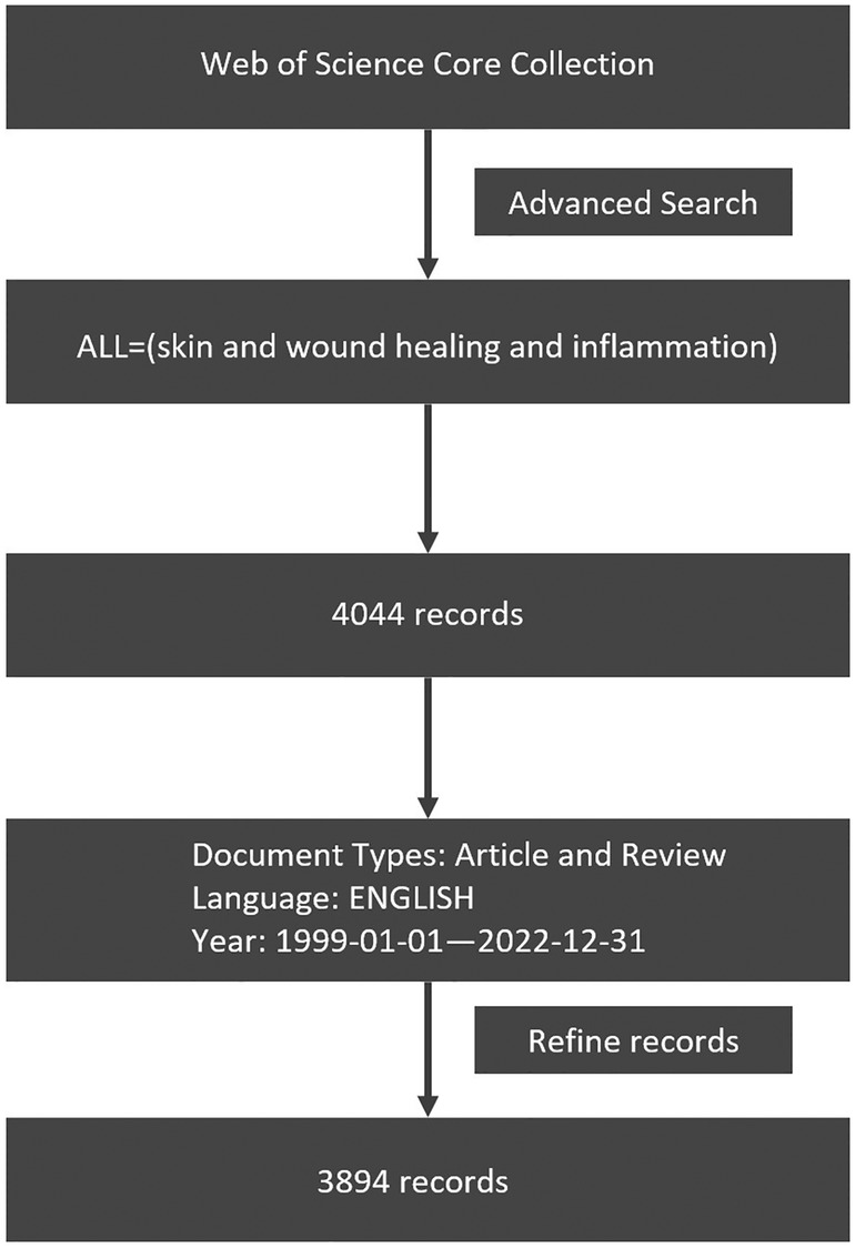
Figure 1. The data collection and retrieval strategy.
The dimensions of this analysis include publication year, author, institution, country and region, journal, keywords, and key references. We downloaded all data imported from the Web of Science (WOS) Core Collection into VOSviewer (version 1.6.18; https://www.vosviewer.com/downloavosviewer) and CiteSpace (version 5.8.R3; https://sourceforge.net/project/citespace/files/latest/do) to perform analyzing and visualizing.
CiteSpace and VOSviewer are the main software that can be used to analyze the scientific knowledge contained in complex data, and display the structure, distribution, and discipline of information through visual methods. Co-occurrence analysis is one method to show the frequency of occurrence of same sentences in one paper to determine their similarities. Therefore, researchers could get to know the research hotspots and future trends. Moreover, co-citation analysis could provide researchers with the first and basic published paper and related knowledge in this specific research area (27). In addition, we used Microsoft Office Excel to analyze the research trends in published papers in the type of tables.
In the figures presented by the visualization map below, each circle represents a node, and the diameter size of the circle represents the frequency of the item that appears in the co-occurrence analysis. The color of a circle is determined by the cluster of categories to which it belongs. The connection between nodes represents the association of the corresponding node, and the strength of the association between nodes is expressed in line width.
The first paper, according to the formula, was published in 1999, and we set the statistical period from 1999 to 2022. The annual publication trends are shown in Figure 2. The number of papers increased from 1999 (n = 31) to 2022 (n = 609). The increase in the number of published papers over the last 2 years indicates that research on skin inflammation and regeneration has received considerable attention.
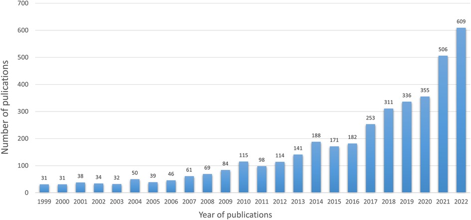
Figure 2. The number of articles per year from 1999 to 2022. The number of literature studies published has grown steadily and peaked in 2022.
Research studies conducted in 99 countries/regions were collected in this study. Figure 3 shows the cooperation between countries/regions. Table 1 lists the top 10 countries with the highest number of publications. The United States (1,089 publications, 57,054 citations) is the most productive country, followed by China (860 publications, 18,269 citations). The United States published the highest number of papers and had the largest number of citations among all countries. Forty-six countries/regions with over 10 papers were divided into six color-coded groups. The largest group consists of 14 countries, focused on the United States, the United Kingdom, and Canada. The above three aspects indicate that the United States is the most influential country.
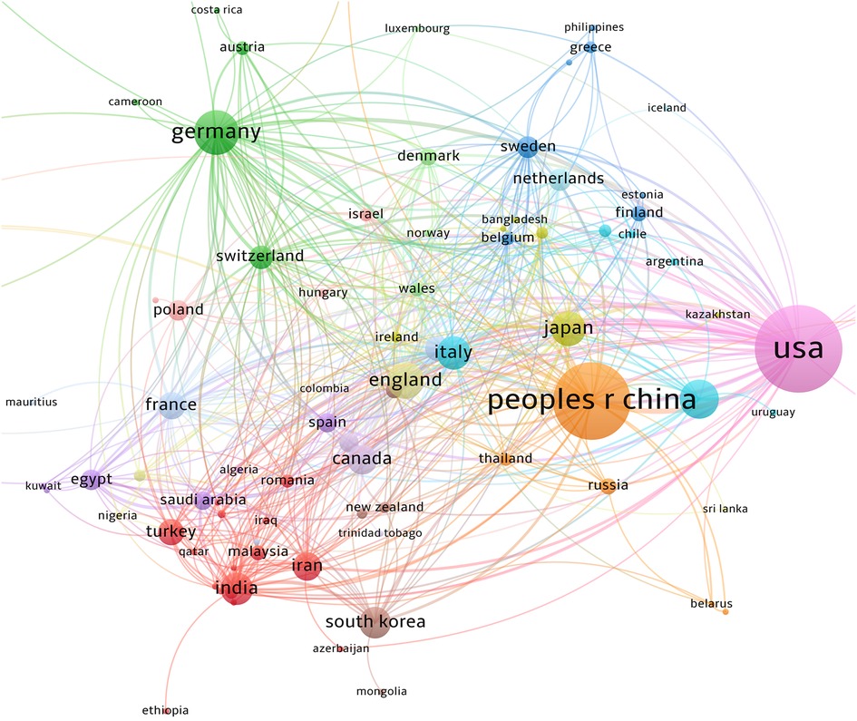
Figure 3. The co-authorship network of countries/regions. A total of 99 countries/regions have participated in the study. China and the United States are the centers of the study. The largest cluster is centered on the United States. China's cooperation between countries and regions is relatively strong.
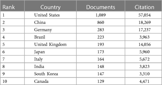
Table 1. The top 10 productive countries/regions.
A total of 3,894 papers on skin inflammation and regeneration were published in 1,184 journals. Table 2 lists the top 10 journals with the most published literature, accounting for 16.2% of the literature (630/3,894). Wound Repair and Regeneration was found to be the most productive journal (111 papers), and Journal of Investigative Dermatology was found to be the most cited journal (8,749 citations). Furthermore, although only three papers were published in Physiological Reviews, they have been cited 3,470 times in total.
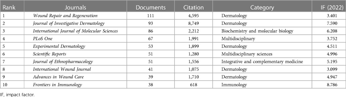
Table 2. The top 10 journals for publications.
Among all papers, 283 were cited over 100 times. Table 3 lists the 10 most cited papers. Regulation of wound healing by growth factors and cytokines published in Physiological Reviews in 2003 is the most cited article (2,422 citations).
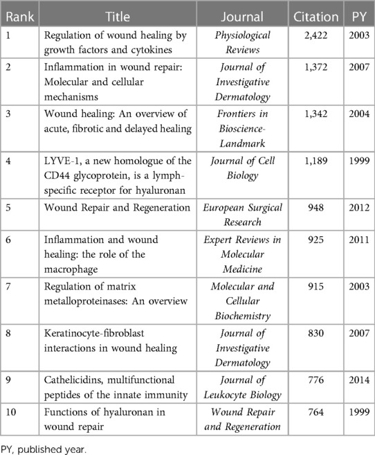
Table 3. The top 10 highest cited articles.
A total of 4,123 institutions have conducted research on skin inflammation and regeneration. Table 4 lists the top 10 institutions with the largest number of publications. Harvard University (43 papers, 3,916 citations) was found to be the most influential institution in this field, with 43 papers and the highest number of citations. Although Shanghai Jiao Tong University has published numerous papers, the number of citations is relatively low (1,621 citations).
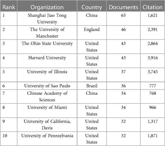
Table 4. The top 10 productive institutions.
The cooperation network of the research institutions is shown in Figure 4. After reviewing institutions with over 10 published papers, we divided the network of co-authors into five color-coded groups, of which the largest two were green and red. The red group consists of 59 universities focused on The University of Manchester, University of Illinois, and Ohio State University. In both the green and red groups, most cooperative relations are limited to China. For example, the universities in the green group are Chinese.
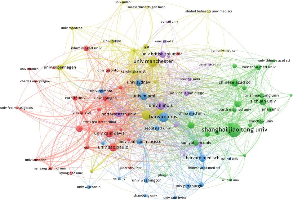
Figure 4. The co-authorship network of institutions. A total of 4,123 institutions participated in the study. The network of co-authors was divided into five clusters of different colors. The red cluster is dominated by American institutions, and the Harvard University has the greatest impact. The green cluster is dominated by Chinese institutions.
A total of 21,144 authors participated in the writing of the 3,894 papers. The top 10 authors with the most published papers are listed in Table 5. The table shows that Werner Sabine (17 publications, 2,076 citations) is the most prolific and cited author in this field. A threshold of six articles was set in VOSviewer, and 103 authors who met the criteria were selected. The results are shown in Figure 5. The nine color-coded groups in the figure represent the most closely linked groups in this field.
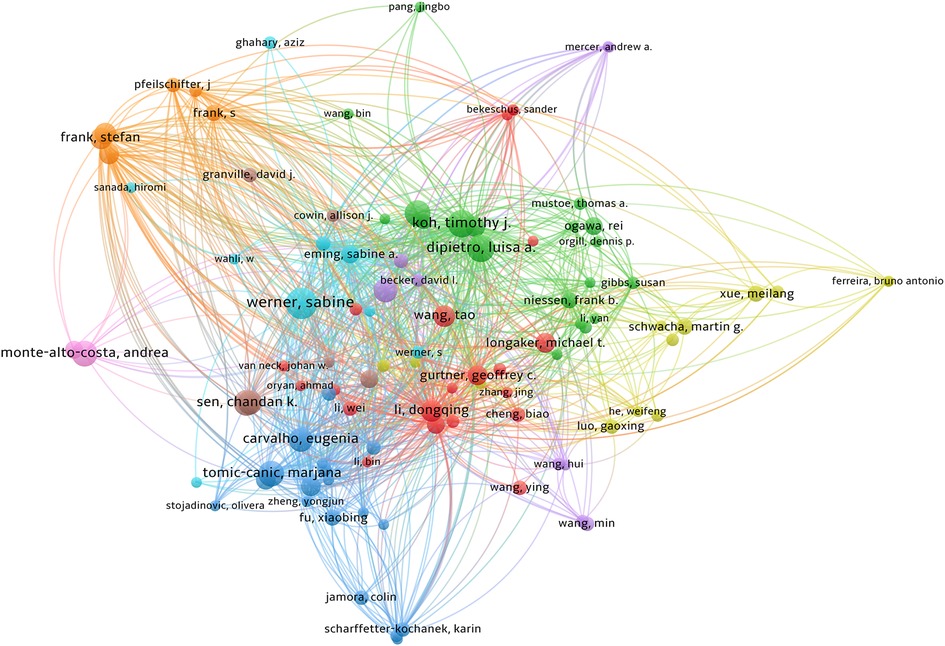
Figure 5. The co-authorship network of authors. A total of 21,144 authors participated in the writing process. The analysis method is Lin Log/modular. The size of the circle represents the influence of the author. Internode connections represent author collaboration and the lines of connection between nodes represent the collaboration between authors.
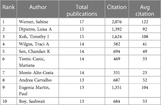
Table 5. The top 10 productive authors.
Following paper co-citation analysis, the 10 most cited papers were selected and are listed in Table 6, which is visualized in Figure 6. The paper titled Mechanisms of disease-Cutaneous wound healing published in the New England Journal of Medicine in 1999 was found to be the most cited (391 citations).
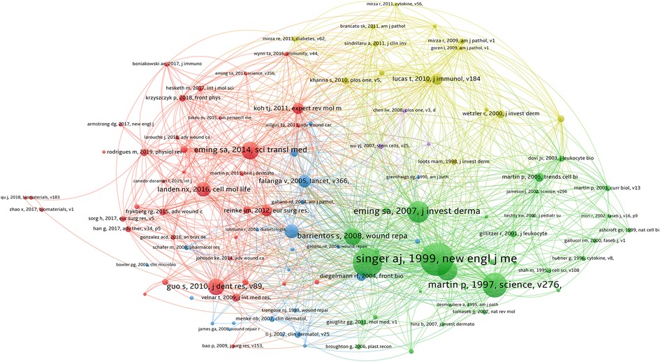
Figure 6. Co-citation analysis of references. The most cited article in co-citation analysis were written by A. J. Singer. Clusters of the same color indicate that the literature studies are related or have some commonalities.
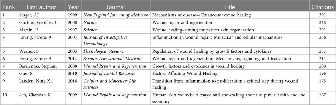
Table 6. The top 10 most co-cited references.
A strong citation burst analysis can identify published papers that have experienced significant changes in their citation situation over a short time period. The red line indicates the duration of the burst, and the intensity of the burst is indicative of the influence of the published paper. Figure 7 shows the first 50 papers with the strongest citation bursts. Wound repair and regeneration: Mechanisms, signaling, and translation published in Science Translational Medicine in 2014 had the highest burst intensity of 41.43. The high-intensity citation burst in this study lasted until 2019. This review article reviews and summarizes recent advances in skin regeneration and presented some beneficial perspectives on promoting wound healing in clinical patients.
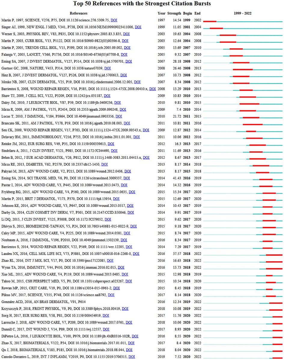
Figure 7. CiteSpace visualization map of top 50 references with the strongest citation bursts. A citation burst is when the citation situation of a paper changes dramatically in a short period of time. The red line indicates the duration of the outbreak, and the intensity of the outbreak indicates the impact of the article. The citation burst is analyzed with CiteSpace.
Keywords summarize the theme and content of the published paper. The frequency analysis of keywords is the most important window for understanding research hotspots and trends. We used VOSviewer to analyze the top 50 keywords (Table 7). After excluding keywords with vague directivity, such as sound healing and skin, we found that the following keywords appeared frequently: angiogenesis (166), diamonds (105), collagen (90), cytokines (76), and keratinocytes (70).
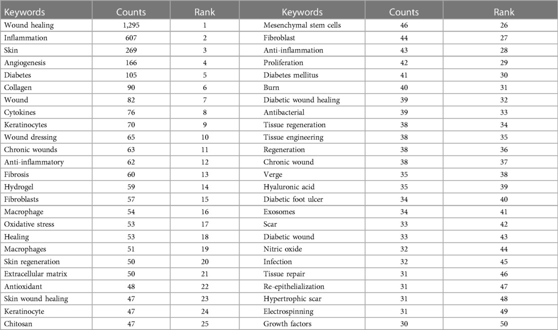
Table 7. The top 50 keywords.
In the Overlay visualization view of VOSviewer (Figure 8), the keywords are colored according to their average occurrence year (AAY). A cooler color indicates that the keywords appeared earlier, while a warmer color indicates that they appeared later. The keywords that recently attracted attention were exosomes (AAY: 2021), hydrogel (AAY: 2020), and wound dressing (AAY: 2019).
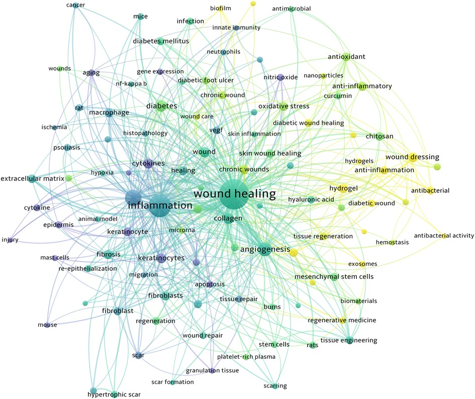
Figure 8. The overlay map of keywords. A node's size indicates how often the keyword appears. Cooler colors indicate earlier occurrences of keywords, and warmer colors indicate later occurrences.
After selecting keywords related to regulatory molecules and pathways, we listed the top 20 keywords with the most frequent occurrence in Table 8, and then visualized them with VOSviewer (Figure 9). The keywords that have appeared most frequently are cytokines; many high-frequency keywords in the table belong to this category, such as VEGF, TGF-β, and TNF-α. Cytokines play an important role in all stages of wound healing. Therefore, studies on the effects of cytokines, growth factors, and downstream effectors in wound healing have always been a hot topic in this field.
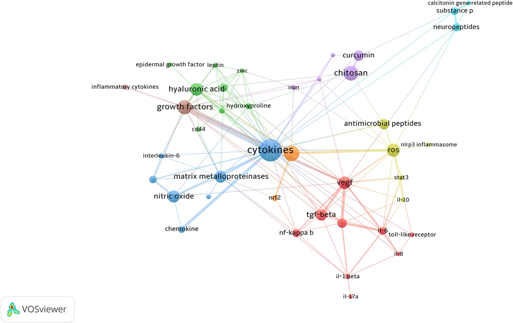
Figure 9. The keywords co-occurrence network visualization. High-frequency keywords with high correlation are shown with the same color, for example, high-frequency keywords of cytokines such as VEGF, TNF-α, and interleukin are shown in red.
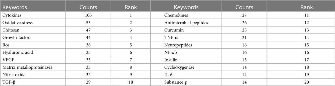
Table 8. The top 20 keywords.
In this study, we used bibliometrics to analyze the current research hotspots and future research trends in the field of skin inflammation and regeneration and found that the number of publications on skin inflammation and regeneration showed an increasing trend over time. Additionally, authors and institutions in the United States, United Kingdom, Canada, and China are at the forefront of research in the field of skin inflammation and regeneration. Finally, a series of advanced research results and treatments for skin inflammation and regeneration are emerging, such as the discovery of key regulatory mechanisms of inflammation, which will provide precise strategies for skin regeneration.
In this study, 3,894 papers on skin inflammation and healing published between January 1, 1999, and December 31, 2022, were searched on WOS. Overall, the evolution of published figures over time is indicative of the importance and progress of related research in the field of skin inflammation and regeneration. Currently, the United States, China, and Germany are the top three countries with the highest number of publications. Citation bursts are focused on the United States, the United Kingdom, and Canada, indicating that the United States is the most influential country. Additionally, Shanghai Jiao Tong University in China, the University of Manchester in England, Ohio State University, Harvard University, and University of Illinois in the United States published the highest number of papers. Harvard University became the most influential institution in this field with 43 papers and the largest number of citations. In terms of global journal quality, the highest number of papers on skin inflammation and regeneration were published in Wound Repair and Regeneration. Frontiers in Immunology is the journal with the highest impact factor (IF), IF 2021 = 8.786.
Dr. Werner of ETH Zurich has published 17 papers with the most citations, ranking himself as the leading author in the field of skin inflammation and regeneration. Dr. Werner in 2003 published one review “Regulation of wound healing by growth factors and cytokines” (28) on Physiological Reviews, which is the most cited paper with 2,422 citations. This review indicates that skin healing is a complex process that involves inflammation and the formation of new tissues. It summarizes numerous approaches focused on growth factors, cytokines, and their receptors or downstream effectors in skin repair. The co-authorship map indicated an increasing pattern of cooperation among researchers interested in skin regeneration. The most cited research is by Dr. Quigley, from Johns Hopkins Hospital, and “Mechanisms of disease: Cutaneous wound healing” (29) published in New England Journal of Medicine, which has been cited 391 times. This article discusses the biology and the role of skin substitutes in wound regeneration. It also indicates that macrophages appear to facilitate wound repair. Furthermore, “Wound repair and regeneration: Mechanisms, signaling, and translation” (30) published in Science Translational Medicine in 2014 has the highest burst intensity of 41.43. This review article reviews and summarizes recent advances in skin regeneration and presents some beneficial perspectives on the promotion of wound healing in clinical patients.
Co-occurrence group analysis was used to analyze the network of keywords studied in skin inflammation and regeneration research. Current research hotspots can be identified based on the keyword frequency. Some of the 50 most common keywords were oxidative stress, collagen, cytokines, and exosomes (31–34). The keyword results showed that the research trend of skin inflammation and regeneration is diversifying, which is related to cell biology and biochemistry (35, 36). Furthermore, analysis of frequency and centrality of keywords revealed that treatments such as “wound dressing,” “tissue engineering,” and “chitosan” appeared earlier (37–39), suggesting that further research on treatment methods is needed. Additionally, keywords such as “exosomes” and “hydrogel” began to attract attention mainly after 2016, suggesting that this research area is the next research direction and hotspot (40, 41).
Numerous studies have indicated that inflammation is an important differentiating factor in wound repair processes (42, 43). Analyzing the key molecules in skin regeneration and inflammation revealed that the frequency of some keywords increased rapidly in recent years. We analyzed and discussed the most commonly used keywords. For example, dendritic epidermal T cells can promote epithelial proliferation by producing keratinocyte growth factor-2 and IL-6, which induce signal transducer and activator of transcription 3 (44, 45). Additionally, damaged epithelial cells secrete alarmin IL-18, which activates resident Tc17 cells to produce the transcription factors GATA3 and IL-13, ultimately promoting wound regeneration (46–48). Furthermore, aberrant inflammation also leads to impaired wound healing, such as wound Tregs curbing Th17 responses that hinder skin healing (49, 50).
In this study, which was completed on March 1, 2023, we collected papers published between January 1, 1999, and December 31, 2022. Consequently, some previously and subsequently published papers with updated results may not be included, making the papers in our database fewer than practical papers. In addition, only articles and reviews were included in this research, as we wanted to control the quality of publications to the maximum possible extent. Therefore, other types of papers might have been overlooked, such as meta-analyses, books, and case reports, which may have advanced achievements in both fundamental and clinical research. Finally, the field of skin inflammation and regeneration covers numerous aspects of the research. Although we retrieved the most relevant publications, some important research results may have been neglected due to the limitations of the bibliometric tools used.
The purpose of this study was to summarize the current research hotspots and future trends in skin inflammation and regeneration. We used bibliometrics to analyze related publications in the field and found that publications on skin inflammation and regeneration show an increasing trend. Furthermore, a series of advanced research results, technology, and treatment for skin regeneration are emerging, such as the research findings of key molecules and mechanisms of inflammation and immunity, which will provide precise information for the treatment of skin injuries. Taken together, this research can enhance our understanding of the current hotspots and future trends in the field of skin inflammation and regeneration and will provide guidelines for fundamental research and clinical treatment.
The original contributions presented in the study are included in the article/Supplementary Material, further inquiries can be directed to the corresponding authors.
S-cW and MH: designed the experiments, interpreted the data, and revised the manuscript. Z-jL: performed the experiments, prepared the figures, and wrote the manuscript. M-jW, Y-tT, and JL: helped to perform the experiments and prepared the figures. All authors contributed to the article and approved the submitted version.
The study was supported by National Natural Science Foundation of China (No. 82101126), Natural Science Foundation of Hunan Province (No. 2021JJ40873), the Hunan Provincial Natural Science Foundation of China (No. 2019JJ50696), Scientific Research Project of Hunan Provincial Health Commission (No. 202203013645), Hunan Provincial Science and Technology Innovation Plan Project (No. 2021SK53527), and the Scientific Research Launch Project for New Employees of the Second Xiangya Hospital of Central South University.
The authors declare that the research was conducted in the absence of any commercial or financial relationships that could be construed as a potential conflict of interest.
All claims expressed in this article are solely those of the authors and do not necessarily represent those of their affiliated organizations, or those of the publisher, the editors and the reviewers. Any product that may be evaluated in this article, or claim that may be made by its manufacturer, is not guaranteed or endorsed by the publisher.
1. Allen EM, Mctague MF, Bay CP, Esposito JG, Von Keudell A, Weaver MJ. The effectiveness of germicidal wipes and ultraviolet irradiation in reducing bacterial loads on electronic tablet devices used to obtain patient information in orthopaedic clinics: evaluation of tablet cleaning methods. J Hosp Infect. (2020) 105:200–4. doi: 10.1016/j.jhin.2020.04.014
2. Nghiem DX, Kazimi N, Mitchell DL, Vink AA, Ananthaswamy HN, Kripke ML, et al. Mechanisms underlying the suppression of established immune responses by ultraviolet radiation. J Invest Dermatol. (2002) 119:600–8. doi: 10.1046/j.1523-1747.2002.01845.x
3. Zang T, Heath K, Etican J, Chen L, Langley D, Holland AJA, et al. Local burn wound environment versus systemic response: comparison of proteins and metabolites. Wound Repair Regen. (2022) 30:560–72. doi: 10.1111/wrr.13042
4. Zhou Z, Xiao J, Huang S, Wu H, Guan S, Wu T, et al. A wet-adhesive carboxymethylated yeast beta-glucan sponge with radical scavenging, bacteriostasis and anti-inflammatory functions for rapid hemostasis. Int J Biol Macromol. (2023) 230:123158. doi: 10.1016/j.ijbiomac.2023.123158
5. Ren H, Zhao F, Zhang Q, Huang X, Wang Z. Autophagy and skin wound healing. Burns Trauma. (2022) 10:tkac003. doi: 10.1093/burnst/tkac003
6. Yang R, Liu F, Wang J, Chen X, Xie J, Xiong K. Epidermal stem cells in wound healing and their clinical applications. Stem Cell Res Ther. (2019) 10:229. doi: 10.1186/s13287-019-1312-z
7. Ebner-Peking P, Krisch L, Wolf M, Hochmann S, Hoog A, Vari B, et al. Self-assembly of differentiated progenitor cells facilitates spheroid human skin organoid formation and planar skin regeneration. Theranostics. (2021) 11:8430–47. doi: 10.7150/thno.59661
8. Tsepkolenko A, Tsepkolenko V, Dash S, Mishra A, Bader A, Melerzanov A, et al. The regenerative potential of skin and the immune system. Clin Cosmet Investig Dermatol. (2019) 12:519–32. doi: 10.2147/CCID.S196364
9. Yan WT, Zhao WJ, Hu XM, Ban XX, Ning WY, Wan H, et al. PANoptosis-like cell death in ischemia/reperfusion injury of retinal neurons. Neural Regen Res. (2023) 18:357–63. doi: 10.4103/1673-5374.346545
10. Yang R, Yang S, Zhao J, Hu X, Chen X, Wang J, et al. Progress in studies of epidermal stem cells and their application in skin tissue engineering. Stem Cell Res Ther. (2020) 11:303. doi: 10.1186/s13287-020-01796-3
11. Metchnikoff E. Elie Metchnikoff (1845–1916), advocate of phagocytosis. JAMA. (1968) 203:139–41. doi: 10.1001/jama.1968.03140020066016
12. Leibovich SJ, Ross R. The role of the macrophage in wound repair. A study with hydrocortisone and antimacrophage serum. Am J Pathol. (1975) 78:71–100. 1109560.1109560
13. Li Z, Gothard E, Coles MC, Ambler CA. Quantitative methods for measuring repair rates and innate-immune cell responses in wounded mouse skin. Front Immunol. (2018) 9:347. doi: 10.3389/fimmu.2018.00347
14. Xiong Y, Mi BB, Lin Z, Hu YQ, Yu L, Zha KK, et al. The role of the immune microenvironment in bone, cartilage, and soft tissue regeneration: from mechanism to therapeutic opportunity. Mil Med Res. (2022) 9:65. doi: 10.1186/s40779-022-00426-8
15. Zhang L, Piipponen M, Liu Z, Li D, Bian X, Niu G, et al. Human skin specific long noncoding RNA HOXC13-AS regulates epidermal differentiation by interfering with Golgi-ER retrograde transport. Cell Death Differ. (2023). doi: 10.1038/s41418-023-01142-z. [Online ahead of print].
16. Hu XM, Li ZX, Zhang DY, Yang YC, Fu SA, Zhang ZQ, et al. A systematic summary of survival and death signalling during the life of hair follicle stem cells. Stem Cell Res Ther. (2021) 12:453. doi: 10.1186/s13287-021-02527-y
17. Khoshnevisan R, Anderson M, Babcock S, Anderson S, Illig D, Marquardt B, et al. NOX1 regulates collective and planktonic cell migration: insights from patients with pediatric-onset IBD and NOX1 deficiency. Inflamm Bowel Dis. (2020) 26:1166–76. doi: 10.1093/ibd/izaa017
18. Kuhns DB, Fink DL, Choi U, Sweeney C, Lau K, Priel DL, et al. Cytoskeletal abnormalities and neutrophil dysfunction in WDR1 deficiency. Blood. (2016) 128:2135–43. doi: 10.1182/blood-2016-03-706028
19. Zhang JH, Wang MJ, Tan YT, Luo J, Wang SC. A bibliometric analysis of apoptosis in glaucoma. Front Neurosci. (2023) 17:1105158. doi: 10.3389/fnins.2023.1105158
20. Zhao X, Akbaritabar A, Kashyap R, Zagheni E. A gender perspective on the global migration of scholars. Proc Natl Acad Sci U S A. (2023) 120:e2214664120. doi: 10.1073/pnas.2214664120
21. Huang R, Jin M, Liu Y, Lu Y, Zhang M, Yan P, et al. Global trends in research of fibroblasts associated with rheumatoid diseases in the 21st century: a bibliometric analysis. Front Immunol. (2023) 14:1098977. doi: 10.3389/fimmu.2023.1098977
22. Zhang JH, Ni SY, Tan YT, Luo J, Wang SC. A bibliometric analysis of PIN1 and cell death. Front Cell Dev Biol. (2022) 10:1043725. doi: 10.3389/fcell.2022.1043725
23. Hu XM, Li ZX, Zhang DY, Yang YC, Zheng SY, Zhang Q, et al. Current research and clinical trends in rosacea pathogenesis. Heliyon. (2022) 8:e10874. doi: 10.1016/j.heliyon.2022.e10874
24. Chen Y, Li Y, Guo L, Hong J, Zhao W, Hu X, et al. Bibliometric analysis of the inflammasome and pyroptosis in brain. Front Pharmacol. (2020) 11:626502. doi: 10.3389/fphar.2020.626502
25. Yan WT, Lu S, Yang YD, Ning WY, Cai Y, Hu XM, et al. Research trends, hot spots and prospects for necroptosis in the field of neuroscience. Neural Regen Res. (2021) 16:1628–37. doi: 10.4103/1673-5374.303032
26. Zheng SY, Hu XM, Huang K, Li ZH, Chen QN, Yang RH, et al. Proteomics as a tool to improve novel insights into skin diseases: what we know and where we should be going. Front Surg. (2022) 9:1025557. doi: 10.3389/fsurg.2022.1025557
27. Romero L, Portillo-Salido E. Trends in sigma-1 receptor research: a 25-year bibliometric analysis. Front Pharmacol. (2019) 10:564. doi: 10.3389/fphar.2019.00564
28. Werner S, Grose R. Regulation of wound healing by growth factors and cytokines. Physiol Rev. (2003) 83:835–70. doi: 10.1152/physrev.2003.83.3.835
29. Singer AJ, Clark RA. Cutaneous wound healing. N Engl J Med. (1999) 341:738–46. doi: 10.1056/NEJM199909023411006
30. Eming SA, Martin P, Tomic-Canic M. Wound repair and regeneration: mechanisms, signaling, and translation. Sci Transl Med. (2014) 6: 265sr266. doi: 10.1126/scitranslmed.3009337
31. Lee SH, An S, Ryu YC, Seo SH, Park S, Lee MJ, et al. Adhesive hydrogel patch-mediated combination drug therapy induces regenerative wound healing through reconstruction of regenerative microenvironment. Adv Healthc Mater. (2023): e2203094. doi: 10.1002/adhm.202203094. [Online ahead of print].36854308
32. Md Fadilah NI, Mohd Abdul Kader Jailani MS, Badrul Hisham MAI, Sunthar Raj N, Shamsuddin SA, Ng MH, et al. Cell secretomes for wound healing and tissue regeneration: next generation acellular based tissue engineered products. J Tissue Eng. (2022) 13:20417314221114273. doi: 10.1177/20417314221114273
33. Nogueira BCF, Campos AK, Alves RS, Sarandy MM, Novaes RD, Esposito D, et al. What is the impact of depletion of immunoregulatory genes on wound healing? A systematic review of preclinical evidence. Oxid Med Cell Longev. (2020) 2020:8862953. doi: 10.1155/2020/8862953
34. Zhang H, Li W, Zhang Q, Zhong R, Li C, Chen Y, et al. Identification of active compounds and molecular mechanisms of Dalbergia tsoi Merr.et Chun to accelerate wound healing. Biomed Pharmacother. (2022) 150:112990. doi: 10.1016/j.biopha.2022.112990
35. Ali N, Hosseini M, Vainio S, Taieb A, Cario-Andre M, Rezvani HR. Skin equivalents: skin from reconstructions as models to study skin development and diseases. Br J Dermatol. (2015) 173:391–403. doi: 10.1111/bjd.13886
36. Qing C. The molecular biology in wound healing & non-healing wound. Chin J Traumatol. (2017) 20:189–93. doi: 10.1016/j.cjtee.2017.06.001
37. Baharlouei P, Rahman A. Chitin and chitosan: prospective biomedical applications in drug delivery, cancer treatment, and wound healing. Mar Drugs. (2022) 20(7):460. doi: 10.3390/md20070460
38. Lin J, Jiao G, Kermanshahi-Pour A. Algal polysaccharides-based hydrogels: extraction, synthesis, characterization, and applications. Mar Drugs. (2022) 20(5):306. doi: 10.3390/md20050306
39. Renuka RR, Julius A, Yoganandham ST, Umapathy D, Ramadoss R, Samrot AV, et al. Diverse nanocomposites as a potential dressing for diabetic wound healing. Front Endocrinol. (2022) 13:1074568. doi: 10.3389/fendo.2022.1074568
40. Frazier T, Alarcon A, Wu X, Mohiuddin OA, Motherwell JM, Carlsson AH, et al. Clinical translational potential in skin wound regeneration for adipose-derived, blood-derived, and cellulose materials: cells, exosomes, and hydrogels. Biomolecules. (2020) 10(10):1373. doi: 10.3390/biom10101373
41. Safari B, Aghazadeh M, Davaran S, Roshangar L. Exosome-loaded hydrogels: a new cell-free therapeutic approach for skin regeneration. Eur J Pharm Biopharm. (2022) 171:50–9. doi: 10.1016/j.ejpb.2021.11.002
42. Castrejon-Comas V, Aleman C, Perez-Madrigal MM. Multifunctional conductive hyaluronic acid hydrogels for wound care and skin regeneration. Biomater Sci. (2023). 11(7):2266–76. doi: 10.1039/d2bm02057b
43. Kacvinska K, Pavlinakova V, Polacek P, Michlovska L, Blahnova VH, Filova E, et al. Accelular nanofibrous bilayer scaffold intrapenetrated with polydopamine network and implemented into a full-thickness wound of a white-pig model affects inflammation and healing process. J Nanobiotechnol. (2023) 21:80. doi: 10.1186/s12951-023-01822-5
44. Deng G, Chen W, Wang P, Zhan T, Zheng W, Gu Z, et al. Inhibition of NLRP3 inflammasome-mediated pyroptosis in macrophage by cycloastragenol contributes to amelioration of imiquimod-induced psoriasis-like skin inflammation in mice. Int Immunopharmacol. (2019) 74:105682. doi: 10.1016/j.intimp.2019.105682
45. Jameson J, Ugarte K, Chen N, Yachi P, Fuchs E, Boismenu R, et al. A role for skin gammadelta T cells in wound repair. Science. (2002) 296:747–9. doi: 10.1126/science.1069639
46. Annunziato F, Romagnani C, Romagnani S. The 3 major types of innate and adaptive cell-mediated effector immunity. J Allergy Clin Immunol. (2015) 135:626–35. doi: 10.1016/j.jaci.2014.11.001
47. Kandikattu HK, Upparahalli Venkateshaiah S, Kumar S, Yadavalli CS, Mishra A. IL-18-mediated neutrophil recruitment promotes acute lung injury in inflammation-mediated chronic pancreatitis. Mol Immunol. (2023) 155:100–9. doi: 10.1016/j.molimm.2023.01.012
48. Yashiro T, Moro K. Crossing the valley of death: toward translational research regarding ILC2. Allergol Int. (2023) 72(2):187–93. doi: 10.1016/j.alit.2022.12.006
49. Crome SQ, Clive B, Wang AY, Kang CY, Chow V, Yu J, et al. Inflammatory effects of ex vivo human Th17 cells are suppressed by regulatory T cells. J Immunol. (2010) 185:3199–208. doi: 10.4049/jimmunol.1000557
Keywords: bibliometric, Citespace, VOSviewer, wound, skin regeneration, inflammation, immune
Citation: Liu Z-j, Wang M-j, Luo J, Tan Y-t, Hou M and Wang S-c (2023) A bibliometric analysis of hotpots and trends for the relationship between skin inflammation and regeneration. Front. Surg. 10:1180624. doi: 10.3389/fsurg.2023.1180624
Received: 6 March 2023; Accepted: 29 March 2023;
Published: 21 April 2023.
Edited by:
Ronghua Yang, Guangzhou First People's Hospital, ChinaReviewed by:
Peng Sun, The Affiliated Hospital of Qingdao University, China© 2023 Liu, Wang, Luo, Tan, Hou and Wang. This is an open-access article distributed under the terms of the Creative Commons Attribution License (CC BY). The use, distribution or reproduction in other forums is permitted, provided the original author(s) and the copyright owner(s) are credited and that the original publication in this journal is cited, in accordance with accepted academic practice. No use, distribution or reproduction is permitted which does not comply with these terms.
*Correspondence: Min Hou aG91bWluMDQyN0Bjc3UuZWR1LmNu Shu-chao Wang d2FuZ3NodWNoYW9AY3N1LmVkdS5jbg==
Specialty Section: This article was submitted to Reconstructive and Plastic Surgery, a section of the journal Frontiers in Surgery
Disclaimer: All claims expressed in this article are solely those of the authors and do not necessarily represent those of their affiliated organizations, or those of the publisher, the editors and the reviewers. Any product that may be evaluated in this article or claim that may be made by its manufacturer is not guaranteed or endorsed by the publisher.
Research integrity at Frontiers
Learn more about the work of our research integrity team to safeguard the quality of each article we publish.