 Christophe Boulay1,2,3*
 Jacques-Olivier Coq2
 Morgan Sangeux1,4
 Guillaume Authier1,2Alexis Ulian1,2
 Maud Pradines3,5
 Marjolaine Baude3,5
 Béatrice Desnous6Jean-Luc Jouve1,2Bernard Parratte1Emilie Peltier1Sébastien Pesenti1Jean-Michel Gracies3,5
- 1Gait Laboratory, Pediatric Orthopaedic Surgery Department, Timone Children’s Hospital, Marseille, France
- 2Institut des Sciences du Mouvement (ISM), Equipe DynamiCC, UMR 7287 CNRS/Aix Marseille Université, Marseille, France
- 3AP-HP, Service de Rééducation Neurolocomotrice, Unité de Neurorééducation, Hôpitaux Universitaires Henri Mondor, Créteil, France
- 4Department of Orthopaedics, University Children’s Hospital, Basel, Switzerland
- 5UR 7377 BIOTN, Laboratoire Analyse et Restauration du Mouvement, Université Paris Est Créteil (UPEC), Créteil, France
- 6Pediatric Neurology Department, Timone Children’s Hospital, Marseille, France
Introduction: Focal vibration therapy (FVT) is increasingly used in the treatment of spastic paresis. In adults, it has been shown to reduce spasticity and to increase torque production from the vibrated muscles by restoring reciprocal inhibition of antagonists, thereby improving overall gait. In children with spastic cerebral palsy (CP), FVT has also been suggested to reduce spasticity, increase torque production and improve gait function, but evidence is limited.
Methods: We report the case of a child with unilateral spastic CP (USCP) and equinus gait (GFMCS II level) with (i) ankle dorsiflexor paresis, (ii) ankle plantar flexor overactivity, especially in gastrosoleus complex and peroneus longus, (iii) spastic myopathy, affecting gastrosoleus complex in particular, and (iv) calf pain seemingly related to muscle overactivity. The child was treated with a two-month program of alternating dorsiflexor and plantar flexor focal vibration therapy (FVT) and botulinum neurotoxin A (BoNT-A) injections into plantar flexors, alongside conventional physiotherapy.
Results and discussion: Clinical evaluations during the two-month program showed (i) improved walking speed (ii) decreased ankle dorsiflexor paresis and ankle plantar flexor overactivity, especially spastic co-contraction and spasticity, (iii) improved passive extensibility in plantar flexors, and (iv) reduced pain. This is the first report of the combination of FVT and BoNT-A injections having promising effects on equinus gait in USCP.
1 Introduction
Cerebral palsy (CP) is a developmental motor disorder affecting about 2.5 children per 1000 live births in developed countries (1), caused by non-progressive brain injury before or shortly after birth, with reduced descending drive that secondarily hinders spinal cord development (2). CP manifests as both a neurological disorder (antagonist overactivity and agonist paresis) and a muscle disorder (spastic myopathy with stiffening of antagonists) (3, 4). At the ankle, there are three main forms of plantar flexor overactivity in children with CP: (i) spastic cocontraction, defined as unwanted, involuntary plantar flexor activation during voluntary dorsiflexor effort, primarily due to misdirection of the supraspinal descending drive, leading to reduced ankle dorsiflexion range of motion (3–6); (ii) spastic dystonia, namely unwanted, involuntary muscle activation at rest, in the absence of stretching or voluntary effort (3, 7), and (iii) spasticity, defined as increased velocity-dependent responses to phasic stretch, detected and measured at rest (3, 8). Children with CP also present with dorsiflexor paresis that is worsened by plantar flexor stretch, known as stretch-sensitive paresis (3, 4, 9).
Two vicious cycles ensue in that context: agonist paresis both causes and becomes worsened by disuse, and spastic myopathy both causes and becomes worsened by overactivity (8, 10). Musculoskeletal deformities acquired during growth have been classically handled by tenotomy or fascia- and/or aponeurosis of muscle(s) release surgery but the underlying spastic myopathy and misdirection of the supraspinal drive remain. Yet, early management, long before growth spurts, is crucial to minimize the emergence of deformities. There is early evidence that focal vibration therapy (FVT) and botulinum neurotoxin A (BoNT-A) injections may improve patient outcomes in this context (11–13).
Repeated sessions of focal vibrations (12) generated with a mechanical or pneumatic device have been reported to reduce resistance to passive movement from the vibrated plantar flexors in paretic patients (14) and to increase voluntary torque production from vibrated dorsiflexors in healthy subjects (15). Furthermore, gait function has been reported to be improved by FVT applied to dorsiflexors in adults after stroke (16), and to plantar flexors in children with CP (12, 17). Vibrations of aponeurosis-tendon junctions activate spindles, inducing primarily Ia excitation, which increases excitability in the corticospinal pathway to the vibrated muscle (17–20) and potentially also to the non-vibrated muscle (18). FVT may therefore be beneficial in children with CP; its safety and efficacy have been suggested in children both for the neuromuscular manifestations of CP and to treat CP-related pain (16, 21–32).
The effects of FVT following BoNT-A injections is unknown but in vitro and clinical data (33, 34) suggest that the increase in muscle temperature and activation induced by FVT may enhance the translocation of the BoNT-A light chain into the presynaptic terminal and thus increase toxin potency and duration in the vibrated muscle (33, 34). Here, we present the case of a child with CP who was treated with plantar flexor FVT before and after BoNT-A injections. The child was evaluated throughout the treatment program using the Five-Step Assessment (FSA) (35), which quantifies the extent of the various components of deforming spastic paresis.
2 Case description
The child, a 5-year-old boy (18.5 kg, 1.09 m) with confirmed diagnosis of unilateral spastic cerebral palsy (USCP) and gross motor functional classification (GMFCS) II, was followed longitudinally for one month of FVT applied to dorsi- and plantar flexor muscles (Vibrasens, Techno Concept, serial number 14-V.01-210, Manosque, France) before and after plantar flexor injections of BoNT-A (Figure 1). During FVT sessions, vibrations were applied alternatingly to the myotendinous junction of the gastrosoleus complex (GSC) and tibialis anterior (TA; see details in Figure 1). On D28, the patient received a total of 15 U/kg abobotulinumtoxin A in gastrocnemius medialis (GM, 60 U), gastrocnemius lateralis (GL, 30 U), peroneus longus (PL, 90 U) and soleus (SOL, 90 U). Injections were performed under local anesthesia with ultrasound guidance.

Figure 1. Details of the two-month treatment program based on focal vibration therapy (FVT)and botulinum neurotoxin A injections. The injections were performed on day 28. Each FVT session involved 10 min of vibration blocks (80 Hz vibration for 10 s every 5 s) applied to dorsiflexors followed by 10 min of the same vibration blocks applied to the plantar flexors, followed by 10 min of alternate vibration blocks applied to dorsiflexors and plantar flexors (80 Hz for 5 s every 5 s). Each session started and ended with a clinical examination (CE), including patient-assessment of illusory movements triggered by applying vibrations to agonist or antagonist muscles.
3 Diagnostic assessment
Clinical assessments, including quantified clinical examination (FSA), electromyography (EMG, data to be reported elsewhere) during walking, and walking speed measurements, were performed before FVT (D0), the morning of the BoNT-A injections (D28), and 29 days after the BoNT-A injections (D57). The patient received 11 FVT sessions between D0 and D28, and another 11 sessions between D28 and D57, as shown in Figure 1. Clinical examinations consisting of FSA measurements and quantified child-perception of illusory movements (20, 36), were performed at the beginning and end of each FVT session to evaluate immediate effects. Conventional physical therapy program for equinus gait continued unchanged during the two-month FVT program. This included plantar flexor stretching and strengthening of command on TA and on plantar flexor muscles once or twice a week.
3.1 Five-step assessment
FSA measurements (first three steps of the FSA) were performed using 0° defined as the position of minimum stretch of the tested muscle (35). Ankle joint range of dorsiflexion measurements against the resistance of the soleus (knee flexed) and of the GSC (knee extended) were performed with the patient in supine position. The range of motion of the ankle was measured between the fibula and the posterior half of the external border of the foot, to cancel effects of any foot pronation during dorsiflexion efforts. Maximum clinical extensibility was estimated by XV1, the angle of arrest under slow stretching. Stretch reflex threshold was quantified by XV3, the angle of catch or clonus under brisk passive stretching. The angle of match between maximal voluntary activation of dorsiflexors and passive and active resistance of the plantar flexors was quantified by XA, i.e., the range of ankle dorsiflexion reached during maximum voluntary ankle dorsiflexion efforts, in the knee extended and knee flexed positions.
3.2 Illusory movements
Illusory movements related to vibration were self-assessed by the child immediately before and after each FVT session (36, 37). Illusory movements were classified as antagonist vibratory response (AVR, the physiological response) if vibrations applied to the myotendinous junction triggered illusory movement in the opposite direction, i.e., felt by the brain as stretch of the vibrated muscle, or tonic vibratory response (TVR) if vibrations applied to the myotendinous junction triggered illusory movement corresponding to the perception of contraction of the vibrated muscle. Results in either direction were reported on an ordinal scale from 0–3, with 0, 1, 2 and 3 indicating no sensation of movement, less than 5° of sensed movement, 5–10° of sensed movement, and more than 10° of sensed movement, respectively.
3.3 Statistics
Mean differences between pre- and post-session FSA variables, before and after BoNT-A injections, were summarized as median (range), and tested for significance using paired t-tests or Wilcoxon tests depending on the conditions. Overall trends in the FSA variables as a function of time before and after BoNT-A injections were fitted using linear regression models and tested for significance using t-tests. Results were summarized as linear regression coefficients with 95% confidence intervals. All analyses were performed in R (version 4.3.2) (38). Statistical significance was defined as p < 0.05.
4 Results
Before BoNT-A injections, all three FSA variables (XV1, XV3, and XA) increased significantly during FVT sessions both in the knee flexed and knee extended positions (Table 1, Figure 2), with average gains of 1–3° for XV1 and XA and 9° for XV3. After BoNT-A injections, the increases in XV1, XV3, and XA between the start and end of each FVT session virtually disappeared, remaining only to a small extent in the knee extended position (Table 1, Figure 2).
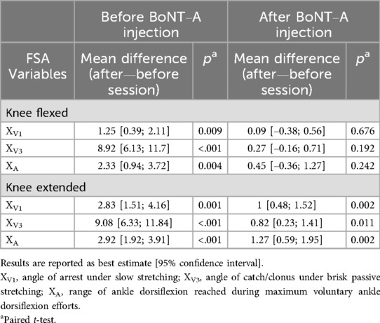
Table 1. Mean differences between post- and pre-session values of Five Step Assessment (FSA) variables, before and after botulinum neurotoxin A (BoNT-A) injection.
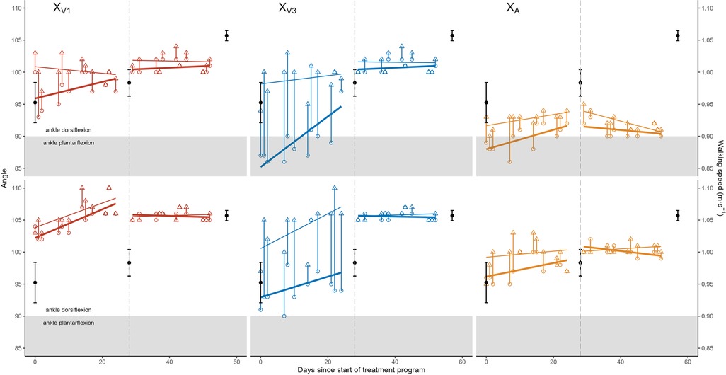
Figure 2. Range of motion (angle in degrees) as a function of time for the Five Step Assessment variables XV1 (red, left), XV3 (blue, middle) and XA (gold, right) in knee extended (top row) and knee extended (bottom row) positions. Pre-focal vibration therapy (FVT) session data are shown as open circles and post-FVT session data are shown as open triangles, with a vertical line linking pre- and post-session values from the same session. Separate lines of best fit are shown through the pre- and post-session values, both before and after botulinum neurotoxin A injections (represented by a vertical dashed line). Walking speeds (secondary y-axis on the right) are shown as solid black points with error bars in all panels.
Overall, Figure 2 shows the cumulative effects of the FVT program that included a general improvement (increase) in FSA variables before BoNT-A injections, up to level that remained approximately constant after BoNT-A injections. The pre-injection increases were significant for pre- and post-session XV1 values in the knee flexed position, post-session XV3 values in the knee flexed position, pre-session XV3 values in the knee extended position, and pre- and post-session XA values in the knee extended position (Table 2). After BoNT-A injections, the only statistically significant variations were small decreases in pre-session XA values in the knee flexed position and in post-session XA values in the knee extended position (Table 2).

Table 2. Linear regression coefficients for pre- and post-session values of FSA variables as a function of time before and after botulinum neurotoxin A (BoNT-A) injection.
Between the beginning and the end of the two-month treatment program (comparing the mean of the first three pre-session values to the mean of the last-three post-session values), the overall angular gains were +2.7° [0.6; 4.7] for XV1, +13.0° [10.9; 15.1] for XV3 and +5.0° [3.7; 6.3] for XA, knee flexed, and +5.3° [−0.9; 11.6] for XV1, +14.0° [11.9; 16.1]° for XV3 and +3.0° [1.7; 4.3] for XA, knee extended. There was an associated +0.11 m/sec (+12%) increase in walking speed from 0.95 ± 0.06 m/sec (mean ± standard error) at the start of the treatment program to 1.06 ± 0.02 m/sec at the end of the program, which was more marked in the second half of the program, after BoNT-A injection (Figure 2). As the treatment program progressed, the child and his parents reported gradual reduction of calf pain.
Illusory movements before BoNT-A injections indicated normal AVR [median level of perceived plantar flexion movement, 3/3; interquartile range (IQR), 0, both before and after FVT sessions] when vibrations were applied to the tibialis anterior (agonist), while vibrations applied to the gastrocsoleus complex (antagonists) triggered no response (median level of perceived plantar flexion movement, 0/3; IQR, 0, both before and after FVT sessions). After BoNT-A injections (the following morning), the child sensed normal AVR when vibrations were applied to the gastrosoleus complex (median, 3/3; IQR, 1.5 before FVT sessions and median 2/3; IQR, 1 after FVT sessions). After these BoNT-A injections into plantar flexors, the child continued to report normal AVR-type illusory movements upon vibration of the tibialis anterior (median, 3/3; IQR, 1, both before and after FVT sessions).
5 Discussion
In this 5-year-old child with USCP, a treatment program comprising 22 alternating dorsi- and plantar flexor FVT sessions over two months with BoNT-A injections in the calf muscles (peroneus longus, soleus, gastrocnemii medialis and lateralis) (39) after one month, alongside continued conventional physiotherapy, was associated with improved walking speed and passive and active dorsiflexion movements and reduced calf pain.
Our results show that FVT sessions had immediate (session-to-session) and longer-term compound effects on clinical variables: increased maximal passive extensibility of the gastrocnemius muscle (increase in XV1), reduced spasticity (increase in XV3), reduced ankle dorsiflexor paresis or reduced passive and active resistance from plantar flexors (increase in XA). In this patient, FVT thus provided significant therapeutic benefits over conventional physiotherapy alone and our results support previous findings that FVT on agonists reduces spasticity in antagonists, perhaps by restoring pre-synaptic inhibition on Ia afferents, and increases agonist torque production, perhaps by increasing the excitability of corticospinal pathways to the vibrated agonists, thereby allowing better regulation between agonists and antagonists (40, 41).
An interesting finding was the quasi-disappearance of the FVT effects following BoNT-A injections into plantar flexors. This may relate to the gamma motoneuron and thus intrafusal blocking due to BoNT-A (42–44), thereby confirming that FVT effects may indeed use spindles as primary medium. The slight increases pursued in the knee extended position highlight the potential benefits of continued FVT in this position. The overall gains achieved for this child over the course of the two-month treatment program compare favorably with those reported by Camerota et al. (21) in a 5-year-old child with CP, after one month of FVT applied directly to the GSC alone (rather than repeated alternate applications to the myotendinous junction of the GSC and TA here).
BoNT-A injections also seemed to have a remarkable effect on patient-assessed illusory movements in this child, who perceived no illusory movement (“TVR-type” response) when vibrations were applied to the gastrocsoleus complex prior to injections, but felt between 5 and 10° of illusory movements into dorsiflexion (the physiological AVR response) after plantar flexor injections. These results may suggest that gastrosoleus complex may have become slightly more extensible (at least in the knee extended position) after injection, with mechanical stretching effects becoming more detectable by the child's brain after injections.
A limitation in this case report is that the post BoNT-A effects were not followed up for a full three months post injection, which might have precluded the evaluation of the full functional effects of the injection (45). In summary, this case highlights the potential benefits of combined FVT, BoNT-A injections and conventional physiotherapy in children with UCSP. Larger studies are required to confirm these preliminary findings.
Data availability statement
The raw data supporting the conclusions of this article will be made available by the authors, without undue reservation.
Ethics statement
The requirement of ethical approval was waived by Portail d'Accès aux Données de Santé Assistance Publique Hôpitaux de Marseille (PADS22-109) for the studies involving humans because none is required under French law for case reports. The studies were conducted in accordance with the local legislation and institutional requirements. Written informed consent for participation in this study was provided by the participants' legal guardians/next of kin. Written informed consent was obtained from the minor(s)' legal guardian/next of kin for the publication of any potentially identifiable images or data included in this article.
Author contributions
CB: Writing – original draft, Writing – review & editing. J-OC: Project administration, Writing – review & editing. MS: Formal Analysis, Writing – original draft, Writing – review & editing. GA: Data curation, Formal Analysis, Investigation, Visualization, Writing – review & editing. AU: Data curation, Formal Analysis, Investigation, Visualization, Writing – review & editing. MP: Data curation, Visualization, Writing – review & editing. MB: Data curation, Visualization, Writing – review & editing. BD: Writing – review & editing. J-LJ: Project administration, Writing – review & editing. BP: Investigation, Visualization, Writing – review & editing. EP: Project administration, Writing – review & editing. SP: Project administration, Writing – review & editing. J-MG: Conceptualization, Methodology, Writing – review & editing.
Funding
The author(s) declare that no financial support was received for the research, authorship, and/or publication of this article.
Acknowledgments
The authors are grateful to the late Jean-Pierre ROLL (Laboratoire de Neurobiologie Humaine, Aix-Marseille Université, France) for his contributions to study design and data collection.
Conflict of interest
The authors declare that the research was conducted in the absence of any commercial or financial relationships that could be construed as a potential conflict of interest.
Generative AI statement
The author(s) declare that no Generative AI was used in the creation of this manuscript.
Publisher's note
All claims expressed in this article are solely those of the authors and do not necessarily represent those of their affiliated organizations, or those of the publisher, the editors and the reviewers. Any product that may be evaluated in this article, or claim that may be made by its manufacturer, is not guaranteed or endorsed by the publisher.
References
1. Chabrier S, Pouyfaucon M, Chatelin A, Bleyenheuft Y, Fluss J, Gautheron V, et al. From congenial paralysis to post-early brain injury developmental condition: where does cerebral palsy actually stand? Ann Phys Rehabil Med. (2020) 63:431–8. doi: 10.1016/j.rehab.2019.07.003
2. Graham HK, Rosenbaum P, Paneth N, Dan B, Lin J-P, Damiano DL, et al. Cerebral palsy. Nat Rev Dis Primers. (2016) 2:15082. doi: 10.1038/nrdp.2015.82
3. Baude M, Nielsen JB, Gracies J-M. The neurophysiology of deforming spastic paresis: a revised taxonomy. Ann Phys Rehabil Med. (2019) 62:426–30. doi: 10.1016/j.rehab.2018.10.004
4. Boulay C, Sangeux M, Authier G, Jacquemier M, Merlo A, Chabrol B, et al. Reduced plantar-flexors extensibility but improved selective motor control associated with age in young children with unilateral cerebral palsy and equinovalgus gait. J Electromyogr Kinesiol. (2022) 65:102665. doi: 10.1016/j.jelekin.2022.102665
5. Ghédira M, Albertsen IM, Mardale V, Loche C-M, Vinti M, Gracies J-M, et al. Agonist and antagonist activation at the ankle monitored along the swing phase in hemiparetic gait. Clin Biomech. (2021) 89:105459. doi: 10.1016/j.clinbiomech.2021.105459
6. Boulay C, Sangeux M, Authier G, Jacquemier M, Merlo A, Chabrol B, et al. Improved gait and radiological measurements after injection of Botulinum toxin into peroneus Longus in young children with USCP and equinovalgus gait. Pediatr Neurol. (2023) 142:1–9. doi: 10.1016/j.pediatrneurol.2023.01.019
7. Lorentzen J, Pradines M, Gracies J-M, Bo Nielsen J. On Denny-Brown’s ‘spastic dystonia’ – What is it and what causes it? Clin Neurophysiol. (2018) 129:89–94. doi: 10.1016/j.clinph.2017.10.023
8. Gracies J-M. Pathophysiology of spastic paresis. II: emergence of muscle overactivity. Muscle Nerve. (2005) 31:552–71. doi: 10.1002/mus.20285
9. Vinti M, Bayle N, Hutin E, Burke D, Gracies J-M. Stretch-sensitive paresis and effort perception in hemiparesis. J Neural Transm. (2015) 122:1089–97. doi: 10.1007/s00702-015-1379-3
10. Gracies J-M. Pathophysiology of spastic paresis. I: paresis and soft tissue changes. Muscle Nerve. (2005) 31:535–51. doi: 10.1002/mus.20284
11. Novak I, Morgan C, Fahey M, Finch-Edmondson M, Galea C, Hines A, et al. State of the evidence traffic lights 2019: systematic review of interventions for preventing and treating children with cerebral palsy. Curr Neurol Neurosci Rep. (2020) 20:1–21. doi: 10.1007/s11910-020-1022-z
12. Ritzmann R, Stark C, Krause A. Vibration therapy in patients with cerebral palsy: a systematic review. Neuropsychiatr Dis Treat. (2018) 14:1607–25. doi: 10.2147/NDT.S152543
13. Dursun N, Bonikowski M, Dabrowski E, Matthews D, Gormley M, Tilton A, et al. Efficacy of repeat AbobotulinumtoxinA (Dysport®) injections in improving gait in children with spastic cerebral palsy. Dev Neurorehabil. (2020) 23:368–74. doi: 10.1080/17518423.2019.1687602
14. Usuki F, Tohyama S. Three case reports of successful vibration therapy of the plantar fascia for spasticity due to cerebral palsy-like syndrome, fetal-type minamata disease. Medicine. (2016) 95:e3385. doi: 10.1097/MD.0000000000003385
15. Souron R, Farabet A, Féasson L, Belli A, Millet GY, Lapole T. Eight weeks of local vibration training increases dorsiflexor muscle cortical voluntary activation. J Appl Physiol. (2017) 122:1504–15. doi: 10.1152/japplphysiol.00793.2016
16. Paoloni M, Mangone M, Scettri P, Procaccianti R, Cometa A, Santilli V. Segmental muscle vibration improves walking in chronic stroke patients with foot drop: a randomized controlled trial. Neurorehabil Neural Repair. (2010) 24:254–62. doi: 10.1177/1545968309349940
17. Steyvers M, Levin O, Van Baelen M, Swinnen SP. Corticospinal excitability changes following prolonged muscle tendon vibration. Neuroreport. (2003) 14:1901–5. doi: 10.1097/01.wnr.0000093296.63079.fa
18. Rosenkranz K, Rothwell JC. Differential effect of muscle vibration on intracortical inhibitory circuits in humans. J Physiol. (2003) 551:649–60. doi: 10.1113/jphysiol.2003.043752
19. Rosenkranz K, Pesenti A, Paulus W, Tergau F. Focal reduction of intracortical inhibition in the motor cortex by selective proprioceptive stimulation. Exp Brain Res. (2003) 149:9–16. doi: 10.1007/s00221-002-1330-3
20. Roll R, Kavounoudias A, Albert F, Legré R, Gay A, Fabre B, et al. Illusory movements prevent cortical disruption caused by immobilization. Neuroimage. (2012) 62:510–9. doi: 10.1016/j.neuroimage.2012.05.016
21. Camerota F, Galli M, Celletti C, Vimercati S, Cimolin V, Tenore N, et al. Quantitative effects of repeated muscle vibrations on gait pattern in a 5-year-old child with cerebral palsy. Case Rep Med. (2011) 2011:359126. doi: 10.1155/2011/359126
22. Celletti C, Camerota F. Preliminary evidence of focal muscle vibration effects on spasticity due to cerebral palsy in a small sample of Italian children. Clin Ter. (2011) 162:e125–128.22041808
23. Katusic A, Alimovic S, Mejaski-Bosnjak V. The effect of vibration therapy on spasticity and motor function in children with cerebral palsy: a randomized controlled trial. NeuroRehabilitation. (2013) 32:1–8. doi: 10.3233/NRE-130817
24. Reyes ML, Hernández M, Holmgren LJ, Sanhueza E, Escobar RG. High-frequency, low-intensity vibrations increase bone mass and muscle strength in upper limbs, improving autonomy in disabled children. J Bone Miner Res. (2011) 26:1759–66. doi: 10.1002/jbmr.402
25. Fontan A, Cignetti F, Nazarian B, Anton J-L, Vaugoyeau M, Assaiante C. How does the body representation system develop in the human brain? Dev Cogn Neurosci. (2017) 24:118–28. doi: 10.1016/j.dcn.2017.02.010
26. Hay L, Redon C. The control of goal-directed movements in children: role of proprioceptive muscle afferents. Hum Mov Sci. (1997) 16:433–51. doi: 10.1016/S0167-9457(97)00005-5
27. Hay L, Bard C, Ferrel C, Olivier I, Fleury M. Role of proprioceptive information in movement programming and control in 5 to 11-year old children. Hum Mov Sci. (2005) 24:139–54. doi: 10.1016/j.humov.2005.05.002
28. Tardieu G, Tardieu C, Lespargot A, Roby A, Bret MD. Can vibration-induced illusions be used as a muscle perception test for normal and cerebral-palsied children? Dev Med Child Neurol. (1984) 26:449–56. doi: 10.1111/j.1469-8749.1984.tb04470.x
29. Noma T, Matsumoto S, Shimodozono M, Etoh S, Kawahira K. Anti-spastic effects of the direct application of vibratory stimuli to the spastic muscles of hemiplegic limbs in post-stroke patients: a proof-of-principle study. J Rehabil Med. (2012) 44:325–30. doi: 10.2340/16501977-0946
30. Casale R, Damiani C, Maestri R, Fundarò C, Chimento P, Foti C. Localized 100 Hz vibration improves function and reduces upper limb spasticity: a double-blind controlled study. Eur J Phys Rehabil Med. (2014) 50:495–504.24651209
31. Costantino C, Galuppo L, Romiti D. Short-term effect of local muscle vibration treatment versus sham therapy on upper limb in chronic post-stroke patients: a randomized controlled trial. Eur J Phys Rehabil Med. (2017) 53:32–40. doi: 10.23736/S1973-9087.16.04211-8
32. Hollins M, McDermott K, Harper D. How does vibration reduce pain? Perception. (2014) 43:70–84. doi: 10.1068/p7637
33. Pirazzini M, Rossetto O, Bertasio C, Bordin F, Shone CC, Binz T, et al. Time course and temperature dependence of the membrane translocation of tetanus and botulinum neurotoxins C and D in neurons. Biochem Biophys Res Commun. (2013) 430:38–42. doi: 10.1016/j.bbrc.2012.11.048
34. Hesse S, Reiter F, Konrad M, Jahnke MT. Botulinum toxin type A and short-term electrical stimulation in the treatment of upper limb flexor spasticity after stroke: a randomized, double-blind, placebo-controlled trial. Clin Rehabil. (1998) 12:381–8. doi: 10.1191/026921598668275996
35. Gracies J-M, Bayle N, Vinti M, Alkandari S, Vu P, Loche CM, et al. Five-step clinical assessment in spastic paresis. Eur J Phys Rehabil Med. (2010) 46:411–21.20927007
36. Roll JP, Gilhodes JC, Tardy-Gervet MF. Perceptive and motor effects of muscular vibrations in the normal human: demonstration of a response by opposing muscles. Arch Ital Biol. (1980) 118:51–71.7458531
37. Calvin-Figuière S, Romaiguère P, Roll JP. Relations between the directions of vibration-induced kinesthetic illusions and the pattern of activation of antagonist muscles. Brain Res. (2000) 881:128–38. doi: 10.1016/s0006-8993(00)02604-4
38. R Core Team. R: a language and environment for statistical computing. (2023). Available online at: https://www.R-project.org/ (Accessed January 9, 2025).
39. Boulay C, Pomero V, Viehweger E, Glard Y, Castanier E, Authier G, et al. Dynamic equinus with hindfoot valgus in children with hemiplegia. Gait Posture. (2012) 36:108–12. doi: 10.1016/j.gaitpost.2012.01.015
40. Gracies J-M. Physiological effects of botulinum toxin in spasticity. Mov Disord. (2004) 19(Suppl 8):S120–128. doi: 10.1002/mds.20065
41. Gracies J-M, Lugassy M, Weisz DJ, Vecchio M, Flanagan S, Simpson DM. Botulinum toxin dilution and endplate targeting in spasticity: a double-blind controlled study. Arch Phys Med Rehabil. (2009) 90:9–16.e2. doi: 10.1016/j.apmr.2008.04.030
42. Filippi GM, Errico P, Santarelli R, Bagolini B, Manni E. Botulinum a toxin effects on rat jaw muscle spindles. Acta Otolaryngol. (1993) 113:400–4. doi: 10.3109/00016489309135834
43. Rosales RL, Arimura K, Takenaga S, Osame M. Extrafusal and intrafusal muscle effects in experimental botulinum toxin-A injection. Muscle Nerve. (1996) 19:488–96. doi: 10.1002/(SICI)1097-4598(199604)19:4%3C488::AID-MUS9%3E3.0.CO;2-8
44. Trompetto C, Bove M, Avanzino L, Francavilla G, Berardelli A, Abbruzzese G. Intrafusal effects of botulinum toxin in post-stroke upper limb spasticity. Eur J Neurol. (2008) 15:367–70. doi: 10.1111/j.1468-1331.2008.02076.x
Keywords: children with cerebral palsy, equinus, focal vibration therapy, botulinum neurotoxin A, five-step assessment
Citation: Boulay C, Coq J-O, Sangeux M, Authier G, Ulian A, Pradines M, Baude M, Desnous B, Jouve J-L, Parratte B, Peltier E, Pesenti S and Gracies J-M (2025) Case Report: Combination of focal vibration therapy and botulinum toxin injections to treat equinus gait in a child with unilateral spastic cerebral palsy. Front. Rehabil. Sci. 6:1454109. doi: 10.3389/fresc.2025.1454109
Received: 1 November 2024; Accepted: 23 January 2025;
Published: 6 February 2025.
Edited by:
Yaprak Cetin, Akdeniz University, TürkiyeReviewed by:
Simona Maria Carmignano, University of Salerno, ItalyMehmet Özkeskin, Ege University, Türkiye
Gokce Yagmur Gunes Gencer, Akdeniz University, Türkiye
Copyright: © 2025 Boulay, Coq, Sangeux, Authier, Ulian, Pradines, Baude, Desnous, Jouve, Parratte, Peltier, Pesenti and Gracies. This is an open-access article distributed under the terms of the Creative Commons Attribution License (CC BY). The use, distribution or reproduction in other forums is permitted, provided the original author(s) and the copyright owner(s) are credited and that the original publication in this journal is cited, in accordance with accepted academic practice. No use, distribution or reproduction is permitted which does not comply with these terms.
*Correspondence: Christophe Boulay, Y2hyaXN0b3BoZS5ib3VsYXlAYXAtaG0uZnI=