 Ting Liu1†‡
 Xiaoqi Ma
 Xiaodong Zhang
94% of researchers rate our articles as excellent or good
Learn more about the work of our research integrity team to safeguard the quality of each article we publish.
Find out more
SYSTEMATIC REVIEW article
Front. Public Health , 19 June 2024
Sec. Clinical Diabetes
Volume 12 - 2024 | https://doi.org/10.3389/fpubh.2024.1342313
Background: Studies have shown that gut dysbiosis contributes to the pathophysiology of type 2 diabetes mellitus (T2DM). Identifying specific gut microbiota dysbiosis may provide insight into the pathogenesis of T2DM.
Purpose: This study investigated the causal relationship between gut microbiota and T2DM using meta-analysis and Mendelian randomization (MR).
Methods: In the first part, we searched for literature on gut microbiota and T2DM, and conducted a meta-analysis. We observed differences in glycosylated hemoglobin and fasting blood glucose levels in both groups. Second, we obtained GWAS data from genome-wide association study database 19 (GWAS). We used two-sample MR analysis to verify the forward and reverse causal associations between gut microbiota and T2DM. Additionally, we selected the European GWAS data from the European Bioinformatics Institute (EBI) as a validation set for external validation of the MR analysis. In the third part, we aimed to clarify which gut microbiota contribute to the degree of causal association between group disorders and T2DM through multivariate MR analysis and Bayesian model averaging (MR-BMA).
Results: 1. According to the meta-analysis results, the glycated hemoglobin concentration in the gut probiotic intervention group was significantly lower than in the control group. Following treatment, fasting blood glucose levels in the intervention group were significantly lower than those in the control group. 2. The results of two samples MR analysis revealed that there were causal relationships between six gut microbiota and T2DM. Genus Haemophilus and order Pasteurellaceae were negatively correlated with T2DM. Genus Actinomycetes, class Melanobacteria and genus Lactobacillus were positively correlated. Reverse MR analysis demonstrated that T2DM and gut microbiota did not have any reverse causal relationship. The external validation data set showed a causal relationship between gut microbiota and T2DM. 3. Multivariate MR analysis and MR-BMA results showed that the independent genus Haemophilus collection had the largest PP.
Conclusion: Our research results suggest that gut microbiota is closely related to T2DM pathogenesis. The results of further MR research and an analysis of the prediction model indicate that a variety of gut microbiota disorders, including genus Haemophilus, are causally related to the development of T2DM. The findings of this study may provide some insight into the diagnosis and treatment of T2DM.
Systematic review registration: https://www.crd.york.ac.uk/PROSPERO
T2DM is one of the most important diseases affecting the health of the global population (1, 2). It will be beneficial to improve the prognosis of patients with T2DM by actively exploring the pathogenesis of the disease. Numerous factors contribute to the development of T2DM, including insulin resistance, insulin secretion defects, changes in the composition of cell membrane lipids, inflammation, gastrogut complications, viral infections, and disturbances in gut microbiota (3–5), among which gut microbiota disorders are a hot topic and target of current research on the pathogenesis of T2DM (6).
Recent studies have shown gut microbiota structure and function changes may participate in T2DM pathogenesis through the “pancreas gut axis” (7–9). The gut microbiota consists of microorganisms that colonize the human intestines. Typically, the gut microbiota consists of anaerobic bacteria and can be classified into six phyla: Firmicutes (Lactobacillus, Enterococcus, Clostridium), Bacteroidetes, Proteobacteria (Enterobacteria), Actinomycetes (Bifidobacterium), Fusobacteria, and Verrucococcus. Of the total gut microbiota, 64%, 23%, 8%, and 3% come from the first four phyla (10). There are several physiological functions performed by gut bacteria and their metabolites, including maintaining the host's gut microecological balance, enhancing immunity, regulating gut motility, affecting nutrition absorption, regulating glucose through gut hormone secretion and activating immunity, and regulating fat metabolism (11–13).
Clinical studies have shown that T2DM patients' microbiota differs significantly from healthy individuals. In T2DM patients, Clostridium and Firmicum were significantly lower than in healthy individuals. It was found, however, that the ratio of Bacteroides to Escherichia coli increased with the decrease of glucose tolerance in the random subjects, which supported the hypothesis that there was an imbalance in the gut microbiota during the onset and development of T2DM (14, 15). Compared to non-T2DM patients, T2DM patients have a higher level of LPS expression. There is evidence that an imbalance of gut microbiota may promote the expression of pro-inflammatory factors such as LPS and inhibit the expression of anti-inflammatory factors, thereby causing inflammation and worsening T2DM (16–19).
Furthermore, the blood sugar concentration of patients with T2DM decreased significantly after taking lactic acid bacteria, indicating that gut microbiota improvement affects blood sugar changes positively. A significant increase in glutathione oxidase and dismutase activity was found in the blood, suggesting gut microbiota improvement affects blood sugar levels. It should be noted that most of the studies mentioned above are clinical cross-sectional studies and cohort studies. Several limitations associated with observational studies make it difficult to establish a causal link between changes in gut microbiota and T2DM.
As an emerging causal inference method, MR analyses GWAS utilizing genetic variation as instrumental variables (IVs) to investigate causal relationships between risk factors and outcomes (20–22). The research process is similar to that of a clinical RCT. The method is widely used in epidemiological causal association studies. In recent years, MR research has made significant advancements. As a result, it positively affects identifying the causal relationship between diseases. However, relatively few studies have focused on the relationship between gut microbiota and T2DM pathogenesis. Consequently, there is no consensus regarding whether gut microbiota contributes to T2DM risk. In a two-sample MR study, Xiang et al. found no evidence that 28 species of gut microbiota were associated with the risk of developing T2DM. According to their study, Streptococcus and Acidaminococcaceae were likely associated with a borderline positive correlation with T2DM risk (23).
In this study, we used meta-analysis to identify a clinical correlation between gut microbiota and T2DM. Then, we used MR research methods to develop a predictive model of gut microbiota for T2DM. The study identified a causal link between gut bacteria and T2DM risk using genetic evidence.
There are three stages to this research, as shown in Figure 1.
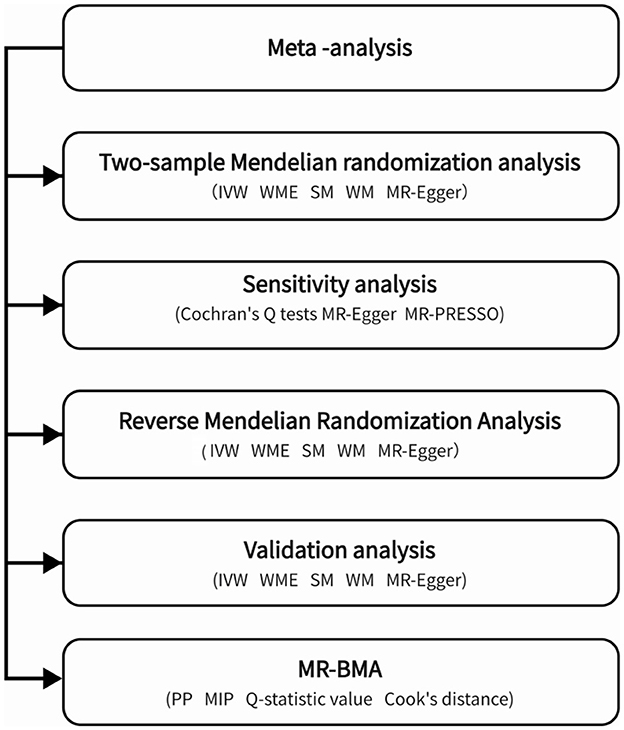
Figure 1. This figure illustrates a flow chart.
Using the subject headings and free words “gut microorganisms,” “type 2 diabetes,” and “clinical randomized controlled trials,” we searched PubMed, Web of Science, Cochrane Library, and Embase databases, respectively. The retrieved documents were not restricted to countries. The language is English, and the period is July 2023.
Inclusion criteria: the study population is T2DM; the research type is a clinical randomized controlled trial; the intervention measures are gut probiotics or similar preparations; and the outcome indicators are fasting blood glucose and glycated hemoglobin levels.
Exclusion criteria: exclude case reports utilizing animal and cell models; literature that has been repeatedly included; literature with incomplete data; unpublished literature; literature that does not include T2DM in the study population; or literature that does not include probiotics or similar preparations in the intervention.
Data extraction and quality evaluation: we imported the literature from four databases into the title list. Using a double-masked method, two investigators screened the literature. They extracted data based on study inclusion and exclusion criteria, including author, year, country, age, sample size, intervention, and outcome measures. We assessed bias risk in randomized controlled trials using a tool developed by the Cochrane Collaboration.
This study was conducted by the Preferred Reporting Items for Systematic Reviews and Meta- Analyses (PRISMA) guidelines. We assessed bias risk in randomized controlled trials using the bias risk assessment tool developed by the Cochrane Collaboration. The PROSPERO registration number was CRD42023444465.
We obtained T2DM and gut microbiota data from the public GWAS database (https://gwas.mrcieu.ac.uk). All data used in this study were publicly available GWAS summary results. The research does not involve ethical or privacy issues; as a result, an ethical review is unnecessary.
This study utilized gut microbial GWAS data from the MiBioGen consortium (24, 25). This study included genome-wide association and gut microbial metagenomic data from 18,340 European populations in 24 cohorts. Genomics-based association studies could be used to examine the genetic association between the relative abundance of gut microorganisms and the genes of the human host. In addition, this study included 271 gut microorganisms for further analysis.
T2DM data from the UK Biobank (UKBB) were used in this study, containing 10,894,596 SNPs in 337,159 individuals, including 335,026 in the control group and 2,133 in the experimental group. In addition, 655,666 Europeans were selected for verification from EBI, including 654,488 experimental subjects and 1,178 control subjects. The dataset contains 5,030,727 SNPs.
The genetic variation selected as an IVs must meet the following three assumptions (26): (1) Correlation assumption: The selected IVs must have a close relationship with T2DM. (2) Independence assumption: IV should not be confounded by confounding factors; (3) Exclusion restriction assumption: IV can only influence T2DM through the gut microbiota, not other channels.
We selected genetic variant SNP sites with genome-wide significance (P < 1 × 10⁻⁵) for T2DM. Select SNPs based on the linkage disequilibrium parameter (r2) threshold of 0.01 and the genetic distance of 139 10,000 kb to guarantee their independence and eliminate the impact of linkage disequilibrium (LD). Our analysis followed the guidelines for conducting MR studies. This report followed the guidelines for Strengthening the Reporting of Studies with MR (STROBE-MR).
In this study, causal effects were estimated using five methods: inverse variance weighting (IVW), MR Egger, weighted median (WME), simple mode (SM), and weighted mode (WM). The IVW method assumes that all genetic variants contain valid IVs. The ratio method calculates the causal effect value of a single instrumental variable. Each estimate is aggregated for weighted linear regression to obtain the total effect value. It is important to note that the MR-Egger method differs from the IVW method in that an intercept term is considered in the regression. The WME method takes advantage of the intermediate effects of all available genetic variants to get an estimate. This is accomplished by weighting the inverse variance of each SNP's association with the outcome. SM and WM are modality-based methods. Modality-based estimation models aggregate SNPs with similar causal effects and return estimates of causal effects for most cluster SNPs. According to WM, every SNP's impact on the cluster is weighted by the inverse variance of its effect (27). This study utilized the IVW method as its preferred method for causal effect estimation due to its higher testing efficiency than the other four MR methods. The results were visualized using forest maps and scatter plots.
Using T2DM as the exposure variable and associated gut microbiota as the outcome variable, we examined whether T2DM has the exact causal relationship with the gut microbiota. In order to assess the causal relationship between the two diseases, MR analysis was conducted once again without modifying either the statistical method or the data source. This study aimed to determine if T2DM affects the gut microbiome. The statistical methods used at this stage are the same as those used at the previous stage.
Selecting 655,666 Europeans from EBI as the validation population, we performed MR analyses again to determine whether the gut microbiome was causally associated with developing different types of T2DM. At this stage, the statistical method remains consistent with the above.
The multivariate MR analysis could simultaneously estimate the causal relationship between multiple risk factors for T2DM. In essence, it is an extension of unit MR analysis. Based on multivariate MR analysis, we used MR Bayesian model averaging (MR-BMA) probability to quantify the degree of causal association between different gut microbiomes and T2DM.
Based on the MR-BMA research method, we predicted the causal importance of the gut microbiota that was significantly related to T2DM and considered the potential pleiotropic effects. A posterior probability (PP) is assigned to each candidate model. After adding PP to each candidate risk factor, we calculated each biomarker's marginal inclusion probability (MIP) and reported each biomarker's model average causal effect (MACE) on T2DM. The best model was preferentially selected based on the PP value ranking of each model (threshold set to 0.02). Finally, we used Q statistics and Cook distance to identify outliers and strong influence points for model diagnosis. The MR-BMA analysis was repeated after these factors were removed.
In this study, quality control was implemented on MR results that had a P < 0.05. The quality control process included sensitivity analysis, heterogeneity testing, and horizontal gene pleiotropy testing. Sensitivity analysis was conducted using the leave-one-out method by deleting individual SNPs in sequence and calculating the combined effect size of the remaining SNPs to evaluate the impact of each SNP on the results. The heterogeneity test uses the Cochran Q test to determine the heterogeneity of SNPs. It also evaluates the possibility of causal effect estimation bias due to SNP measurement errors resulting from differing analysis platforms, experimental conditions, and sample populations. In horizontal pleiotropy testing, the intercept term of MR-Egger regression was used to assess whether IVs impact outcomes through pathways other than exposure.
We completed Meta-analysis with RevMan 5.3 and STATA 16.0 statistical software, and we conducted MR analysis using R packages such as “Mendelian Randomization” and “Two Sample MR.” Furthermore, Multivariate MR analysis uses the R language code and functions related to MR-MBA publicly released on GitHub (https://github.com/verena-zuber/demo~AMD).
We retrieved 1,378 documents from four databases: PubMed, Web of Science, Cochrane Library, and Embase. Ultimately, we included six documents after screening them according to inclusion and exclusion criteria. A flow chart of the screening process was shown in Figure 2. The six included in the literature Kanazawa et al. (28), Chaiyasut et al. (29), Khalili et al. (30), Rustanti et al. (31), Toejing et al. (32), Firouzi et al. (33) were all RCT trials, and their interventions included gut probiotics.
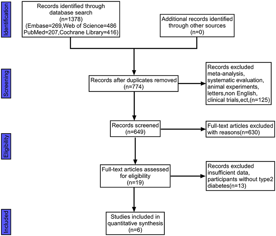
Figure 2. This figure illustrates the flow chart of literature screening.
The outcome indicators of six of the included studies were glycated hemoglobin and fasting blood glucose. A meta-analysis using a fixed effects model demonstrated that the glycated hemoglobin of the intervention group was significantly lower than that of the control group after treatment (WMD = −0.108, 95%CI: −0.314–0.098, P = 0.305). There was no statistical heterogeneity among the studies (I2 = 0, P = 0.486). In the intervention group, fasting blood glucose levels were significantly lower than in the control group (WMD = −13.83, 95%CI: −18.831– −8.835, P < 0.05). Fasting blood glucose results were not statistically heterogeneous (I2 = 0, P = 0.639) (Figure 3). All studies were analyzed for sensitivity, and the results showed no significant differences. A publication bias analysis was also conducted using Begg's test: glycosylated hemoglobin (P = 0.647) and fasting blood glucose (P = 0.0229), indicating no publication bias present.
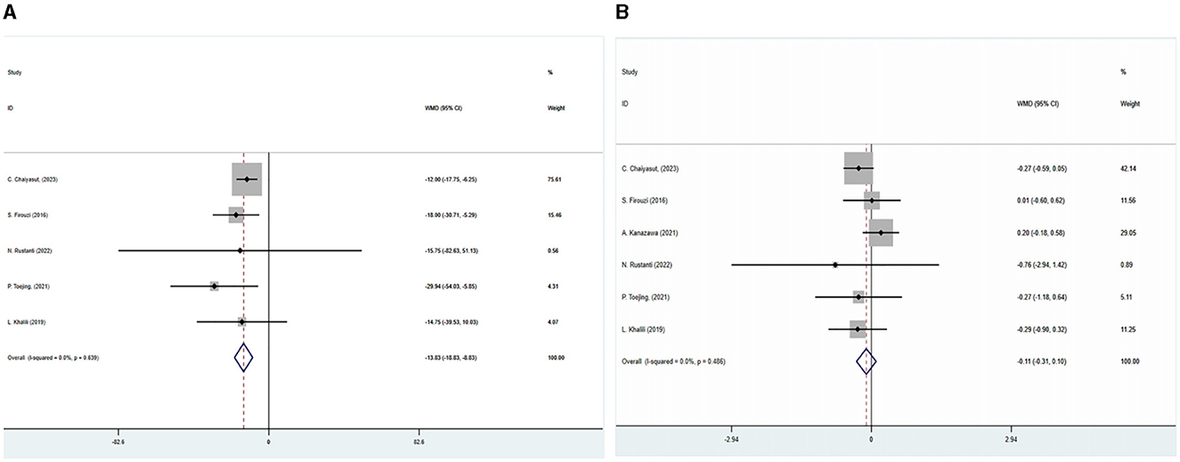
Figure 3. This figure displays a forest plot illustrating the impact of the combination of SGLT2 inhibitors with metformin on glycated, hemoglobin and fasting blood glucose levels were analyzed using a fixed-effects model, and the results were expressed as weighted mean difference (WMD) and 95% confidence interval (CI).
As exposures for the study, 271 gut microbes were selected from GWAS data on gut microbes involving 5,959 healthy Europeans. This study examined the relative abundance of 271 gut microorganism species, including 10 phyla, 16 classes, 22 orders, 48 families, 98 genera, and 77 species. A two-sample MR study was conducted using 271 species of gut microorganisms and 2,770 SNPs for the study, which was screened based on P < 1 × 10 – 5 and linkage disequilibrium thresholds.
Five TSMR methods were employed to analyze the causal relationship between the relative abundance of 271 species of gut microorganisms and T2DM, and the IVW method was primarily used to analyze the results.
Based on the IVW method, five gut microorganisms were associated with T2DM. These five types of gut microorganisms include three genera, one class and one order, each belonging to genus Haemophilus (OR = 0.997, 95%CI: −0.004–−0.002, P = 0.003), genus Actinobacteria (OR = 1.003, 95%CI: 0.002–0.005, P = 0.004), order Pasteurellaceae (OR = 0.998, 95%CI: −0.003–−0.001, P = 0.009), class Melanobacteria (OR = 1.001, 95%CI: 0.001–0.002, P = 0.034), genus Lactobacillus (OR = 1.001, 95%CI: −0.00001–0.002, P = 0.035).
Based on the Q test results, it was determined that there was no heterogeneity among the included SNPs, of which the five gut microbiota were (P = 0.642, P = 0.942, P = 0.256, P = 0.700, P = 0.186). According to the MR-Egger regression intercept, no horizontal pleiotropy exists in the association between gut microbiota and T2DM. These were genus Haemophilus (MR-Egger intercept = −0.0003, P = 0.272), genus Actinobacteria (MR-Egger intercept = 0.0002, P = 0.588), order Pasteurellaceae (MR- Egger intercept = −0.0003, P = 0.112), class Melanobacteria (MR-Egger intercept = −0.00004, P = 0.864), genus Lactobacillus (MR-Egger intercept = 0.0003, P = 0.295) (Figures 4, 5). According to the MR results of the retention method, no SNPs significantly impacted the effect estimation of gut microbiota and T2DM. As a result, the causal relationship appeared stable (Table 1).
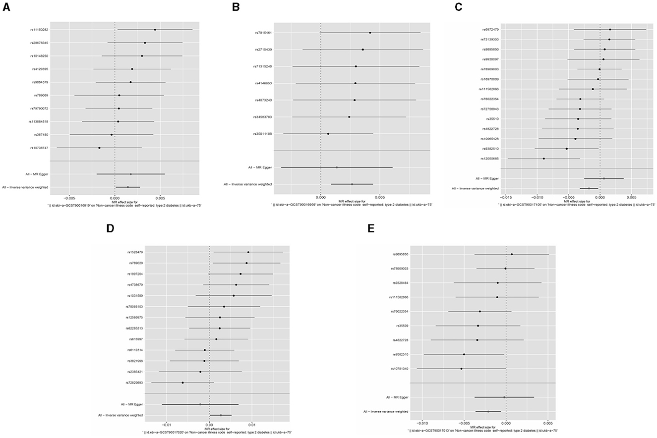
Figure 4. Forest map of Mendelian randomization analysis of gut microbes and T2DM.
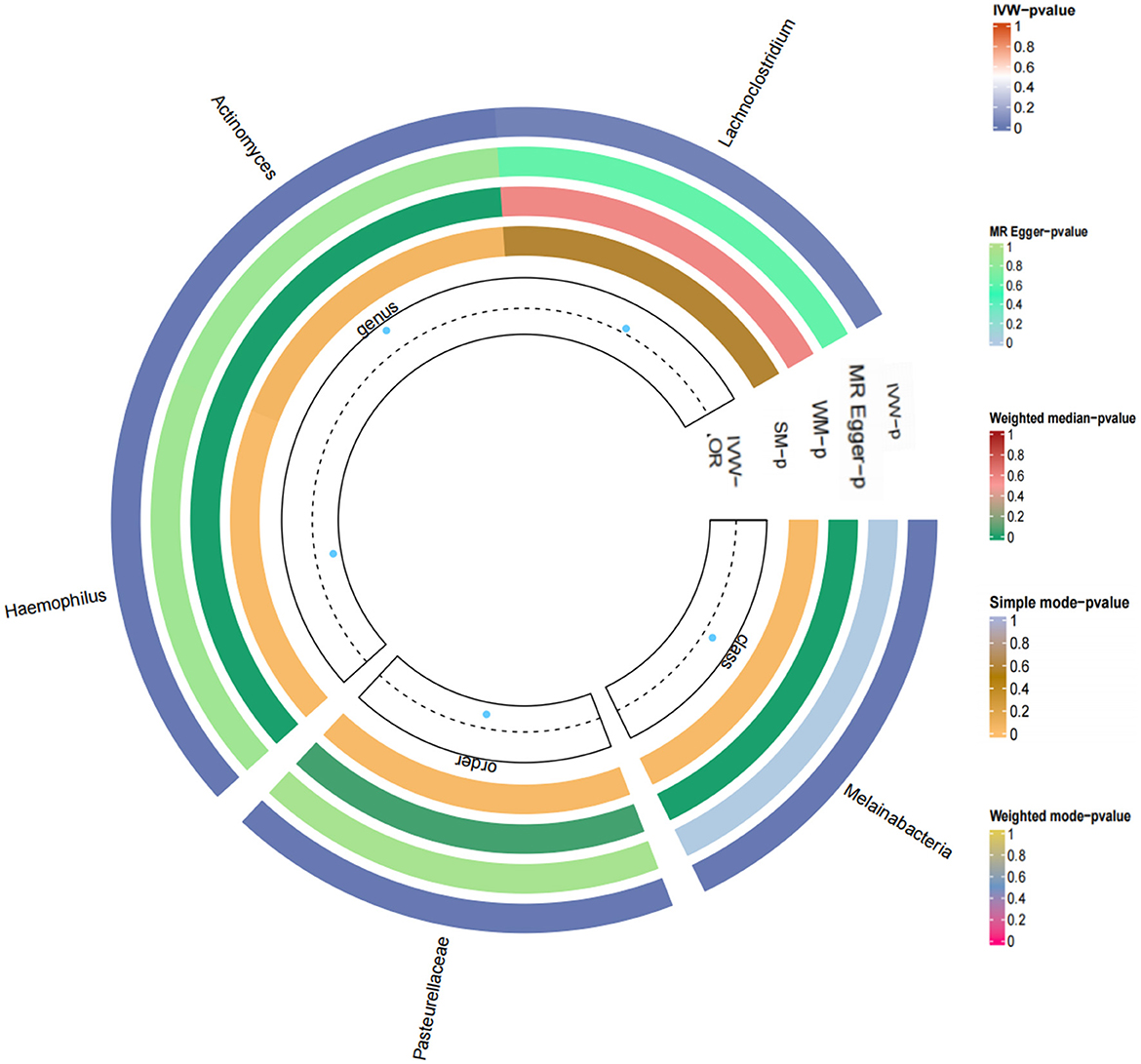
Figure 5. Loop diagram of the Mendelian randomization analysis of gut microbes and T2DM.
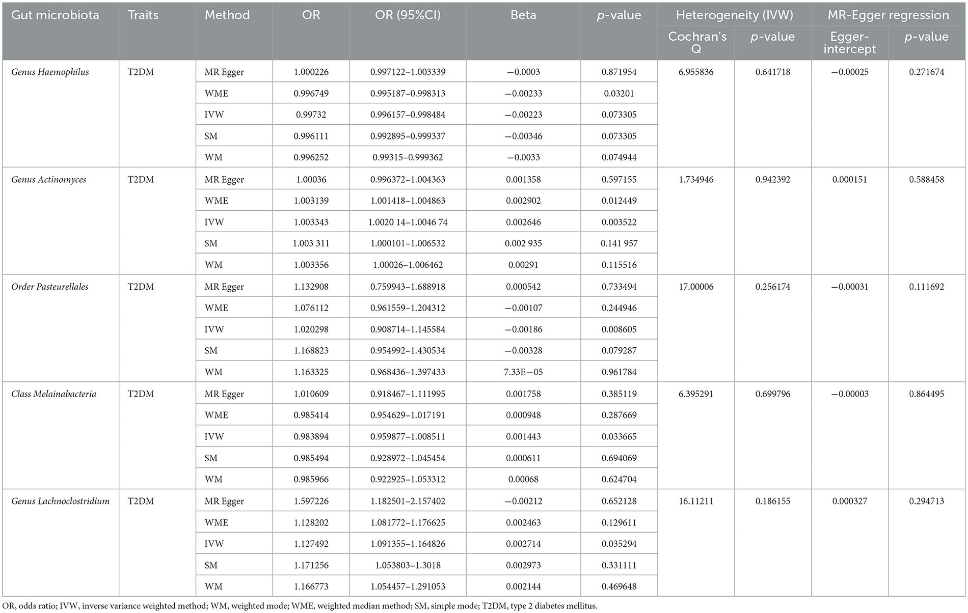
Table 1. Summary of the results of two-sample Mendelian randomization analysis of gut microbiota and T2DM.
We again conducted a two-sample Mendelian randomized study; T2DM was used as an exposure factor, and six related gut microorganisms were selected as outcome factors. The results indicated no causal link between T2DM and gut microbiota. Genus Haemophilus (OR = 5.555, 95%CI: 0.002–17,959.04, P = 0.678), genus Actinobacteria (OR = 0.021, 95%CI: 2.74E-06–160.7864, P = 0.397), order Pasteurellaceae (OR = 8.029, 95%CI: 0.003–2,292.23, P = 0.608), class Melanobacteria (OR = 0.031, 95%CI: 1.33E-06–706.659, P = 0.497), genus Lactobacillus (OR = 1.112, 95%CI: 0.001–1026.964, P = 0.976). The study suggests that T2DM and gut microbiota do not have any causal relationship (Figure 6).
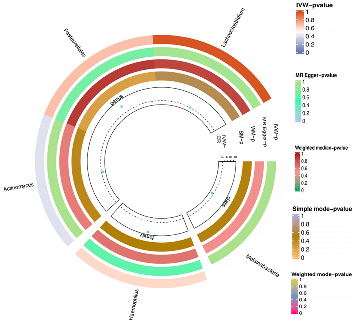
Figure 6. Loop diagram of the gut microbiome and T2DM in reverse Mendelian randomized analysis.
As the validation population for the Two-Sample MR analysis verification, 655,666 European individuals were selected from EBI. The results of the IVW method showed that 223 gut microorganisms are causally related to T2DM. We used the Q test to exclude heterogeneity and the MR-Egger regression method to exclude gut microorganisms that exhibit horizontal pleiotropy.
Ultimately, 22 types of gut microorganisms were found. These included genus Streptococcus (P = 1.08E-60), phylum Actinobacteria (P = 1.77E-16), genus Eubacterium (P = 4.04E-14), genus Ruminococcus (P = 1.42E-10), genus VerrucomicrobiaUCG003 (P = 1.57E-09) and UCG010 (P = 2.51E-09), family Clostridiaceae (P = 9.61E-09), genus Butyrivibrio (P = 2.57E-07), genus Lachnospira (P = 4.23E-07), genus Faecalibacterium (P = 2.01E-06), Eubacteriumredox (P = 2.67E-06), genus Candida (P = 1.76E-05), genus Ruminococcus (P = 7.68E-05), genus Thielella (P = 0.0001), genus Anaerobacteria (P = 0.0003), genus Rikenella (P = 0.0007), family Christeniaceae (P = 0.0010), genus Eisenbergella (P = 0.0024), genus Lactococcus (P = 0.0026), phylum Firmicutes (P = 0.0043) and two unknown bacterial groups (P = 3.84E-10, P = 9.61E-09). According to the results, certain gut microbiota might be responsible for T2DM (Figure 7).
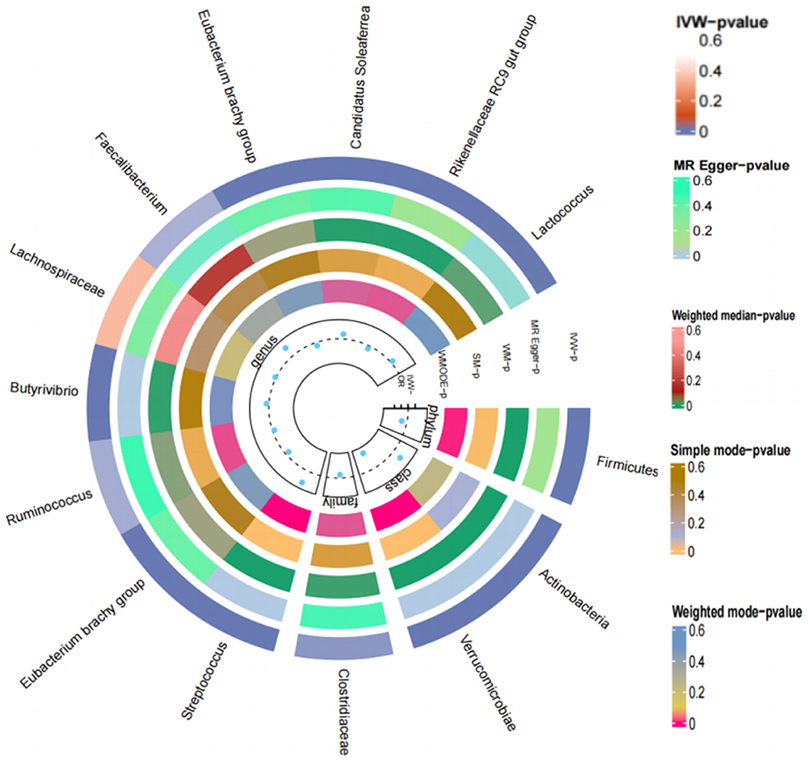
Figure 7. Loop diagram of the Mendelian randomization validation analysis of gut microbes and T2DM.
We performed MR-BMA analysis on six types of gut microbiota and included 55 SNPs in the analysis. Fourteen SNPs were deleted because the Cook distance exceeded 10, three SNPs were removed because the Q statistic exceeded the threshold, and 38 SNPs were retained and included in the model. Based on the MR-BMA model, the six gut microbiota were organized and combined into ten groups. We performed a multivariate MR analysis using weighted regression for each group of gut microbiota. There were six groups with PP > 0.02, and the optimal model of the genus Haemophilus had the highest (PP = 0.447).
Furthermore, we identified other optimal models (PP > 0.02) for the genus Lactobacillus, genus Actinomycetes, order Pasteurellaceae, and class Melanobacteria. The PP was then added up for each candidate's risk factor. The MIP of a risk factor represents the probability that it will be a causal determinant of disease risk, which was the risk factor with the high levels of causal evidence (MIP = 0.449), and the specific value was shown in Table 2.
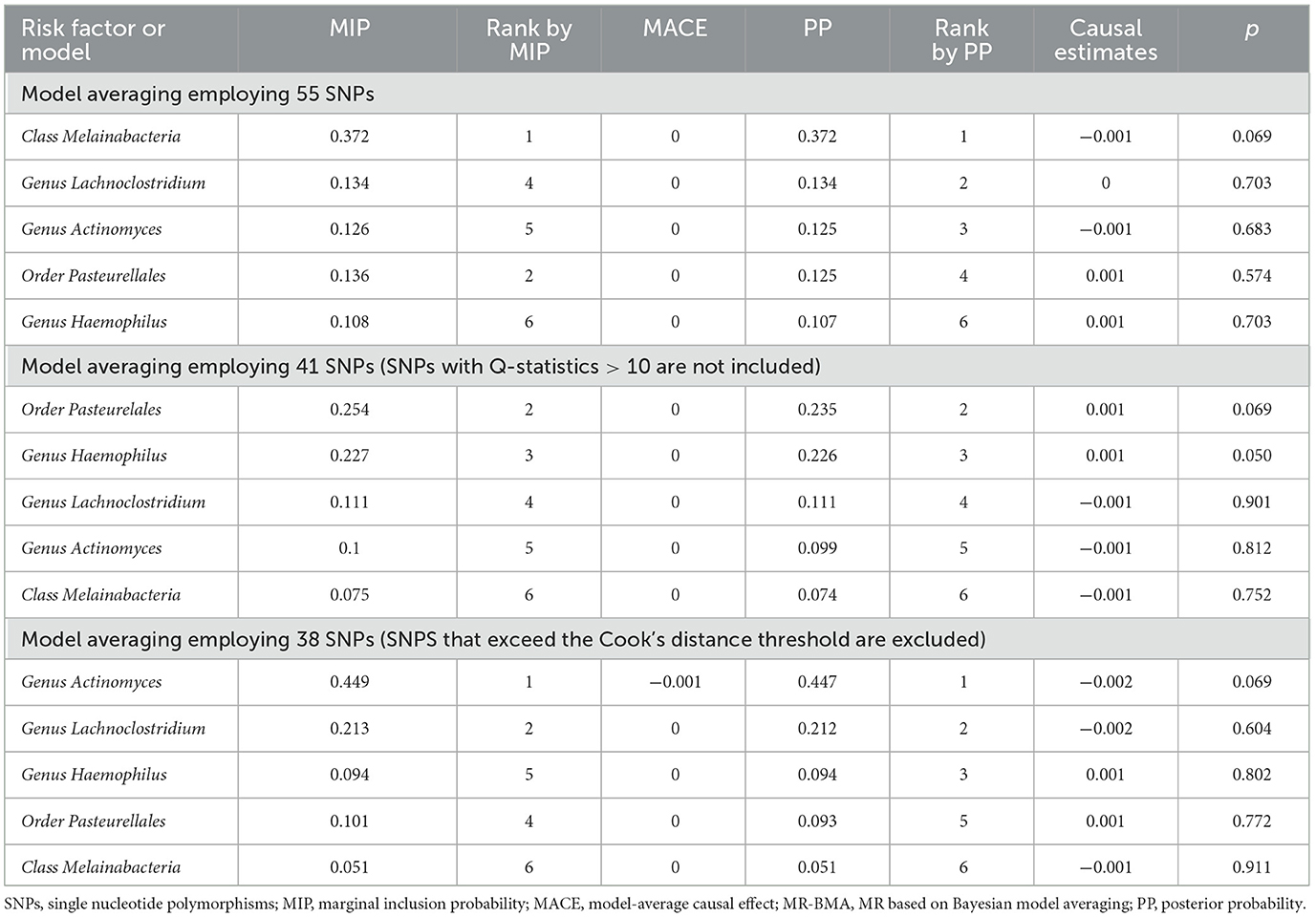
Table 2. Ranking of gut microbiota for T2DM using MR-BMA.
In this study, we used meta-analysis to observe that T2DM patients who took gut probiotics had lower fasting blood sugar and glycated hemoglobin levels than the control group. Our meta-analysis suggests that regulating gut microbiota disorders may delay T2DM progression. It is unclear, however, which specific gut microbiota disorders maybe involved in T2DM pathogenesis. For this issue, we used MR research methods further to study the causal relationship between gut microbiota and T2DM. The results of the two-sample MR analysis showed that five gut microbiota are causally related to T2DM, among which the genus Haemophilus and order Pasteurellaceae were negatively correlated with T2DM; genus Actinomycetes, class Melanobacteria, and genus Lactobacillus were positively correlated. Reverse MR analysis demonstrated that T2DM and gut microbiota do not have any reverse causal relationship. Additionally, we selected data from EBI for external verification. MR analysis showed that 22 types of gut microbiota were causally related to T2DM. Based on the external validation data set, this study suggests a causal relationship between gut microbiota and T2DM.
Evidence shows gut microbiota structure and function changes are associated with T2DM (34). The pathophysiology of T2DM is influenced by gut microbiota and its related metabolites, including blood sugar metabolism, insulin resistance, and chronic inflammation. Recent studies have demonstrated an alteration in the gut microbiota of T2DM patients. Compared to the general population, T2DM patients had higher genus Lactobacillus (35), Enterococcus, and Clostridium levels in the phylum Firmicutes. The genus Bacteroidetes was decreasing in the phylum Bacteroides. In the phylum Actinobacteria, the number of genus Bifidobacteria and genus Rochella decreases, and the gut microbiota changes signaling pathways that affect lipid and glucose metabolism, resulting in low-level inflammation, insulin resistance, and ultimately contributing to T2DM (15). Additionally, T2DM patients with better blood sugar control had a decrease in Enterobacteriaceae and Enterococcus compared with patients with poor blood sugar control. Furthermore, a significant improvement in insulin resistance was observed, with increased Bifidobacterium, Bacteroidetes, and bacteria that produced butyrate and propionate.
Generally, the mechanism by which gut microbiota disorder may contribute to T2DM includes three components (36): First, these gut microbiota can decrease the composition of short-chain fatty acid- producing bacteria in diabetic subjects, affect the metabolism of bile acids, and increase the microbiota in the gut tract (37). Second, they may prevent the recovery of the gut mucosal barrier, a process related to consuming potential gut pathogens and weakening digestion enzymes. As a result, it increases diabetes risk by promoting the production of trimethylamine nitrogen oxide (TMAO), which results in cholesterol accumulation and insulin resistance (38). Furthermore, these gut microbiotas can affect the progression of diabetes as well as other chronic diseases, such as systemic lupus erythematosus (39), rheumatoid arthritis (40), inflammatory bowel disease (41), chronic kidney disease (42), and ischemic stroke (43).
Numerous studies have demonstrated a close relationship between changes in gut microbiota structure and function and T2DM. However, given the diversity of gut microbiota, it is still unclear what type of gut microbiota disorder contributes to T2DM development. Previously, Xiang et al. (23) used MR research to demonstrate that Streptococcus and Acidaminococcaceae may have a critical positive correlation with T2DM risk. Our validation data set also revealed a positive association between Streptococcus and T2DM, consistent with Xiang et al.'s findings. Based on the research of Xiang et al., we expanded the types of gut microbiota and ranked the exposure factors. According to further MR study results, genus Haemophilus and order Pasteurellaceae were negatively correlated with T2DM and might play a protective role; genus Actinomycetes, class Melanobacteria, and genus Lactobacillus were positively correlated with T2DM, and they might be associated with T2DM. Additionally, the MR-BMA predictions suggested that the genus Haemophilus and T2DM had a significant causal relationship.
Some studies have demonstrated a close relationship between the genus Haemophilus and T2DM (44, 45). As stated by Nuli et al. (46), people with impaired glucose tolerance would have a higher proportion of order Pasteurellaceae and genus Haemophilus than those with normal glucose tolerance. Nevertheless, Zhang et al. (47) and Zhong et al. (48) found a reduction in the proportion of genus Haemophilus in new-onset diabetes. Studies have shown (49) that decreasing the abundance of genus Haemophilus in the intestine may produce short-chain fatty acids (SCFAs), such as butyric acid, propionic acid, and acetic acid, which may increase gut permeability (50). Several inflammatory factors, including lipopolysaccharides (LPS) and chyle particles, could pass through the gut epithelial barrier and activate downstream inflammatory pathways and pro-inflammatory cytokine cascades, resulting in a chronic systemic inflammatory response, impaired glucose metabolism, increasing insulin resistance, and eventually contributing to diabetes. In addition, A decrease in the number of genus Haemophilus species in the gut tract could also increase the production of uremic toxins (51, 52), including indenyl sulfate and p-cresol sulfate, which might lead to mitochondrial dysfunction, podocyte damage, thickening of the glomerular basement membrane, and complications such as diabetes kidney disease and coronary heart disease.
The results of our MR study suggest that the genus Actinobacteria and genus Lactobacillus may contribute to the progression of diabetes. The relevant research indicated that genus Actinomycetes and genus Lactobacilli in the intestine are associated with chronic inflammatory diseases, including T2DM. According to recent research (53–55), patients with T2DM have reduced amounts and proportions of bacteria from the genus Actinobacteria in their gut microbiota. In addition, the genus Lactobacilli positively correlates with fasting blood glucose and HbA1c levels. Several studies have shown that genus Lactobacilli levels were significantly higher among type 2 diabetics than healthy individuals (56, 57). Interestingly, we observed that order Pasteurellaceae and class Melanobacteria were associated with T2DM. More clinical and basic research is needed to determine the specific pathogenesis of these gut microbiota and T2DM.
This study has several advantages: First, it includes clinical and genetic studies. We used various research methods to elucidate gut microbiota and T2DM correlation. These researches included meta-analysis, two-sample MR analysis, and reverse validation with different data sets. In addition, we used MR-BMA torank the strength of the causal association between gut microbiota and T2DM. Second, we clarify the causal relationship between specific gut microbiota imbalances and T2DM. In this paper, we propose novel ideas and methods for studying the mechanism of the “pancreas-gut axis”.
Somethings could be improved in this study: Firstly, We could not further perform reverse MR analysis due to insufficient SNPs. The results of our study did not allow us to conclude that gut microbiota and T2DM were mutually related. Secondly, the MR results of the validation data set used in this article differ from those of the original data set. This may be due to the use of different consortium data. Lastly, although causal associations derived from MR studies have a certain amount of research value, they still require additional clinical and basic research confirmation. In light of this, we should be cautious when interpreting relevant research results.
Our research results suggest that gut microbiota is closely related to T2DM pathogenesis. The results of further MR research and an analysis of the prediction model indicate that a variety of gut microbiota disorders, including genus Haemophilus, are causally related to the development of T2DM. The findings of this study may provide some insight into the diagnosis and treatment of T2DM.
The original contributions presented in the study are included in the article/Supplementary material, further inquiries can be directed to the corresponding author.
TL: Writing – review & editing. YC: Data curation, Software, Validation, Visualization, Writing – original draft. NL: Visualization, Writing – original draft. XM: Data curation, Formal analysis, Visualization, Writing – original draft. J-aF: Writing – review & editing. XZ: Conceptualization, Supervision, Writing – review & editing.
The author(s) declare that no financial support was received for the research, authorship, and/or publication of this article.
We express our gratitude to all scholars who participated in the design, creation, retrieval, analysis, writing, and revision of the study.
The authors declare that the research was conducted in the absence of any commercial or financial relationships that could be construed as a potential conflict of interest.
All claims expressed in this article are solely those of the authors and do not necessarily represent those of their affiliated organizations, or those of the publisher, the editors and the reviewers. Any product that may be evaluated in this article, or claim that may be made by its manufacturer, is not guaranteed or endorsed by the publisher.
The Supplementary Material for this article can be found online at: https://www.frontiersin.org/articles/10.3389/fpubh.2024.1342313/full#supplementary-material
1. Ahmad E, Lim S, Lamptey R, Webb DR, Davies MJ. Type 2 diabetes. Lancet. (2022) 400:1803–20. doi: 10.1016/S0140-6736(22)01655-5
2. Chatterjee S, Khunti K, Davies MJ. Type 2 diabetes. Lancet. (2017) 389:2239–51. doi: 10.1016/S0140-6736(17)30058-2
3. Uehara K, Santoleri D, Whitlock AEG, Titchenell PM. Insulin regulation of hepatic lipid homeostasis. Compr Physiol. (2023) 13:4785–809. doi: 10.1002/cphy.c220015
4. Lu S, Li YR, Qian ZJ, Zhao TS, Feng ZW, Weng XG, et al. Role of the inflammasome in insulin resistance and type 2 diabetes mellitus. Front Immunol. (2023) 14:1052756. doi: 10.3389/fimmu.2023.1052756
5. Hasanian-Langroudi F, Ghasemi A, Hedayati M, Siadat SD, Tohidi M. Novel insight into the effect of probiotics in the regulation of the most important pathways involved in the pathogenesis of type 2 diabetes mellitus. Probiot Antimicrob Proteins. (2023) 16:829–844. doi: 10.1007/s12602-023-10056-8
6. Rayner CK, Horowitz M. Agonism of receptors in the gut-pancreas axis in type 2 diabetes: are two better than one? Lancet. (2018) 391:2577–8. doi: 10.1016/S0140-6736(18)30936-X
7. Nasr NE, Sadek KM. Role and mechanism(s) of incretin-dependent therapies for treating diabetes mellitus. Environ Sci Pollut Res. (2022) 29:18408–22. doi: 10.1007/s11356-022-18534-2
8. Prada-Oliveira JA, Camacho-Ramirez A, Salas-Alvarez J, Campos-Martinez FJ, Lechuga-Sancho AM, Almorza-Gomar D, et al. GLP-1 mediated improvement of the glucose tolerance in the T2DM GK rat model after massive jejunal resection. Ann Anat. (2019) 223:1–7. doi: 10.1016/j.aanat.2019.01.007
9. Zhang Z, Tanaka I, Pan Z, Ernst PB, Kiyono H, Kurashima Y. Gut homeostasis and inflammation: Gut microbiota at the crossroads of pancreas-gut barrier axis. Eur J Immunol. (2022) 52:1035–46. doi: 10.1002/eji.202149532
10. Zitvogel L. Ayyoub M, Routy B, Kroemer G. Microbiome and anticancer immunosurveillance. Cell. (2016) 165:276–87. doi: 10.1016/j.cell.2016.03.001
11. Adak A, Khan MR. An insight into gut microbiota and its functionalities. Cell Mol Life Sci. (2019) 76:473–93. doi: 10.1007/s00018-018-2943-4
12. Cheng ZL, Zhang L, Yang L, Chu HK. The critical role of gut microbiota in obesity. Front Endocrinol. (2022) 13:1025706. doi: 10.3389/fendo.2022.1025706
13. Afzaal M, Saeed F, Shah YA, Hussain M, Rabail R, Socol CT, et al. Human gut microbiota in health and disease: unveiling the relationship. Front Microbiol. (2022) 13:999001. doi: 10.3389/fmicb.2022.999001
14. Gurung M, Li ZP, You H, Rodrigues R, Jump DB, Morgun A, et al. Role of gut microbiota in type 2 diabetes pathophysiology. Ebiomedicine. (2020) 51:102590. doi: 10.1016/j.ebiom.2019.11.051
15. Sabatino A, Regolisti G, Cosola C, Gesualdo L, Fiaccadori E. Gut microbiota in type 2 diabetes and chronic kidney disease. Curr Diab Rep. (2017) 17:16. doi: 10.1007/s11892-017-0841-z
16. Dong Y, Wang P, Yang XC, Chen ML, Li J. Potential of gut microbiota forlipopolysaccharide biosynthesis in European women with type 2 diabetes based on metagenome. Front Cell Dev Biol. (2022) 10:1027413. doi: 10.3389/fcell.2022.1027413
17. Zhang LL, Chu JJ, Hao WH, Zhang JJ, Li HB, Yang CJ, et al. Gut microbiota and type 2 diabetes mellitus: association, mechanism, and translational applications. Mediat Inflammat. (2021) 2021:5110276. doi: 10.1155/2021/5110276
18. Salgaço MK, Oliveira LGS, Costa GN, Bianchi F, Sivieri K. Relationship between gut microbiota, probiotics, and type 2 diabetes mellitus. Appl Microbiol Biotechnol. (2019) 103:9229–38. doi: 10.1007/s00253-019-10156-y
19. Vatanen T, Kostic AD, d'Hennezel E, Siljander H, Franzosa EA, Yassour M, et al. Variation in microbiome LPS immunogenicity contributes to autoimmunity in humans. Cell. (2016) 165:842–53. doi: 10.1016/j.cell.2016.05.056
20. Greenland S. An introduction to instrumental variables for epidemiologists. Int J Epidemiol. (2000) 29:722–9. doi: 10.1093/ije/29.4.722
21. Birney E. Mendelian randomization. Cold Spring Harb Perspect Med. (2022) 12:a041302. doi: 10.1101/cshperspect.a041302
22. Davey Smith G, Hemani G. Mendelian randomization: genetic anchors for causal inference in epidemiological studies. Hum Mol Genet. (2014) 23:R89–98. doi: 10.1093/hmg/ddu328
23. Xiang K, Zhang JJ, Xu YY, Zhong X, Ni J, Pan HF. Genetically predicted causality of 28 gut microbiome families and type 2 diabetes mellitus risk. Front Endocrinol. (2022) 13:780133. doi: 10.3389/fendo.2022.780133
24. Kurilshikov A, Medina-Gomez C, Bacigalupe R, Radjabzadeh D, Wang J, Demirkan A, et al. Large-scale association analyses identify host factors influencing human gut microbiome composition. Nat Genet. (2021) 53:156. doi: 10.1038/s41588-020-00763-1
25. MiBioGen. (2022) Available online at: https://mibiogen.gcc.rug.nl/ (accessed September 16, 2022).
26. Sheehan NA, Didelez V, Burton PR, Tobin MD. Mendelian randomisation and causal inference in observational epidemiology. PLoS Med. (2008) 5:e177. doi: 10.1371/journal.pmed.0050177
27. Rasooly D, Peloso GM. Two-sample multivariable Mendelian randomization analysis using R. Curr Prot. (2021) 1:e335. doi: 10.1002/cpz1.335
28. Kanazawa A, Aida M, Yoshida Y, Kaga H, Katahira T, Suzuki L, et al. Effects of synbiotic supplementation on chronic inflammation and the gut microbiota in obese patients with type 2 diabetes mellitus: a randomized controlled study. Nutrients. (2021) 13:558. doi: 10.3390/nu13020558
29. Chaiyasut C, Sivamaruthi BS, Lailerd N, Sirilun S, Thangaleela S, Khongtan S, et al. Influence of Bifidobacterium breve on the glycaemic control, lipid profile and microbiome of type 2 diabetic subjects: a preliminary randomized clinical trial. Pharmaceuticals. (2023) 16:695. doi: 10.3390/ph16050695
30. Khalili L, Alipour B, Asghari Jafar-Abadi M, Faraji I, Hassanalilou T, Mesgari Abbasi M, et al. The effects of lactobacillus caseion glycemic response, serum sirtuin1 and fetuin-A levels in patients with type 2 diabetes mellitus: a randomized controlled trial. Iran Biomed J. (2019) 23:68–77. doi: 10.29252/.23.1.68
31. Rustanti N, Murdiati A, Juffrie M, Rahayu ES. Effect of probiotic Lactobacillus plantarum dad-13 on metabolic profiles and gut microbiota in type 2 diabetic women: a randomized double-blind controlled trial. Microorganisms. (2022) 10:1806. doi: 10.3390/microorganisms10091806
32. Toejing P, Khampithum N, Sirilun S, Chaiyasut C, Lailerd N. Influence of Lactobacillus paracasei HII01 supplementation on glycemia and inflammatory biomarkers in type 2 diabetes: a randomized clinical trial. Foods. (2021) 10:1455. doi: 10.3390/foods10071455
33. Firouzi S, Majid HA, Ismail A, Kamaruddin NA, Barakatun-Nisak MY. Effect of multi-strain probiotics (multi-strain microbial cell preparation) on glycemic control and other diabetes-related outcomes in people with type 2 diabetes: a randomized controlled trial. Eur JNutr. (2017) 56:1535–50. doi: 10.1007/s00394-016-1199-8
34. Jeon J, Jang J, Park K. Effects of consuming calcium-rich foods on the incidence of type 2 diabetes mellitus. Nutrients. (2018) 11:31. doi: 10.3390/nu11010031
35. Huang Y, Wang Z, Ma H, Ji S, Chen Z, Cui Z, et al. Dysbiosis and implication of the gut microbiota in diabetic retinopathy. Front Cell Infect Microbiol. (2021) 11:646348. doi: 10.3389/fcimb.2021.646348
36. Long J, Cai Q, Steinwandel M, Hargreaves MK, Bordenstein SR, Blot WJ, et al. Association of oral microbiome with type 2 diabetes risk. J Periodontal Res. (2017) 52:636–43. doi: 10.1111/jre.12432
37. Schippa S, Conte MP. Dysbiotic events in gut microbiota: impact on human health. Nutrients. (2014) 6:5786–805. doi: 10.3390/nu6125786
38. Oellgaard J, Winther SA, Hansen TS, Rossing P, von Scholten BJ. Trimethylamine N-oxide (TMAO) as a new potential therapeutic target for insulin resistance and cancer. Curr Pharm Des. (2017) 23:3699–712. doi: 10.2174/1381612823666170622095324
39. Xiang K, Wang P, Xu Z, Hu YQ, He YS, Chen Y, et al. Causal effects of gut microbiome on systemic lupus erythematosus: a two-sample Mendelian randomization study. Front Immunol. (2021) 12:667097. doi: 10.3389/fimmu.2021.667097
40. Attur M, Scher JU, Abramson SB, Attur M. Role of gut dysbiosis and nutrition in rheumatoid arthritis. Cells. (2022) 11:2436. doi: 10.3390/cells11152436
41. Xu Q, Ni JJ, Han BX, Yan SS, Wei XT, Feng GJ, et al. Causal relationship between gut microbiota and autoimmune diseases: a two-sample Mendelian randomization study. Front Immunol. (2021) 12:746998. doi: 10.3389/fimmu.2021.746998
42. Li N, Wang Y, Wei P, Min Y, Yu M, Zhou G, et al. Causal effects of specific gut microbiota on chronic kidney diseases and renal function-a two-sample Mendelian randomization study. Nutrients. (2023) 15:360. doi: 10.3390/nu15020360
43. Meng C, Deng P, Miao R, Tang H, Li Y, Wang J, et al. Gut microbiome and risk of ischaemic stroke: a comprehensive Mendelian randomization study. Eur J Prev Cardiol. (2023) 30:613–20. doi: 10.1093/eurjpc/zwad052
44. Sun K, Gao Y, Wu H, Huang X. The causal relationship between gut microbiota and type 2 diabetes: atwo- sample Mendelian randomized study. Front Public Health. (2023) 11:1255059. doi: 10.3389/fpubh.2023.1255059
45. Letchumanan G, Abdullah N, Marlini M, Baharom N, Lawley B, Omar MR, et al. Gut microbiota composition in prediabetes and newly diagnosed type 2 diabetes: a systematic review of observational studies. Front Cell Infect Microbiol. (2022) 12:943427. doi: 10.3389/fcimb.2022.943427
46. Nuli R, Cai J, Kadeer A, Zhang Y, Mohemaiti P. Integrative analysis toward different glucose tolerance- related gut microbiota and diet. Front Endocrinol (Lausanne). (2019) 10:295. doi: 10.3389/fendo.2019.00295
47. Zhang X, Shen D, Fang Z, Jie Z, Qiu X, Zhang C, et al. Human gut microbiota changes reveal the progression of glucose intolerance. PLoS ONE. (2013) 8:e71108. doi: 10.1371/journal.pone.0071108
48. Zhong H, Ren H, Lu Y, Fang C, Hou G, Yang Z, et al. Distinct gut metagenomics and metaproteomics signatures in prediabetics and treatment-naïve type 2 diabetics. EBioMedicine. (2019) 47:373–83. doi: 10.1016/j.ebiom.2019.08.048
49. Tanase DM, Gosav EM, Neculae E, Costea CF, Ciocoiu M, Hurjui LL, et al. Role of gut microbiota on onset and progression of microvascular complications of type 2 diabetes (T2DM). Nutrients. (2020) 12:3719. doi: 10.3390/nu12123719
50. De D, Nayak T, Chowdhury S, Dhal PK. Insights of host physiological parameters and gut microbiome of Indian type 2 diabetic patients visualized via metagenomics and machine learning approaches. Front Microbiol. (2022) 13:914124. doi: 10.3389/fmicb.2022.914124
51. Hsiao EY, McBride SW, Hsien S, Sharon G, Hyde ER, McCue T, et al. Microbiota modulate behavioral and physiological abnormalities associated with neurodevelopmental disorders. Cell. (2013) 155:1451–63. doi: 10.1016/j.cell.2013.11.024
52. Kikuchi K, Saigusa D, Kanemitsu Y, Matsumoto Y, Thanai P, Suzuki N, et al. Gut microbiome-derived phenyl sulfate contributes to albuminuria in diabetic kidney disease. Nat Commun. (2019) 10:1835. doi: 10.1038/s41467-019-09735-4
53. Wei M, Gu E, Luo J, Zhang Z, Xu D, Tao X, et al. Enterococcus hirae WEHI01 isolated from a healthy Chinese infant ameliorates the symptoms of type 2 diabetes by elevating the abundance of Lactobacillales in rats. J Dairy Sci. (2020) 103:2969–81. doi: 10.3168/jds.2019-17185
54. Zhao X, Zhang Y, Guo R, Yu W, Zhang F, Wu F, et al. The alteration in composition and function of gut microbiome in patients with type 2 diabetes. J Diabetes Res. (2020) 2020:8842651. doi: 10.1155/2020/8842651
55. Lambeth SM, Carson T, Lowe J, Ramaraj T, Leff JW, Luo L, et al. Composition, diversity and abundance of gut microbiome in prediabetes and type 2 diabetes. J Diabetes Obes. (2015) 2:1–7. doi: 10.15436/2376-0949.15.031
56. Sedighi M, Razavi S, Navab-Moghadam F, Khamseh ME, Alaei-Shahmiri F, Mehrtash A, et al. Comparison of gut microbiota in adult patients with type 2 diabetes and healthy individuals. Microb Pathog. (2017) 111:362–9. doi: 10.1016/j.micpath.2017.08.038
Keywords: T2DM, meta-analysis, two-sample MR analysis, multi-sample MR analysis, MR-BMA gut microbiota, MR-BMA
Citation: Liu T, Cao Y, Liang N, Ma X, Fang J-a and Zhang X (2024) Investigating the causal association between gut microbiota and type 2 diabetes: a meta-analysis and Mendelian randomization. Front. Public Health 12:1342313. doi: 10.3389/fpubh.2024.1342313
Received: 21 November 2023; Accepted: 27 May 2024;
Published: 19 June 2024.
Edited by:
Reza Rastmanesh, American Physical Society, United StatesReviewed by:
Eugenia Bezirtzoglou, Democritus University of Thrace, GreeceCopyright © 2024 Liu, Cao, Liang, Ma, Fang and Zhang. This is an open-access article distributed under the terms of the Creative Commons Attribution License (CC BY). The use, distribution or reproduction in other forums is permitted, provided the original author(s) and the copyright owner(s) are credited and that the original publication in this journal is cited, in accordance with accepted academic practice. No use, distribution or reproduction is permitted which does not comply with these terms.
*Correspondence: Xiaodong Zhang, enhkc3BsQDE2My5jb20=
†These authors have contributed equally to this work
‡ORCID: Ting Liu orcid.org/0000-0001-6276-4063
Yang Cao orcid.org/0000-0003-0646-8088
Ning Liang orcid.org/0000-0002-6659-3334
Xiaoqi Ma orcid.org/0009-0007-2323-5493
Jing-ai Fang orcid.org/0000-0002-2659-6477
Xiaodong Zhang orcid.org/0000-0003-3210-1407
Disclaimer: All claims expressed in this article are solely those of the authors and do not necessarily represent those of their affiliated organizations, or those of the publisher, the editors and the reviewers. Any product that may be evaluated in this article or claim that may be made by its manufacturer is not guaranteed or endorsed by the publisher.
Research integrity at Frontiers
Learn more about the work of our research integrity team to safeguard the quality of each article we publish.