- 1School of Life Sciences, Central China Normal University, Key Laboratory for Geographical Process Analysis and Simulation, Wuhan, China
- 2Bio-Resources Key Laboratory of Shaanxi Province, Shaanxi University of Technology, Hanzhong, China
The chloroplast genome (plastome) of angiosperms (particularly photosynthetic members) is generally highly conserved, although structural rearrangements have been reported in a few lineages. In this study, we revealed Corydalis to be another unusual lineage with extensive large-scale plastome rearrangements. In the four newly sequenced Corydalis plastomes that represent all the three subgenera of Corydalis, we detected (1) two independent relocations of the same five genes (trnV-UAC-rbcL) from the typically posterior part of the large single-copy (LSC) region to the front, downstream of either the atpH gene in Corydalis saxicola or the trnK-UUU gene in both Corydalis davidii and Corydalis hsiaowutaishanensis; (2) relocation of the rps16 gene from the LSC region to the inverted repeat (IR) region in Corydalis adunca; (3) uniform inversion of an 11–14 kb segment (ndhB-trnR-ACG) in the IR region of all the four Corydalis species (the same below); (4) expansions (>10 kb) of IR into the small single-copy (SSC) region and corresponding contractions of SSC region; and (5) extensive pseudogenizations or losses of 13 genes (accD, clpP, and 11 ndh genes). In addition, we also found that the four Corydalis plastomes exhibited elevated GC content in both gene and intergenic regions and high number of dispersed repeats. Phylogenomic analyses generated a well-supported topology that was consistent with the result of previous studies based on a few DNA markers but contradicted with the morphological character-based taxonomy to some extent. This study provided insights into the evolution of plastomes throughout the three Corydalis subgenera and will be of value for further study on taxonomy, phylogeny, and evolution of Corydalis.
Introduction
The chloroplast genome (plastome) of angiosperms (particularly photosynthetic members) is generally highly conserved in terms of structural organization, gene content, and gene arrangement (Palmer, 1985; Wicke et al., 2011; Ruhlman and Jansen, 2014; Mower and Vickrey, 2018). Plastome rearrangements, if they occurred, tend to be relatively minor. Large-scale rearrangement is rare, but it occasionally exists in a few lineages, such as Campanulaceae (Knox et al., 1993; Cosner et al., 2004; Knox, 2014; Knox and Li, 2017; Uribe-Convers et al., 2017, etc.), Geraniaceae (Palmer et al., 1987; Chumley et al., 2006; Guisinger et al., 2011; Weng et al., 2014, 2017; Röschenbleck et al., 2016, etc.), Fabaceae (Kolodner and Tewari, 1979; Palmer and Thompson, 1981; Lavin et al., 1990; Doyle et al., 1996; Cai et al., 2008; Martin et al., 2014; Schwarz et al., 2015; Wang et al., 2017, etc.), Oleaceae (Lee et al., 2007), Asteraceae (Jansen and Palmer, 1987; Kim et al., 2005; Sablok et al., 2019, etc.), Plantaginaceae (Zhu et al., 2016; Kwon et al., 2019; Asaf et al., 2020, etc.), and Poaceae (Palmer and Thompson, 1982; Doyle et al., 1992; Michelangeli et al., 2003; Burke et al., 2016; Liu et al., 2020, etc.). Given that the structure of the two already known plastomes from the genus Corydalis was highly variable (Wu et al., 2020; Xu and Wang, 2020), this genus is expected to represent an overlooked lineage with extensive large-scale plastome rearrangements. However, understanding of the pattern, origin, and evolution of plastome rearrangements within Corydalis is presently limited by the paucity of plastome sequences.
Corydalis (∼465 species) is the largest genus within Papaveraceae and one of the largest genera in Chinese flora (Wu et al., 1999; Zhang et al., 2008). Corydalis species, with a wide distribution in north temperate regions, are particularly diverse in the Hengduan Mountains and Qinghai–Tibet Plateau and adjacent areas (Wu et al., 1999; Zhang et al., 2008). Along with the uplift of the Hengduan Mountains and Qinghai–Tibet Plateau, Corydalis species have experienced intensive and rapid differentiation, and their plastomes must have also undergone a series of genetic shift to adapt to the drastically changed environment. Corydalis may represent an appropriate group to explore how the plastome content and structure have varied in a fine scale in the evolution history, when the unusual plastome rearrangements have originated, and why those changes have happened. In addition, some Corydalis plants have the potential to be exploited as medicine for their anti-hepatitis, antitumor, cardiovascular disease treatment, and pain-releasing effects (Luo et al., 1984; Editorial Board of Chinese Tibetan Medicine, 1996; Chinese Pharmacopoeia Commission, 2015; Zhang B. et al., 2016, etc.). However, the complexity of their morphological characters has greatly challenged our understanding of their taxonomy, ecology, evolution, and utilization. Based on morphological characters and/or a few molecular markers, previous studies (Fedde, 1936; Lidén, 1986; Lidén et al., 1995, 1997; Wu et al., 1996, 1999; Wang, 2006; Zhang et al., 2008; Zhang Z.X. et al., 2016; Jiang et al., 2018; Ren et al., 2018; Xu and Wang, 2018, etc.) have made positive contributions to the taxonomy and systematics of Corydalis. Until now, a robust backbone phylogeny of this genus, which is instructive for taxonomy and systematics, is still not completed due to the lack of enough genetic resources.
Complete plastome data have been applied to resolve long-standing controversies at different taxonomic levels (e.g., Jansen et al., 2007; Moore et al., 2007, 2010; Ma et al., 2014; Barrett et al., 2016; Zhai et al., 2019). The highly divergent regions of the plastome can be identified as DNA barcodes for future phylogenetic and population genetic analyses. Meanwhile, the plastome rearrangements can also be useful phylogenetic markers, because they typically lack homoplasy and are easily identified (Jansen and Palmer, 1987; Downie and Palmer, 1992; Doyle et al., 1996; Cosner et al., 2004). So far, only two Corydalis plastomes were formally, but simply, reported (Wu et al., 2020; Xu and Wang, 2020).
In the present study, we newly sequenced the plastomes of four Corydalis species representing all the three subgenera of Corydalis and conducted detailed comparative genomic analyses of them with the two previously reported Corydalis plastomes and the rest of the Ranunculales plastomes as well. Specifically, we aimed to (1) determine the plastome structure, gene content, and gene arrangement of all the three subgenera of Corydalis; (2) explore the pattern, origin, evolution, and phylogenetic utility of plastome rearrangements in Corydalis; (3) evaluate the effectiveness of complete plastome in phylogenetic analyses; and (4) screen the highly informative plastome DNA regions for future Sanger-based studies.
Materials and Methods
Taxon Sampling, DNA Extraction, Library Construction, and Sequencing
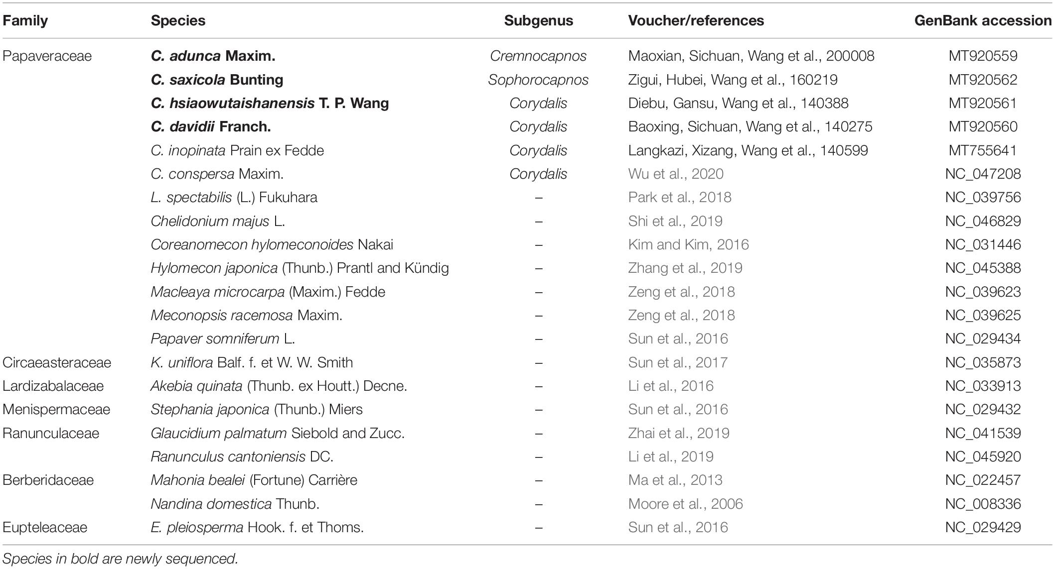
Samples of four Corydalis species (i.e., Corydalis adunca Maxim., Corydalis saxicola Bunting, Corydalis hsiaowutaishanensis T. P. Wang, and Corydalis davidii Franch.), representing all the three subgenera of Corydalis, were collected from their wild population (Table 1). Voucher specimens were deposited in the herbarium of the Central China Normal University (CCNU), Wuhan, China. Fresh leaves were dried in the wild using silica gel and preserved at −20°C until DNA extraction. Plants were identified following the treatment of Corydalis in the Flora of China (Zhang et al., 2008). DNA extraction, library preparation, and sequencing were conducted at Novogene (Tianjin, China). Total DNA was isolated using the modified cetyl trimethylammonium bromide (CTAB) method (Doyle and Doyle, 1987). DNA degradation and contamination were monitored on 1% agarose gels. DNA concentration was measured using a Qubit 2.0 fluorometer (Life Technologies, CA, United States). Approximately 1.5 μg of each DNA sample was fragmented by sonication to an average size of 350 bp. Sequencing libraries were generated using the NEBNext Ultra DNA Library Prep Kit for Illumina (NEB, United States) following the manufacturer’s recommendations. Libraries were sequenced using Illumina NovaSeq 6000 (Illumina, San Diego, CA, United States) with 2 × 150 bp paired-end reads.
Together with the above four newly sequenced samples, we included another two previously published Corydalis plastomes in our analyses (Corydalis conspersa Maxim., NC_047208; Wu et al., 2020; and Corydalis inopinata Prain ex Fedde MT755641; Xu and Wang, 2020). Another five already-reported Corydalis plastomes were not included, because the one plastome of Corydalis trisecta Franch. (MK713939; Kanwal et al., 2020) was assigned as unverified in the National Center for Biotechnology Information (NCBI), and the other four plastomes from C. saxicola and Corydalis tomentella Franch. sequenced in a preprint paper (Ren et al., 2020) may have not sustained to final verification. Besides, the rest of the Ranunculales plastomes (267, excluding unverified sequences and source sequences of the reference sequences) in NCBI (accessed on June 5, 2020) were also included in our analyses. In total, 273 plastomes were used in the present study.
Plastome Assembly and Annotation
The adaptors in raw data were removed, and low-quality sequences were trimmed using fastp v0.20.1 (Chen et al., 2018) with default parameters. Read quality of clean reads was assessed using FastQC v0.11.9 (Andrews, 2010). The clean reads were assembled using GetOrganelle v1.6.2 (Jin et al., 2018) with the chloroplast genome of C. inopinata (MT755641) as reference. Another assembly for each Corydalis species was performed using NOVOPlasty (Dierckxsens et al., 2017), with matK sequence (MH319908) of Corydalis temulifolia as seed, to confirm the GetOrganelle assemblies. The clean reads were mapped to the draft genome using BWA v0.7.17 (Li, 2013), filtered using SAMtools 1.10 (Li et al., 2009), and visualized using IGV 2.8.0 (Robinson et al., 2011) to check the concatenation of contigs. Furthermore, the rearrangements and quadripartite junction sites in the four newly sequenced Corydalis plastomes were verified with PCR and Sanger sequencing. The newly designed primers and their location on the plastomes were listed in Supplementary Table S1.
The complete chloroplast genomes were annotated using PGA (Qu et al., 2019), with the plastome of Euptelea pleiosperma Hook. f. et Thoms. (NC_029429), Papaver somniferum L. (NC_029434), and Lamprocapnos spectabilis (L.) Fukuhara (NC_039756) as reference. All tRNAs were verified by ARAGORN v1.2.38 (Laslett and Canback, 2004) and tRNAscan-SE v2.0 (Lowe and Chan, 2016). The annotation results were contrasted with the three annotation reference plastomes and with another three model plant plastomes [Arabidopsis thaliana (L.) Heynh., NC_000932; Nicotiana tabacum L., NC_001879; and Gossypium hirsutum L., NC_007944] and adjusted when necessary. The schematic diagrams of the chloroplast genomes were drawn in OGDRAW v1.3.1 (Greiner et al., 2019). All the newly annotated genomes were submitted to NCBI, and the accession numbers are shown in Table 1.
The GC content, large single-copy (LSC) size, inverted repeat (IR) size, small single-copy (SSC) size, and total genome size of all the 273 plastomes used in the present study were counted using Biopython v1.77 (Cock et al., 2009) and drawn in boxplot using Matplotlib v3.2.1 (Hunter, 2007). In addition, we also calculated GC content of each of the 97 genes (not including intron) shared between Corydalis plastomes and other representative Ranunculales plastomes which are listed in Table 1.
Genome Structure Analyses
To determine synteny and identify possible rearrangements, we compared the four newly sequenced and two previously published Corydalis plastomes (those six plastomes were hereafter referred to as “the Corydalis plastomes”) with the plastome of E. pleiosperma (NC_029429) using Mauve 2.4.0 (Darling et al., 2004) with the “progressiveMauve” algorithm. We employed E. pleiosperma as reference, because it exhibited typical angiosperm quadripartite plastome structure and was sister to the remaining Ranunculales. The Mauve result was manually modified to make the notable rearrangements clear and concise. To assess the expansion/contraction of the IR regions in detail, we compared the single-copy (SC)/IR junctions and their adjacent genes of the Corydalis plastomes with the E. pleiosperma plastome. The schematic diagram was manually modified on the basis of the plastome gene map drawn in OGDRAW. The whole sequence similarity among the Corydalis plastomes was comparatively analyzed and plotted using mVISTA (Frazer et al., 2004) in Shuffle-LAGAN mode, with default parameters and with the plastome of C. conspersa (NC_047208) as reference.
Repetitive Sequence Analyses
Three types of repetitive sequence were searched for in the Corydalis plastomes, that is, simple sequence repeats (SSR), tandem repeat, and dispersed repeat. Before those analyses were performed, one copy of the IR (IRa) was removed. The SSRs were detected using MISA v2.0 (Thiel et al., 2003) with the minimum numbers of repeats set to 10, 6, 5, 5, 5, and 5 for mononucleotide, dinucleotide, trinucleotide, tetranucleotide, pentanucleotide, and hexanucleotide repeats, respectively. Tandem repeats were detected using the Tandem Repeats Finder v4.09 (Benson, 1999). The alignment parameters match, mismatch, indel, minimum alignment score, and maximum period size were set to 2, 7, 7, 80, and 500, respectively. Four types of dispersed repeat (forward, reverse, complement, and palindromic) were detected using REPuter (Kurtz et al., 2001) with the minimal repeat size set to 30 bp and with a Hamming distance of 1.
Positive Selection Analyses
To investigate selection pressures from the environment on the Corydalis plastomes, we calculated non-synonymous (Ka), synonymous (Ks), and Ka/Ks ratios of 66 protein coding genes shared among the Corydalis plastomes, with the plastome of L. spectabilis (NC_039756) as reference. L. spectabilis was chosen as the reference, because it is closest to Corydalis among the taxa whose complete plastome have been reported. The protein-coding DNA sequences were extracted and translated into protein sequences using Biopython v1.77 (Cock et al., 2009). Protein sequences of each gene were aligned using MAFFT v7.450 (Katoh and Standley, 2013) and then converted into their corresponding codon-based nucleotide alignments by the Perl script PAL2NAL v14 (Suyama et al., 2006). Then Ka, Ks, and Ka/Ks ratios were calculated using the KaKs Calculator version 2.0 (Wang et al., 2010) with default parameters, except that we used the 11th genetic code (-c 11). The two anomalously large Ka/Ks values (Ka/Ks = 50), which represent extremely low synonymous substitutions in the alignment, were changed to 0. The ratios Ka/Ks > 1, Ka/Ks = 1, and Ka/Ks < 1 suggest positive selection, neutral selection, and purifying selection, respectively.
Phylogenetic Analyses
In total, 21 plastomes were included in our phylogenetic analyses, including all six Corydalis plastomes and 15 representatives of Ranunculales plastomes (Table 1). Among them, E. pleiosperma (NC_029429), which was sister to the rest of the Ranunculales, was used as an outgroup to root the trees. The data matrix contains coding regions of 64 protein-coding genes and four rRNA genes, which were shared among the 21 plastomes. Those genes including infA gene, rps15 gene, and the genes listed in Table 3 were excluded, because of pseudogenization or loss of the genes in one or more analyzed plastomes. Codon-based nucleotide alignments of each individual genes were aligned using MAFFT v7.450 (Katoh and Standley, 2013) and PAL2NAL v14 (Suyama et al., 2006) collectively. After manual adjustment, the nucleotide alignments were concatenated. The optimal partitioning scheme and corresponding best fit substitution model for Bayesian inference (BI) analysis were determined using PartitionFinder v2.1.1 (AICc criterion and greedy algorithm for all the mrbayes models, Lanfear et al., 2016) with the concatenated dataset initially partitioned by gene and codon position. Another three similar PartitionFinder analyses was lunched with only one model, GTR, GTR + G, or GTR + I + G; the resulting partitioning schemes with the lowest AICc values were used in maximum likelihood (ML) analysis. Three different phylogenetic methods, that is, BI, ML, and maximum parsimony (MP), were performed. The BI analysis was performed using MrBayes v3.2.7 (Ronquist et al., 2012), with the partitioned dataset and corresponding substitution model, one cold and three heated chains for one-million-generation Markov chains, sampling every 100 generations, and 25% discarded as burn-in. The ML analysis was performed using RAxML v8.2.12 (Stamatakis, 2014) with the partitioned dataset, GTRGAMMA model, and 1,000 bootstrap replicates. The MP analysis was performed using PAUP 4.0a166 (Swofford, 2003), with the directly concatenated dataset, heuristic search with 1,000 random taxon stepwise addition sequences, tree bisection reconnection branch swapping, and 1,000 bootstrap replications.
Results
Sequencing Results and Characteristics of the Newly Sequenced Corydalis Plastomes
For the four newly sampled Corydalis species, 18,463,522–22,599,330 raw 150 nt paired-end reads (Q30 > 90%) were obtained, with their average coverage depth ranging from 1,273 to 7,154-folds (Table 2). The plastomes assembled using GetOrganelle and NOVOPlasty were identical. All the Sanger sequencing results were also identical to the corresponding sequences of the assembled plastomes. The four newly sequenced complete plastomes ranged in length from 165,416 to 196,128 bp (Table 2). The four newly sequenced Corydalis plastomes exhibited the typical angiosperm quadripartite structure, with two IR regions (39,867–47,226 bp each) separating the LSC (85,352–94,289 bp) and SSC (330–12,086 bp) regions. The chloroplast genome schematic diagram of C. davidii is taken as an example and shown in Figure 1. The GC content of the four newly sequenced Corydalis plastomes ranged from 40.21 to 41.03% (Table 2). As viewed from the boxplots, the GC content, LSC size (except for C. davidii and C. hsiaowutaishanensis), IR size, and total genome size (except for C. davidii) of the four newly sequenced Corydalis plastomes were all among the largest, while their SSC sizes were all among the smallest within Ranunculales (Figure 2). The GC content of C. adunca (41.03%) is the highest, the LSC size of C. saxicola (94,289 bp) is the largest, and the SSC size of C. davidii (330 bp) is the smallest ever recorded in this order. For the 97 shared genes in the four newly sequenced Corydalis plastomes, 71.65% (278 out of 388) exhibited elevated GC content, as compared with the average GC content of 15 representatives of Ranunculales plastomes (Supplementary Table S2). The four newly sequenced Corydalis plastomes contained 101–112 unique genes, consisting of 67–78 protein-coding genes, 30 tRNA genes, and four rRNA genes, and 0–7 pseudogenes (Table 2). In total, 13 genes (accD, clpP, and 11 ndh genes) were detected to be pseudogenized or lost in one or more Corydalis plastomes (Table 3). The accD gene was lost in the four newly sequenced Corydalis plastomes, and the clpP gene was lost in C. hsiaowutaishanensis. C. adunca contained truncated versions of ndhC, ndhB, ndhF, ndhD, ndhI, ndhH, and ndhA. C. davidii contained truncated versions of ndhB, ndhD, ndhE, and ndhH but lacked sequences for ndhJ, ndhK, ndhC, ndhF, ndhG, ndhI, and ndhA. Besides, the rrn16 gene was duplicated in C. adunca, and the psaI gene was duplicated in both C. saxicola and C. hsiaowutaishanensis.

Table 2. Sequencing results (raw data), genome structure, and gene content of the four newly sequenced Corydalis plastomes.
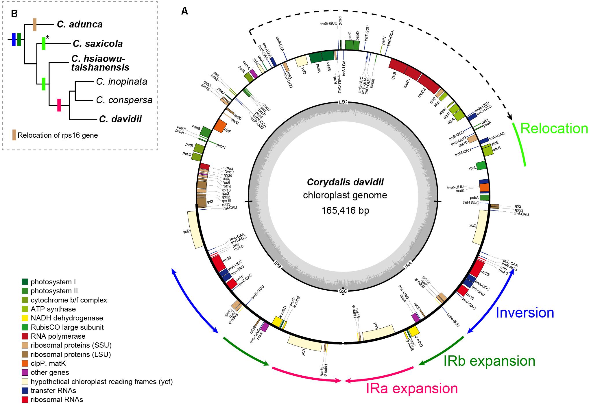
Figure 1. (A) Circular gene map of C. davidii. Genes inside and outside the map are transcribed in clockwise and counterclockwise directions, respectively. Genes belonging to different functional groups are color coded. Tick lines indicate the extent of the inverted repeats (IRA and IRB). The ring of bar graphs on the inner circle indicates the GC content in dark gray, with the line representing 50%. (B) Distributions of rearrangements in other Corydalis species. Species in bold are newly sequenced. Color of the boxes is the same with the rearrangements annotated around the circular gene map or indicated under the phylogenetic tree. Asterisks indicate a similar relocation, but downstream of the atpH gene.
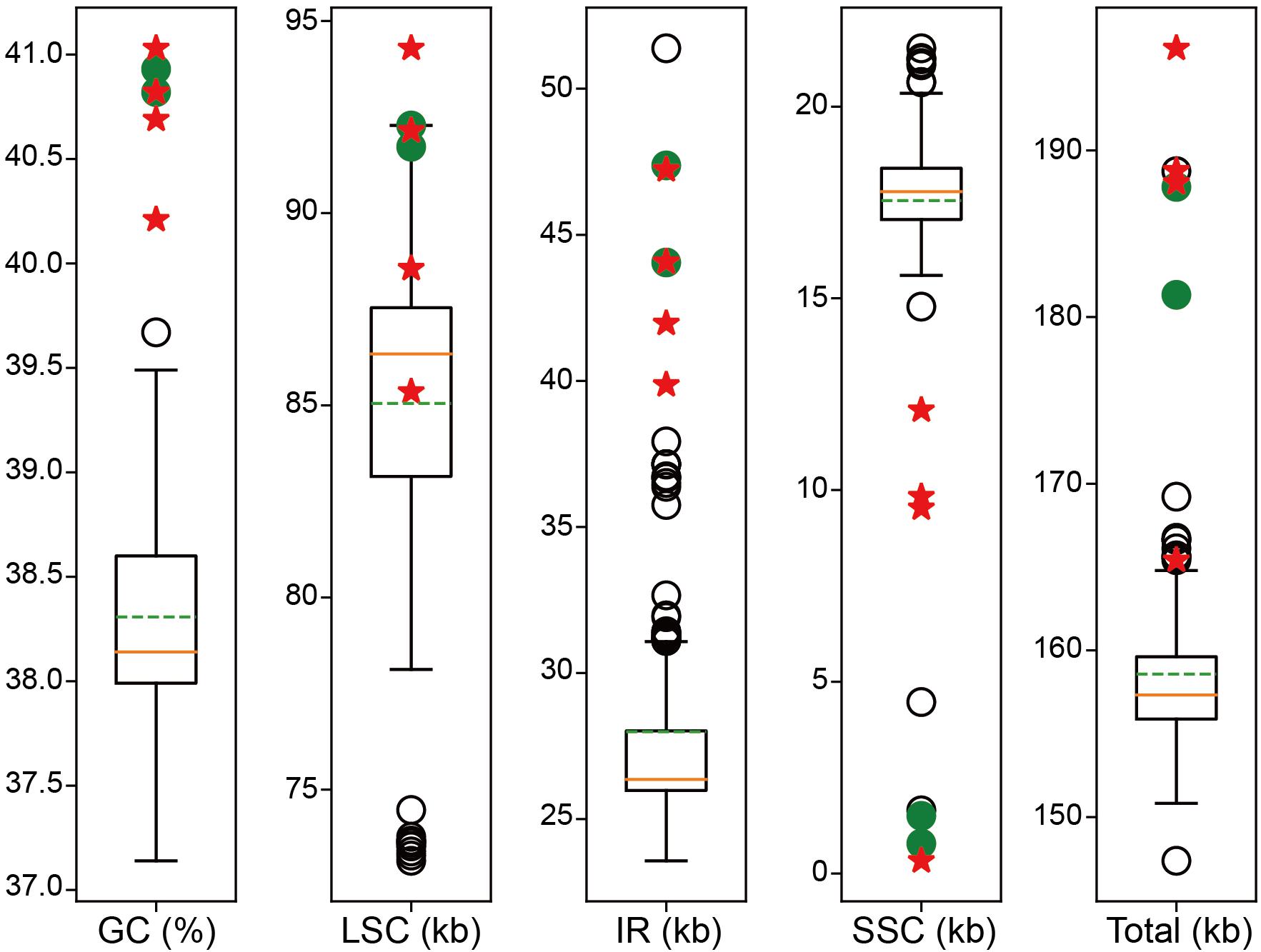
Figure 2. Boxplot distribution of GC content, LSC size, IR size, SSC size, and total genome size of the Corydalis plastomes relative to Ranunculales. The orange solid lines indicate the median. The green dotted lines indicate the mean value. The red five-pointed stars indicate the newly sequenced Corydalis plastomes. The green solid circles indicate previously published Corydalis plastomes. Empty circles indicate the rest outliers.
Genome Structure Variations
Mauve alignment identified 16 locally collinear blocks, from which five unusual rearrangements in the four newly sequenced Corydalis plastomes were deduced (Figure 3).
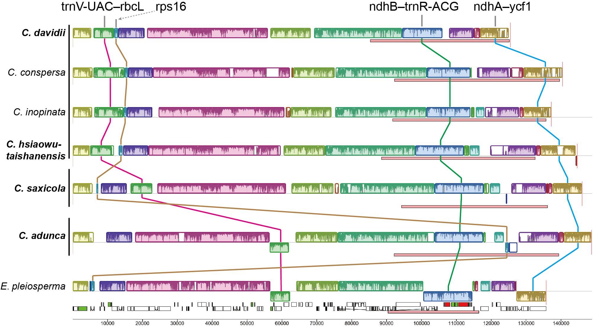
Figure 3. Structural alignments of Corydalis and E. pleiosperma plastomes using Mauve. Colored blocks represent locally collinear blocks (LCBs), and the blocks connected by lines indicate homology. Blocks drawn below the horizontal line indicate sequences found in an inverted orientation. Pink boxes below each plastome indicate IR regions. The vertical lines after species names, from top to bottom, indicate subg. Corydalis, subg. Sophorocapnos, and subg. Cremnocapnos, respectively. Species in bold are newly sequenced.
Firstly, one block (∼6–7 kb) contains five genes (trnV-UAC, trnM-CAU, atpE, atpB, and rbcL) and the associated non-coding sequences, relocated from the typically posterior part of the LSC region to the front. In C. adunca (subg. Cremnocapnos), those five genes displayed a typical angiosperm location, within the 56–62 kb region, downstream of the ndhC gene. In C. saxicola (subg. Sophorocapnos), those five genes relocated to the 17–23 kb region, downstream of the atpH gene. In C. davidii and C. hsiaowutaishanensis (subg. Corydalis), those five genes relocated to the 5–12 kb region, downstream of the trnK-UUU gene.
Secondly, one small block (∼1 kb) contained only the rps16 gene relocated from the LSC region to downstream of the ndhF gene in the IR region in C. adunca. Meanwhile, one duplicate of the rrn16 gene in C. adunca transferred to the original rps16 gene loci (not shown in the Mauve result).
Thirdly, one block (∼11–14 kb) in the IR region contains 11 genes (ndhB-trnR-ACG), inverted uniformly in the four newly sequenced Corydalis plastomes.
Fourthly, the IRs expanded greatly, with the absorption of more than half of the SSC region in different types (Figure 4). The IRb region expanded downstream of the pseudogenized ndhI gene in C. adunca or the middle of the ndhA gene in C. saxicola or the middle of the ndhI gene in C. hsiaowutaishanensis, resulting in the transfer of 9–10 genes (though some were pseudogenized) from the SSC region to the IR region. In contrast, in C. davidii, IRb expanded downstream of the pseudogenized ndhE gene, and successively, IRa expanded downstream of the pseudogenized ndhH gene. As a result, the IR regions of the four newly sequenced Corydalis plastomes ranged from 39,867 to 44,070 bp in length, which were more than 10 kb longer than the average (28,127 bp) of Ranunculales plastomes.
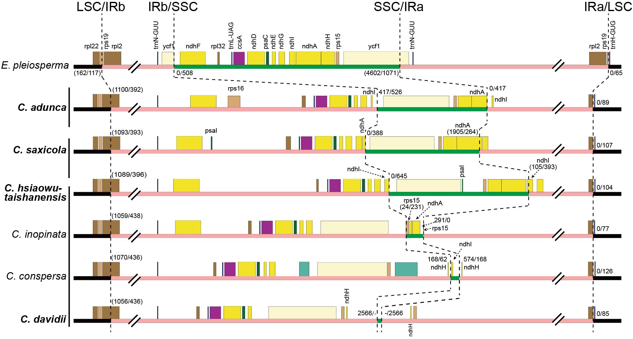
Figure 4. Comparison of LSC, IR, and SSC boundaries among Corydalis and E. pleiosperma plastomes. The black, pink, and green thick lines indicate LSC, IR, and SSC, respectively. The numbers before and after the slash indicate the distance of the nearest genes relative to the junction site or the length of the front and latter parts of the genes that span the junction site when they appeared within a parenthesis. The vertical lines after species names, from top to bottom, indicate subg. Cremnocapnos, subg. Sophorocapnos, and subg. Corydalis, respectively. Species in bold are newly sequenced.
Lastly, the SSC regions were inverted uniformly in the four newly sequenced Corydalis plastomes.
Except for the IR–SSC boundary variations mentioned above, the IR shrank slightly at the LSC/IRb border, from the middle of the rps19 gene to the middle of the rpl2 gene (Figure 4), creating a 392–436 bp truncated rpl2 gene copy (pseudogene) in the IRa region of the four newly sequenced Corydalis plastomes.
The alignment and conserved regions of the Corydalis plastomes are shown in Figure 5. Overall, they exhibited high similarity, especially in the gene regions, with the exception that some specific regions that involved plastome rearrangements were highly divergent. For the detected conserved regions, the average identity values of untranslated (tRNA and rRNA), exon, intron, and intergenic regions were 99.22, 93.33, 93.0, and 88.27%, respectively. Some intron regions (rps16, rpl16, and trnK-UUU) and intergenic regions (trnQ-UUG-psbK, psbK-psbI, atpH-atpI, rpoB-trnC-GCA, trnC-GCA-petN, psbM-trnD-GUC, trnD-GUC-trnY-GUA, trnE-UUC-trnT-GGU, trnT-GGU-psbD, trnT-UGU-trnL-UAA, rbcL-atpB, psaI-ycf4, petA-psbJ, and psbE-petL), which exhibited some extent of divergence, were identified, and they have the potential to be DNA markers in future phylogenetic analyses.
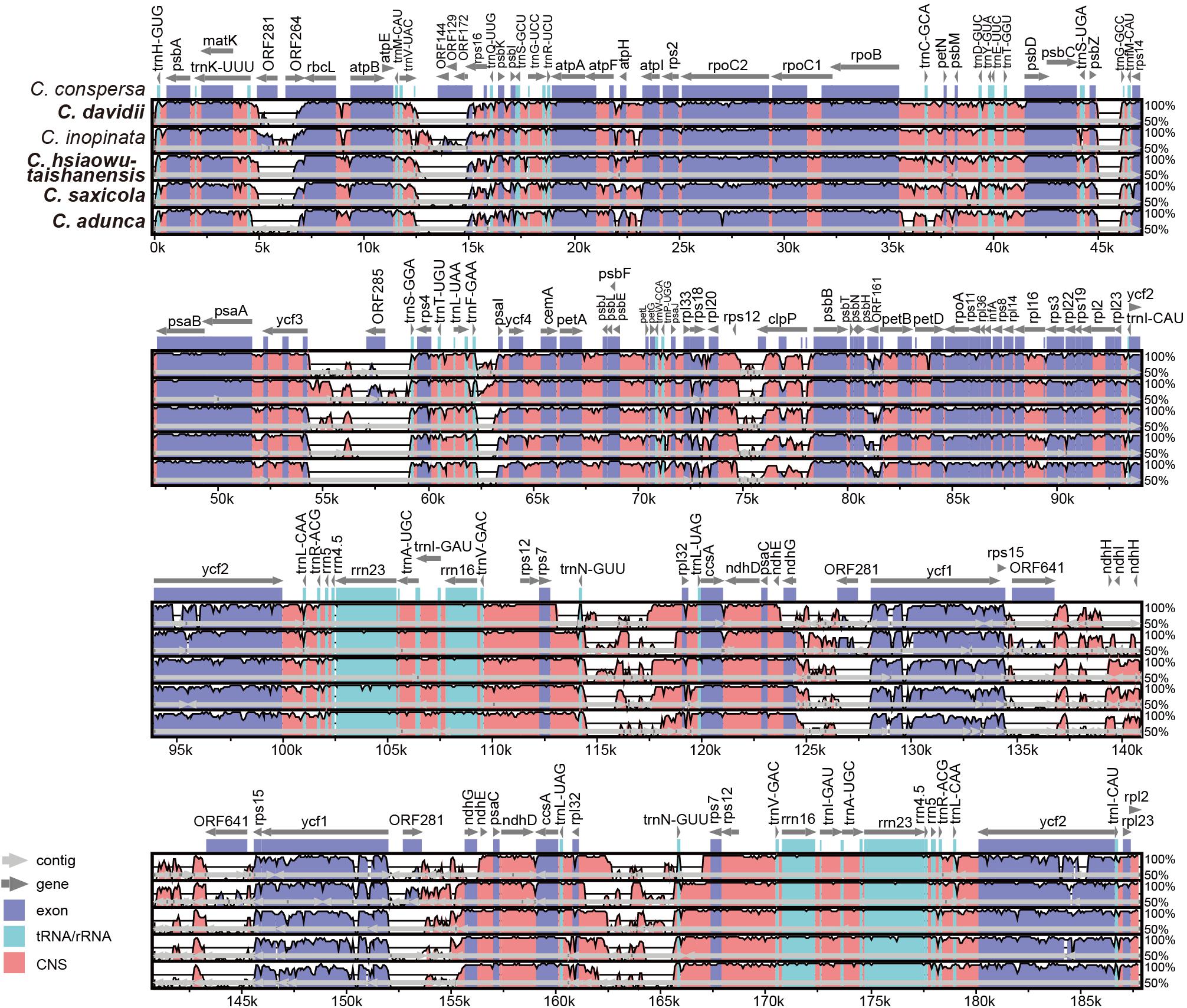
Figure 5. Comparative analyses of plastome differences in the four Corydalis plastomes. The dark blue regions represent exons, light-blue regions represent untranslated regions (tRNA and rRNA), and pink regions represent non-coding sequences (CNS). The vertical scale shows the percentage of identity, varying from 50 to 100%. Species in bold are newly sequenced.
Repeat Sequences
In total, we detected 150 SSRs including 135 mononucleotide repeats and 15 dinucleotide repeats in the four newly sequenced Corydalis plastomes (not including IRa; Table 4). Almost all mononucleotide repeats were composed of A/T (94.07%), while only 5.93% were composed of C/G. The 15 dinucleotide repeats consisted of 14 AT/TA repeats and one AG repeat. The total numbers of SSRs in the four newly sequenced Corydalis plastomes were similar to those of C. inopinata, C. conspersa, and L. spectabilis. It was, however, larger than that of P. somniferum and smaller than that of E. pleiosperma. In total, 26–28 tandem repeats and 156–274 dispersed repeats were detected in each of the four newly sequenced Corydalis plastomes (Table 4). C. adunca has the most dispersed repeats (274), which were ∼16 times of E. pleiosperma (17) and ∼30 times of P. somniferum (9).

Table 4. Numbers of SSR, tandem repeat, and dispersed repeat in the plastomes of Corydalis species and related species.
Nucleotide Substitution Rates
The Ka/Ks ratios of the 66 protein-coding genes are shown in the bar graph (Figure 6). The majority (85.1%) of Ka/Ks ratios was between 0 and 0.5. The average Ka/Ks ratio for all those genes was 0.26. Higher Ka/Ks values were detected mainly in the rpl and rps gene families. The Ka/Ks ratios of four genes (psaI, rpl23, rpl36, and rps7) were greater than 1 in one or more pairwise comparisons. In total, 13 Ka/Ks ratios, that is, those of all the four genes in C. adunca; psaI, rpl36, and rps7 in C. saxicola; rpl36 and rps7 in both C. hsiaowutaishanensis and C. conspersa; and psaI and rps7 in C. davidii, were larger than 1.
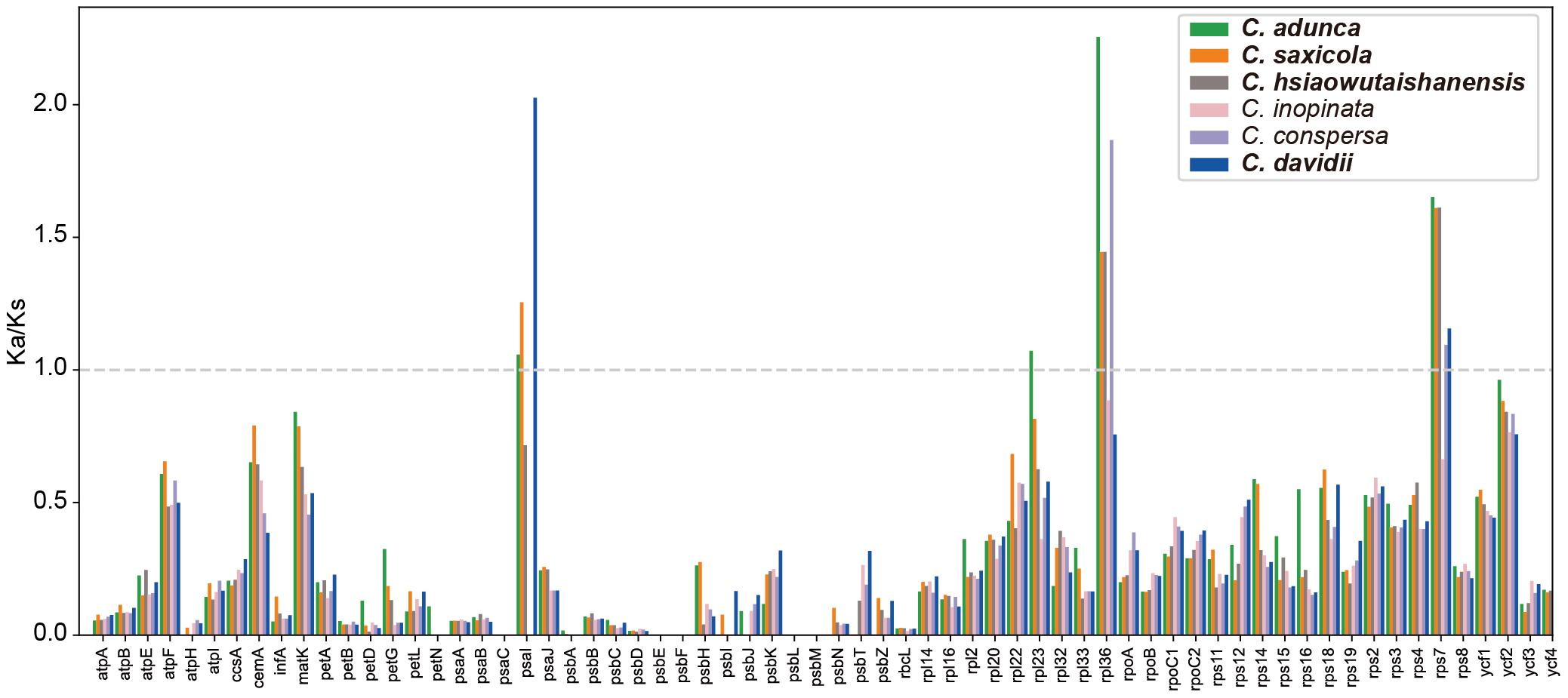
Figure 6. The Ka/Ks ratios of 66 protein-coding genes of the Corydalis plastomes relative to L. spectabilis. Species in bold are newly sequenced.
Phylogenetic Analyses
The data matrix used in phylogenetic analyses consisted of 63,027 nucleotide sites; of these, 8,642 (13.7%) were parsimony informative. The PartitionFinder2 analyses using mrbayes models partitioned the concatenated dataset into 70 subsets and 14 substitution models (Supplementary Table S3). The AICc values of another three independent PartitionFinder2 analyses using GTR, GTR + G, or GTR + I + G were 524,755.977691, 518,714.822997, and 518,513.525426, respectively. The partitioning scheme determined using GTR + I + G (62 subsets; Supplementary Table S4) has the lowest AICc value and was used in ML analysis. The three phylogenetic analyses (BI, ML, and MP) generated identical topologies, and the vast majority branches received full support. Thus, only the BI phylogenetic tree is presented in Figure 7. Three clades can be defined in the phylogenetic tree, that is, the Eupteleaceae clade, the Papaveraceae clade, and the clade composed of the rest of the Ranunculales families. Within Papaveraceae, two sub-clades corresponding to the subfamilies (Papaveroideae and Fumarioideae) were resolved with full support. Corydalis belongs to the Fumarioideae sub-clade, and all lineages within Corydalis were fully supported. The four newly sequenced Corydalis species, that is, C. adunca (sect. Strictae), C. saxicola (sect. Thalictrifoliae), C. hsiaowutaishanensis (sect. Dactylotuber), and C. davidii (sect. Davidianae), served as the successively diverged lineages in this genus.
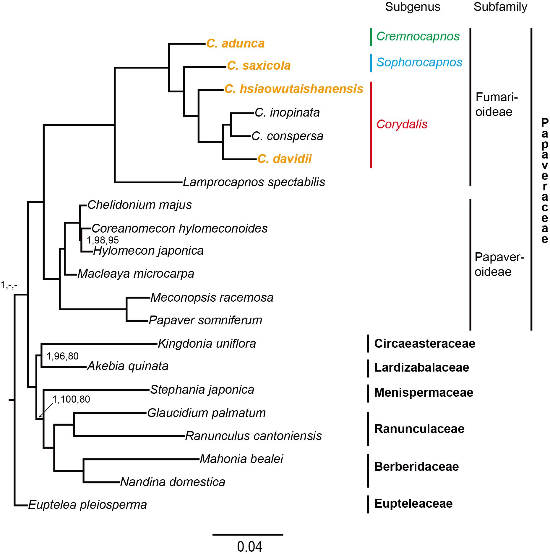
Figure 7. Phylogenetic tree conducted using BI methods. The numbers above branches represent BI posterior probability, ML bootstrap support, and MP bootstrap support, respectively. The full support values were not indicated.
Discussion
Abnormal Genome Sizes, Reduced Gene Composition, Elevated GC Content, and High Number of Repeat Sequences
The LSC size, IR size, SSC size, and total genome size of the Corydalis plastomes were proven to be abnormally larger or smaller than those of the other Ranunculales, though a few exceptions existed (Figure 2). Several factors, for example, gene duplications, gene losses, and IR expansions, may be responsible for the variation in Corydalis plastome sizes. Among them, the expansions of IR into the SSC region (for details, see section “Various IR Expansions”) have contributed the most to the increase of plastome size. Gene losses (and/or pseudogenizations), on the contrary, have partially counteracted the increase of plastome size. Owing to its pseudogenization or loss of all eight ndh genes in the SSC and IR regions, C. davidii had the smallest IR and total plastome size, albeit its IR expansion was most pronounced in Corydalis.
With respect to gene content, the Corydalis plastomes exhibited extensive pseudogenization or loss of some of the 13 genes (11 ndh, accD, and clpP genes). The 11 chloroplast ndh genes that encode NADH dehydrogenase subunits are essential for the function of chloroplast and are widely present in angiosperms (Martin and Sabater, 2010). However, we found that 7–11 of the ndh genes were either pseudogenized or lost in the plastome of C. adunca, C. davidii, C. inopinata, and C. conspersa. The pseudogenization and/or loss of ndhJ, ndhK, and ndhC were possibly associated with the adjacent relocation event in the LSC region, while the pseudogenization and/or loss of the rest of the ndh genes were possibly associated with the IR boundary shift, as evidenced in another Ranunculales species (Kingdonia uniflora, Sun et al., 2017), where similar plastome rearrangements were also accompanied by pseudogenization and/or loss of ndh genes. In a phylogenetic perspective, the pseudogenization or loss of ndh genes in the Corydalis and Ranunculales may have occurred independently after the split of those species, which was probably a similar case found in the research of Orchidaceae (Lin et al., 2015). It is necessary to use more species to elucidate the pattern and mechanism of pseudogenization and loss of ndh genes within Corydalis. Another unusual case is the uniform loss of the accD gene in the Corydalis plastomes. We speculate that the loss of the accD gene probably occurred in the common ancestor of the Corydalis species. The accD gene encodes one of four subunits of the acetyl-CoA carboxylase enzyme (ACC), which is a rate-limiting enzyme in the first committed step of fatty acid synthesis (Cronan and Waldrop, 2002; Sasaki and Nagano, 2004). The loss of the accD gene is lethal, as evidenced by the study of tobacco (Kode et al., 2005). Within angiosperms, the lost accD has been found to relocate to the nucleus in some species, such as Trifolium repens (Magee et al., 2010), some Campanulaceae species (Rousseau et al., 2013), and Platycodon grandiflorum (Hong et al., 2017). We are not sure whether the plastid-encoded accD gene is lost entirely or functionally transferred to the nucleus in Corydalis, but there must be some kind of compensatory mechanism that can explain their loss from the plastome.
The GC content of the Corydalis plastomes (40.21–41.03%) was slightly higher than the average of Ranunculales (38.31%). It seems not unusual, but when examining the NCBI RefSeq database (accessed on October 19, 2020), we can find that only ∼130 plastomes out of more than 5,000 land plant plastomes have GC content higher than 40%. The increase of GC content in the Corydalis plastomes has affected not only the gene regions but also the intergenic and intron regions. This is because the GC content of the gene region and intergenic and intron regions of the Corydalis plastomes has increased, by 0.8–1.75% and 3.32–4.76%, respectively, as compared with the average of the 15 representatives of Ranunculales plastomes. It is well known that the GC base pair is more stable than the AT. Thus, the overall increase of GC content may potentially enhance the stability of the plastomes and consequently contribute to their adaptation to some harsh conditions. Here, the two largest GC contents within Ranunculales, kept by C. adunca and C. inopinata, may have contributed to their adaptation to the Qinghai–Tibet Plateau.
The pairwise Ka/Ks ratios were widely used as an effective way to detect positive selection or adaptive evolution in plant species (Wang et al., 2010; Gao et al., 2019). For the protein-coding genes in the Corydalis plastomes, the majority (85.1%) of Ka/Ks ratios obtained in our analyses were between 0 and 0.5, which was consistent with the result of previous researches (Nei et al., 2010; Yang et al., 2020). This suggests that the majority of genes in the Corydalis plastomes were probably under purifying selection. The rpl and rps gene families, which are involved in self-replication, showed higher Ka/Ks ratios compared with the majority of the rest of the genes in the Corydalis plastome. This may contribute to their adaptation to diverse habitats. In the present study, only four genes (psaI, rpl23, rpl36, and rps7) showed Ka/Ks ratios greater than 1 (Figure 6) in one or more pairwise comparisons, indicating that they may undergo some selective pressure. All four genes (psaI, rpl23, rpl36, and rps7) and three genes (except for rpl23) in the plastomes of C. adunca and C. saxicola, respectively, showed Ka/Ks ratios greater than 1. Considering that these two species readily grow in dry habitats, which is different from the rest of the Corydalis species and L. spectabilis, we speculate that the chloroplast functional genes may have facilitated their adaptation to such a dry environment.
Another unique feature of the Corydalis plastomes was its high number of repeat sequences, especially the dispersed repeats (81–280), which can be ∼30 times of the number of the dispersed repeats in P. somniferum (9). Some researchers have suggested that repeat sequences may contribute to plastome rearrangement (Ogihara et al., 1988; Milligan et al., 1989; Cole et al., 2018). In the present study, we also detected that the dispersed repeat sequences often located around the loci that involved rearrangement.
Rare Gene Relocations and Inversions
Among the various chloroplast genome rearrangements, relocation is probably the least common in angiosperms (Mower and Vickrey, 2018). Until now, it was only reported within a few lineages, such as Oleaceae (Lee et al., 2007), Campanulaceae (Knox, 2014; Knox and Li, 2017; Uribe-Convers et al., 2017), and Ranunculaceae (Liu et al., 2018). In the present study, a 5 kb segment containing five genes (trnV-UAC-rbcL), which were traditionally located downstream of the ndhC gene, has unconventionally inverted and relocated either downstream of the atpH gene in C. saxicola (subg. Sophorocapnos) or downstream of the trnK-UUU gene in both C. davidii and C. hsiaowutaishanensis (subg. Corydalis) (Figure 3). The latter relocation event was also detected in the two previously published Corydalis plastomes (subg. Corydalis; Wu et al., 2020; Xu and Wang, 2020). In a phylogenetic view, it seems that the locations of those five genes (trnV-UAC-rbcL) were subgenus specific, and the relocation event probably occurred after the divergence of subg. Cremnocapnos. Moreover, subg. Sophorocapnos and subg. Corydalis displayed different types of trnV-UAC-rbcL location, suggesting that the two relocation events occurred independently in these two subgenera. Until now, the relocation of those five genes (trnV-UAC-rbcL) was only reported from Corydalis within Ranunculales. Research from some Oleaceae species (Lee et al., 2007) revealed that the plastome relocation resulted from two overlapping inversions: the first inversion inverted the entire segment, and the second restored some part to its original gene order. It is possible that the relocation of those five genes (trnV-UAC-rbcL) in Corydalis is also the result of two overlapping inversions. Putatively, the first inversion for subg. Corydalis is ∼50 kb, comprising rps16-rbcL, which translocates trnV-UAC-rbcL to downstream of trnK-UUU, and the second inversion restores the subsequent genes (ndhC-rps16) to their original order. Additionally, an inversion of trnQ-UUG-rbcL, which was similar to the first “putative” subg. Corydalis inversion, has been reported in two Ranunculales species (Circaeaster agrestis Maxim. and K. uniflora, Sun et al., 2017). It is likely that this “putative” inversion may also be found in some Corydalis species. There were reports of similar relocations of trnV-UAC-rbcL to downstream the psbA gene in at least 22 genera of Campanulaceae (Knox, 2014; Knox and Li, 2017; Uribe-Convers et al., 2017). Significantly, similar plastome rearrangements have happened independently in those two distantly related lineages. Elucidating the origin, evolution, and mechanism under such similar but independent genome rearrangements seems an exciting topic to explore in further comparative genomic studies.
One segment that contains 11 genes (ndhB-trnR-ACG) in the IR region, accounting for about 60% of the typical IR region, inverted uniformly in the Corydalis plastomes and L. spectabilis plastome (Park et al., 2018). This inversion was shared by all the sequenced Fumarioideae plastomes, but it was absent from the subfamily Papaveroideae. This indicated that the inversion has occurred after the divergence of those two Papaveraceae subfamilies and existed in the common ancestor of Lamprocapnos and Corydalis. In the nearby region of the above inversion, C. adunca, which was sister to the rest of the Corydalis, was characterized by a unique relocation of the rps16 gene from the LSC region to the IR region and by the transfer of a replicate of its rrn16 gene to the typical rps16 gene site. Whether those two rearrangements occurred in the plastome of C. adunca simultaneously or independently, extra hidden plastome rearrangements must have happened in the evolutionary history. L. spectabilis, a species that diverged before Corydalis within the subfamily Fumarioideae, also exhibited some extra rearrangements around the inverted ndhB-trnR-ACG segment (Park et al., 2018). Those portend that the common ancestor of Corydalis and Lamprocapnos may possess a variable IR region. With a variable IR region in the ancestor, it may be interesting to research why only the inversion of the ndhB-trnR-ACG segment in the IR regions was kept throughout all those species of the subfamily Fumarioideae.
The inversion in the SSC regions, by the way, has been detected in the Corydalis plastomes, as compared with the plastome of E. pleiosperma and most other angiosperms. However, it is probably not a noticeable feature, because previous studies have found that most plastomes often occur in two distinct haplotypes (with equal frequency) differing in the orientation of SC regions (Palmer, 1983; Wang and Lanfear, 2019).
Various IR Expansions
The IR regions of angiosperm plastomes usually start around the rps19 gene and terminate almost uniformly at either downstream of the trnN-GUU or truncated ycf1 gene. IR expansion has been reported in some lineages, but it was usually into the LSC region (e.g., in Ma et al., 2013; Sun et al., 2013; Liu et al., 2018; Mower and Vickrey, 2018; Zhai et al., 2019). The IR/SSC junctions were thought to be relatively stable (Sun et al., 2016; Zhu et al., 2016; Mower and Vickrey, 2018; Park et al., 2018). In the present study, we found that the IR of the Corydalis plastomes expanded markedly at the IR/SSC boundaries (Figures 3, 4), which has resulted in the second to seventh largest IR sizes and the three smallest and sixth to eighth smallest SSC sizes in Ranunculales. The three earlier diverged Corydalis species (C. adunca, C. saxicola, and C. hsiaowutaishanensis; Figure 7) have only IRb/SSC expansion, while C. inopinata, C. conspersa, and C. davidii have a further IRa/SSC expansion. L. spectabilis, which also belongs to the subfamily Fumarioideae (Papaveraceae) but diverged earlier (Figure 7) than Corydalis, was characterized with the expansion of IRa into the SSC region. Thus, three types of IR/SSC expansion can be defined within the subfamily Fumarioideae, that is, IRa/SSC expansion, IRb/SSC expansion, and IRb/SSC + IRa/SSC expansion. While in the subfamily Papaveroideae (Papaveraceae), no IR expansion was observed. Hereby, we can deduce that IR expansion occurred after the diversification of those two Papaveraceae subfamilies. After a further examination of the IR/SSC boundary within Corydalis, we found that the adjacent genes differed between lineages. These results suggest that more than one IR expansion event has occurred and that each lineage has its specific IR expansion history. More extensive sampling is needed to elucidate the IR expansion pattern within Corydalis.
Phylogenetic Implications and New DNA Markers
The plastid phylogenomic analyses generated highly supported phylogeny with three distinct clades (i.e., Eupteleaceae, Papaveraceae, and the rest of the Ranunculales families), which is consistent with the results of previous studies (Wang et al., 2009; Pérez-Gutiérrez et al., 2015; Sauquet et al., 2015; Sun et al., 2016). All the Corydalis lineages were fully supported in the phylogenetic tree, which indicates that the application of complete plastome data can improve the resolution of the phylogeny of Corydalis. This suggests that the complete plastome data have the potential to be employed in the construction of a robust phylogeny for Corydalis, which would be instructive for resolving the taxonomic controversy in this genus. Importantly, we resolved C. adunca (sect. Strictae) to be a lineage with early divergence in this genus and C. hsiaowutaishanensis (sect. Dactylotuber) to be an earlier diverged lineage in subg. Corydalis. This was consistent with the results of previous studies based on two DNA markers (rps16 and matK; Wang, 2006) but contradicted with the morphological character-based taxonomy to some extent (Wu et al., 1996, 1999).
Highly variable DNA markers are useful, especially in fast species identification and wide-range phylogenetic analyses. In the present study, the introns of rps16, rpl16, and trnK-UUU and intergenic regions of trnQ-UUG-psbK, psbK-psbI, atpH-atpI, rpoB-trnC-GCA, etc., which exhibited some extent of divergence, were identified and have great potential to be exploited as DNA markers. Some of them have already been used as markers in previous phylogenetic analyses of Corydalis (e.g., Wang, 2006; Zhang Z.X. et al., 2016; Jiang et al., 2018; Ren et al., 2018), while the rest is in need of exploitation in the future. Nevertheless, they should be used with caution in Corydalis, because genes that stayed in the SC region or relocated to the IR region usually obtain different substitution rates (Zhu et al., 2016), which may mislead the phylogenetic reference. For example, the rps16 gene, which has been used in the previous phylogenetic analysis of Corydalis (Lidén et al., 1997; Wang, 2006), was found to have relocated to IR in C. adunca, while it still remained in the LSC region in the rest of the sequenced Corydalis species.
Conclusion
We provided insights into the plastome structure of Corydalis and resolved Corydalis to be an overlooked lineage that exhibited some extraordinary rearrangements (relocations, inversions, IR expansions, pseudogenizations, gene losses, gene duplications, etc.). Although similar rearrangements have occurred multiple times in the history of angiosperm, it is noteworthy that all those rearrangements existed in a single genus. The plastome data also turned out to be effective in enhancing the resolution of phylogenies in Corydalis, and it could be alternatively employed to construct a robust phylogeny for Corydalis in further studies. The results obtained in this study may be valuable for further study on the taxonomy, phylogeny, and evolution of Corydalis, a taxonomically difficult but fascinating genus.
Data Availability Statement
The datasets generated for this study can be found in National Center for Biotechnology Information (NCBI) under the accession numbers: MT920559–MT920562.
Author Contributions
XX performed the analyses and drafted the manuscript. DW provided the suggestions on structuring the manuscript and the main points of the discussion and revised the manuscript. Both authors conceived the study, obtained the samples, designed the experiments, read, and approved the final manuscript.
Funding
This work was supported by grants from the National Natural Science Foundation of China (31170310), Science and Technology Basic Work (2013FY112100 and 2013FY112300), the Special Foundation for the Specimen Platform of China, teaching specimen sub-platform, http://mnh.scu.edu.cn/(2005DKA21403-JK), Fundamental Research Funds for the Central Universities (Excellent doctoral dissertation development program of CCNU, 202050185085), and funds from the Shaanxi Provincial Bio-Resource Key Laboratory, Shaanxi University of Technology (SLGPT2019KF03-02).
Conflict of Interest
The authors declare that the research was conducted in the absence of any commercial or financial relationships that could be construed as a potential conflict of interest.
Supplementary Material
The Supplementary Material for this article can be found online at: https://www.frontiersin.org/articles/10.3389/fpls.2020.600354/full#supplementary-material
References
Andrews, S. (2010). FastQC: A Quality Control Tool for High Throughput Sequence Data. Available online at: http://www.bioinformatics.babraham.ac.uk/projects/fastqc/ (accessed 21 April 2020).
Asaf, S., Khan, A. L., Lubna, K. A., Khan, A., Khan, G., Lee, I.-G., et al. (2020). Expanded inverted repeat region with large scale inversion in the first complete plastid genome sequence of Plantago ovata. Sci. Rep. 10:3881. doi: 10.1038/s41598-020-60803-y
Barrett, C. F., Baker, W. J., Comer, J. R., Conran, J. G., Lahmeyer, S. C., LeebensMack, J. H., et al. (2016). Plastid genomes reveal support for deep phylogenetic relationships and extensive rate variation among palms and other commelinid monocots. New Phytol. 209, 855–870. doi: 10.1111/nph.13617
Benson, G. (1999). Tandem repeats finder: a program to analyze DNA sequences. Nucleic Acids Res. 27, 573–580. doi: 10.1093/nar/27.2.573
Burke, S. V., Lin, C. S., Wysocki, W. P., Clark, L. G., and Duvall, M. R. (2016). Phylogenomics and plastome evolution of tropical forest Grasses (Leptaspis, Streptochaeta: Poaceae). Front. Plant Sci. 7:1993. doi: 10.3389/fpls.2016.01993
Cai, Z. Q., Guisinger, M., Kim, H. G., Ruck, E., Blazier, J. C., McMurtry, V., et al. (2008). Extensive reorganization of the plastid genome of Trifolium subterraneum (Fabaceae) is associated with numerous repeated sequences and novel DNA insertions. J. Mol. Evol. 67, 696–704. doi: 10.1007/s00239-008-9180-7
Chen, S. F., Zhou, Y. Q., Chen, Y. R., and Gu, J. (2018). fastp: an ultra-fast all-in-one FASTQ preprocessor. Bioinformatics 34, i884–i890. doi: 10.1093/bioinformatics/bty560
Chinese Pharmacopoeia Commission (2015). Pharmacopoeia of the People’s Republic of China, Vol. 1. Beijing: China Medical Science Press.
Chumley, T. W., Palmer, J. D., Mower, J. P., Fourcade, H. M., Calie, P. J., Boore, J. L., et al. (2006). The complete chloroplast genome sequence of Pelargonium × hortorum: organization and evolution of the largest and most highly rearranged chloroplast genome of land plants. Mol. Biol. Evol. 23, 2175–2190. doi: 10.1093/molbev/msl089
Cock, P. J. A., Antao, T., Chang, J. T., Chapman, B. A., Cox, C. J., Dalke, A., et al. (2009). Biopython: freely available Python tools for computational molecular biology and bioinformatics. Bioinformatics 25, 1422–1423. doi: 10.1093/bioinformatics/btp163
Cole, L. W., Guo, W., Mower, J. P., and Palmer, J. D. (2018). High and variable rates of repeat-mediated mitochondrial genome rearrangement in a genus of plants. Mol. Biol. Evol. 35, 2773–2785. doi: 10.1093/molbev/msy176
Cosner, M. E., Raubeson, L. A., and Jansen, R. K. (2004). Chloroplast DNA rearrangements in Campanulaceae: phylogenetic utility of highly rearranged genomes. BMC Evol. Biol. 4:27. doi: 10.1186/1471-2148-4-27
Cronan, J. E., and Waldrop, G. L. (2002). Multi-subunit acetyl-CoA carboxylase. Prog. Lipid Res. 41, 407–435. doi: 10.1016/S0163-7827(02)00007-3
Darling, A. C. E., Mau, B., Blattner, F. R., and Perna, N. T. (2004). Mauve: multiple alignment of conserved genomic sequence with rearrangements. Genome Res. 14, 1394–1403. doi: 10.1101/gr.2289704
Dierckxsens, N., Mardulyn, P., and Smits, G. (2017). NOVOPlasty: de novo assembly of organelle genomes from whole genome data. Nucleic Acids Res. 45:e18. doi: 10.1093/nar/gkw955
Downie, S. R., and Palmer, J. D. (1992). “Use of chloroplast DNA rearrangements in reconstructing plant phylogeny,” in Plant Molecular Systematics, eds P. Soltis, D. Soltis, and J. J. Doyle (New York, NY: Chapman and Hall), 14–35. doi: 10.1007/978-1-4615-3276-7_2
Doyle, J. J., Davis, J. I., Soreng, R. J., Garvin, D., and Anderson, M. J. (1992). Chloroplast DNA inversions and the origin of the grass family (Poaceae). Proc. Natl. Acad. Sci. U.S.A. 89, 7722–7726. doi: 10.1073/pnas.89.16.7722
Doyle, J. J., and Doyle, J. L. (1987). A rapid DNA isolation procedure for small quantities of freshf tissue. Phytochem. Bull. 19, 11–15.
Doyle, J. J., Doyle, J. L., Ballenger, J. A., and Palmer, J. D. (1996). The Distribution and Phylogenetic Significance of a 50-kb Chloroplast DNA Inversion in the Flowering Plant Family Leguminosae. Mol. Phylogenet. Evol. 5, 429–438. doi: 10.1006/mpev.1996.0038
Editorial Board of Chinese Tibetan Medicine (1996). Chinese Tibetan medicine. Shanghai: Shanghai Science and Technology Press.
Fedde, F. (1936). “Papaveraceae,” in Die natuerlichen Pflanzenfamilien, 2nd Edn, eds A. Engler and K. Prantl (Leipzig: W. Engelmann), 5–145.
Frazer, K. A., Pachter, L., Poliakov, A., Rubin, E. M., and Dubchak, I. (2004). VISTA: computational tools for comparative genomics. Nucleic Acids Res. 32, W273–W279. doi: 10.1093/nar/gkh458
Gao, L. Z., Liu, Y. L., Zhang, D., Li, W., Gao, J., Liu, Y., et al. (2019). Evolution of Oryza chloroplast genomes promoted adaptation to diverse ecological habitats. Commun. Biol. 2:278. doi: 10.1038/s42003-019-0531-2
Greiner, S., Lehwark, P., and Bock, R. (2019). OrganellarGenomeDRAW (OGDRAW) version 1.3.1: expanded toolkit for the graphical visualization of organellar genomes. Nucleic Acids Res. 47, W59–W64. doi: 10.1093/nar/gkz238
Guisinger, M. M., Kuehl, J. V., Boore, J. L., and Jansen, R. K. (2011). Extreme reconfiguration of plastid genomes in the angiosperm family geraniaceae: rearrangements, repeats, and codon usage. Mol. Biol. Evol. 28, 583–600. doi: 10.1093/molbev/msq229
Hong, C. P., Park, J., Lee, Y., Lee, M., Park, S. G., Uhm, Y., et al. (2017). accD nuclear transfer of Platycodon grandiflorum and the plastid of early Campanulaceae. BMC Genomics 18:607. doi: 10.1186/s12864-017-4014-x
Hunter, J. D. (2007). Matplotlib: A 2D graphics environment. Comput. Sci. Eng. 9, 90–95. doi: 10.1109/MCSE.2007.55
Jansen, R. K., Cai, Z., Raubeson, L. A., Daniell, H., Leebens-Mack, J., Müller, K. F., et al. (2007). Analysis of 81 genes from 64 plastid genomes resolves relationships in angiosperms and identifies genome-scale evolutionary patterns. Proc. Natl. Acad. Sci. U.S.A. 104, 19369–19374. doi: 10.1073/pnas.0709121104
Jansen, R. K., and Palmer, J. D. (1987). A chloroplast DNA inversion marks an ancient evolutionary split in the sunflower family (Asteraceae). Proc. Natl. Acad. Sci. U.S.A. 84, 5818–5822. doi: 10.1073/pnas.84.16.5818
Jiang, L., Li, M. H., Zhao, F. X., Chu, S. S., Zhang, L. P., Xu, T., et al. (2018). Molecular identification and taxonomic implication of herbal species in genus Corydalis (Papaveraceae). Molecules 23, 1–10. doi: 10.3390/molecules23061393
Jin, J. J., Yu, W. B., Yang, J. B., Song, Y., Yi, T. S., and Li, D. Z. (2018). GetOrganelle: a simple and fast pipeline for de novo assemble of a complete circular chloroplast genome using genome skimming data. bioRxiv [Preprint]. doi: 10.1101/256479
Kanwal, N., Zhang, X., Afzal, N., Yang, J., Li, Z. H., and Zhao, G. F. (2020). Complete chloroplast genome of a Chinese endemic species Corydalis trisecta Franch. (Papaveraceae). Mitochondrial DNA B Resour. 4, 2291–2292. doi: 10.1080/23802359.2019.1627930
Katoh, K., and Standley, D. M. (2013). MAFFT multiple sequence alignment software version 7: improvements in performance and usability. Mol. Biol. Evol. 30, 772–780. doi: 10.1093/molbev/mst010
Kim, H. W., and Kim, K. J. (2016). Complete plastid genome sequences of Coreanomecon hylomeconoides Nakai (Papaveraceae), a Korea endemic genus. Mitochondrial DNA B Resour. 1, 601–602. doi: 10.1080/23802359.2016.1209089
Kim, K. J., Choi, K. S., and Jansen, R. K. (2005). Two chloroplast DNA inversions originated simultaneously during the early evolution of the sunflower family (Asteraceae). Mol. Biol. Evol. 22, 1783–1792. doi: 10.1093/molbev/msi174
Knox, E. B. (2014). The dynamic history of plastid genomes in the Campanulaceae sensu lato is unique among angiosperms. Proc. Natl. Acad. Sci. U.S.A. 111, 11097–11102. doi: 10.1073/pnas.1403363111
Knox, E. B., Downie, S. R., and Palmer, J. D. (1993). Chloroplast genome rearrangements and the evolution of giant lobelias from herbaceous ancestors. Mol. Biol. Evol. 10, 414–430. doi: 10.1093/oxfordjournals.molbev.a040017
Knox, E. B., and Li, C. (2017). The East Asian origin of the giant lobelias. Am. J. Bot. 104, 924–938. doi: 10.3732/ajb.1700025
Kode, V., Mudd, E. A., Iamtham, S., and Day, A. (2005). The tobacco plastid accD gene is essential and is required for leaf development. Plant J. 44, 237–244. doi: 10.1111/j.1365-313X.2005.02533.x
Kolodner, R., and Tewari, K. K. (1979). Inverted repeats in chloroplast DNA from higher plants. Proc. Natl. Acad. Sci. U.S.A. 76, 41–45. doi: 10.1073/pnas.76.1.41
Kurtz, S., Choudhuri, J. V., Ohlebusch, E., Schleiermacher, C., Stoye, J., and Giegerich, R. (2001). REPuter: the manifold applications of repeat analysis on a genomic scale. Nucleic Acids Res. 29, 4633–4642. doi: 10.1093/nar/29.22.4633
Kwon, W., Kim, Y., Park, C. H., and Park, J. (2019). The complete chloroplast genome sequence of traditional medical herb, Plantago depressa Willd. (Plantaginaceae). Mitochondrial DNA B Resour. 4, 437–438. doi: 10.1080/23802359.2018.1553530
Lanfear, R., Frandsen, P. B., Wright, A. M., Senfeld, T., and Calcott, B. (2016). PartitionFinder 2: new methods for selecting partitioned models of evolution for molecular and morphological phylogenetic analyses. Mol. Biol. Evol. 34, 772–773. doi: 10.1093/molbev/msw260
Laslett, D., and Canback, B. (2004). ARAGORN, a program to detect tRNA genes and tmRNA genes in nucleotide sequences. Nucleic Acids Res. 32, 11–16. doi: 10.1093/nar/gkh152
Lavin, M., Doyle, J. J., and Palmer, J. D. (1990). Evolutionary significance of the loss of the chloroplast-DNA inverted repeat in the Leguminosae subfamily Papilionoideae. Evolution 44, 390–402. doi: 10.1111/j.1558-5646.1990.tb05207.x
Lee, H. L., Jansen, R. K., Chumley, T. W., and Kim, K. J. (2007). Gene relocations within chloroplast genomes of Jasminum and Menodora (Oleaceae) are due to multiple, overlapping inversions. Mol. Biol. Evol. 24, 1161–1180. doi: 10.1093/molbev/msm036
Li, B., Li, Y. D., Cai, Q. F., Lin, F. R., Huang, P., and Zheng, Y. Q. (2016). Development of chloroplast genomic resources for Akebia quinata (Lardizabalaceae). Conserv. Genet. Resour. 8, 447–449. doi: 10.1007/s12686-016-0593-0
Li, H. (2013). Aligning Sequence reads, Clone Sequences and Assembly Contigs with BWA-MEM. arXiv, 1303.3997v2 [q-bio.GN]. Available online at: https://arxiv.org/abs/1303.3997 (accessed May 15, 2020).
Li, H., Handsaker, B., Wysoker, A., Fennell, T., Ruan, J., Homer, N., et al. (2009). The sequence alignment/map (SAM) format and SAMtools. Bioinformatics 25, 2078–2079. doi: 10.1093/bioinformatics/btp352
Li, T. J., Fu, X. C., Deng, H. S., Han, X. J., Wen, F., and Xu, L. L. (2019). The complete chloroplast genome of Ranunculus Cantoniensis. Mitochondrial DNA B Resour. 4, 1095–1096. doi: 10.1080/23802359.2019.1586483
Lidén, M. (1986). Synopsis of Fumarioideae with a monograph of the tribe Fumarieae. Opera Bot. 88, 1–133. doi: 10.1017/s0264180100000217
Lidén, M., Fukuhara, T., and Axberg, T. (1995). Phylogeny of Corydalis, ITS and morphology. Plant Syst. Evol. 9, 183–188. doi: 10.1007/978-3-7091-6612-3_17
Lidén, M., Fukuhara, T., Rylander, J., and Oxelman, B. (1997). Phylogeny and classification of Fumariaceae, with emphasis on Dicentra s. l., based on the chloroplast gene rps16 intron. Plant Syst. Evol. 206, 411–420. doi: 10.1007/BF00987960
Lin, C. S., Chen, J. J., Huang, Y. T., Chan, M. T., Daniell, H., Chang, W. J., et al. (2015). The location and translocation of ndh genes of chloroplast origin in the Orchidaceae family. Sci. Rep. 5:9040. doi: 10.1038/srep09040
Liu, H., He, J., Ding, C., Lyu, R., Pei, L., Cheng, J., et al. (2018). Comparative analysis of complete chloroplast genomes of Anemoclema, Anemone, Pulsatilla, and Hepatica revealing structural variations among genera in tribe anemoneae (Ranunculaceae). Front. Plant Sci. 9:1097. doi: 10.3389/fpls.2018.01097
Liu, Q., Li, X., Li, M. Z., Xu, W. K., Schwarzacher, T., and Heslop-Harrison, J. S. (2020). Comparative chloroplast genome analyses of Avena: insights into evolutionary dynamics and phylogeny. BMC Plant Biol. 20:406. doi: 10.1186/s12870-020-02621-y
Lowe, T. M., and Chan, P. P. (2016). tRNAscan-SE On-line: integrating search and context for analysis of transfer RNA genes. Nucleic Acids Res. 44, W54–W57. doi: 10.1093/nar/gkw413
Luo, D. S., Feng, C. H., and Xia, G. C. (1984). The resources of the Tibetan drugs in Qinghai-Xizang Plateau — Preliminary studies on the plants of Corydalis. Zhong Cao Yao 15, 33–36.
Ma, J., Yang, B., Zhu, W., Sun, L., Tian, J., and Wang, X. (2013). The complete chloroplast genome sequence of Mahonia bealei (Berberidaceae) reveals a significant expansion of the inverted repeat and phylogenetic relationship with other angiosperms. Gene 528, 120–131. doi: 10.1016/j.gene.2013.07.037
Ma, P. F., Zhang, Y. X., Zeng, C. X., Guo, Z. H., and Li, D. Z. (2014). Chloroplast phylogenomic analyses resolve deep-level relationships of an intractable bamboo tribe Arundinarieae (Poaceae). Syst. Biol. 63, 933–950. doi: 10.1093/sysbio/syu054
Magee, A. M., Aspinall, S., Rice, D. W., Cusack, B. P., Semon, M., Perry, A. S., et al. (2010). Localized hypermutation and associated gene losses in legume chloroplast genomes. Genome Res. 20, 1700–1710. doi: 10.1101/gr.111955.110
Martin, G. E., Rousseau-Gueutin, M., Cordonnier, S., Lima, O., Michon-Coudouel, S., Naquin, D., et al. (2014). The first complete chloroplast genome of the Genistoid legume Lupinus luteus: evidence for a novel major lineage-specific rearrangement and new insights regarding plastome evolution in the legume family. Ann. Bot. 113, 1197–1210. doi: 10.1093/aob/mcu050
Martin, M., and Sabater, B. (2010). Plastid ndh genes in plant evolution. Plant Physiol. Biochem. 48, 636–645. doi: 10.1016/j.plaphy.2010.04.009
Michelangeli, F. A., Davis, J. I., and Stevenson, D. W. (2003). Phylogenetic relationships among Poaceae and related families as inferred from morphology, inversions in the plastid genome, and sequence data from the mitochondrial and plastid genomes. Am. J. Bot. 90, 93–106. doi: 10.3732/ajb.90.1.93
Milligan, B. G., Hampton, J. N., and Palmer, J. D. (1989). Dispersed repeats and structural reorganization in subclover chloroplast DNA. Mol. Biol. Evol. 6, 355–368. doi: 10.1093/oxfordjournals.molbev.a040558
Moore, M. J., Bell, C. D., Soltis, P. S., and Soltis, D. E. (2007). Using plastid genome-scale data to resolve enigmatic relationships among basal angiosperms. Proc. Natl. Acad. Sci. U.S.A. 104, 19363–19368. doi: 10.1073/pnas.0708072104
Moore, M. J., Dhingra, A., Soltis, P. S., Shaw, R., Farmerie, W. G., Folta, K. M., et al. (2006). Rapid and accurate pyrosequencing of angiosperm plastid genomes. BMC Plant Biol. 6:17. doi: 10.1186/1471-2229-6-17
Moore, M. J., Soltis, P. S., Bell, C. D., Burleigh, J. G., and Soltis, D. E. (2010). Phylogenetic analysis of 83 plastid genes further resolves the early diversification of eudicots. Proc. Natl. Acad. Sci. U.S.A. 107, 4623–4628. doi: 10.1073/pnas.0907801107
Mower, J. P., and Vickrey, T. L. (2018). Structural diversity among plastid genomes of land plants. Adv. Bot. Res. 85, 263–292. doi: 10.1016/bs.abr.2017.11.013
Nei, M., Suzuki, Y., and Nozawa, M. (2010). The neutral theory of molecular evolution in the genomic Era. Annu. Rev. Genomics Hum. Genet. 11, 265–289. doi: 10.1146/annurev-genom-082908-150129
Ogihara, Y., Terachi, T., and Sasakuma, T. (1988). Intramolecular recombination of chloroplast genome mediated by short direct-repeat sequences in wheat species. Proc. Natl. Acad. Sci. U.S.A. 85, 8573–8577. doi: 10.1073/pnas.85.22.8573
Palmer, J. D. (1983). Chloroplast DNA exists in two orientations. Nature 301, 92–93. doi: 10.1038/301092a0
Palmer, J. D. (1985). Comparative organization of chloroplast genomes. Annu. Rev. Genet. 19, 325–354. doi: 10.1146/annurev.ge.19.120185.001545
Palmer, J. D., Nugent, J. M., and Herbon, L. A. (1987). Unusual structure of geranium chloroplast DNA: a triple-sized repeat, extensive gene duplications, multiple inversions, and new repeat families. Proc. Natl. Acad. Sci. U.S.A. 84, 769–773. doi: 10.1073/pnas.84.3.769
Palmer, J. D., and Thompson, W. F. (1981). Rearrangements in the chloroplast genomes of mung bean and pea. Proc. Natl. Acad. Sci. U.S.A. 78, 5533–5537. doi: 10.1073/pnas.78.9.5533
Palmer, J. D., and Thompson, W. F. (1982). Chloroplast DNA rearrangements are more frequent when a large inverted repeat sequence is lost. Cell 29, 537–550. doi: 10.1016/0092-8674(82)90170-2
Park, S., An, B., and Park, S. (2018). Reconfiguration of the plastid genome in Lamprocapnos spectabilis: IR boundary shifting, inversion, and intraspecific variation. Sci. Rep. 8:13568. doi: 10.1038/s41598-018-31938-w
Pérez-Gutiérrez, M. A., Romero-García, A. T., Fernández, M. C., Blanca, G., Salinas-Bonillo, M. J., and Suárez-Santiago, V. N. (2015). Evolutionary history of fumitories (subfamily fumarioideae, papaveraceae): an old story shaped by the main geological and climatic events in the northern hemisphere. Mol. Phylogenet. Evol. 88, 75–92. doi: 10.1016/j.ympev.2015.03.026
Qu, X. J., Moore, M. J., Li, D. Z., and Yi, T. S. (2019). PGA: a software package for rapid, accurate, and fexible batch annotation of plastomes. Plant Methods 15:50. doi: 10.1186/s13007-019-0435-7
Ren, F. M., Wang, L. Q., Li, Y., Zhuo, W., Xu, Z. C., Guo, H. J., et al. (2020). Highly variable chloroplast genome from two endangered Papaveraceae lithophytes Corydalis saxicola and C. tomentella. Res. Square [Preprint]. doi: 10.21203/rs.3.rs-18411/v1
Ren, F. M., Wang, Y. W., Xu, Z. C., Li, Y., Xin, T. Y., Zhou, J. G., et al. (2018). DNA barcoding of Corydalis, the most taxonomically complicated genus of Papaveraceae. Ecol. Evol. 9, 1934–1945. doi: 10.1002/ece3.4886
Robinson, J. T., Thorvaldsdóttir, H., Winckler, W., Guttman, M., Lander, E. S., Getz, G., et al. (2011). Integrative genomics viewer. Nat. Biotechnol. 29, 24–26. doi: 10.1038/nbt.1754
Ronquist, F., Teslenk, M., Van der Mark, P., Ayres, D., Darling, A., Höhna, S., et al. (2012). MrBayes 3.2: efficient Bayesian phylogenetic inference and model choice across a large model space. Syst. Biol. 61, 539–542. doi: 10.1093/sysbio/sys029
Röschenbleck, J., Wicke, S., Weinl, S., Kudla, J., and Müller, K. F. (2016). Genus-wide screening reveals four distinct types of structural plastid genome organization in Pelargonium (Geraniaceae). Genome Biol. Evol. 9, 64–76. doi: 10.1093/gbe/evw271
Rousseau, G. M., Huang, X., Higginson, E., Ayliffe, M., Day, A., and Timmis, J. N. (2013). Potential functional replacement of the plastidic acetyl-CoA carboxylase subunit (accD) gene by recent transfers to the nucleus in some angiosperm lineages. Plant Physiol. 161, 1918–1929. doi: 10.1104/pp.113.214528
Ruhlman, T. A., and Jansen, R. K. (2014). “The plastid genomes of flowering plants,” in Chloroplast Biotechnology: Methods and Protocols, ed. P. Maliga (New York, NY: Springer), 3–38. doi: 10.1007/978-1-62703-995-6_1
Sablok, G., Amiryousefi, A., He, X., Hyvönen, J., and Poczai, P. (2019). Sequencing the plastid genome of giant ragweed (Ambrosia trifida, Asteraceae) from a herbarium specimen. Front. Plant Sci. 10:218. doi: 10.3389/fpls.2019.00218
Sasaki, Y., and Nagano, Y. (2004). Plant acetyl-CoA carboxylase: structure, biosynthesis, regulation, and gene manipulation for plant breeding. Biosci. Biotechnol. Biochem. 68, 1175–1184. doi: 10.1271/bbb.68.1175
Sauquet, H., Carrive, L., Poullain, N., Sannier, J., Damerval, C., and Nadot, S. (2015). Zygomorphy evolved from disymmetry in Fumarioideae (Papaveraceae, Ranunculales): new evidence from an expanded molecular phylogenetic framework. Ann. Bot. 115, 895–914. doi: 10.1093/aob/mcv020
Schwarz, E. N., Ruhlman, T. A., Sabir, J. S. M., Hajrah, N. H., Alharbi, N. S., Al-Malki, A. L., et al. (2015). Plastid genome sequences of legumes reveal parallel inversions and multiple losses of rps16 in papilionoids. J. Syst. Evol. 53, 458–468. doi: 10.1111/jse.12179
Shi, L. C., Chen, H. M., Jiang, M., Wang, L. Q., Wu, X., Huang, L. F., et al. (2019). CPGAVAS2, an integrated plastome sequence annotator and analyzer. Nucleic Acids Res. 47, W65–W73. doi: 10.1093/nar/gkz345
Stamatakis, A. (2014). RAxML version 8: a tool for phylogenetic analysis and post-analysis of large phylogenies. Bioinformatics 30, 1312–1313. doi: 10.1093/bioinformatics/btu033
Sun, Y. X., Moore, M. J., Lin, N., Adelalu, K. F., Meng, A., Jian, S., et al. (2017). Complete plastome sequencing of both living species of Circaeasteraceae (Ranunculales) reveals unusual rearrangements and the loss of the ndh gene family. BMC Genomics 18:592. doi: 10.1186/s12864-017-3956-3
Sun, Y. X., Moore, M. J., Meng, A., Soltis, P. S., Soltis, D. E., Li, J., et al. (2013). Complete plastid genome sequencing of trochodendraceae reveals a significant expansion of the inverted repeat and suggests a paleogene divergence between the two extant species. PLoS One 8:e60429. doi: 10.1371/journal.pone.0060429
Sun, Y. X., Moore, M. J., Zhang, S. J., Soltis, P. S., Soltis, D. E., Zhao, T. T., et al. (2016). Phylogenomic and structural analyses of 18 complete plastomes across nearly all families of early-diverging eudicots, including an angiosperm-wide analysis of IR gene content evolution. Mol. Phylogenet. Evol. 96, 93–101. doi: 10.1016/j.ympev.2015.12.006
Suyama, M., Torrents, D., and Bork, P. (2006). PAL2NAL: robust conversion of protein sequence alignments into the corresponding codon alignments. Nucleic Acids Res. 34, W609–W612. doi: 10.1093/nar/gkl315
Swofford, D. L. (2003). PAUP. Phylogenetic Analysis Using Parsimony (∗and Other Methods). Version 4. Sunderland: Sinauer Associates Press.
Thiel, T., Michalek, W., Varshney, R., and Graner, A. (2003). Exploiting EST databases for the development and characterization of genederived SSR-markers in barley (Hordeum vulgare L.). Theor. Appl. Genet. 106, 411–422. doi: 10.1007/s00122-002-1031-0
Uribe-Convers, S., Carlsen, M. M., Lagomarsino, L. P., and Muchhala, N. (2017). Phylogenetic relationships of Burmeistera (Campanulaceae: Lobelioideae): Combining whole plastome with targeted loci data in a recent radiation. Mol. Phylogenet. Evol. 107, 551–563. doi: 10.1016/j.ympev.2016.12.011
Wang, D. P., Zhang, Y. B., Zhang, Z., Zhu, J., and Yu, J. (2010). KaKs_Calculator 2.0: a toolkit incorporating gamma-series methods and sliding window strategies. Genom. Proteom. Bioinform. 8, 77–80. doi: 10.1016/S1672-0229(10)60008-3
Wang, W., and Lanfear, R. (2019). Long-reads reveal that the chloroplast genome exists in two distinct versions in most plants. Genome Biol. Evol. 11, 3372–3381. doi: 10.1093/gbe/evz256
Wang, W., Lu, A. M., Ren, Y., Endress, M. E., and Chen, Z. D. (2009). Phylogeny and classification of Ranunculales: evidence from four molecular loci and morphological data. Perspect. Plant Ecol. Evol. Syst. 11, 81–110. doi: 10.1016/j.ppees.2009.01.001
Wang, Y. H., Qu, X. J., Chen, S. Y., Li, D. Z., and Yi, T. S. (2017). Plastomes of Mimosoideae structural and size variation, sequence divergence, and phylogenetic implication. Tree Genet. Genomes 13:41. doi: 10.1007/s11295-017-1124-1
Wang, Y. W. (2006). Systematics of Corydalis DC. (Fumariaceae). Ph.D thesis, Institute of Botany, the Chinese Academy of Sciences, Beijing.
Weng, M. L., Blazier, J. C., Govindu, M., and Jansen, R. K. (2014). Reconstruction of the ancestral plastid genome in geraniaceae reveals a correlation between genome rearrangements, repeats, and nucleotide substitution rates. Mol. Biol. Evol. 31, 645–659. doi: 10.1093/molbev/mst257
Weng, M. L., Ruhlman, T. A., and Jansen, R. K. (2017). Expansion of inverted repeat does not decrease substitution rates in Pelargonium plastid genomes. New Phytol. 214, 842–851. doi: 10.1111/nph.14375
Wicke, S., Schneeweiss, G. M., dePamphilis, C. W., Müller, K. F., and Quandt, D. (2011). The evolution of the plastid chromosome in land plants: gene content, gene order, gene function. Plant Mol.Biol. 76, 273–297. doi: 10.1007/s11103-011-9762-4
Wu, J., Lin, P. C., Guo, Y. P., and Liu, M. D. (2020). The complete chloroplast genome of Corydalis conspersa. Mitochondrial DNA B Resour. 5, 1977–1978. doi: 10.1080/23802359.2020.1756944
Wu, Z. Y., Zhuang, X., and Su, Z. Y. (1996). The systematic evolution of Corydalis in relation to florogenesis and floristic regionalization in the world. Acta Bot. Yunnan. 18, 241–267.
Wu, Z. Y., Zhuang, X., and Su, Z. Y. (1999). “Corydalis DC,” in Flora Reipublicae Popularis Sinicae, Tomus 32, ed. Delecti Flora Reipublicae Popularis Sinicae Agendae Academiae Sinicae (Beijing: Science Press), 106–479 [in Chinese].
Xu, X. D., and Wang, D. (2018). Corydalis ternatifolia belongs to C. sect. Asterostigmata, not C. sect. Incisae (Papaveraceae): Evidence from morphological and phylogenetic study. Phytotaxa 382, 193–203. doi: 10.11646/phytotaxa.382.2.4
Xu, X. D., and Wang, D. (2020). Characterization of the complete chloroplast genome of Corydalis inopinata Prain ex Fedde (Papaveraceae). Mitochondrial DNA B Resour. 5, 3302–3303. doi: 10.1080/23802359.2020.1814887
Yang, X., Xie, D. F., Chen, J. P., Zhou, S. D., Yu, Y., and He, X. J. (2020). Comparative analysis of the complete chloroplast genomes in Allium Subgenus Cyathophora (Amaryllidaceae): phylogenetic relationship and adaptive evolution. Biomed Res. Int. 2020:1732586. doi: 10.1155/2020/1732586
Zeng, C. X., Hollingsworth, P. M., Yang, J., He, Z. S., Zhang, Z. R., Li, D. Z., et al. (2018). Genome skimming herbarium specimens for DNA barcoding and phylogenomics. Plant Methods 14:43. doi: 10.1186/s13007-018-0300-0
Zhai, W., Duan, X. S., Zhang, R., Guo, C. C., Li, L., Xu, G. X., et al. (2019). Chloroplast genomic data provide new and robust insights into the phylogeny and evolution of the Ranunculaceae. Mol. Phylogenet. Evol. 135, 12–21. doi: 10.1016/j.ympev.2019.02.024
Zhang, B., Huang, R. Z., Hua, J., Liang, H., Pan, Y. M., Dai, L. M., et al. (2016). Antitumor lignanamides from the aerial parts of Corydalis saxicola. Phytomedicine 23, 1599–1609. doi: 10.1016/j.phymed.2016.09.006
Zhang, M. L., Su, Z. Y., and Lidén, M. (2008). “Corydalis DC,” in Flora of China, Vol. 7, eds Z. Y. Wu, P. H. Raven, and D. Y. Hong (Beijing: Science Press), 295–427.
Zhang, Y., Lee, J., Liu, X., and Sun, Z. (2019). The first complete chloroplast genome of Hylomecon japonica and its phylogenetic position within Papaveraceae. Mitochondrial DNA B Resour. 4, 2349–2350. doi: 10.1080/23802359.2019.1573125
Zhang, Z. X., Wang, D., and Yang, X. (2016). The taxonomic position of Corydalis parviflora Su & Lidén (Papaveraceae), a genetically distinct species: evidence from cpDNA and nDNA sequences. Biochem. Syst. Ecol. 67, 134–141. doi: 10.1016/j.bse.2016.06.003
Keywords: Corydalis, plastome, rearrangement, relocation, inversion, IR expansion, pseudogenization
Citation: Xu X and Wang D (2021) Comparative Chloroplast Genomics of Corydalis Species (Papaveraceae): Evolutionary Perspectives on Their Unusual Large Scale Rearrangements. Front. Plant Sci. 11:600354. doi: 10.3389/fpls.2020.600354
Received: 29 August 2020; Accepted: 21 December 2020;
Published: 27 January 2021.
Edited by:
Hervé Sauquet, Royal Botanic Gardens and Domain Trust, AustraliaReviewed by:
Yanxia Sun, Wuhan Botanical Garden, Chinese Academy of Sciences, ChinaDomingos Cardoso, Federal University of Bahia, Brazil
Copyright © 2021 Xu and Wang. This is an open-access article distributed under the terms of the Creative Commons Attribution License (CC BY). The use, distribution or reproduction in other forums is permitted, provided the original author(s) and the copyright owner(s) are credited and that the original publication in this journal is cited, in accordance with accepted academic practice. No use, distribution or reproduction is permitted which does not comply with these terms.
*Correspondence: Dong Wang, ZHdAbWFpbC5jY251LmVkdS5jbg==; Mzk1MzUxOTk4QHFxLmNvbQ==
†ORCID: Xiaodong Xu, orcid.org/0000-0001-8218-1068; Dong Wang, orcid.org/0000-0003-1874-7690
 Xiaodong Xu
 Dong Wang1,2*†