 Tammyris Helena Rebecchi Silveira1
 Fabiano Beraldi Calmasini2
 Mariana Gonçalves de Oliveira1
 Fernando Ferreira Costa3
 Fábio Henrique Silva1*
- 1Laboratory of Pharmacology, São Francisco University Medical School, Bragança Paulista, Brazil
- 2Escola Paulista de Medicina, Department of Pharmacology, Universidade Federal de São Paulo, São Paulo, Brazil
- 3Hematology and Hemotherapy Center, University of Campinas, Campinas, Brazil
Men with sickle cell disease (SCD) frequently experience priapism, defined as prolonged, painful erections occurring without sexual arousal or desire. This urological emergency can lead to penile fibrosis and permanent erectile dysfunction if not treated adequately. Due to its complex pathophysiology, there is currently no effective preventative treatment for this condition. Recent studies have highlighted the dysfunction of the nitric oxide (NO) and cyclic guanosine monophosphate (cGMP) pathway in erectile tissues as a critical mechanism in developing priapism in SCD. Additionally, further research indicates that intravascular hemolysis promotes increased smooth muscle relaxation in the corpus cavernosum and that excess heme may significantly contribute to priapism in SCD. Pharmacological treatments should ideally target the pathophysiological basis of the disease. Agents that reduce excess free heme in the plasma have emerged as potential therapeutic candidates. This review explores the molecular mechanisms underlying the excess of heme in SCD and its contribution to developing priapism. We discuss pharmacological approaches targeting the excess free heme in the plasma, highlighting it as a potential therapeutic target for future interventions in managing priapism.
1 Introduction
Sickle cell disease (SCD) is a genetic disorder that results in the production of abnormal hemoglobin, known as hemoglobin S (HbS) (Kavanagh et al., 2022). This aberrant hemoglobin causes red blood cells to deform into a sickle shape under stressful conditions, leading to numerous health complications (Kato et al., 2018). Among the acute manifestations of SCD are vaso-occlusive crises, characterized by the blockage of blood flow due to these sickled cells, which results in pain, potential organ damage, and an elevated risk of infection (Kavanagh et al., 2022).
Another severe complication of SCD is priapism, defined as prolonged, painful erections that occur without sexual arousal or desire (Bivalacqua et al., 2022). The median age of onset for priapism in SCD patients is 15 years, and up to 48% of men with the disease may experience at least one episode during their lifetime (Arduini and Trovó de Marqui, 2018). This condition represents a urological emergency requiring prompt diagnosis and treatment to prevent irreversible damage to the erectile tissue, potentially leading to permanent erectile dysfunction due to penile fibrosis (Musicki and Burnett, 2020). Studies indicate that approximately 30% of men experiencing recurrent priapism episodes develop erectile dysfunction (Mantadakis et al., 1999; Adeyoju et al., 2002; Alvaia et al., 2020). Furthermore, priapism leads to psychological stress and diminishes the quality of life.
Despite the frequent occurrence of priapism, existing treatment modalities tend to be reactive rather than preventive and are typically applied too late during a priapism episode. Currently, there is limited focus on developing preventative strategies for priapism in individuals with SCD (Bivalacqua et al., 2022). Although there has been notable progress in delineating the intricate pathophysiological processes underlying priapism, additional research is necessary to develop preventative therapies that address the pathophysiological basis of the disease (Musicki and Burnett, 2020).
The pathology of SCD is closely associated with intravascular hemolysis. Clinical studies have demonstrated a positive correlation between priapism and elevated levels of intravascular hemolysis in male patients with SCD (Nolan et al., 2005; Kato et al., 2006; Cita et al., 2016). The process of intravascular hemolysis leads to the release of hemoglobin into the plasma (Reiter et al., 2002). Hemoglobin present in the plasma or interstitial space can rapidly undergo oxidation, producing methemoglobin (HbFe3+), which readily releases its heme group, leading to the excessive release of heme into the bloodstream (Bunn and Jandl, 1968; Reiter et al., 2002; Gbotosho et al., 2021). Elevated levels of plasma heme contribute to the pathophysiology of SCD by driving an inflammatory response, vaso-occlusion, and the formation of reactive oxygen species (ROS) (Belcher et al., 2014; Belcher et al., 2018b).
In the context of priapism in SCD, the role of heme is particularly significant. This mini-review explores the molecular mechanisms underlying the excess of heme in SCD and its contribution to the development of priapism. Furthermore, we discuss pharmacological approaches targeting the excess of free heme in the plasma, highlighting it as a potential therapeutic target for future interventions for the management of priapism.
2 Nitric oxide signaling in penile erection
Penile erection is the result of a complex interplay among vascular, neural, and hormonal factors (MacDonald and Burnett, 2021). Central to this process is nitric oxide (NO), which serves as the primary mediator of erection. The release of NO from vascular endothelium and penile nerve terminals is crucial for the initiation of an erection. Once released, NO diffuses into the adjacent smooth muscle cell, which binds to the ferrous heme group of soluble guanylate cyclase (sGC-Fe2+) (Andersson, 2011). This binding triggers the conversion of GTP to cGMP, a secondary messenger that plays a central role in the erection process. Elevated levels of cGMP activate cGMP-dependent protein kinase, which modulates several proteins responsible for muscle relaxation, including myosin light chain phosphatase and potassium channels (Andersson, 2011). Activating these proteins decreases intracellular calcium levels, facilitating smooth muscle relaxation and, consequently, the dilation of penile vessels, which is essential for erection development (Andersson, 2011). Moreover, the dynamics between the production of cGMP and its degradation by a specific enzyme, phosphodiesterase type 5 (PDE5), are fundamental for maintaining the necessary balance for an adequate erection. PDE5 hydrolyzes cGMP to GMP, which is a crucial step that leads to the termination of an erection (MacDonald and Burnett, 2021). This balance between synthesis and degradation ensures that erections occur in a regulated and efficient manner (MacDonald and Burnett, 2021).
3 Nitric oxide pathway dysfunctions in SCD-associated priapism pathophysiology
Experimental research has shown that priapism associated with SCD primarily arises from decreased bioavailability of NO and the consequent downregulation of PDE5 function (Champion et al., 2005; Lagoda et al., 2014; Silva et al., 2016b). The reduction in NO bioavailability in the erectile tissue in SCD is attributed to several alterations, including reduced expression and activity of endothelial NO synthase (eNOS), increased production of ROS that inactivates NO, and excess plasma hemoglobin that reacts with NO (Bivalacqua et al., 2013; Musicki et al., 2020; Pereira et al., 2022; 2023; 2024a). This downregulation of PDE5 in the smooth muscle of the corpus cavernosum is attributed to reduced basal levels of cGMP as PDE5 expression is positively regulated by cGMP levels (Corbin et al., 2000; Lin et al., 2002; Champion et al., 2005; Lagoda et al., 2014; Silva et al., 2016b; Musicki et al., 2018; 2020; Pereira et al., 2022). A decrease in PDE5 protein expression has been observed in patients with SCD who suffer from priapism and in SCD mouse models (Champion et al., 2005; Lagoda et al., 2014; Silva et al., 2016a). With reduced PDE5 activity, when NO activates GCs to produce cGMP, an excessive accumulation of cGMP occurs in the erectile tissue, following sexual stimulation or during nocturnal erections (Anele et al., 2015). This excess of cGMP leads to an exaggerated relaxation of the smooth muscle in the corpus cavernosum, potentially resulting in priapism (Pereira et al., 2024a). In vitro functional studies have demonstrated enhanced nitrergic relaxation (elicited by electrical field stimulation), as well as both endothelium-dependent (mediated by acetylcholine) and endothelium-independent (induced by NO donors) relaxation in the corpus cavernosum of transgenic mouse models for SCD, specifically the “Berkeley” and “Townes” models (Mi et al., 2008; Claudino et al., 2009; Silva et al., 2016a; Silva et al., 2016b; Musicki et al., 2018; Pereira et al., 2022; Pinheiro et al., 2022). These findings are associated with reduced PDE5 expression (Champion et al., 2005; Silva et al., 2016b; Musicki et al., 2018; Pereira et al., 2022).
4 The role of free heme in the pathophysiology of sickle cell disease
Under physiological conditions, haptoglobin and hemopexin are the primary plasma proteins responsible for protecting the body against the accumulation of free hemoglobin and heme in the plasma. In the plasma, haptoglobin binds to free hemoglobin, while hemopexin binds to the heme group, forming haptoglobin–hemoglobin and hemopexin–heme complexes. These complexes are metabolized by macrophages in the reticuloendothelial system and hepatocytes (Smith and Morgan, 1979; Hvidberg et al., 2005). However, in SCD, the extensive release of hemoglobin into the plasma leads to depleting haptoglobin levels, consequently elevating free hemoglobin concentration in the bloodstream (Reiter et al., 2002; Schaer et al., 2013). Free hemoglobin in the plasma or interstitial space can release its heme group into the plasma (Schaer et al., 2013). In patients with SCD, the levels of haptoglobin and hemopexin are significantly reduced because the capacity of these protective systems is overwhelmed in hemolytic conditions (Muller-Eberhard et al., 1968; Schaer et al., 2013; Santiago et al., 2018). This results in high residual levels of hemolysis products in circulation, posing a substantial oxidative and pro-inflammatory risk (Muller-Eberhard et al., 1968; Schaer et al., 2013; Santiago et al., 2018).
Plasma heme is as a potent inducer of inflammation and is recognized as an erythrocytic danger-associated molecular pattern (eDAMP) molecule (Gbotosho et al., 2021). This molecule significantly contributes to a pro-inflammatory state, promoting various complications of SCD, such as vaso-occlusion and acute lung injury (Figueiredo et al., 2007; Bozza and Jeney, 2020). In murine models of SCD, it has been demonstrated that heme can activate vascular endothelium through toll-like receptor 4 (TLR4) (Belcher et al., 2014). Activation of this receptor triggers the production of inflammatory mediators, including interleukins IL-1, IL-6, and IL-8, as well as ROS. This process also leads to the release of von Willebrand factor and P-selectin, which are involved in blood coagulation and cellular adhesion, thereby promoting vascular stasis and vaso-occlusion (Belcher et al., 2014). Moreover, extracellular heme exposure in SCD can increase the expression of placental growth factor (PlGF) and IL-6 (Kapetanaki et al., 2019; Gbotosho et al., 2020). These elevations have significant implications for the secretion of endothelin-1 and the development of pulmonary hypertension (Sundaram et al., 2010). Elevated levels of these molecules can lead to renal and cardiac dysfunction due to sustained inflammation and oxidative stress within the vascular system (Nath et al., 2018; Rubio-Navarro et al., 2019; Gbotosho et al., 2020; Gbotosho et al., 2021).
In summary, these findings highlight the critical role of plasma heme in exacerbating the inflammatory state and the related complications in SCD, underscoring the importance of targeting these pathways for therapeutic interventions.
5 Implications of heme excess for priapism in sickle cell disease
Heme is a fundamental biological molecule that plays a critical role in various physiological and biochemical processes essential to life. Despite its essential functions and benefits, heme can also contribute to pathogenesis under conditions of imbalance or stress (Gbotosho et al., 2021). Excessive release of heme, resulting from hemolysis or tissue damage, can exacerbate oxidative stress, promote inflammation, and activate pathological immune responses (Vinchi et al., 2013; Belcher et al., 2014; Kapetanaki et al., 2019; Gbotosho et al., 2020; Menon et al., 2022). Consequently, heme metabolism and regulation are meticulously controlled within the organism to maintain homeostasis and prevent tissue damage. To counteract the toxicity resulting from hemolysis, heme is metabolized by two enzymes, inducible heme oxygenase-1 (HO-1) and constitutive heme oxygenase-2 (HO-2) (Thomsen et al., 2013). This metabolic process produces carbon monoxide (CO), biliverdin, and iron, thereby reducing the harmful effects of hemolysis (Chiabrando et al., 2014).
CO, produced through the degradation of heme, influences multiple cellular signaling pathways (Chiabrando et al., 2014). Despite its notoriety as a toxic environmental pollutant, endogenously produced CO exerts significant physiological effects, including the induction of vasodilation (Stone and Marletta, 1994; Kozma et al., 1997; Wang et al., 1997). This effect is mediated through the activation of sGC, an enzyme that catalyzes the conversion of guanosine triphosphate (GTP) into cGMP (Stone and Marletta, 1994). cGMP acts as a crucial secondary messenger that promotes the relaxation of vascular smooth muscle, thereby facilitating vasodilation and contributing to the regulation of blood pressure (Stec et al., 2008). The activation of the CO-sGC-cGMP pathway promotes vasodilation independently of endothelial mechanisms (Wang et al., 1997; Lu et al., 2022). Furthermore, in the corpus cavernosum of rats, CO has been shown to produce a concentration-dependent relaxation of the smooth muscle through an activation-dependent mechanism involving the sGC-cGMP pathway (Ushiyama et al., 2004; Decaluwé et al., 2017).
In animal arteries, the heme group induces concentration-dependent relaxation by activating the CO-sGC pathway (Kozma et al., 1997; Hosein et al., 2002). Until recently, the effect of heme on erectile tissue remained unexplored. A pioneering study has now demonstrated, for the first time, that heme induces concentration-dependent relaxation in the corpus cavernosum through the HO-CO-sGC-cGMP signaling pathway (Pereira et al., 2024b). Furthermore, excess heme has been shown to potentiate the relaxation triggered by the NO-sGC pathway in erectile tissue, stimulated by agents such as acetylcholine, electric field stimulation, and NO donors (Pereira et al., 2024b). These findings are similar to those observed in SCD mice (Mi et al., 2008; Claudino et al., 2009; Silva et al., 2016a). Moreover, another study from our group has revealed that the induction of intravascular hemolysis in mice leads to a priapism phenotype characterized by an increased expression of HO-1, which is associated with enhanced relaxation of the corpus cavernosum stimulated by the NO-cGMP pathway (Iacopucci et al., 2022). This suggests that CO produced by HO-1 plays a crucial role in the increased relaxation of the CC (Iacopucci et al., 2022).
Given these findings, it is imperative to conduct studies with human corpus cavernosum tissues to investigate the relaxant effect of heme and validate these mechanisms in a clinical setting. Such studies would provide essential insights and potentially confirm the therapeutic targets identified in the animal models. Additionally, molecular studies should be carried out on erectile tissue from patients with SCD to confirm the increased expression of HO-1 and further understand the interaction between heme metabolism and the pathological mechanisms leading to priapism. This would not only enhance our understanding of the disease pathology but also aid in the development of targeted treatments for managing priapism in patients with SCD.
In summary, the findings underscore the pivotal role of heme metabolism in vascular physiology and its pathological implications in SCD. By delineating the mechanisms through which heme and its metabolic byproduct, CO, modulate the relaxation of the corpus cavernosum, this research offers important understanding of the pathophysiology of priapism in SCD. The ability of heme to induce relaxation via the HO-CO-sGC-cGMP pathway highlights a novel therapeutic avenue for managing priapism. Modulating this pathway could provide a targeted strategy to alleviate symptoms, contributing to a better quality of life for patients. Continued research into the molecular interactions of heme in vascular tissues is advocated to translate these findings into clinical therapies.
6 Therapeutic targeting of heme in priapism treatment
The role of heme in the pathophysiology of SCD presents a compelling opportunity for therapeutic intervention (Gbotosho et al., 2021). Emerging approaches that focus on modulating the heme levels and its downstream effects hold promise for addressing this complication. Strategies are being investigated to reduce excess free heme in the bloodstream and mitigate its detrimental effects. One promising avenue involves the use of heme scavengers, such as hemopexin (Vallelian et al., 2022). Hemopexin, a plasma protein that binds free heme, neutralizes its pro-oxidant and pro-inflammatory effects, thereby preventing pathological consequences (Vallelian et al., 2022). Preclinical studies have demonstrated the significant therapeutic potential of hemopexin in reducing the complications of SCD.
Hemopexin treatment has decreased the release of P-selectin and von Willebrand factor, which are critical mediators of vaso-occlusion and inflammation (Belcher et al., 2018a). In murine models of SCD, hemopexin effectively inhibited these processes, thereby reducing the incidence of vaso-occlusive events and associated inflammation (Belcher et al., 2018a; Gentinetta et al., 2022). Additionally, hemopexin positively impacts endothelial function and cardiovascular health. Vinchi et al. (2013) demonstrated that hemopexin administration improved endothelial dysfunction, corrected cardiac alterations, and decreased mean arterial pressure. The treatment increased eNOS activity in the aortas of these mice while reducing oxidative and nitrosative stress, which are significant contributors to endothelial damage (Vinchi et al., 2013). Hemopexin treatment also improved cardiopulmonary dysfunction in murine SCD models. Hemopexin administration dose dependently attenuated pulmonary fibrosis and oxidative modifications in the lung and myocardium of the right ventricle, highlighting its potential to mitigate pulmonary and cardiac complications associated with SCD (Buehler et al., 2021). A recent study reported that Townes SCD mice exhibited higher levels of free heme in the serum, levels of lipid peroxidation, and increased cardiomyopathy, which were effectively corrected by hemopexin treatment (Menon et al., 2022). This suggests that hemopexin can restore cardiac function and reduce cardiac stress in SCD (Menon et al., 2022).
Moreover, hemopexin deficiency has been identified as a risk factor for acute kidney injury in SCD. Studies have shown that hemopexin treatment can prevent acute kidney injury by mitigating the adverse effects of free heme on renal tissues (Ofori-Acquah et al., 2020). Collectively, these studies underscore the therapeutic potential of hemopexin in managing various complications of SCD, including vaso-occlusion, inflammation, endothelial dysfunction, cardiopulmonary abnormalities, and acute kidney injury. Future research and clinical trials are warranted to translate these preclinical benefits into effective treatments for patients with SCD.
Chronic treatment with hemopexin has been shown to reverse pathophysiological mechanisms associated with priapism, such as endothelial dysfunction, decreased NO bioavailability, and increased oxidative stress (Vinchi et al., 2013; Belcher et al., 2018a; Ofori-Acquah et al., 2020; Buehler et al., 2021; Gentinetta et al., 2022; Menon et al., 2022). As a result, hemopexin has emerged as a promising therapeutic option for managing priapism associated with SCD. Additionally, given the experimental evidence that excess heme and intravascular hemolysis can generate a priapism phenotype, hemopexin treatment may reduce the heme levels in the plasma, decreasing its availability to enter the HO-CO-sGC-cGMP pathway. This reduction can help mitigate excessive relaxation in the corpus cavernosum, potentially preventing the onset of priapism (Figure 1). These strategies aim to mitigate the adverse effects of free heme and offer a targeted approach to address the underlying pathophysiological mechanisms of priapism in SCD. Future preclinical studies are essential to confirm these findings and validate hemopexin as a therapeutic intervention for patients with SCD who experience recurrent priapism. Establishing the efficacy and safety of hemopexin through these studies will pave the way for its use in clinical settings, ultimately improving patient outcomes and quality of life.
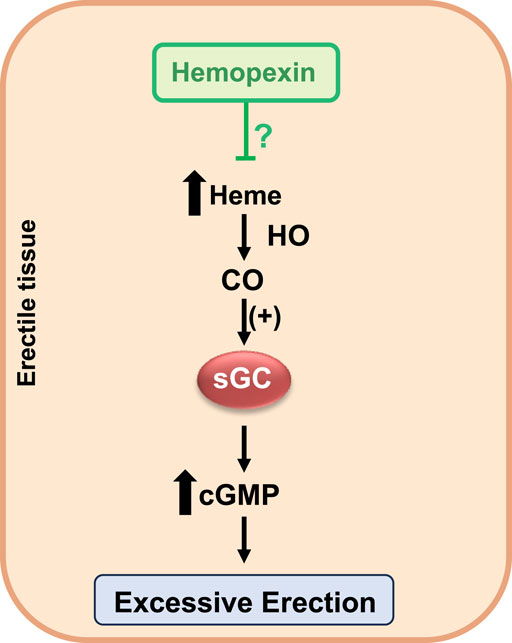
Figure 1. Hemopexin as a candidate for priapism prevention in SCD.
Another strategy to reduce plasma heme levels is through treatment with haptoglobin. Haptoglobin functions by sequestering excess plasma hemoglobin, thereby reducing the release of the heme group from hemoglobin (Buehler et al., 2021). Recently, a study published by our research group demonstrated that haptoglobin treatment reversed the priapism phenotype in SCD mice, characterized by excessive relaxation of the corpus cavernosum mediated by NO-sGC pathway stimulation (Pereira et al., 2022). Additionally, haptoglobin treatment reduced oxidative and nitrosative stress (Pereira et al., 2022).
Considering these findings, haptoglobin and hemopexin offer multifaceted therapeutic strategies that address the root causes of various SCD complications, providing hope for better management and outcomes for patients with this debilitating condition. Future clinical studies must validate these findings and establish haptoglobin and hemopexin as standard treatments for SCD-related complications.
Hydroxyurea, the first drug approved by the FDA in 1998 for treating SCD, has consistently demonstrated significant benefits in reducing the frequency and severity of vaso-occlusive crises and enhancing the overall quality of life for SCD patients (Charache et al., 1995; Tshilolo et al., 2019). The primary mechanism of action of hydroxyurea is to induce the production of fetal hemoglobin (HbF), which inhibits the polymerization of hemoglobin S and, thus, reduces red blood cell sickling and hemolysis (Cokic et al., 2003). Clinical studies have reported that hydroxyurea can decrease markers of intravascular hemolysis, such as plasma hemoglobin levels and lactate dehydrogenase (Chenou et al., 2021). However, hydroxyurea treatment did not reduce the plasma heme concentrations in patients with SCD (Chenou et al., 2021). Few clinical studies have reported the beneficial effects of hydroxyurea treatment in men with SCD (Saad et al., 2004; Anele et al., 2014). A preclinical study reported that hydroxyurea treatment did not alter the priapism phenotype in transgenic SCD mice, suggesting that its effectiveness in preventing priapism through the reduction of hemolysis might be limited (Pereira et al., 2023).
L-glutamine, approved in 2017, has been recognized for its role in reducing pain crises in patients with SCD. L-glutamine plays a critical role in regulating oxidative stress, which is a pivotal contributor to the pathophysiology of SCD (Niihara et al., 2018). A clinical study reported that L-glutamine reduces lactate dehydrogenase levels, a marker of intravascular hemolysis (Elenga et al., 2022). However, the effects of L-glutamine on plasma heme and priapism have yet to be investigated.
Voxelotor, approved in 2019, binds to HbS and inhibits its polymerization, thereby mitigating the sickling of red blood cells and reducing hemolysis (Vichinsky et al., 2013). A clinical study reported that voxelotor treatment increased hemoglobin levels and decreased indirect bilirubin levels in SCD patients, which is a hemolysis-associated biomarker (Vichinsky et al., 2013). However, the effects of voxelotor on plasma heme and its role in priapism have yet to be investigated, representing a critical gap in current SCD treatment research.
Crizanlizumab, approved in 2019, inhibits the P-selectin adhesive pathway, which is activated during inflammation, leading to a reduced frequency of pain crises in patients with SCD (Ataga et al., 2017). A clinical study has reported that crizanlizumab treatment does not alter hemolysis markers such as hemoglobin levels, lactate dehydrogenase, haptoglobin levels, reticulocyte counts, and indirect bilirubin (Ataga et al., 2017). These findings suggest that the observed clinical benefits derived from P-selectin inhibition do not involve a reduction in hemolysis. A clinical trial is underway to evaluate the efficacy and safety of crizanlizumab in SCD patients with priapism (NCT03938454).
Given the pivotal role of heme in the pathophysiology of SCD, it is imperative to design clinical trials that assess the effects of medications on plasma heme levels and priapism. Among the currently approved drugs for SCD, voxelotor shows the most evident effect in reducing hemolysis due to its mechanism of action. However, considering the complex pathophysiology of SCD, the future management of priapism will likely require a combination of therapies rather than solely relying on monotherapy. Future clinical trials should explore the synergy of combining therapies such as hydroxyurea, voxelotor, crizanlizumab, and L-glutamine with new pharmacological agents. Such approaches could greatly enhance treatment efficacy and directly influence the management of priapism, offering a more comprehensive solution for patients suffering from this debilitating complication.
7 Conclusion
Priapism in SCD presents significant clinical challenges due to its complex pathophysiology, demanding urgent and effective treatments to improve the patient’s quality of life. Accumulating evidence underscores the roles of intravascular hemolysis and excess heme in contributing to the priapism phenotype, emphasizing the need for targeted therapeutic strategies. In this context, haptoglobin and hemopexin have emerged as promising agents, showing the potential to mitigate the adverse effects of free heme and playing a crucial role in managing this debilitating complication. Although treatments like hydroxyurea, L-glutamine, voxelotor, and crizanlizumab have demonstrated benefits in reducing pain crises and managing general SCD symptoms, their impacts on plasma heme levels and direct effects on priapism remain less well understood. This identifies a critical gap in the current treatment paradigms and highlights the necessity for continued research and development of combination therapies. Such approaches should aim not only to control hemolysis but also to reduce heme levels, offering a more targeted and comprehensive relief from priapism, thereby potentially transforming patient outcomes in SCD.
Author contributions
TR: writing–original draft and writing–review and editing. FC: writing–original draft and writing–review and editing. MO: writing–original draft and writing–review and editing. FF: writing–original draft and writing–review and editing. FS: conceptualization, writing–original draft, and writing–review and editing.
Funding
The author(s) declare that financial support was received for the research, authorship, and/or publication of this article. This work was supported by the São Paulo Research Foundation (Grant numbers: 2017/08122-9, 2019/18886-1).
Conflict of interest
The authors declare that the research was conducted in the absence of any commercial or financial relationships that could be construed as a potential conflict of interest.
Publisher’s note
All claims expressed in this article are solely those of the authors and do not necessarily represent those of their affiliated organizations, or those of the publisher, the editors, and the reviewers. Any product that may be evaluated in this article, or claim that may be made by its manufacturer, is not guaranteed or endorsed by the publisher.
References
Adeyoju A. B., Olujohungbe A. B. K., Morris J., Yardumian A., Bareford D., Akenova A., et al. (2002). Priapism in sickle-cell disease; incidence, risk factors and complications - an international multicentre study. BJU Int. 90, 898–902. doi:10.1046/j.1464-410x.2002.03022.x
Alvaia M. A., Maia H. A. A. da S., Nelli A. de M., Guimarães C. O. S., Carvalho E. S. de S., Netto J. M. B., et al. (2020). Prevalence of priapism in individuals with sickle cell disease and implications on male sexual function. Einstein (Sao Paulo) 18, eAO5070. doi:10.31744/einstein_journal/2020AO5070
Andersson K.-E. (2011). Mechanisms of penile erection and basis for pharmacological treatment of erectile dysfunction. Pharmacol. Rev. 63, 811–859. doi:10.1124/pr.111.004515
Anele U. A., Le B. V., Resar L. M. S., Burnett A. L. (2015). How I treat priapism. Blood 125, 3551–3558. doi:10.1182/blood-2014-09-551887
Anele U. A., Mack A. K., Resar L. M. S., Burnett A. L. (2014). Hydroxyurea therapy for priapism prevention and erectile function recovery in sickle cell disease: a case report and review of the literature. Int. Urol. Nephrol. 46, 1733–1736. doi:10.1007/s11255-014-0737-7
Arduini G. A. O., Trovó de Marqui A. B. (2018). Prevalence and characteristics of priapism in sickle cell disease. Hemoglobin 42, 73–77. doi:10.1080/03630269.2018.1452760
Ataga K. I., Kutlar A., Kanter J., Liles D., Cancado R., Friedrisch J., et al. (2017). Crizanlizumab for the prevention of pain crises in sickle cell disease. N. Engl. J. Med. 376, 429–439. doi:10.1056/NEJMoa1611770
Belcher J. D., Chen C., Nguyen J., Abdulla F., Zhang P., Nguyen H., et al. (2018a). Haptoglobin and hemopexin inhibit vaso-occlusion and inflammation in murine sickle cell disease: role of heme oxygenase-1 induction. PLoS One 13, e0196455. doi:10.1371/journal.pone.0196455
Belcher J. D., Chen C., Nguyen J., Milbauer L., Abdulla F., Alayash A. I., et al. (2014). Heme triggers TLR4 signaling leading to endothelial cell activation and vaso-occlusion in murine sickle cell disease. Blood 123, 377–390. doi:10.1182/blood-2013-04-495887
Belcher J. D., Gomperts E., Nguyen J., Chen C., Abdulla F., Kiser Z. M., et al. (2018b). Oral carbon monoxide therapy in murine sickle cell disease: beneficial effects on vaso-occlusion, inflammation and anemia. PLoS One 13, e0205194. doi:10.1371/journal.pone.0205194
Bivalacqua T. J., Allen B. K., Brock G. B., Broderick G. A., Chou R., Kohler T. S., et al. (2022). The diagnosis and management of recurrent ischemic priapism, priapism in sickle cell patients, and non-ischemic priapism: an AUA/SMSNA guideline. J. Urol. 208, 43–52. doi:10.1097/JU.0000000000002767
Bivalacqua T. J., Musicki B., Hsu L. L., Berkowitz D. E., Champion H. C., Burnett A. L. (2013). Sildenafil citrate-restored eNOS and PDE5 regulation in sickle cell mouse penis prevents priapism via control of oxidative/nitrosative stress. PLoS One 8, e68028. doi:10.1371/journal.pone.0068028
Bozza M. T., Jeney V. (2020). Pro-inflammatory actions of heme and other hemoglobin-derived DAMPs. Front. Immunol. 11, 1323. doi:10.3389/fimmu.2020.01323
Buehler P. W., Swindle D., Pak D. I., Ferguson S. K., Majka S. M., Karoor V., et al. (2021). Hemopexin dosing improves cardiopulmonary dysfunction in murine sickle cell disease. Free Radic. Biol. Med. 175, 95–107. doi:10.1016/j.freeradbiomed.2021.08.238
Bunn H. F., Jandl J. H. (1968). Exchange of heme among hemoglobins and between hemoglobin and albumin. J. Biol. Chem. 243, 465–475. doi:10.1016/s0021-9258(18)93628-8
Champion H. C., Bivalacqua T. J., Takimoto E., Kass D. A., Burnett A. L. (2005). Phosphodiesterase-5A dysregulation in penile erectile tissue is a mechanism of priapism. PNAS 102, 1661–1666. doi:10.1073/pnas.0407183102
Charache S., Terrin M. L., Moore R. D., Dover G. J., Barton F. B., Eckert S. V., et al. (1995). Effect of hydroxyurea on the frequency of painful crises in sickle cell anemia. Investigators of the Multicenter Study of Hydroxyurea in Sickle Cell Anemia. N. Engl. J. Med. 332, 1317–1322. doi:10.1056/NEJM199505183322001
Chenou F., Hounkpe B. W., Domingos I. de F., Tonassé W. V., Batista T. H. C., Santana R. M., et al. (2021). Effect of hydroxyurea therapy on intravascular hemolysis and endothelial dysfunction markers in sickle cell anemia patients. Ann. Hematol. 100, 2669–2676. doi:10.1007/s00277-021-04636-3
Chiabrando D., Vinchi F., Fiorito V., Mercurio S., Tolosano E. (2014). Heme in pathophysiology: a matter of scavenging, metabolism and trafficking across cell membranes. Front. Pharmacol. 5, 61. doi:10.3389/fphar.2014.00061
Cita K.-C., Brureau L., Lemonne N., Billaud M., Connes P., Ferdinand S., et al. (2016). Men with sickle cell anemia and priapism exhibit increased hemolytic rate, decreased red blood cell deformability and increased red blood cell aggregate strength. PLoS One 11, e0154866. doi:10.1371/journal.pone.0154866
Claudino M. A., Franco-Penteado C. F., Corat M. A. F., Gimenes A. P., Passos L. A. C., Antunes E., et al. (2009). Increased cavernosal relaxations in sickle cell mice priapism are associated with alterations in the NO-cGMP signaling pathway. J. Sex. Med. 6, 2187–2196. doi:10.1111/j.1743-6109.2009.01337.x
Cokic V. P., Smith R. D., Beleslin-Cokic B. B., Njoroge J. M., Miller J. L., Gladwin M. T., et al. (2003). Hydroxyurea induces fetal hemoglobin by the nitric oxide-dependent activation of soluble guanylyl cyclase. J. Clin. Investig. 111, 231–239. doi:10.1172/JCI16672
Corbin J. D., Turko I. V., Beasley A., Francis S. H. (2000). Phosphorylation of phosphodiesterase-5 by cyclic nucleotide-dependent protein kinase alters its catalytic and allosteric cGMP-binding activities. Eur. J. Biochem. 267, 2760–2767. doi:10.1046/j.1432-1327.2000.01297.x
Decaluwé K., Pauwels B., Boydens C., Thoonen R., Buys E. S., Brouckaert P., et al. (2017). Erectile dysfunction in heme-deficient nitric oxide-unresponsive soluble guanylate cyclase knock-in mice. J. Sex. Med. 14, 196–204. doi:10.1016/j.jsxm.2016.12.007
Elenga N., Loko G., Etienne-Julan M., Al-Okka R., Adel A. M., Yassin M. A. (2022). Real-World data on efficacy of L-glutamine in preventing sickle cell disease-related complications in pediatric and adult patients. Front. Med. (Lausanne) 9, 931925. doi:10.3389/fmed.2022.931925
Elliott V., Hoppe C. C., Ataga K. I., Ware Russell E., Videlis N., El-Beshlawy A., et al. (2019). A phase 3 randomized trial of voxelotor in sickle cell disease. N. Engl. J. Med. 381, 509–519. doi:10.1056/NEJMoa1903212
Figueiredo R. T., Fernandez P. L., Mourao-Sa D. S., Porto B. N., Dutra F. F., Alves L. S., et al. (2007). Characterization of heme as activator of Toll-like receptor 4. J. Biol. Chem. 282, 20221–20229. doi:10.1074/jbc.M610737200
Gbotosho O. T., Kapetanaki M. G., Ghosh S., Villanueva F. S., Ofori-Acquah S. F., Kato G. J. (2020). Heme induces IL-6 and cardiac hypertrophy genes transcripts in sickle cell mice. Front. Immunol. 11, 1910. doi:10.3389/fimmu.2020.01910
Gbotosho O. T., Kapetanaki M. G., Kato G. J. (2021). The worst things in life are free: the role of free heme in sickle cell disease. Front. Immunol. 11, 561917. Available at:. doi:10.3389/fimmu.2020.561917
Gentinetta T., Belcher J. D., Brügger-Verdon V., Adam J., Ruthsatz T., Bain J., et al. (2022). Plasma-derived hemopexin as a candidate therapeutic agent for acute vaso-occlusion in sickle cell disease: preclinical evidence. J. Clin. Med. 11, 630. doi:10.3390/jcm11030630
Hosein S., Marks G. S., Brien J. F., McLaughlin B. E., Nakatsu K. (2002). An extracellular source of heme can induce a significant heme oxygenase mediated relaxation in the rat aorta. Can. J. Physiol. Pharmacol. 80, 761–765. doi:10.1139/y02-086
Hvidberg V., Maniecki M. B., Jacobsen C., Højrup P., Møller H. J., Moestrup S. K. (2005). Identification of the receptor scavenging hemopexin-heme complexes. Blood 106, 2572–2579. doi:10.1182/blood-2005-03-1185
Iacopucci A. P. M., da Silva Pereira P., Pereira D. A., Calmasini F. B., Pittalà V., Reis L. O., et al. (2022). Intravascular hemolysis leads to exaggerated corpus cavernosum relaxation: implication for priapism in sickle cell disease. FASEB J. 36, e22535. doi:10.1096/fj.202200867R
Kapetanaki M. G., Gbotosho O. T., Sharma D., Weidert F., Ofori-Acquah S. F., Kato G. J. (2019). Free heme regulates Placenta Growth Factor through NRF2-antioxidant response signaling. Free Radic. Biol. Med. 143, 300–308. doi:10.1016/j.freeradbiomed.2019.08.009
Kato G. J., McGowan V., Machado R. F., Little J. A., Taylor J., Morris C. R., et al. (2006). Lactate dehydrogenase as a biomarker of hemolysis-associated nitric oxide resistance, priapism, leg ulceration, pulmonary hypertension, and death in patients with sickle cell disease. Blood 107, 2279–2285. doi:10.1182/blood-2005-06-2373
Kato G. J., Piel F. B., Reid C. D., Gaston M. H., Ohene-Frempong K., Krishnamurti L., et al. (2018). Sickle cell disease. Nat. Rev. Dis. Prim. 4, 18010. doi:10.1038/nrdp.2018.10
Kavanagh P. L., Fasipe T. A., Wun T. (2022). Sickle cell disease: a review. JAMA 328, 57–68. doi:10.1001/jama.2022.10233
Kozma F., Johnson R. A., Nasjletti A. (1997). Role of carbon monoxide in heme-induced vasodilation. Eur. J. Pharmacol. 323, R1–R2. doi:10.1016/s0014-2999(97)00145-3
Lagoda G., Sezen S. F., Hurt K. J., Cabrini M. R., Mohanty D. K., Burnett A. L. (2014). Sustained nitric oxide (NO)-releasing compound reverses dysregulated NO signal transduction in priapism. FASEB J. 28, 76–84. doi:10.1096/fj.13-228817
Lin C.-S., Chow S., Lau A., Tu R., Lue T. F. (2002). Human PDE5A gene encodes three PDE5 isoforms from two alternate promoters. Int. J. Impot. Res. 14, 15–24. doi:10.1038/sj.ijir.3900802
Lu W., Yang X., Wang B. (2022). Carbon monoxide signaling and soluble guanylyl cyclase: facts, myths, and intriguing possibilities. Biochem. Pharmacol. 200, 115041. doi:10.1016/j.bcp.2022.115041
MacDonald S. M., Burnett A. L. (2021). Physiology of erection and pathophysiology of erectile dysfunction. Urol. Clin. North Am. 48, 513–525. doi:10.1016/j.ucl.2021.06.009
Mantadakis E., Cavender J. D., Rogers Z. R., Ewalt D. H., Buchanan G. R. (1999). Prevalence of priapism in children and adolescents with sickle cell anemia. J. Pediatr. Hematol. Oncol. 21, 518–522. doi:10.1097/00043426-199911000-00013
Menon A. V., Liu J., Tsai H. P., Zeng L., Yang S., Asnani A., et al. (2022). Excess heme upregulates heme oxygenase 1 and promotes cardiac ferroptosis in mice with sickle cell disease. Blood 139, 936–941. doi:10.1182/blood.2020008455
Mi T., Abbasi S., Zhang H., Uray K., Chunn J. L., Xia L. W., et al. (2008). Excess adenosine in murine penile erectile tissues contributes to priapism via A2B adenosine receptor signaling. J. Clin. Investigation 118, 1491–1501. doi:10.1172/JCI33467
Muller-Eberhard U., Javid J., Liem H. H., Hanstein A., Hanna M. (1968). Brief report: plasma concentrations of hemopexin, haptoglobin and heme in patients with various hemolytic diseases. Blood 32, 811–815. doi:10.1182/blood.v32.5.811.811
Musicki B., Burnett A. L. (2020). Mechanisms underlying priapism in sickle cell disease: targeting and key innovations on the preclinical landscape. Expert Opin. Ther. Targets 24, 439–450. doi:10.1080/14728222.2020.1745188
Musicki B., Karakus S., Akakpo W., Silva F. H., Liu J., Chen H., et al. (2018). Testosterone replacement in transgenic sickle cell mice controls priapic activity and upregulates PDE5 expression and eNOS activity in the penis. Andrology 6, 184–191. doi:10.1111/andr.12442
Musicki B., Karakus S., La Favor J. D., Chen H., Silva F. H., Sturny M., et al. (2020). TSPO ligand FGIN-1-27 controls priapism in sickle cell mice via endogenous testosterone production. J. Cell. Physiol. 236, 3073–3082. doi:10.1002/jcp.30075
Nath K. A., Belcher J. D., Nath M. C., Grande J. P., Croatt A. J., Ackerman A. W., et al. (2018). Role of TLR4 signaling in the nephrotoxicity of heme and heme proteins. Am. J. Physiol. Ren. Physiol. 314, F906-F914–F914. doi:10.1152/ajprenal.00432.2017
Niihara Y., Miller S. T., Kanter J., Lanzkron S., Smith W. R., Hsu L. L., et al. (2018). A phase 3 trial of l-glutamine in sickle cell disease. N. Engl. J. Med. 379, 226–235. doi:10.1056/NEJMoa1715971
Nolan V. G., Wyszynski D. F., Farrer L. A., Steinberg M. H. (2005). Hemolysis-associated priapism in sickle cell disease. Blood 106, 3264–3267. doi:10.1182/blood-2005-04-1594
Ofori-Acquah S. F., Hazra R., Orikogbo O. O., Crosby D., Flage B., Ackah E. B., et al. (2020). Hemopexin deficiency promotes acute kidney injury in sickle cell disease. Blood 135, 1044–1048. doi:10.1182/blood.2019002653
Pereira D. A., Calmasini F. B., Costa F. F., Burnett A. L., Silva F. H. (2024a). Nitric oxide resistance in priapism associated with sickle cell disease: mechanisms, therapeutic challenges, and future directions. J. Pharmacol. Exp. Ther., JPET-MR–2023-001962. JPET-MR-2023-001962. doi:10.1124/jpet.123.001962
Pereira D. A., Pereira D. A., Pereira P. da S., Silveira T. H. R., Calmasini F. B., Reis L. O., et al. (2023). Hydroxyurea does not reverse functional alterations of the nitric oxide-cGMP pathway associated with priapism phenotype in corpus cavernosum from sickle cell mouse. PLOS ONE 18, e0292706. doi:10.1371/journal.pone.0292706
Pereira D. A., Pereira D. A., Silveira T. H. R., Calmasini F. B., Burnett A. L., Costa F. F., et al. (2024b). Heme-induced corpus cavernosum relaxation and its implications for priapism in sickle cell disease: a mechanistic insight. Andrology. doi:10.1111/andr.13599
Pereira P. da S., Pereira D. A., Calmasini F. B., Reis L. O., Brinkman N., Burnett A. L., et al. (2022). Haptoglobin treatment contributes to regulating nitric oxide signal and reduces oxidative stress in the penis: a preventive treatment for priapism in sickle cell disease. Front. Physiol. 13, 961534. doi:10.3389/fphys.2022.961534
Pinheiro A. K., Pereira D. A., Dos Santos J. L., Calmasini F. B., Alexandre E. C., Reis L. O., et al. (2022). Resveratrol-nitric oxide donor hybrid effect on priapism in sickle cell and nitric oxide-deficient mouse. PLoS One 17, e0269310. doi:10.1371/journal.pone.0269310
Reiter C. D., Wang X., Tanus-Santos J. E., Hogg N., Cannon R. O., Schechter A. N., et al. (2002). Cell-free hemoglobin limits nitric oxide bioavailability in sickle-cell disease. Nat. Med. 8, 1383–1389. doi:10.1038/nm1202-799
Rubio-Navarro A., Vázquez-Carballo C., Guerrero-Hue M., García-Caballero C., Herencia C., Gutiérrez E., et al. (2019). Nrf2 plays a protective role against intravascular hemolysis-mediated acute kidney injury. Front. Pharmacol. 10, 740. doi:10.3389/fphar.2019.00740
Saad S. T. O., Lajolo C., Gilli S., Marques Júnior J. F. C., Lima C. S., Costa F. F., et al. (2004). Follow-up of sickle cell disease patients with priapism treated by hydroxyurea. Am. J. Hematol. 77, 45–49. doi:10.1002/ajh.20142
Santiago R. P., Guarda C. C., Figueiredo C. V. B., Fiuza L. M., Aleluia M. M., Adanho C. S. A., et al. (2018). Serum haptoglobin and hemopexin levels are depleted in pediatric sickle cell disease patients. Blood Cells Mol. Dis. 72, 34–36. doi:10.1016/j.bcmd.2018.07.002
Schaer D. J., Buehler P. W., Alayash A. I., Belcher J. D., Vercellotti G. M. (2013). Hemolysis and free hemoglobin revisited: exploring hemoglobin and hemin scavengers as a novel class of therapeutic proteins. Blood 121, 1276–1284. doi:10.1182/blood-2012-11-451229
Silva F. H., Claudino M. A., Calmasini F. B., Alexandre E. C., Franco-Penteado C., Burnett A. L., et al. (2016a). Sympathetic hyperactivity, increased tyrosine hydroxylase and exaggerated corpus cavernosum relaxations associated with oxidative stress plays a major role in the penis dysfunction in Townes sickle cell mouse. PLoS ONE 11, e0166291. doi:10.1371/journal.pone.0166291
Silva F. H., Karakus S., Musicki B., Matsui H., Bivalacqua T. J., Dos Santos J. L., et al. (2016b). Beneficial effect of the nitric oxide donor compound 3-(1,3-dioxoisoindolin-2-yl)benzyl nitrate on dysregulated phosphodiesterase 5, NADPH oxidase and nitrosative stress in the sickle cell mouse penis: implication for priapism treatment. J. Pharmacol. Exp. Ther. 359, 230–237. doi:10.1124/jpet.116.235473
Smith A., Morgan W. T. (1979). Haem transport to the liver by haemopexin. Receptor-mediated uptake with recycling of the protein. Biochem. J. 182, 47–54. doi:10.1042/bj1820047
Stec D. E., Drummond H. A., Vera T. (2008). Role of carbon monoxide in blood pressure regulation. Hypertension 51, 597–604. doi:10.1161/HYPERTENSIONAHA.107.097154
Stone J. R., Marletta M. A. (1994). Soluble guanylate cyclase from bovine lung: activation with nitric oxide and carbon monoxide and spectral characterization of the ferrous and ferric states. Biochemistry 33, 5636–5640. doi:10.1021/bi00184a036
Sundaram N., Tailor A., Mendelsohn L., Wansapura J., Wang X., Higashimoto T., et al. (2010). High levels of placenta growth factor in sickle cell disease promote pulmonary hypertension. Blood 116, 109–112. doi:10.1182/blood-2009-09-244830
Thomsen J. H., Etzerodt A., Svendsen P., Moestrup S. K. (2013). The haptoglobin-CD163-heme oxygenase-1 pathway for hemoglobin scavenging. Oxid. Med. Cell. Longev. 2013, 523652. doi:10.1155/2013/523652
Tshilolo L., Tomlinson G., Williams T. N., Santos B., Olupot-Olupot P., Lane A., et al. (2019). Hydroxyurea for children with sickle cell anemia in sub-saharan africa. N. Engl. J. Med. 380, 121–131. doi:10.1056/NEJMoa1813598
Ushiyama M., Morita T., Kuramochi T., Yagi S., Katayama S. (2004). Erectile dysfunction in hypertensive rats results from impairment of the relaxation evoked by neurogenic carbon monoxide and nitric oxide. Hypertens. Res. 27, 253–261. doi:10.1291/hypres.27.253
Vallelian F., Buehler P. W., Schaer D. J. (2022). Hemolysis, free hemoglobin toxicity, and scavenger protein therapeutics. Blood 140, 1837–1844. doi:10.1182/blood.2022015596
Vinchi F., De Franceschi L., Ghigo A., Townes T., Cimino J., Silengo L., et al. (2013). Hemopexin therapy improves cardiovascular function by preventing heme-induced endothelial toxicity in mouse models of hemolytic diseases. Circulation 127, 1317–1329. doi:10.1161/CIRCULATIONAHA.112.130179
Keywords: corpus cavernosum, heme, heme oxygenase, hemopexin, haptoglobin
Citation: Silveira THR, Calmasini FB, de Oliveira MG, Costa FF and Silva FH (2024) Targeting heme in sickle cell disease: new perspectives on priapism treatment. Front. Physiol. 15:1435220. doi: 10.3389/fphys.2024.1435220
Received: 19 May 2024; Accepted: 26 June 2024;
Published: 17 July 2024.
Edited by:
Tiago L. Duarte, Universidade do Porto, PortugalReviewed by:
David Archer, Emory University, United StatesCopyright © 2024 Silveira, Calmasini, de Oliveira, Costa and Silva. This is an open-access article distributed under the terms of the Creative Commons Attribution License (CC BY). The use, distribution or reproduction in other forums is permitted, provided the original author(s) and the copyright owner(s) are credited and that the original publication in this journal is cited, in accordance with accepted academic practice. No use, distribution or reproduction is permitted which does not comply with these terms.
*Correspondence: Fábio Henrique Silva, ZmFiaW9oc2lsdmE4N0BnbWFpbC5jb20=