 Timon Ax1,2*
 Bergita Ganse3,4
 Fabian N. Fries1,5
 Nóra Szentmáry5
 Cintia S. de Paiva6
 Francesc March de Ribot7
 Slade O. Jensen2,8
 Berthold Seitz1
 Thomas J. Millar9
- 1Department of Ophthalmology, Saarland University Medical Center, Homburg/Saar, Germany
- 2School of Medicine, Western Sydney University, Sydney, NSW, Australia
- 3Werner Siemens-Endowed Chair for Innovative Implant Development (Fracture Healing), Departments and Institutes of Surgery, Saarland University, Homburg/Saar, Germany
- 4Department of Trauma, Hand and Reconstructive Surgery, Departments and Institutes of Surgery, Saarland University, Homburg/Saar, Germany
- 5Dr. Rolf M. Schwiete Center for Limbal Stem Cell and Aniridia Research, Saarland University, Homburg/Saar, Germany
- 6Ocular Surface Center, Department of Ophthalmology, Baylor College of Medicine, Cullen Eye Institute, Houston, TX, United States
- 7Department of Ophthalmology, Otago University, Dunedin, New Zealand
- 8Antimicrobial Resistance and Mobile Elements Group, Ingham Institute of Applied Medical Research, Sydney, NSW, Australia
- 9Beyond 700 Pty Ltd, Sydney, NSW, Australia
Long-duration spaceflight can have adverse effects on human health. One of the most common ocular conditions experienced by astronauts is dry eye disease (DED). Symptoms of DED include feelings of eye irritation, eye strain, foreign body sensation and blurred vision. Over 30% of International Space Station expedition crew members reported irritation and foreign body sensation. We reviewed the current literature on the prevalence and mechanisms of DED in astronauts and its potential implications for long-duration spaceflight, including the influence of environmental factors, such as microgravity and fluid shift on tear film physiology in space. DED has negative effects on astronaut performance, which is why there is a need for further research into the pathophysiology and countermeasures. As an in-flight countermeasure, neurostimulation seems to be among the most promising options.
1 Introduction
Upon return to Earth, over 30% of International Space Station (ISS) expedition crew members and approximately 15% of shuttle crew members reported experiencing signs or symptoms of dry eye disease (DED) while in space (Gibson and Duncan, 2009; Reschke et al., 2016). DED is an umbrella term encompassing a variety of diseases affecting the ocular surface with tear film dysfunction and inflammation as their shared feature (Aggarwal and Galor, 2018). Common symptoms include eye strain, burning or stinging, grittiness, itchiness, redness, sensitivity to light, and blurry and reduced vision, all of which are frequently reported by astronauts (Szczotka-Flynn et al., 2019; Meer et al., 2023). In the US, a recent meta-analysis estimated a DED prevalence of 8.1% for the entire population (McCann et al., 2022). Given the medical testing and excellent health of spaceflight participants prior to flights, the high incidence of DED might be related to the microgravity exposure. Sleep and mood disorders associated with DED have been reported on Earth (Ayaki et al., 2016; Kitazawa et al., 2018; Zhou et al., 2022), and are also both known to affect crew performance (Wu et al., 2018; Stuster, 2021). Thus, on long-duration flights, DED will need to be addressed because visual acuity and comfort are critical for performing space-related tasks. However, the pathophysiology of DED in microgravity and possible countermeasures are unknown. Despite its high incidence and operational relevance, DED was not mentioned in the latest consensus paper concerning the directions to identify risks and mitigation strategies for ocular changes in human spaceflight (Seidler et al., 2022). For these reasons, the aim of the present review paper was to summarize the current evidence for the clinical relevance, pathophysiology, and possible countermeasures of DED in microgravity.
2 Physiology of the tear film
The tear film is a thin fluid film covering the cornea and bulbar conjunctiva. The three main tear film components are mucins, water, and lipids (Yokoi et al., 2014). The mucins are embedded in the apical membrane of ocular surface cells and are also secreted into the tear film by specialized conjunctival goblet cells (Spurr-Michaud et al., 2007; Hodges and Dartt, 2013). The aqueous portion of the tear film comprising a mixture of proteins, water, and ions is secreted by the lacrimal glands (Green-Church et al., 2008) and the lipids are secreted by glands (Meibomian glands) in the eyelids (Bron et al., 2004). Besides maintaining sharp vision, the tear film keeps the eye healthy by providing microbial protection and nutrients (McDermott, 2013).
3 Tear film dynamics and effects of microgravity
3.1 Tear film structure
In the scientific community, current consensus is that the tear film should be regarded as a whole, rather than three separate layers (Georgiev et al., 2019). Together, the three layers combined enable the final tear film to be only ∼ 3 µm thick, and to cover a relatively vast area (∼2 cm2; or ∼2 × 108µm2) without collapsing (Tsubota and Nakamori, 1995). This means that molecular rather than gravitational forces dominate its spread and stability (Jones et al., 2005). However, gravity is likely to have a direct impact on the tear menisci, i.e., the thin bands of tear fluid between the eyeball and lid margins. Gravitational effects cause a steepened curvature and lower tear volume in the upper meniscus and vice versa in the lower meniscus (Su et al., 2015; Maki et al., 2020). It is, however, unclear whether changes in tear film components in space are responsible for the DED complaints reported by astronauts. Neither tear film sampling of astronauts, nor analyses of tear film spread have been performed to date.
3.2 Blinking
Blinking is a cycle of movement that helps to distribute and remove the tear film. The opening part of the blink cycle is responsible for spreading the aqueous and meibum (lipid) to form an even tear film over the eye (Harrison et al., 2008). The closing phase of a blink (∼100 ms) is faster than the opening phase (∼200 ms) (Kwon et al., 2013; Mak et al., 2016; Rodriguez et al., 2018; McMonnies, 2021). Tear film spreading is completed in ∼1 s after cessation of the upstroke of the blink (Ring et al., 2012). During the open-eye part of the blink cycle, the tear film evidently forms a gel and becomes isolated from the reservoir of tears in the upper and lower tear menisci (Miller et al., 2002; Yokoi et al., 2014; Georgiev et al., 2019). Lubricity between the eyelid and ocular surface in part is maintained by specialized stratified epithelium that covers the lid margin at the point of closest contact (lid wiper). Changes to this region of the eyelid have been associated with signs of poor tear film performance (Korb et al., 2005).
The fast-closing part of the blink cycle is needed to fluidize the tear film because it shows non-Newtonian viscosity i.e., the higher the shear force the less viscous the tears (Pult et al., 2015b; Arshinoff et al., 2021; Recchioni et al., 2022). Blinking also cleans the ocular surface by removing tears via the tear menisci into the puncta, lacrimal canaliculi and lacrimal sac (McDonald and Brubaker, 1971). Dust and microbe removal from the eye surface is likely to be even more important in microgravity where large dust particles and dense objects such as metal filings do not settle (Haines et al., 2019; Jahn et al., 2021). In fact, astronauts commonly report corneal foreign bodies (Meer et al., 2023).
Blink speed, frequency and fullness contribute to normal tear film formation. Studying the nature of blinking is difficult because of differences in defining a complete/full blink (Collins et al., 2006; Pult et al., 2013; Wang et al., 2018) and blink frequency varies with alertness, conversation, the nature of visual task being undertaken, mood, sex, and ethnicity (Yolton et al., 1994; Tsubota et al., 2002; Himebaugh et al., 2009; Wang and Craig, 2019). Natural blinking changes upon conscious command, and the characteristics of purposeful blinking are very individual (McMonnies, 2020). Eyelid contact in full blinks compresses the Meibomian glands, increases secretion and spreads oils onto the eyelid margins (Korb et al., 1994). Nevertheless, as little as 10% of blinks need to have the lids fully touching along their length (full in amplitude), to maintain a stable tear film (Shew et al., 2022). Mathematical and analog models of blinking have not been able to predict all the clinical observations (Braun and Fitt, 2003; Pult et al., 2015a). While it is known that being in microgravity significantly affects gaze behavior via the vestibular system, blink behavior has not yet been studied (Clément, 2011; Reschke et al., 2016). Given the above, we hypothesize that if blinking remains normal the tear film should form properly in microgravity.
3.3 The effect of cephalad fluid shift (CFS) on the tear film
In microgravity, fluid redistributes from the lower half of the body to the head causing tissue swelling and increases in intraocular and cranial pressure (Nelson et al., 2014). The effects of cephalad fluid shift (CFS) have been well studied in relation to retinal thickness, axial length, anterior chamber depth, and refraction, and the changes to eye health have been labeled as spaceflight-associated neuro-ocular syndrome (SANS) (Lee et al., 2020; Macias et al., 2020). CFS also causes swelling of the eyelids and an upward shift in eyebrow position (Schneider et al., 2016; Karlin et al., 2021). Thereby, the nasolacrimal drainage apparatus is affected. Swelling of the eyelids may change the pressure that they exert onto the ocular surface (lid wiper), and the size and shape of the tear menisci; both could influence the spread of the tear film (Yamamoto et al., 2016; van Setten, 2020). Swelling will affect tear drainage because normally in the open eye, the upper and lower canaliculi are relaxed and fill with aqueous. The common canaliculus is constricted and then, during a blink, the aqueous flows into the common canaliculus because the upper and lower canaliculi constrict and the common canaliculus relaxes (Kakizaki et al., 2013). From there, the tears flow into the lacrimal sac which is surrounded by a cavernous body. It is rich in a vascular plexus that is important for regulating tear-outflow by dilating and constricting (Paulsen et al., 2000; Ayub et al., 2003). CFS is expected to change the vascular plexus of the cavernous body resulting in functional nasolacrimal duct obstruction and tear retention. Tear drainage from the lacrimal sac to the inferior meatus of the nose is also affected by gravity (Chavis et al., 1978; Sahlin and Chen, 1997). Insufficient drainage means the tear film is not replaced leading to the accumulation of toxins and inflammatory agents (Pflugfelder et al., 2002; de Paiva and Pflugfelder, 2004). Partial gravity (e.g., Moon or Mars gravity) is expected to induce similar changes but to a lesser degree.
4 Evaluation of tear film problems in space
Diagnosing tear film disorders on Earth has proven to be cumbersome because the tear film is complex, invisible, and small in volume. Sampling and bright light causes reflex tearing, and there is discordance between signs and symptoms of DED (Behrens et al., 2006; Sullivan et al., 2014; Bartlett et al., 2015; Wolffsohn et al., 2017), e.g., symptoms have been associated with non-ocular pain, depression, and post-traumatic stress disorder (Galor et al., 2015). Moreover, different clinics vary in their cut-off values to distinguish a normal tear film from an unhealthy one. Consequently, diagnosis is often uncertain and inconsistent. To minimize this problem, the TFOS DEWS II (Dry Eye Workshop) expert panel recommended an initial questionnaire, followed by tests for any one of three possible markers of a loss of tear film homeostasis (tear film stability, hyperosmolarity or corneal staining). Some examples are non-invasive breakup time (NIBUT), osmolarity, and ocular surface staining with fluorescein and lissamine green (Wolffsohn et al., 2017). A slit lamp is used for many of these tests, and it can be used for evaluating other ocular tissues (cornea, iris, lens, retina). However, the availability of diagnostic and treatment tools will be limited in long-haul spaceflights due to payload (volume and mass) and opportunities to replace diagnostic products or treatments are scarce.
The current diagnostic equipment on the ISS includes an ophthalmoscope with downlink video function as well as a blue light filter and fluorescein strips. A slit lamp is not available, but astronauts are trained to use an ophthalmoscope to perform a complete primary ocular examination with lid eversion. Fluid shift may confound the diagnosis due to congestion of conjunctival blood vessels, causing “red eye” (Marshburn et al., 2019). Installing more advanced ocular surface diagnosis technology on the ISS or on future spaceships and stations would present a challenge. Automated NIBUT (non-invasive tear breakup time), tear film interferometry and osmolarity test devices are too bulky or use disposables and are therefore unsuitable for being part of the payload. Rather, small, multipurpose systems and self-administered testing should be considered. Infrared imaging, for example, has shown potential as a non-invasive and disposable-free diagnostic tool and has already been used on the ISS for other purposes (Howell et al., 2008; Shah and Galor, 2021). Screen and smartphone-based tests for blinking and evaluating the eye are developing rapidly, and some of these could be considered as they become available (Wolffsohn et al., 2018; Okumura et al., 2022).
5 Treatment of tear film problems in space
Eyedrops are the most common form of treatment for tear problems on Earth (Jones et al., 2017). These will be inappropriate for long-haul spaceflights because of payload and use-by dates. The application of eyedrops in microgravity would also be a major issue because “dropping” them into the eye is challenging. The fluid must be “wicked” into the eye using surface tension forces by making direct contact with the dropper bottle, thus contaminating the bottle in the process. Ointments and gels are easier to apply and are currently in use on the ISS for eye infections but would impair vision when used for lubrication. Therefore, other methods related to restoring the physiology of the tear film should be used. One possible option is ocular neurostimulation. While early devices used electricity and disposables (TrueTear, Allergan Inc.), current technology (iTear100, Olympic Ophthalmics Inc.; Figure 1) externally stimulates the nasociliary nerve through vibration to immediately induce lacrimation and potentially Meibomian gland secretion (Farhangi et al., 2019; Ji et al., 2020). This means that one device could be used by the entire crew. Other “low tech” treatments which have proven effective on Earth are blink exercises, heating and massage of Meibomian glands, and cleaning the area around the eyes (Bitton et al., 2019; McMonnies, 2021). However, unlike neurostimulation these need to be repeated on a regular basis to start providing relief.
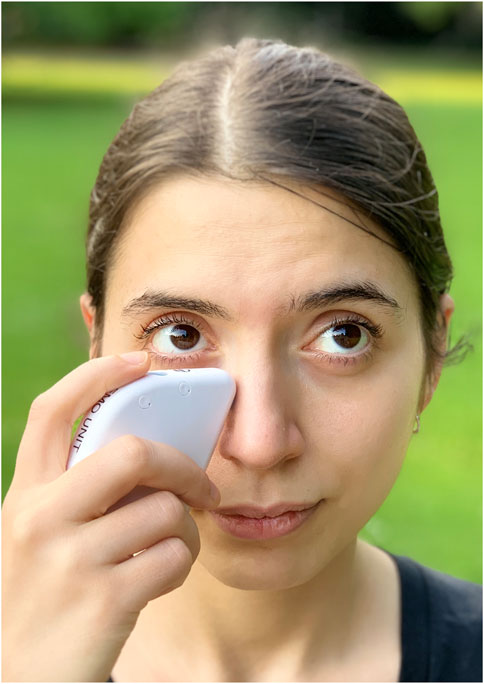
FIGURE 1. Application of iTear100 neurostimulation system.
6 Environmental hazards associated with space travel
The interior of spacecraft and space stations are likely to have an impact on the tear film. This is because of strong air currents (air exchanged 45–77 times per hour in crew quarters) coupled with a relative humidity of about 60% (ISS), increased CO2 exposure, artificial lighting, cosmic radiation, and extensive screen work (Gamus et al., 1994; Scull et al., 1998; Uchino et al., 2013; Stenger et al., 2017; Georgescu et al., 2021) which increase tear film evaporation and inflammation of the ocular surface. These factors need to be addressed to minimize the risk of ocular surface damage.
Astronauts are either inside a spaceship or space station, or in special extravehicular activity (EVA) suits with no opportunity to move to a different environment. That way, they are constantly exposed to aerosols and bioaerosols (airborne particles containing microbes such as bacteria, fungi, and viruses) which accumulate in the cabin (Harrad et al., 2023). Larger particles which would settle out on Earth also harbor higher concentrations of microbes (Haines et al., 2019). Furthermore, there is research pointing towards alterations of virulence of pathogens in microgravity (Velez Justiniano et al., 2023).
On the ISS, high-efficiency particle air filters remove particles with a minimum efficiency of 99.97%, which diminishes the chance of these particles entering astronauts’ eyes. The ISS also has eye-wash devices available to flush debris from the eye (James and Ryder, 2019).
On Earth, extensive use of visual display terminals (VDT), such as computer screens, is a major cause of DED. VDT use adversely affects blinking and Meibomian glands, NIBUT, ocular comfort and causes signs of ocular surface damage (Fjaervoll et al., 2022). Breaks from using screens and blink exercises could provide relief (Wolffsohn et al., 2023).
Sleep is needed for the ocular surface to regenerate (Li et al., 2022). Lack of sleep or poor sleep quality are risk factors of DED (Ayaki et al., 2016; Galor et al., 2023). Astronauts often report difficulty falling asleep and staying asleep, and disruptions to their circadian rhythms. This is mainly due to the lack of a natural day-night cycle, as well as the noise and lighting conditions (especially blue light). To mitigate these issues, astronauts typically follow a carefully designed sleep schedule and may use sleep aids such as eye masks and earplugs (Allen et al., 2016; Barger et al., 2020). Recently, dynamic solid-state lighting was installed on the ISS as a countermeasure for circadian misalignment and sleep disruption (Brainard et al., 2016; Rahman et al., 2022).
In addition to sleep disorders, behavioral and mood disorders occur with increasing mission length (Clement et al., 2020). They are associated with dry eye and vice versa, though the exact mechanism is yet to be determined. One possible mechanism is that symptoms of DED affect the daily lives of patients, thus affecting their mood (An and Kim, 2022; Tsai et al., 2022; Galor et al., 2023). The management of mood disorders in space is complex and ranges from astronaut selection as a preventative measure to in-flight private videoconferences with behavioral health specialists (Sipes et al., 2021).
7 Discussion
Current evidence suggests that DED will be an issue during long-haul spaceflights, and it will need to be addressed to prevent potential consequences for astronaut health and performance. Experience from ISS crew indicates that ∼30% of astronauts are affected (Reschke et al., 2016), and this warrants systematic investigation that includes the type of DED afflicting astronauts, identification of factors contributing to it, and modes of treatment suitable for space travel. Blinking behavior and tear film dynamics could be studied on a parabolic flight to uncover immediate effects of microgravity. Long-term effects, on the other hand, could be investigated by performing tear film sampling on astronauts in addition to blink and tear film tests.
With NASA planning a return to the Moon as soon as 2024, the potential dangers of the lunar environment need to be accounted for (Smith et al., 2020). Extraterrestrial dust is a potential threat, e.g., acute exposure to lunar dust causes ocular irritation (Meyers et al., 2012) but is not directly toxic. Lunar dust tends to be especially jagged due to the absence of weathering (James and Kahn-Mayberry, 2011; Pohlen et al., 2022; Miranda et al., 2023). In the Apollo missions, numerous astronauts reported ocular erythematous and watery eyes with decreased vision after exposure to lunar dust following its introduction to the lunar module upon return from EVAs (Scheuring et al., 2008). Martian dust is potentially a bigger threat to ocular health because it contains reactive perchlorates (Davila et al., 2013; Crotts, 2014).
Goggles could be worn by astronauts when acute exposure is expected. Once exposed, while eye irrigation would help, repeated irrigation, e.g., after every EVA, would most likely remove the protective mucin cover of the ocular surface increasing the risk of DED and infection (Naderi et al., 2020). However, 1 month of daily eye irrigation with a commercial eye wash solution containing vitamins, chondroitin sulfate and other supplements did not show detrimental effects (Yazu et al., 2019). It is yet to be determined if repeated flushing with recycled water as used in the ISS would have any detrimental effects on eye health (Straub et al., 2013). Alternatively, neurostimulation (see above) to promote tearing and natural flushing would be a possibility worth investigating.
In conclusion, there are still significant knowledge gaps when it comes to ocular surface health in space. These should be addressed by future research.
Author contributions
TA: Writing–original draft. BG: Writing–review and editing. FF: Writing–review and editing. NS: Writing–review and editing. CP: Writing–review and editing. FR: Writing–review and editing. SJ: Writing–review and editing. BS: Writing–review and editing. TM: Writing–original draft.
Funding
The authors declare that no financial support was received for the research, authorship, and/or publication of this article.
Conflict of interest
Author TM is employed by Beyond 700 Pty Ltd.
The remaining authors declare that the research was conducted in the absence of any commercial or financial relationships that could be construed as a potential conflict of interest.
Publisher’s note
All claims expressed in this article are solely those of the authors and do not necessarily represent those of their affiliated organizations, or those of the publisher, the editors and the reviewers. Any product that may be evaluated in this article, or claim that may be made by its manufacturer, is not guaranteed or endorsed by the publisher.
References
Aggarwal S., Galor A. (2018). What's new in dry eye disease diagnosis? Curr. Adv. challenges 7, F1000 Faculty Rev-1952. doi:10.12688/f1000research.16468.1
Allen C. S., Danielson R. W., Allen J. R. (2016). “Acoustics and audition,” in Space physiology and medicine, 169–196.
An Y., Kim H. (2022). Sleep disorders, mental health, and dry eye disease in South Korea. Sci. Rep. 12 (1), 11046. doi:10.1038/s41598-022-14167-0
Arshinoff A., Hofmann I., Nae H. (2021). Role of rheology in tears and artificial tears. J. Cataract. Refract Surg. 47 (5), 655–661. doi:10.1097/j.jcrs.0000000000000508
Ayub M., Thale A. B., Hedderich J., Tillmann B. N., Paulsen F. P. (2003). The cavernous body of the human efferent tear ducts contributes to regulation of tear outflow. Invest. Ophthalmol. Vis. Sci. 44 (11), 4900–4907. doi:10.1167/iovs.03-0493
Ayaki M., Kawashima M., Negishi K., Kishimoto T., Mimura M., Tsubota K. (2016). Sleep and mood disorders in dry eye disease and allied irritating ocular diseases. Sci. Rep. 6, 22480. doi:10.1038/srep22480
Barger L. K., Dinges D. F., Czeisler C. A. (2020). “Sleep and circadian effects of space,” in Handbook of bioastronautics. Editors L. R. Young,, and J. P. Sutton (Cham: Springer), 1–7.
Bartlett J. D., Keith M. S., Sudharshan L., Snedecor S. J. (2015). Associations between signs and symptoms of dry eye disease: a systematic review. Clin. Ophthalmol. 9, 1719–1730. doi:10.2147/OPTH.S89700
Behrens A., Doyle J. J., Stern L., Chuck R. S., McDonnell P. J., Azar D. T., et al. (2006). Dysfunctional tear syndrome: a Delphi approach to treatment recommendations. Cornea 25 (8), 900–907. doi:10.1097/01.ico.0000214802.40313.fa
Bitton E., Ngo W., Dupont P. (2019). Eyelid hygiene products: a scoping review. Cont. Lens Anterior Eye 42 (6), 591–597. doi:10.1016/j.clae.2019.09.008
Brainard G. C., Barger L. K., Soler R. R., Hanifin J. P. (2016). The development of lighting countermeasures for sleep disruption and circadian misalignment during spaceflight. Curr. Opin. Pulm. Med. 22 (6), 535–544. doi:10.1097/MCP.0000000000000329
Braun R. J., Fitt A. D. (2003). Modelling drainage of the precorneal tear film after a blink. Math. Med. Biol. 20 (1), 1–28. doi:10.1093/imammb/20.1.1
Bron A. J., Tiffany J. M., Gouveia S. M., Yokoi N., Voon L. W. (2004). Functional aspects of the tear film lipid layer. Exp. Eye Res. 78 (3), 347–360. doi:10.1016/j.exer.2003.09.019
Chavis R. M., Welham R. A., Maisey M. N. (1978). Quantitative lacrimal scintillography. Arch. Ophthalmol. 96 (11), 2066–2068. doi:10.1001/archopht.1978.03910060454013
Clement G. R., Boyle R. D., George K. A., Nelson G. A., Reschke M. F., Williams T. J., et al. (2020). Challenges to the central nervous system during human spaceflight missions to Mars. J. Neurophysiol. 123 (5), 2037–2063. doi:10.1152/jn.00476.2019
Clément G. (2011). “The neuro-sensory system in space,” in Fundamentals of space medicine (New York, NY: Springer), 95–142.
Collins M. J., Iskander D. R., Saunders A., Hook S., Anthony E., Gillon R. (2006). Blinking patterns and corneal staining. Eye Contact Lens 32 (6), 287–293. doi:10.1097/01.icl.0000224551.58399.9a
Crotts A. (2014). The New moon: water, exploration, and future habitation. New York: NY Cambridge University Press.
Davila A. F., Willson D., Coates J. D., McKay C. P. (2013). Perchlorate on Mars: a chemical hazard and a resource for humans. Int. J. Astrobiol. 12 (4), 321–325. doi:10.1017/s1473550413000189
de Paiva C. S., Pflugfelder S. C. (2004). Tear clearance implications for ocular surface health. Exp. Eye Res. 78 (3), 395–397. doi:10.1016/j.exer.2003.08.003
Farhangi M., Cheng A. M., Baksh B., Sarantopoulos C. D., Felix E. R., Levitt R. C., et al. (2019). Effect of non-invasive intranasal neurostimulation on tear volume, dryness and ocular pain. Br. J. Ophthalmol. 104, 1310–1316. doi:10.1136/bjophthalmol-2019-315065
Fjaervoll K., Fjaervoll H., Magno M., Noland S. T., Dartt D. A., Vehof J., et al. (2022). Review on the possible pathophysiological mechanisms underlying visual display terminal-associated dry eye disease. Acta Ophthalmol. 100 (8), 861–877. doi:10.1111/aos.15150
Galor A., Britten-Jones A. C., Feng Y., Ferrari G., Goldblum D., Gupta P. K., et al. (2023). TFOS Lifestyle: impact of lifestyle challenges on the ocular surface. Ocul. Surf. 28, 262–303. doi:10.1016/j.jtos.2023.04.008
Galor A., Zlotcavitch L., Walter S. D., Felix E. R., Feuer W., Martin E. R., et al. (2015). Dry eye symptom severity and persistence are associated with symptoms of neuropathic pain. Br. J. Ophthalmol. 99 (5), 665–668. doi:10.1136/bjophthalmol-2014-306057
Gamus D., Weschler Z., Greenberg S., Romano A. (1994). Decreased tear secretion in Chernobyl children: external eye disorders in children subjected to long-term low-dose radiation. Adv. Exp. Med. Biol. 350, 513–516. doi:10.1007/978-1-4615-2417-5_85
Georgescu M. R., Meslem A., Nastase I., Sandu M. (2021). Numerical and experimental study of the International Space Station crew quarters ventilation. J. Build. Eng. 41, 102714. doi:10.1016/j.jobe.2021.102714
Georgiev G. A., Eftimov P., Yokoi N. (2019). Contribution of mucins towards the physical properties of the tear film: a modern update. Int. J. Mol. Sci. 20 (24), 6132. doi:10.3390/ijms20246132
Gibson C. R., Duncan J. M. (2009). “Background: preflight screening, in-flight capabilities, & postflight testing,” in NASA papilledema summit. No. JSC-CN-18652 ).
Green-Church K. B., Nichols K. K., Kleinholz N. M., Zhang L., Nichols J. J. (2008). Investigation of the human tear film proteome using multiple proteomic approaches. Mol. Vis. 14, 456–470.
Haines S. R., Bope A., Horack J. M., Meyer M. E., Dannemiller K. C. (2019). Quantitative evaluation of bioaerosols in different particle size fractions in dust collected on the International Space Station (ISS). Appl. Microbiol. Biotechnol. 103 (18), 7767–7782. doi:10.1007/s00253-019-10053-4
Harrad S., Abdallah M.A.-E., Drage D., Meyer M. (2023). Persistent organic contaminants in dust from the international space station. Environ. Sci. Technol. Lett. 10, 768–772. doi:10.1021/acs.estlett.3c00448
Harrison W. W., Begley C. G., Liu H., Chen M., Garcia M., Smith J. A. (2008). Menisci and fullness of the blink in dry eye. Optom. Vis. Sci. 85 (8), 706–714. doi:10.1097/OPX.0b013e318181ae02
Himebaugh N. L., Begley C. G., Bradley A., Wilkinson J. A. (2009). Blinking and tear break-up during four visual tasks. Optom. Vis. Sci. 86 (2), E106–E114. doi:10.1097/OPX.0b013e318194e962
Hodges R. R., Dartt D. A. (2013). Tear film mucins: front line defenders of the ocular surface; comparison with airway and gastrointestinal tract mucins. Exp. Eye Res. 117, 62–78. doi:10.1016/j.exer.2013.07.027
Howell P. A., Winfree W. P., Cramer K. E. (2008). On-orbit passive thermography. Nondestruct. Test. Eval. 23 (3), 195–210. doi:10.1080/10589750701855171
Jahn L. G., Bland G. D., Monroe L. W., Sullivan R. C., Meyer M. E. (2021). Single-particle elemental analysis of vacuum bag dust samples collected from the International Space Station by SEM/EDX and sp-ICP-ToF-MS. Aerosol Sci. Technol. 55 (5), 571–585. doi:10.1080/02786826.2021.1874610
James J., Kahn-Mayberry N. (2011). 318 risk of adverse health effects from lunar dust exposure. Physics.
James J. T., Ryder V. E. (2019). “Health effects of atmospheric contamination,” in Principles of clinical medicine for space flight (New York, NY: Springer), 153–174.
Ji M. H., Moshfeghi D. M., Periman L., Kading D., Matossian C., Walman G., et al. (2020). Novel extranasal tear stimulation: pivotal study results. Transl. Vis. Sci. Technol. 9 (12), 23. doi:10.1167/tvst.9.12.23
Jones L., Downie L. E., Korb D., Benitez-Del-Castillo J. M., Dana R., Deng S. X., et al. (2017). TFOS DEWS II management and therapy report. Ocul. Surf. 15 (3), 575–628. doi:10.1016/j.jtos.2017.05.006
Jones M. B., Please C. P., McElwain D. L., Fulford G. R., Roberts A. P., Collins M. J. (2005). Dynamics of tear film deposition and draining. Math. Med. Biol. 22 (3), 265–288. doi:10.1093/imammb/dqi012
Kakizaki H., Takahashi Y., Miyazaki H., Nakamura Y. (2013). Movement of internal canalicular orifice in association with blinking: direct observation after dacryocystorhinostomy. Am. J. Ophthalmol. 156 (5), 1051–1055. doi:10.1016/j.ajo.2013.06.021
Karlin J. N., Farajzadeh J., Stacy S., Esfandiari M., Rootman D. B. (2021). The effect of zero gravity on eyelid and brow position. Ophthalmic Plast. Reconstr. Surg. 37 (6), 592–594. doi:10.1097/IOP.0000000000001961
Kitazawa M., Sakamoto C., Yoshimura M., Kawashima M., Inoue S., Mimura M., et al. (2018). The relationship of dry eye disease with depression and anxiety: a naturalistic observational study. Transl. Vis. Sci. Technol. 7 (6), 35. doi:10.1167/tvst.7.6.35
Korb D. R., Baron D. F., Herman J. P., Finnemore V. M., Exford J. M., Hermosa J. L., et al. (1994). Tear film lipid layer thickness as a function of blinking. Cornea 13 (4), 354–359. doi:10.1097/00003226-199407000-00012
Korb D. R., Herman J. P., Greiner J. V., Scaffidi R. C., Finnemore V. M., Exford J. M., et al. (2005). Lid wiper epitheliopathy and dry eye symptoms. Eye Contact Lens 31 (1), 2–8. doi:10.1097/01.icl.0000140910.03095.fa
Kwon K. A., Shipley R. J., Edirisinghe M., Ezra D. G., Rose G., Best S. M., et al. (2013). High-speed camera characterization of voluntary eye blinking kinematics. J. R. Soc. Interface 10 (85), 20130227. doi:10.1098/rsif.2013.0227
Lee A. G., Mader T. H., Gibson C. R., Tarver W., Rabiei P., Riascos R. F., et al. (2020). Spaceflight associated neuro-ocular syndrome (SANS) and the neuro-ophthalmologic effects of microgravity: a review and an update. NPJ Microgravity 6, 7. doi:10.1038/s41526-020-0097-9
Li S., Tang L., Zhou J., Anchouche S., Li D., Yang Y., et al. (2022). Sleep deprivation induces corneal epithelial progenitor cell over-expansion through disruption of redox homeostasis in the tear film. Stem Cell Rep. 17 (5), 1105–1119. doi:10.1016/j.stemcr.2022.03.017
Macias B. R., Patel N. B., Gibson C. R., Samuels B. C., Laurie S. S., Otto C., et al. (2020). Association of long-duration spaceflight with anterior and posterior ocular structure changes in astronauts and their recovery. JAMA Ophthalmol. 138 (5), 553–559. doi:10.1001/jamaophthalmol.2020.0673
Mak F. H., Harker A., Kwon K. A., Edirisinghe M., Rose G. E., Murta F., et al. (2016). Analysis of blink dynamics in patients with blepharoptosis. J. R. Soc. Interface 13 (116), 20150932. doi:10.1098/rsif.2015.0932
Maki K. L., Braun R. J., Barron G. A. (2020). The influence of a lipid reservoir on the tear film formation. Math. Med. Biol. 37 (2), 363–388. doi:10.1093/imammb/dqz018
Marshburn T. H., Lindgren K. N., Moynihan S. (2019). “Acute care,” in Principles of clinical medicine for space flight. Editors M. R. Barratt, E. S. Baker, and S. L. Pool (New York, NY: Springer), 457–480.
McCann P., Abraham A. G., Mukhopadhyay A., Panagiotopoulou K., Chen H., Rittiphairoj T., et al. (2022). Prevalence and incidence of dry eye and meibomian gland dysfunction in the United States: a systematic review and meta-analysis. JAMA Ophthalmol. 140 (12), 1181–1192. doi:10.1001/jamaophthalmol.2022.4394
McDermott A. M. (2013). Antimicrobial compounds in tears. Exp. Eye Res. 117, 53–61. doi:10.1016/j.exer.2013.07.014
McDonald J. E., Brubaker S. (1971). Meniscus-induced thinning of tear films. Am. J. Ophthalmol. 72 (1), 139–146. doi:10.1016/0002-9394(71)91604-7
McMonnies C. W. (2021). Diagnosis and remediation of blink inefficiency. Cont. Lens Anterior Eye 44 (3), 101331. doi:10.1016/j.clae.2020.04.015
McMonnies C. W. (2020). The clinical and experimental significance of blinking behavior. J. Optom. 13 (2), 74–80. doi:10.1016/j.optom.2019.09.002
Meer E., Grob S., Antonsen E. L., Sawyer A. (2023). Ocular conditions and injuries, detection and management in spaceflight. NPJ Microgravity 9 (1), 37. doi:10.1038/s41526-023-00279-y
Meyers V. E., Garcia H. D., Monds K., Cooper B. L., James J. T. (2012). Ocular toxicity of authentic lunar dust. BMC Ophthalmol. 12, 26. doi:10.1186/1471-2415-12-26
Miller K. L., Polse K. A., Radke C. J. (2002). Black-line formation and the "perched" human tear film. Curr. Eye Res. 25 (3), 155–162. doi:10.1076/ceyr.25.3.155.13478
Miranda S., Marchal S., Cumps L., Dierckx J., Kruger M., Grimm D., et al. (2023). A Dusty Road for Astronauts. Biomedicines 11 (7). doi:10.3390/biomedicines11071921
Naderi K., Gormley J., O'Brart D. (2020). Cataract surgery and dry eye disease: a review. Eur. J. Ophthalmol. 30 (5), 840–855. doi:10.1177/1120672120929958
Nelson E. S., Mulugeta L., Myers J. G. (2014). Microgravity-induced fluid shift and ophthalmic changes. Life (Basel) 4 (4), 621–665. doi:10.3390/life4040621
Okumura Y., Inomata T., Midorikawa-Inomata A., Sung J., Fujio K., Akasaki Y., et al. (2022). DryEyeRhythm: a reliable and valid smartphone application for the diagnosis assistance of dry eye. Ocul. Surf. 25, 19–25. doi:10.1016/j.jtos.2022.04.005
Paulsen F. P., Thale A. B., Hallmann U. J., Schaudig U., Tillmann B. N. (2000). The cavernous body of the human efferent tear ducts: function in tear outflow mechanism. Invest. Ophthalmol. Vis. Sci. 41 (5), 965–970.
Pflugfelder S. C., Solomon A., Dursun D., Li D. Q. (2002). Dry eye and delayed tear clearance: "a call to arms. Adv. Exp. Med. Biol. 506, 739–743. doi:10.1007/978-1-4615-0717-8_104
Pohlen M., Carroll D., Prisk G. K., Sawyer A. J. (2022). Overview of lunar dust toxicity risk. NPJ Microgravity 8 (1), 55. doi:10.1038/s41526-022-00244-1
Pult H., Korb D. R., Murphy P. J., Riede-Pult B. H., Blackie C. (2015a). A new model of central lid margin apposition and tear film mixing in spontaneous blinking. Cont. Lens Anterior Eye 38 (3), 173–180. doi:10.1016/j.clae.2015.01.012
Pult H., Riede-Pult B. H., Murphy P. J. (2013). A new perspective on spontaneous blinks. Ophthalmology 120 (5), 1086–1091. doi:10.1016/j.ophtha.2012.11.010
Pult H., Tosatti S. G., Spencer N. D., Asfour J. M., Ebenhoch M., Murphy P. J. (2015b). Spontaneous blinking from a tribological viewpoint. Ocul. Surf. 13 (3), 236–249. doi:10.1016/j.jtos.2014.12.004
Rahman S. A., Kent B. A., Grant L. K., Clark T., Hanifin J. P., Barger L. K., et al. (2022). Effects of dynamic lighting on circadian phase, self-reported sleep and performance during a 45-day space analog mission with chronic variable sleep deficiency. J. Pineal Res. 73 (4), e12826. doi:10.1111/jpi.12826
Recchioni A., Mocciardini E., Ponzini E., Tavazzi S. (2022). Viscoelastic properties of the human tear film. Exp. Eye Res. 219, 109083. doi:10.1016/j.exer.2022.109083
Reschke M. F., Clément G., Thorson S. L., Harm D. L., Mader T. H., Dudley A. M., et al. (2016). “Neurology,” in Space physiology and medicine. Editors A. Nicogossian, R. Williams, C. Huntoon, C. Doarn, J. Polk, and V. Scheider (New York, NY: Springer), 245–282.
Ring M. H., Rabensteiner D. F., Horwath-Winter J., Boldin I., Horantner R., Haslwanter T. (2012). Introducing a new parameter for the assessment of the tear film lipid layer. Invest. Ophthalmol. Vis. Sci. 53 (10), 6638–6644. doi:10.1167/iovs.12-10257
Rodriguez J. D., Lane K. J., Ousler G. W., Angjeli E., Smith L. M., Abelson M. B. (2018). Blink: characteristics, controls, and relation to dry eyes. Curr. Eye Res. 43 (1), 52–66. doi:10.1080/02713683.2017.1381270
Sahlin S., Chen E. (1997). Gravity, blink rate, and lacrimal drainage capacity. Am. J. Ophthalmol. 124 (6), 758–764. doi:10.1016/s0002-9394(14)71692-7
Scheuring R. A., Jones J. A., Novak J. D., Polk J. D., Gillis D. B., Schmid J., et al. (2008). The Apollo Medical Operations Project: recommendations to improve crew health and performance for future exploration missions and lunar surface operations. Acta Astronaut. 63 (7-10), 980–987. doi:10.1016/j.actaastro.2007.12.065
Schneider V. S., Charles J. B., Conkin J., Prisk G. K. (2016). “Cardiopulmonary system: aeromedical considerations,” in Space physiology and medicine (New York, NY: Springer), 227–244.
Scull T., Devin M., Bedard T. (1998). “International space station temperature and humidity control subassembly hardware, control and performance description,” in SAE technical paper series. SAE international.
Seidler R. D., Stern C., Basner M., Stahn A. C., Wuyts F. L., Zu Eulenburg P. (2022). Future research directions to identify risks and mitigation strategies for neurostructural, ocular, and behavioral changes induced by human spaceflight: a NASA-ESA expert group consensus report. Front. Neural Circuits 16, 876789. doi:10.3389/fncir.2022.876789
Shah A. M., Galor A. (2021). Impact of ocular surface temperature on tear characteristics: current insights. Clin. Optom. (Auckl) 13, 51–62. doi:10.2147/OPTO.S281601
Shew W., Muntz A., Dean S. J., Pult H., Wang M. T. M., Craig J. P. (2022). Blinking and upper eyelid morphology. Cont. Lens Anterior Eye 45 (6), 101702. doi:10.1016/j.clae.2022.101702
Sipes W., Holland A., Beven G. (2021). “Managing behavioral health in space,” in Handbook of bioastronautics. Editors L. R. Young,, and J. P. Sutton (Switzerland: Springer Chem), 425–436.
Smith M., Craig D., Herrmann N., Mahoney E., Krezel J., McIntyre N., et al. (2020). “The artemis program: an overview of NASA's activities to return humans to the moon,” in 2020 IEEE aerospace conference (MT, USA: IEEE): Big Sky).
Spurr-Michaud S., Argueso P., Gipson I. (2007). Assay of mucins in human tear fluid. Exp. Eye Res. 84 (5), 939–950. doi:10.1016/j.exer.2007.01.018
Stenger M. B., Traver W. J., Brunstetter T., Gibson C. R. (2017). Evidence report: risk of spaceflight associated neuro-ocular syndrome (SANS). [Online]. Available: https://ntrs.nasa.gov/search.jsp?R=20180000936 ([Accessed].
Straub J. E., Plumlee D. K., Schultz J. R., McCoy J. T. (2013). “Potable water quality for international space station expeditions 30-33,” in 43rd international conference on environmental systems (Tucson, AZ: American Institute of Aeronautics and Astronautics).
Stuster J. W. (2021). “Behavioral challenges of space exploration,” in Handbook of bioastronautics (Switzerland: Springer Cham), 385–397.
Su T. Y., Ho W. T., Lu C. Y., Chang S. W., Chiang H. K. (2015). Correlations among ocular surface temperature difference value, the tear meniscus height, Schirmer's test and fluorescein tear film break up time. Br. J. Ophthalmol. 99 (4), 482–487. doi:10.1136/bjophthalmol-2014-305183
Sullivan B. D., Crews L. A., Messmer E. M., Foulks G. N., Nichols K. K., Baenninger P., et al. (2014). Correlations between commonly used objective signs and symptoms for the diagnosis of dry eye disease: clinical implications. Acta Ophthalmol. 92 (2), 161–166. doi:10.1111/aos.12012
Szczotka-Flynn L. B., Maguire M. G., Ying G. S., Lin M. C., Bunya V. Y., Dana R., et al. (2019). Impact of dry eye on visual acuity and contrast sensitivity: dry eye assessment and management study. Optom. Vis. Sci. 96 (6), 387–396. doi:10.1097/OPX.0000000000001387
Tsai C. Y., Jiesisibieke Z. L., Tung T. H. (2022). Association between dry eye disease and depression: an umbrella review. Front. Public Health 10, 910608. doi:10.3389/fpubh.2022.910608
Tsubota K., Miyake M., Matsumoto Y., Shintani M. (2002). Visual protective sheet can increase blink rate while playing a hand-held video game. Am. J. Ophthalmol. 133 (5), 704–705. doi:10.1016/s0002-9394(02)01389-2
Tsubota K., Nakamori K. (1995). Effects of ocular surface area and blink rate on tear dynamics. Arch. Ophthalmol. 113 (2), 155–158. doi:10.1001/archopht.1995.01100020037025
Uchino M., Yokoi N., Uchino Y., Dogru M., Kawashima M., Komuro A., et al. (2013). Prevalence of dry eye disease and its risk factors in visual display terminal users: the Osaka study. Am. J. Ophthalmol. 156 (4), 759–766. doi:10.1016/j.ajo.2013.05.040
van Setten G. B. (2020). Impact of attrition, intercellular shear in dry eye disease: when cells are challenged and neurons are triggered. Int. J. Mol. Sci. 21 (12), 4333. doi:10.3390/ijms21124333
Velez Justiniano Y. A., Goeres D. M., Sandvik E. L., Kjellerup B. V., Sysoeva T. A., Harris J. S., et al. (2023). Mitigation and use of biofilms in space for the benefit of human space exploration. Biofilm 5, 100102. doi:10.1016/j.bioflm.2022.100102
Wang M. T. M., Craig J. P. (2019). Natural history of dry eye disease: perspectives from inter-ethnic comparison studies. Ocul. Surf. 17 (3), 424–433. doi:10.1016/j.jtos.2019.03.004
Wang M. T. M., Tien L., Han A., Lee J. M., Kim D., Markoulli M., et al. (2018). Impact of blinking on ocular surface and tear film parameters. Ocul. Surf. 16 (4), 424–429. doi:10.1016/j.jtos.2018.06.001
Wolffsohn J. S., Arita R., Chalmers R., Djalilian A., Dogru M., Dumbleton K., et al. (2017). TFOS DEWS II diagnostic methodology report. Ocul. Surf. 15 (3), 539–574. doi:10.1016/j.jtos.2017.05.001
Wolffsohn J. S., Craig J. P., Vidal-Rohr M., Huarte S. T., Ah Kit L., Wang M. (2018). Blink Test enhances ability to screen for dry eye disease. Cont. Lens Anterior Eye 41 (5), 421–425. doi:10.1016/j.clae.2018.06.003
Wolffsohn J. S., Lingham G., Downie L. E., Huntjens B., Inomata T., Jivraj S., et al. (2023). TFOS Lifestyle: impact of the digital environment on the ocular surface. Ocul. Surf. 28, 213–252. doi:10.1016/j.jtos.2023.04.004
Wu B., Wang Y., Wu X., Liu D., Xu D., Wang F. (2018). On-orbit sleep problems of astronauts and countermeasures. Mil. Med. Res. 5 (1), 17. doi:10.1186/s40779-018-0165-6
Yamamoto Y., Shiraishi A., Sakane Y., Ohta K., Yamaguchi M., Ohashi Y. (2016). Involvement of eyelid pressure in lid-wiper epitheliopathy. Curr. Eye Res. 41 (2), 171–178. doi:10.3109/02713683.2015.1009636
Yazu H., Kozuki N., Dogru M., Shibasaki A., Fujishima H. (2019). The effect of long-term use of an eyewash solution on the ocular surface mucin layer. Int. J. Mol. Sci. 20 (20), 5078. doi:10.3390/ijms20205078
Yokoi N., Bron A. J., Georgiev G. A. (2014). The precorneal tear film as a fluid shell: the effect of blinking and saccades on tear film distribution and dynamics. Ocul. Surf. 12 (4), 252–266. doi:10.1016/j.jtos.2014.01.006
Yolton D. P., Yolton R. L., López R., Bogner B., Stevens R., Rao D., et al. (1994). The effects of gender and birth control pill use on spontaneous blink rates. J. Am. Optom. Assoc. 65 (11), 763–770.
Keywords: dry eye, tear film, international space station, blink, eyelid, lacrimal duct, microgravity, spaceflight
Citation: Ax T, Ganse B, Fries FN, Szentmáry N, de Paiva CS, March de Ribot F, Jensen SO, Seitz B and Millar TJ (2023) Dry eye disease in astronauts: a narrative review. Front. Physiol. 14:1281327. doi: 10.3389/fphys.2023.1281327
Received: 22 August 2023; Accepted: 03 October 2023;
Published: 19 October 2023.
Edited by:
Nathaniel J. Szewczyk, Ohio University, United StatesReviewed by:
Jennifer Craig, The University of Auckland, New ZealandCopyright © 2023 Ax, Ganse, Fries, Szentmáry, de Paiva, March de Ribot, Jensen, Seitz and Millar. This is an open-access article distributed under the terms of the Creative Commons Attribution License (CC BY). The use, distribution or reproduction in other forums is permitted, provided the original author(s) and the copyright owner(s) are credited and that the original publication in this journal is cited, in accordance with accepted academic practice. No use, distribution or reproduction is permitted which does not comply with these terms.
*Correspondence: Timon Ax, YXh0aTAwMDAxQHN0dWQudW5pLXNhYXJsYW5kLmRl