 Yongmei Feng1
 Stefan Grotegut2Predrag Jovanovic3Valentina Gandin4Steven H. Olson2
 Rabi Murad1Anne Beall5Sharon Colayco5Paul De-Jesus5Sumit Chanda5†
 Brian P. English4Robert H. Singer4Michael Jackson2Ivan Topisirovic3
 Ze’ev A. Ronai1*
- 1Cancer Center at Sanford Burnham Prebys Medical Discovery Institute, La Jolla, CA, United States
- 2Conrad Prebys Center for Chemical Genomics at Sanford Burnham Prebys Medical Discovery Institute, La Jolla, CA, United States
- 3Lady Davis Institute, SMBD Jewish General Hospital, Gerald Bronfman Department of Oncology and Division of Experimental Medicine, McGill University, Montreal, QC, Canada
- 4Janelia Research Campus, Howard Hughes Medical Institute, Ashburn, VA, United States
- 5Immunology and Infectious Disease Center at Sanford Burnham Prebys Medical Discovery Institute, La Jolla, CA, United States
A Corrigendum on
Inhibition of coronavirus HCoV-OC43 by targeting the eIF4F complex
by Feng Y, Grotegut S, Jovanovic P, Gandin V, Olson SH, Murad R, Beall A, Colayco S, De-Jesus P, Chanda S, English BP, Singer RH, Jackson M, Topisirovic I and Ronai ZA (2022). Front. Pharmacol. 13:1029093. doi: 10.3389/fphar.2022.1029093
In the published article, there was an error in Figure 4 as published. One of the lanes (referring to eIF4E) was mistakenly replaced with a control lane (third panel from the top in Figure 4A). The corrected Figure 4 and its caption appear below.
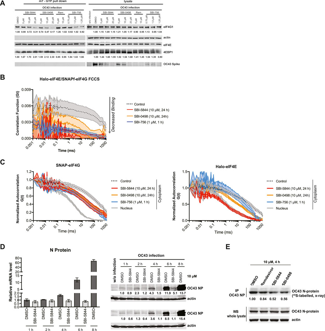
Figure 4. SBI-5844 and SBI-0498 attenuates translation initiation complex function. (A) Western blot analysis of indicated proteins prepared from A549 cells infected with OC43 and treated with indicated compounds. Cell lysates were incubated with m7GTP-agarose beads to capture the eIF4F complex. Shown is western blot of both total cell lysates (right panel) and m7GTP-agarose bound proteins (left panel) immunoblotted with indicated antibodies. Each protein band was quantified by ImageJ, normalized to actin (right panel) or eIF4E (left panel) as indicated. (B) Simultaneous diffusion of endogenous JF585Halo-eIF4E and JF646SNAPf-eIF4G1 throughout the focal volume was analyzed in living cells by dual color cross-correlation spectroscopy in the indicated conditions (Mean ± SEM; N = 10). Cross-correlation, due to eIF4F:eIF4G interaction, was detected in the cytoplasm of cells treated with vehicle control. Minimal cross-correlation was detected upon treatment with SBI-756, SBI-5844 or SBI-0498. (C) Diffusion of endogenous JF646SNAPf-eIF4G1 (left panel) and JF585Halo-eIF4E (right panel) was measured by fluorescence correlation spectroscopy in living cells treated with vehicle control or indicated compounds, in the indicated cellular compartment (Mean ± SEM; N = 10). Nuclear temporal autocorrelation depicts diffusion of free-diffusing JF646SNAPf-eIF4G1 (upper panel) and JF585Halo-eIF4E (lower panel). The autocorrelation curves were normalized for visual comparison of shape. (D) Vero E6 cells were infected with OC43 and treated with SBI-5844 for indicated times. RNA and protein were extracted and subjected to RT-qPCR analysis for virus RNA (left, mean ± SD, n = 3) and WB for virus N-protein (right). Each protein band was quantified by ImageJ, normalized to actin levels as indicated in numbers shown under the blots. (E) Vero E6 cells were infected with OC43 for 24 h. Cells were then treated with indicated compounds and radiolabeled with 35S-methionineand and 35S-cysteine for newly synthesized protein for 4 h. OC43 N-protein was immunoprecipitated (IP), separated by SDS-PAGE, transferred to PVDF membrane, and exposed to x-ray film. Each band was quantified by ImageJ, normalized to OC43 N-protein level as indicated under the blots.
The authors apologize for this error and state that this does not change the scientific conclusions of the article in any way. The original article has been updated.
Publisher’s note
All claims expressed in this article are solely those of the authors and do not necessarily represent those of their affiliated organizations, or those of the publisher, the editors and the reviewers. Any product that may be evaluated in this article, or claim that may be made by its manufacturer, is not guaranteed or endorsed by the publisher.
Keywords: COVID-19, OC43, SARS-CoV-2, coronavirus, eIF4F, translation initiation complex, Vero E6, A549
Citation: Feng Y, Grotegut S, Jovanovic P, Gandin V, Olson SH, Murad R, Beall A, Colayco S, De-Jesus P, Chanda S, English BP, Singer RH, Jackson M, Topisirovic I and Ronai ZA (2024) Corrigendum: Inhibition of coronavirus HCoV-OC43 by targeting the eIF4F complex. Front. Pharmacol. 15:1528449. doi: 10.3389/fphar.2024.1528449
Received: 14 November 2024; Accepted: 26 November 2024;
Published: 19 December 2024.
Edited and reviewed by:
Heike Wulff, University of California, Davis, United StatesCopyright © 2024 Feng, Grotegut, Jovanovic, Gandin, Olson, Murad, Beall, Colayco, De-Jesus, Chanda, English, Singer, Jackson, Topisirovic and Ronai. This is an open-access article distributed under the terms of the Creative Commons Attribution License (CC BY). The use, distribution or reproduction in other forums is permitted, provided the original author(s) and the copyright owner(s) are credited and that the original publication in this journal is cited, in accordance with accepted academic practice. No use, distribution or reproduction is permitted which does not comply with these terms.
*Correspondence: Ze’ev A. Ronai, emVldkByb25haWxhYi5uZXQ=
†Present address: Sumit Chanda, Department of Immunology and Microbiology, Scripps Research, La Jolla, CA, United States