
94% of researchers rate our articles as excellent or good
Learn more about the work of our research integrity team to safeguard the quality of each article we publish.
Find out more
REVIEW article
Front. Pharmacol., 03 July 2024
Sec. Ethnopharmacology
Volume 15 - 2024 | https://doi.org/10.3389/fphar.2024.1365639
This article is part of the Research TopicReviews in Ethnopharmacology: 2023View all 31 articles
 Chen-Fei Fu1†
 Jian-Long Li1†Jia-Wei Chen2Hao Liang3Wen-Rui Zhao4Shi-Yu He5Xiao-Wei Ma4*Xiao-Fan Yang4*He-Lin Wang6*
Sepsis is a complex syndrome characterized by multi-organ dysfunction, due to the presence of harmful microorganisms in blood which could cause mortality. Complications associated with sepsis involve multiple organ dysfunction. The pathogenesis of sepsis remains intricate, with limited treatment options and high mortality rates. Traditional Chinese medicine (TCM) has consistently demonstrated to have a potential on various disease management. Its complements include reduction of oxidative stress, inhibiting inflammatory pathways, regulating immune responses, and improving microcirculation. Traditional Chinese medicine can mitigate or even treat sepsis in a human system. This review examines progress on the use of TCM extracts for treating sepsis through different pharmacological action and its mechanisms. The potential targets of TCM extracts and active ingredients for the treatment of sepsis and its complications have been elucidated through molecular biology research, network pharmacology prediction, molecular docking analysis, and visualization analysis. Our aim is to provide a theoretical basis and empirical support for utilizing TCM in the treatment of sepsis and its complications while also serving as a reference for future research and development of sepsis drugs.
The management of sepsis has emerged as a critical scientific challenge, necessitating urgent attention as a significant global public health crisis (Groote and Meersch-Dini, 2022). The extensive heterogeneity observed in host, pathogen, and the environment contributes significantly to disease development (Jones et al., 2021). Despite recent advancements in sepsis treatment and advanced life support, mortality rate from septic shock remains approximately is 50% (Vincent et al., 2019). The ongoing pursuit of improved treatment resulted in research for development of alternative therapies to manage sepsis.
In traditional Chinese medicine, sepsis is commonly classified as one of the exogenous febrile diseases documented in ancient Chinese medical literature, including the Treatise on Febrile Diseases, Treatise on Epidemic Febrile Diseases, and other relevant publications (Fan et al., 2020). The theory of traditional Chinese medicine (TCM) emphasizes that the treatment of sepsis should focus on clearing heat and detoxification, restoring normal bowel movement, promoting blood circulation and removing blood stasis, as well as strengthening immune system. According to the concept of entirety and the method of treatment with syndrome differentiation of TCM, the clinical efficacy of sepsis was significantly improved and the case fatality rate effectively reduced (Zhao et al., 2017). The collection, processing, and preparation of Chinese herbal medicine adhere to the fundamental principles of TCM, which boasts into a rich history for both theory and practice when it comes to treating critically ill patients. The practitioners of TCM have expertise for identification of symptoms related to acute and critical illnesses, as well as formulating appropriate TCM treatments. Traditional Chinese medicine can serve as an adjunctive therapy in the management of sepsis due to its advantageous characteristics, including a complex composition, multiple targets of action, and involvement in various signaling pathways (Usmani et al., 2021). It exhibits diverse effects such as anti-inflammatory properties, improvement of microcirculation, alleviation of gastrointestinal dysfunction, and enhancement of the immune system. Moreover, it may even confer protection against organ damage caused by sepsis (Usmani et al., 2021). The efficacy and safety of employing this evidence-based TCM treatment in conjunction with intensive sepsis therapy increasingly acknowledged by the medical community and patients. Traditional Chinese medicine has played a crucial role in the management of COVID-19 patients, providing us novel insights into the merits and significance of employing traditional Chinese medicine for sepsis treatment (Lyu et al., 2021). Traditional Chinese medicine has been incorporated into the standard treatment for sepsis (Wang X.-H. et al., 2022), including medications such as Xuebijing injection (XBJ), Shenfu injection, Qingwen Baidu decoction, Xuanbai Chengqi decoction (XBCQ), and Qingmai decoction (QWBD). The significant advancements have been made in the exploration of the efficacy and mechanism of action for TCM to sepsis treatment, with an increasing number of studies corroborating the prominent role in the prevention and management of sepsis (Zheng et al., 2023). Although there have been review articles discussing the research progress of traditional Chinese medicine compounds and monomer extracts in the treatment of sepsis and multi-organ injury (Song et al., 2023), this review merely presents a superficial list of various monomer extracts used for sepsis treatment, lacking a comprehensive analysis and discussion on their therapeutic mechanisms. It is noteworthy that our review commences with an exploration of the etiology and pathogenesis of sepsis, subsequently delving into the therapeutic strategies for sepsis utilizing traditional Chinese medicine extracts based on their respective mechanisms of action. This approach facilitates a comprehensive understanding of TCM’s therapeutic process for sepsis, as TCM typically addresses diseases through multiple pathways and targets. Categorizing interventions based on their mechanism rather than solely relying on TCM extracts enhances our comprehension and cognition of TCM’s efficacy in treating sepsis. Our review establishes correlations between the mechanisms of traditional Chinese medicine extracts for treating sepsis and its pathogenesis, thereby emphasizing the logical basis behind TCM disease management and bolstering its persuasiveness. Consequently, this review serves as a valuable reference for future development of sepsis medications.
Sepsis is a life-threatening syndrome of multiple organ dysfunction caused by dysregulation in the body’s immune response to infection (Sygitowicz and Sitkiewicz, 2021). The mortality rate of patients with sepsis can exceed 30%–35% in the absence of timely and effective intervention, as a result of an increased inflammatory response caused by immunosuppression or primary infection (Vincent et al., 2019). The complications induced by sepsis involve multi-organ dysfunction such as pulmonary impairment, cerebral injury, renal impairment, hepatic impairment, and myocardial damage (Sun et al., 2020; Zhou and Liao, 2021; Chang et al., 2022; Sekino et al., 2022; Zhao et al., 2022). The high mortality rate resulting from sepsis is primarily attributed to the occurrence of multi-organ failure induced by the disease. The pathogenesis of sepsis is highly intricate at the cellular and molecular level, which encompasses various pathophysiologic processes such as inflammation imbalance, immune dysfunction, mitochondrial damage, coagulation disorders, neuroendocrine immune network abnormalities, endoplasmic reticulum stress, and autophagy, ultimately resulting in multi-organ dysfunction (Huang et al., 2019a). According to the conventional perspective, following an initial phase of excessive inflammation, a subsequent phase of diminished inflammation, partially attributed to the release of anti-inflammatory cytokines, leads to profound immunosuppression (Sheth et al., 2019). Currently, preliminary results indicated that the stages of inflammation promotion and immune suppression may occur concurrently (Zhong and Yin, 2023), rendering the interaction between host and pathogen conducive for disease development. The high-risk factors for sepsis patients encompass advanced age, gender, presence of immunosuppressive diseases, medications, history of cancer, diabetes mellitus, alcohol misuse, and the utilization of indwelling urinary catheters (Tokuda et al., 2023). The severity of sepsis depends on the host’s age, comorbidities, and immune status, as well as pathogenic factors such as toxicity, microbial species, and infection burden (Olinder et al., 2022). In addition to pathogen-related factors, there may also be host genetic factors that could increase the risk of developing sepsis (Mukherjee et al., 2019). The latest guidelines released by the “Surviving Sepsis Campaign” in October 2021 further emphasize the implementation of sepsis screening for standardized treatment, enhance infection control and optimize antimicrobial drug utilization, as well as recommend novel treatment techniques and concepts. The current international clinical guidelines primarily include rapid screening and early diagnosis, accurate identification and control of infection sources and pathogenic microorganisms, timely and effective antibiotic treatment and fluid resuscitation, management of hemodynamics, and mechanical ventilation (Fitridge and Thompson, 2007).
Despite some advancements in the research and treatment of sepsis in recent years, a comprehensive understanding of its pathogenesis and key mechanisms is still lacking, necessitating further improvements in targeted therapies. However, lack of effective treatment methods hinders the management of cellular and tissue organ damage resulted from sepsis. The treatment of sepsis remains a global challenge, involving the prevention of multiple infections, management of inflammatory responses and immune-mediated damage, as well as addressing gastrointestinal dysfunction and coagulation disorders. (Arefian et al., 2017; Cecconi et al., 2018; Marik, 2018).
The fundamental principle of TCM is rooted in the “holistic concept,” which serves as a guiding principle, while the “syndrome differentiation” acts as the diagnostic and therapeutic method. These two theories direct the management of sepsis with TCM (Li and Xu, 2011). The “holistic concept” posits that the human body functions as an integrated organic entity, emphasizing the synergistic interplay among multiple organs to safeguard against disease and maintain homeostasis. The integration of “syndrome differentiation” and “disease differentiation” involves understanding the location, etiology, nature, and interplay between “positive” and “evil” factors, reflecting the pathological changes. The aforementioned statement also aligns with the concept of “personalized medicine” in contemporary medical practice, which holds immense significance in the realm of diagnosis and treatment. Western medicine research has shifted from a one-sided focus on eradicating disease-causing microorganisms to regulating the body’s immune response. It now emphasizes the intrinsic connection between organs in the whole body, as evidenced by theories such as liver-kidney syndrome, heart-kidney syndrome, and lung-intestinal axis. Traditional Chinese medicine is used to dynamically adjust multiple systems in patients, aligning with the holistic concept of Chinese medicine (Liu et al., 2022). Due to variations in individual characteristics, pathogenic factors, and disease duration, patients exhibit diverse responses to drug therapy. Additionally, each sepsis patient presents distinct clinical manifestations, disease progression levels, and alterations in disease mechanisms. Therefore, it is essential to employ diverse methods and treatment modalities tailored to the individual, timing, and location, aligning with the diagnostic and therapeutic approach of Chinese medicine known as “syndrome differentiation.”
The TCM treatment of sepsis can be summarized as “Four Syndromes and Four Methods” based on the etiology and pathogenesis. The “Four Syndromes” include blood stasis, toxic heat, viscera and qi obstruction as well as acute deficiency. Correspondingly, the “Four Methods” are activating blood circulation to remove stasis, clearing heat and detoxifying, regulating the lower energizer while nourishing the root (Xu and Lu, 2018). The improvement of microcirculation corresponds to the traditional Chinese medicine approach of “activating blood circulation to resolve stasis.” In sepsis, microcirculatory disorders are a significant pathological change characterized by inadequate capillary perfusion and sluggish blood flow. The objective of the “activating blood circulation to resolve stasis” in TCM is to enhance blood circulation, particularly microcirculation, thereby alleviating tissue hypoxia and ischemia, preventing the formation of microcirculatory thrombosis, as well as halting further progression of disseminated intravascular coagulation (DIC). The inhibition of inflammatory mediator release aligns with the treatment principle of “clearing heat and detoxifying.” A significant amount of inflammatory mediators such as tumor necrosis factor and interleukins are released during sepsis, resulting in systemic inflammation of the body. Traditional Chinese medicine’s approach to “clearing heat and detoxification” can mitigate inflammatory response through the utilization of specific TCM possessing heat-clearing and detoxifying properties, thereby suppressing excessive release of inflammatory mediators. Promoting the elimination of endotoxins is akin to the principle of “regulating the lower energizer” in traditional Chinese medicine. In sepsis, bacterial endotoxins accumulate within the body, exacerbating the condition. For septic patients presenting with symptoms such as constipation and abdominal distension, TCM that facilitate bowel movement and heat expulsion can be employed for sepsis treatment. Modern medicine acknowledges that the intestines serve as a major reservoir of bacteria in the human body. During severe infections like septicemia, stress weakens or damages intestinal barrier function, allowing a significant influx of bacteria and endotoxins into circulation via the portal vein and intestinal lymphatic system, resulting in gut-derived endotoxemia and bacterial translocation. The principle underlying both immune regulation and the “nourishing the root” method in traditional Chinese medicine is similar. Sepsis often leads to suppression or disruption of the body’s immune function. In traditional Chinese medicine, the emphasis on nourishing the spleen, kidneys, and other organs aims to enhance immune function, which can be seen as analogous to modern medical immunomodulatory treatments.
Combined with the clinical manifestations, the “method of supporting and consolidating the essence” should be considered as a fundamental treatment approach for “septic shock” or “sepsis recovery,” predominantly relying on TCM that promote and consolidate vital energy. “Activating blood circulation to remove stasis” is a treatment method used when shock is combined with DIC or to prevent the occurrence of DIC, primarily using traditional Chinese medicine that promotes blood circulation and resolves stagnation. “Clearing heat and detoxifying” and “regulating the lower energizer” are therapeutic approaches employed to combat pathogenic infections, while also serving as preventive measures against septic shock. These methods primarily focus on utilizing TCM with heat-removing properties to clear heat and toxins from the body’s lower internal organs (Li and Wang, 2017; Ling et al., 2023). The Chinese herbal medicine types that clear heat and detoxify, promote blood circulation, remove blood stasis, and strengthen the foundation of treatment are effective in treating sepsis. The “Four Syndromes and Four Methods” provide a convenient way to identify infectious shock in TCM. The four syndromes can simultaneously correspond to the various stages of pathology in Western medicine, thereby enhancing the precision in drug identification and utilization (Jing et al., 2022). The treatment of sepsis in TCM aims not only to eliminate pathogenic factors, but also to restore internal balance and coordination within the organism (Liu et al., 2022). It regulates the functions of internal organs, promoting harmony between the five viscera and six bowels, allowing the body to achieve an optimal state through medication for self-regulation. This ultimately restores internal homeostasis and realizes the delicate interplay of yin and yang.
Traditional Chinese medicine extracts and its active components possess distinct chemical structures, precise therapeutic effects, and facilitate quality control. These compounds offer “individualized therapies” for different stages of sepsis development through multiple pathways and targets, thereby promoting body equilibrium (Han and Wu, 2010; Li et al., 2013). This review provides a comprehensive overview of the advancements in sepsis treatment research, focusing on the extraction and isolation of bioactive compounds from natural sources. These compounds are obtained through inhibiting the release of inflammatory mediators, facilitating endotoxin elimination, enhancing microcirculation, and modulating immune responses.
The mechanisms action of TCM extracts in the treatment of sepsis are systematically summarized and presented in Figures 1, 2. The extracts of traditional Chinese medicine regulate diverse mechanisms through various signaling pathways to enhance sepsis management, exemplifying the multifaceted approaches and targets characteristic of TCM in disease treatment.
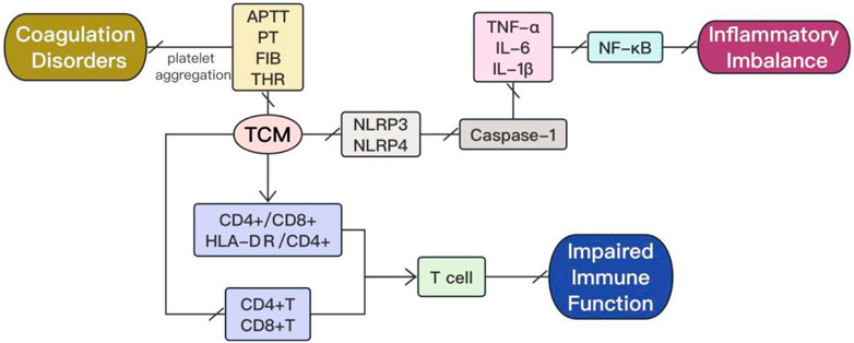
Figure 1. The mechanism actions in treatment of sepsis with TCM extracts involve the modulation of inflammatory imbalance, impaired immune function, and coagulation disorders.
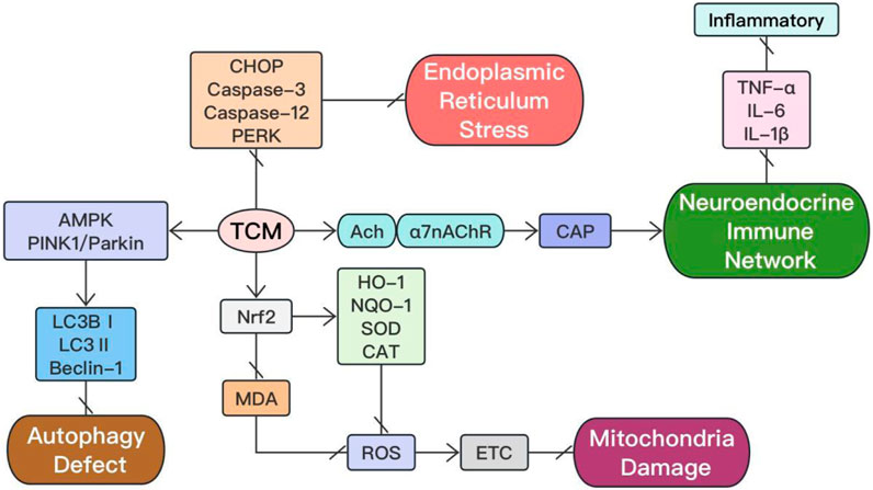
Figure 2. The other mechanism actions in treatment of sepsis with TCM extracts.
Inflammatory imbalance serves as the fundamental basis for the pathogenesis and progression of sepsis. The pathogenic microorganisms, including bacteria, fungi, parasites, and viruses, elicit responses throughout the course of sepsis. The initial acute response triggered by the host in response to an invasive pathogen typically involves phagocytosis of the pathogen by macrophages and production of a variety of pro-inflammatory cytokines. This leads to the onset of a cytokine storm and activation of the intrinsic immune system (Lin et al., 2022). The activation of the intrinsic immune system is clearly mediated by pattern-recognition receptor, which detect damage-associated molecular pattern (DAMP) or pathogen-associated molecular pattern (PAMP) and subsequently upregulate inflammation-related genes, initiating a cascade of immune cell activations (Raymond et al., 2017). In the immune response to sepsis, exogenous factors derived from pathogens, such as lipopolysaccharides, and endogenous factors released by injured cells both play a crucial role. Figure 3 shows the inflammatory imbalance process during sepsis. The findings of various studies have demonstrated the effective inhibition of inflammatory factor release, regulation of pro-inflammatory and anti-inflammatory responses, antagonism against endotoxins, inhibition of hyperfibrinolysis, as well as modulation of the expression of inflammatory signaling pathways by herbal monomers (Li et al., 2013). These mechanisms contribute to the protection of septic organs’ function and improvement in patient prognosis. The characteristics of TCM extracts by the modulation of inflammatory imbalance on sepsis are summarized in Table 1.
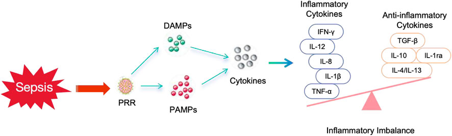
Figure 3. The inflammatory imbalance process during sepsis.
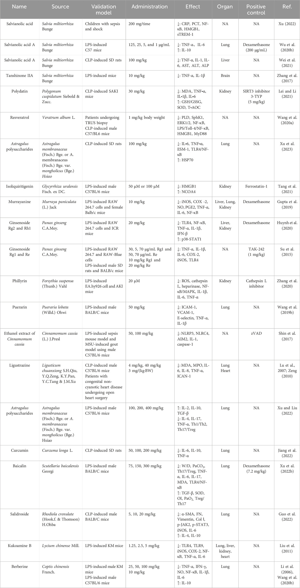
Table 1. Characteristics of TCM extracts by the modulation of inflammatory imbalance on sepsis.
The active components salvianolic acid and tanshinone, derived from the Chinese herbal medicine Salvia miltiorrhiza Bunge, possess biological effects such as enhancing circulation, exerting anti-inflammatory and antioxidant effects (Wang J. et al., 2019). These components can potentially serve as therapeutic agents against the release of inflammatory and oxidative mediators during sepsis and microcirculatory disorders in shock (Yuan J. et al., 2021). Xu (2022) discovered that the incorporation of salvianolic acid into conventional fluid resuscitation can effectively suppress the inflammatory and stress responses during sepsis combined with shock. Additionally, it has been shown to significantly enhance blood oxygen metabolism in pediatric patients with sepsis combined with shock. Wu S. et al. (2020) utilized lipopolysaccharide (LPS)-induced macrophage RAW264.7 and a murine sepsis model to assess the impact of metabolites isolated from extracts of the aerial parts of S. miltiorrhiza, specifically salvianolic acid A (administered via intraperitoneal injection at concentrations of 125, 25, 5, and 1 μg/mL), on sepsis. The cellular activity was evaluated using the methyl thiazolyl tetrazolium (MTT) assay, while the levels of inflammatory factors were measured in both cells and mouse livers through enzyme-linked immunosorbent assay (ELISA) analysis. The results demonstrated that salvianolic acid A increased cellular activity in all dosage groups. Moreover, the levels of inflammatory factors TNF-α and IL-6 were significantly or markedly reduced, while the level of IL-10 was significantly elevated in all dosage groups (except for the 5 μg/mL group). These findings suggest that salvianolic acid A effectively mitigated the impact of LPS on cellular activity. The effects of intraperitoneal administration of salvianolic acid A at a dosage of 100 mg/kg on liver injury in rats with cecal ligation and puncture (CLP) were analyzed and investigated (Wei et al., 2021). The expression levels of inflammatory markers TNF-α, IL-1, IL-6, as well as liver injury markers aspartate aminotransferase (AST), alanine aminotransferase (ALT), and alkaline phosphatase (ALP) were detected using ELISA. Additionally, liver tissues from the rats were subjected to pathological observation. The results demonstrated that compared to the sepsis control group, the salvianolic acid experimental group exhibited a significant reduction in levels of both liver injury markers and inflammatory markers. Zhang et al. (2017) investigated the interventional effects of tanshinone IIA (10 mg/kg) on cerebral microcirculation and neuroinflammatory responses in LPS-induced septic mice. Utilizing the BI2000 microcirculation image processing system, researchers observed that blood flow velocity of microvessels and microarteries in the soft meninges at different phases of each group’s mice through an open cranial window. Additionally, ELISA was employed to detect the levels of inflammatory factors in mouse brain hippocampal tissue. The results demonstrated a significant acceleration in both micro-artery and micro-vein blood flow velocity within the drug group compared to that of the model group, along with significantly reduced levels of TNF-α and IL-1β in cerebral hippocampus tissue when compared to those found in the model group. These findings indicated that tanshinone IIA could enhance cerebral microblood flow velocity, mitigate neuroinflammatory reactions, and exert protective effects on the brain.
Lei and Li (2021) reported that polydatin significantly reduced serum levels of inflammatory factors in SAKI mice. Wang B. et al. (2020) demonstrated that resveratrol exerts regulatory effects on downstream signaling molecules of Toll-like receptor 4 (TLR-4), sphingosine kinase 1 (SphK1), extracellular signal-regulated kinase 1/2 (ERK1/2), and NF-κB, leading to a reduction in cytokine production by human primary monocytes stimulated with LPS through phospholipase D (PLD). Moreover, it downregulates myeloid differentiation factor 88 (MyD88), which inhibits the LPS/Toll-4/NF-κB signaling pathway, thereby attenuating the production of cytokines, chemokines, and high mobility group protein B1 (HMGB1) by human primary monocytes stimulated with LPS and exerting anti-inflammatory effects.
Xu et al. (2023) discovered that Astragalus polysaccharides (APS), a kind of polysaccharides obtained from the roots of A. membranaceus (Fisch.) Bge. or A. membranaceus (Fisch.) Bge. var. mongholicus (Bge.) Hsiao, mitigated the acute inflammatory response in septic rats. Tang et al. (2021) observed that the utilization of isoliquiritigenin reduced the expression of HMGB1 and inhibited ferroptosis in a sepsis-induced model of AKI, thereby attenuating the inflammatory response.
The major compound isolated from Murraya paniculata (L.) Jack is murrayanine, which has been reported to possess significant antioxidant (Abu Bakar et al., 2007), anti-inflammatory (Gupta et al., 2010), and immunomodulatory activities (Shah et al., 2008). Gupta et al. (2019) discovered that murrayanine inhibited the expression of inducible nitric-oxide synthase (iNOS) and cyclooxygenase (COX-2), reduces the production of nitric-oxide (NO), prostaglandin E2 (PGE2), TNF-α, and IL-6, decreases NF-κB activity, ameliorates LPS-induced lung, liver and kidney injuries, as well as increases survival rates in mice with LPS-induced sepsis models. These findings demonstrated that murrayanine was an ideal natural compound and a new chemical entity for treating sepsis and other inflammatory diseases.
The anti-inflammatory effects of ginseng have been demonstrated in a variety of diseases (Kim et al., 2018; Xu H. et al., 2022). Ginseng contains a diverse range of bioactive compounds, and its therapeutic potential has been associated with neuromodulation, anticancer activity, lipid modulation, and antithrombotic activity (Ru et al., 2015). However, there is currently no definitive study comparing the relative efficacy of different components of ginseng in the treatment of sepsis. A study suggested that treatments with ginsenoside Rg2 and Rh1 might exhibit greater efficacy compared to single-component treatments (Huynh et al., 2020). Similarly, another study demonstrated that the combination of ginsenoside Rg1 and Re in the treatment of septic mice effectively reduced LPS-induced hyperthermia, leukocyte counts, and serum levels of pro-inflammatory mediators. Furthermore, this combination even exhibited a 90% increase in survival rate among lethally septic shocked mice. Subsequent analysis revealed that the effective anti-inflammatory effect could be attributed to the different distribution locations of these components (Rg1 being located both intracellularly and extracellularly while Re is located extracellularly), suggesting that combining various components of ginseng might yield superior therapeutic outcomes (Su et al., 2015).
Phillyrin exhibited inhibitory effects on reactive oxygen species (ROS) production, reduced the expression levels of histone L and heparinase both in vitro and in vivo, and suppressed the secretion of inflammatory cytokines to ameliorate renal function in AKI mice (Zhang et al., 2020). The isoflavone compound puerarin has demonstrated its ability to dilate coronary blood vessels, enhanced myocardial metabolism, exhibited antioxidant and anti-inflammatory activities, protect vascular endothelial function, and improve microcirculation (Wei et al., 2014). Puerarin pretreatment at a dose of 50 mg/kg for 4 days significantly attenuated the serum levels of intercellular adhesion molecule-1 (ICAM-1), vascular cell adhesion molecule-1 (VCAM-1), and E-selectin, as well as TNF-α and IL-1β in mice with LPS-induced endotoxemia (Wang S. et al., 2019). These findings suggested that puerarin effectively suppressed the release of inflammatory factors and adhesion molecules, thereby ameliorating LPS-induced vascular endothelial cell injury.
Modern studies have confirmed the therapeutic effect of Cinnamomum cassia (L.) J.Presl in sepsis by inhibiting the activation of inflammatory vesicles. Shin et al. (2017) utilized bone marrow-derived macrophage BMDMs to investigate the modulatory effect of 50 and 100 mg/kg C. cassia ethanol extract on inflammatory vesicle activation. They also established a mouse model of LPS-induced sepsis and a monosodium urate-induced gout model, observing that the compound increased the survival rate of mice with LPS-induced septic shock while inhibiting the activation of nucleotide-binding domain, leucine-rich repeat, and pyrin domain-containing protein 3 (NLRP3), NLRP4, and melanoma-deficiency factor 2 inflammatory vesicles through inhibition of apoptosis-associated granulocyte-like proteins oligomerization within CARD structural domains, consequently suppressing IL-1β and caspase-1 secretion.
A study replicating a mouse model of sepsis by CLP demonstrated that ligustrazine significantly attenuated the pulmonary inflammatory response during sepsis, leading to a significant reduction in plasma levels of IL-6 in ligustrazine-treated septic mice (Zeng, 2010). Clinical studies have also revealed that ligustrazine effectively decreased peripheral blood levels of IL-8 and TNF-α in patients undergoing extracorporeal circulation, potentially contributing to the management of systemic inflammatory response syndrome during extracorporeal circulation (Lu et al., 2007).
The relevant studies have demonstrated that Astragalus polysaccharide (APS) exhibits diverse pharmacological activities, including anti-inflammatory, antioxidant, anti-atherosclerotic, apoptosis inhibition, and ischemia-hypoxia protection (Zhou et al., 2018). Consequently, it is extensively employed in clinical settings as a crucial immunomodulator or antioxidant (Zhang et al., 2019). The sepsis mouse model was created using LPS and divided them into groups, with APS administered at different doses and saline interventions respectively (Xu and Liu, 2022). The results demonstrated a significant improvement in the imbalance of Th1/Th2 and Th17/Treg cells in septic mice treated with APS. Additionally, there was an increase in the levels of anti-inflammatory factors such as IL-2, IL-10, and TGF-β, while pro-inflammatory factors including IL-4, IL-17, and TNF-α were downregulated. Furthermore, a significant dose-dependent effect was observed.
Curcumin exhibited significant potential in reducing the levels of IL-6, IL-10, and TNF-α within the lung tissue of septic rats (Jiang et al., 2022). This suggested that curcumin effectively ameliorated inflammation levels, alleviated immune suppression, and mitigated damage to lung tissues in septic rats.
Baicalin (BA), a flavonoid compound found in the root of Scutellaria baicalensis Georgi, exhibits various pharmacological effects such as lipid-lowering, anti-inflammatory, antibacterial, antioxidant and immunomodulatory activities. The mice were induced with sepsis through intraperitoneal injection of LPS, followed by treatment with baicalin to mitigate lung injury (Xu L. et al., 2022). The results demonstrated that BA inhibited TLR4/NF-κB pathway activation, suppressed inflammatory response, promoted Treg/Th17 balance shift towards Treg cells, reduced lung tissue damage and improved lung function. The effect was dose-dependent.
Salidroside, the active component found in Rhodiola crenulate (Hook.f. & Thomson) H.Ohba, exhibits pharmacological effects that include anti-aging, anti-inflammatory, immunomodulatory, and antioxidant properties. A murine model of acute lung injury with sepsis was established through CLP (Guo et al., 2022). The findings demonstrated that salidroside attenuated the pulmonary response in CLP-induced septic mice by downregulating the expression of JAK2 and STAT3, while significantly reducing the levels of iNOS and IL-6 in lung tissue. Moreover, there was a significant increase in IL-4 and IL-10 levels, indicating that salidroside ameliorates lung injury in CLP-induced septic mice by modulating inflammatory factors within the lung tissue.
Kukoamine B is an alkaloid component primarily derived from the Chinese medicinal plant Lycium chinense Mill. In a study, the affinity of kukoamine B towards LPS and CpG DNA was investigated, along with its inhibitory effect on the release of TNF-α and IL-6 from RAW264.7 cells both independently and in combination (Liu et al., 2011). The findings demonstrated that kukoamine B exhibited significant neutralization of multiple pathogenic molecules and effectively suppressed the induced inflammatory response. Notably, kukoamine B represents the first reported natural product-derived inhibitor targeting both LPS and CpG DNA.
Berberine, a bioactive alkaloid isolated from Chinese herbal medicine Coptis chinensis Franch., has been shown to reduce plasma levels of TNF-α, IFN-γ and NO in mice with sepsis (Li et al., 2006). Recent domestic research also confirmed that berberine could inhibit the activation of NF-κB signal transduction and further supports its use as a single drug for treating sepsis patients along with other drugs (Wang Y. et al., 2020).
In conclusion, TCM extracts with anti-inflammatory properties effectively suppress the release of inflammatory factors, regulate pro-inflammatory/anti-inflammatory balance, and modulate the expression of inflammatory signaling pathways in organs affected by sepsis (show Figure 1). Therefore, these TCM extracts hold tremendous potential for treating sepsis.
The dysfunction of immune function is a crucial factor contributing to the occurrence and progression of sepsis. During the inflammatory response in sepsis, neutrophils interact with and migrate across endothelial cells under the influence of chemokines that are attracted to the site of inflammation. At this location, they recognize and engulf pathogens, release various active factors and proteolytic enzymes, ultimately eliminating the pathogen (Shen et al., 2017). Severe impairment of the immune system is a common pathological change observed in sepsis. During the initial cytokine storm phase, a significant number of sepsis patients may succumb to death. While survivors may experience immune suppression, leading to an inability to clear primary infections and reactivation of latent viruses. Sepsis-induced immunosuppression affects both innate and adaptive immunity. The immunosuppressive response following sepsis has been characterized as a compensatory anti-inflammatory response syndrome, which is regulated by co-stimulatory molecules like CD80/B7-1 that are produced through activation of the TLR signaling pathway. Additionally, this response involves the conversion of naive T cells into cytokine-induced regulatory T cells, resulting in decreased expression of antigen-presenting associated transcription factors (e.g., CD80/B7-1) and IRF4/MUM1 (Honma et al., 2005; Negishi et al., 2005; Pepin et al., 2018). Traditional Chinese medicine has the ability to regulate the functions of immune cells, including their composition, differentiation, activation, secretion, and killing (Yang and Fan, 2021). This enables it to enhance the body’s innate resistance against pathogens and prevent cytokine storms. Specifically, it can interrupt and reverse the immunosuppressive state associated with severe sepsis, thereby improving prognosis and preventing further damage caused by pathogenic toxins (Wang and Chen, 2017). The characteristics of TCM extracts by the modulation of impaired immune function on sepsis are summarized in Table 2.
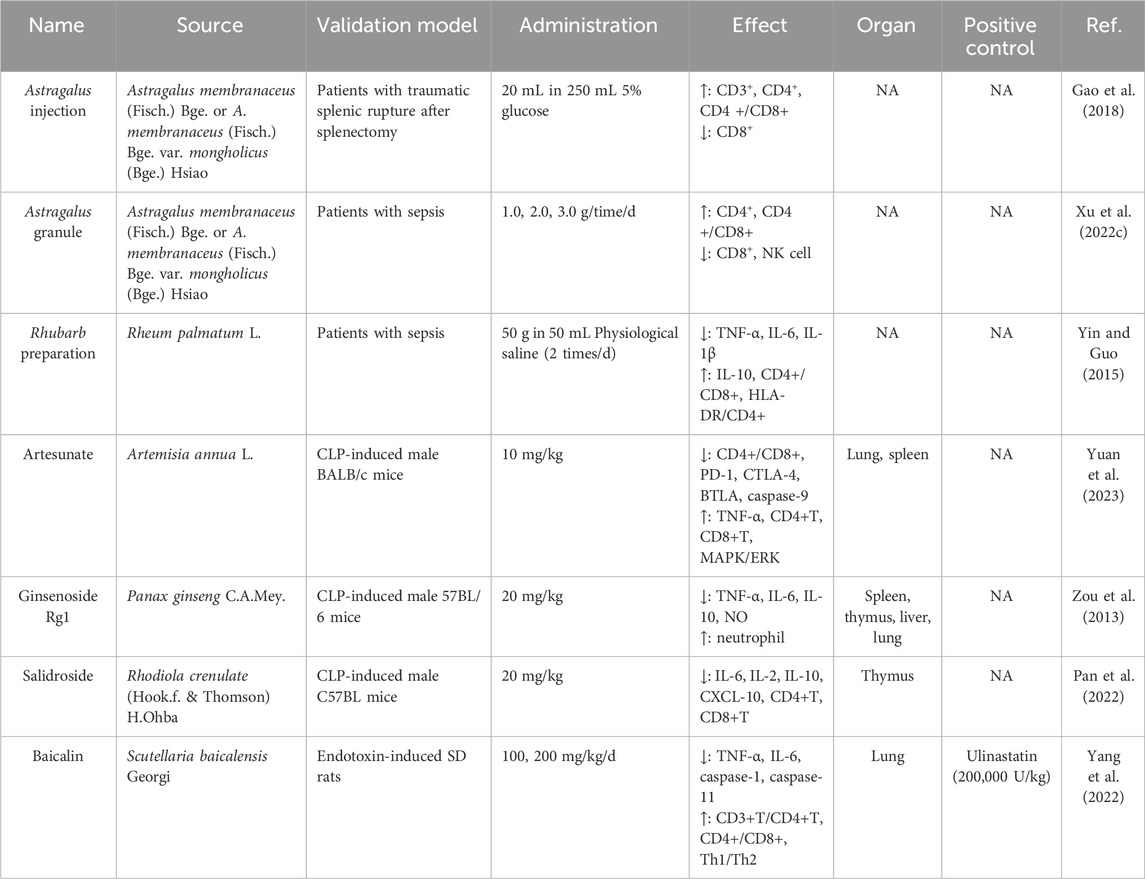
Table 2. Characteristics of TCM extracts by the modulation of impaired immune function on sepsis.
The advanced study have demonstrated that Astragalus injection exhibits a significant improvement effect on T lymphocyte subsets in patients with traumatic splenectomy, while maintaining high levels of drug safety (Gao et al., 2018). This suggested that Astragalus injection possessed the ability to modulate various immune active cells, thereby ameliorating immune imbalance through metabolic regulation and exerting dual regulatory effects on immune function. The immunomodulatory effects of high-dose Huangqi granules was further validated by promoting lymphocyte proliferation and regulating the proportion of lymphocyte subsets through rigorous investigations (Xu X. et al., 2022).
Yin and Guo (2015) utilized Rhubarb preparation for small intestine perfusion via naso-intestinal tube and observed a significant reduction in the levels of TNF-α, IL-6, and IL-1β among sepsis patients. Simultaneously, there was an increase in the levels of IL-10, as well as improvements in CD4+/CD8+ ratios and human leukocyte antigen-DR (HLA-DR)/CD4+ expression. These findings provided evidence supporting the immunomodulatory effects of Rhubarb preparation on sepsis patients.
An immunosuppressive mouse model of CL-induced sepsis-related secondary bacterial infection was established, demonstrating that artesunate could reverse immunosuppression not only through innate immune regulation but also through adaptive immune regulation (Yuan et al., 2023). These immunosuppressive effects on the adaptive immune response were achieved by increasing the number of T cells through inhibition of CD4+ and CD8+ T cell apoptosis. Artesunate inhibited the expression of programmed cell death protein-1 (PD-1), cytotoxic T lymphocyte antigen-4 (CTLA-4), and B and T lymphocyte attenuator (BTLA) receptors while activating the MAPK/ERK signaling pathway as its potential mechanism. Furthermore, it was found that artesunate can inhibit caspase-9 expression and activation, potentially specifically targeting the mitochondrial apoptosis pathway in T cells. These findings suggested that artesunate had the potential to function as a modulator for both T cell number and function, making it a promising candidate for treating immunosuppressive diseases.
The intravenous administration of 20 mg/kg ginsenoside Rg1 to CLP mice resulted in an increase in peritoneal neutrophil counts (Zou et al., 2013). Additionally, the application of ginsenoside demonstrated inhibitory effects on thymus and spleen lymphocyte apoptosis, thereby enhancing bacterial clearance rate and survival rate. These findings suggested that ginsenoside Rg1 had the potential to enhance innate immunity, promote maintenance of adaptive immunity, and provide effective protection against sepsis.
Salidroside is derived from the dried roots and rhizome of R. crenulate (Hook.f. & Thomson) H.Ohba, possessing anti-cancer, antioxidant, anti-inflammatory, and immunomodulatory properties (Magani et al., 2020). Researches indicated its significant role in improving neuroinflammation (Wang et al., 2018), inhibiting renal fibrosis (Li et al., 2019), ameliorating fatty liver conditions (Zheng et al., 2018), and suppressing cancer cell proliferation (Zhu et al., 2020). Pan et al. (2022) established a sepsis mouse model induced by CLP and divided the mice into groups. They were treated with either salidroside or saline solution. The administration of salidroside significantly reduced the expression of inflammatory factors TNF-α, IL-6, IFN-γ, as well as inhibitory factor IL-10 in the spleen of septic mice. This suggested that salidroside could inhibit the inflammatory response in the spleen of septic mice and subsequently regulate their splenic immune function. Additionally, salidroside was found to inhibit CXC chemokine ligand-10 (CXCL-10) expression in the spleen of mice with sepsis, thereby reducing infiltration by immune cells and improving splenic immune status.
After baicalin intervention, there were a decrease in the elevated levels of TNF-α and IL-6 in the serum of septic rats, as well as an increase in the percentages of CD3+ T lymphocytes, CD4+ T lymphocytes, CD4+/CD8+ ratio, and Th1/Th2 ratio in peripheral blood (Yang et al., 2022). No significant difference between the high-dose baicalin group and ulinastatin group was observed when compared to each other. These findings suggested that baicalin had the potential to downregulate inflammatory factors and improve immune disorders in septic rats.
Traditional Chinese medicine extracts can regulate the body’s immune function and improve immune disorders (show Figure 1). They play a crucial role in reversing the immunosuppressive state of severe sepsis and enhancing the body’s own resistance.
The current research has identified the interaction between inflammation and coagulation as a pivotal mechanism in the pathogenesis of sepsis. The inflammatory response in sepsis triggers a coagulation reaction and initiates coagulation, while the ensuing coagulation response further amplifies the inflammatory process (Ma et al., 2019). Under normal circumstances, three key physiological factors regulate the activation of coagulation through anticoagulant pathway systems: the tissue factor pathway inhibitor system, the activated protein C (APC) system, and the antithrombotic system that controls coagulation activation (Petäjä, 2011). The three pathways in sepsis exhibit varying degrees of disruption. Owing to impaired protein synthesis, the coagulation inhibitor pathways demonstrate sustained depletion and protein degradation at low levels. The downregulation of thrombomodulin (TM) and endothelial protein C receptor expression is attributed to the conversion of protein C to APC under inflammatory conditions (Levi and Poll, 2015). The maximal activation of endogenous fibrinolysis and coagulation is significantly attenuated in sepsis, wherein the production of fibrinogen activators (i.e., tissue plasminogen activator (t-PA)) and urokinase-type plasminogen activator (u-PA) by vascular endothelial cell storage sites leads to an augmented stimulation of fibrinogen activation and release of subquantified plasmin, while this effect is counteracted by the sustained increase inhibitor-1 (PAI-1) (Biemond et al., 1995). The Chinese medicines extracts can exert their effects on various components including blood vessels, endothelial cells, platelets, and coagulation factors. They possess the ability to inhibit platelet aggregation and activation, thereby improving sepsis-associated coagulation disorders and reducing the incidence of DIC, ultimately enhancing the quality of life for sepsis patients (Jin et al., 2022). The characteristics of TCM extracts by the modulation of coagulation disorders on sepsis are summarized in Table 3.

Table 3. Characteristics of TCM extracts by the modulation of coagulation disorders on sepsis.
Persicae semen extract possessed inhibitory effects on platelet aggregation, the release of various inflammatory cytokines, inflammation-induced vascular permeability, and pulmonary vascular endothelial damage reduction (Yi et al., 2016). Huangqi (Astragalus) and Danggui (Angelica) were revealed the ability to induce relaxation in airway smooth muscle, scavenge free radicals, inhibit platelet aggregation, and suppress the release of inflammatory mediators such as thromboxane (Li et al., 2015). These actions effectively counteract microthrombosis and confer a protective effect on the vascular endothelium. In another study, breviscapine effectively enhanced the regulation of cellular coagulation and fibrinolysis (Li Z. et al., 2017).
A sepsis model was established by inducing CLP in SD rats (Li and Jin, 2016). Simultaneously, safflower yellow A was administered at various doses, revealing that a low dose of safflower yellow A effectively attenuated the prolongation of prothrombin time (PT) and activated partial thromboplastin time (APTT). Furthermore, it significantly suppressed the expression levels of serum TF, IL-1β, and TNF-α, while enhancing the expression level of serum tissue factor pathway inhibitor (TFPI). These findings suggested that a low dose of safflower yellow A could alleviate the reciprocal amplification between coagulation and inflammation during sepsis, ameliorate sepsis-induced coagulation dysfunction, regulate inflammatory factors’ expression, delay organ dysfunction syndrome onset, and exert protective effects against lung injury in septic conditions.
The therapeutic effects of emodin on cardiovascular responses induced by diesel exhaust particles (DEP) were investigated in a mouse model (Nemmar et al., 2015). They discovered that emodin significantly reduced the levels of inflammatory factors such as leukocytes, TNF-α, and IL-1. Moreover, it effectively inhibited platelet aggregation in vitro, prevented DEP-induced shortening of APTT and PT, while improved the pre-thrombotic state of small cerebral arteries and veins. The intravenous administration of emodin-6-O-b-D-glucoside in CLP model mice effectively attenuated the release of endothelial cell protein c receptor (EPCR) induced by CLP through inhibition of TNF-α converting enzyme (TACE) expression, thereby exerting a modulatory effect on anticoagulation (Lee et al., 2013).
CLP-introduced rats with sepsis exhibited a hypocoagulable tendency attributed to the attenuation of coagulation factors and fibrinogen function (Lin et al., 2018). Moreover, it was observed that the endogenous coagulation factor function and fibrinogen function of CLP rats improved, leading to a reduction in 24-h mortality rate following intervention with emodin (50 mg/kg.d). After baicalin intervention, a significant reduction in the levels of APTT, PT, and fibrinogen (FIB) in the sepsis group of rats was observed, particularly in the high dose group of baicalin (Yang et al., 2022). These findings suggested that baicalin effectively ameliorated coagulation disorders in septic rats and disrupted the coagulation-inflammation vicious cycle that mutually exacerbated each other.
Therefore, TCM extracts hold back platelet aggregation and activation, improve coagulation dysfunction in sepsis, as well as reduce the incidence of disseminated intravascular coagulation and multiple organ dysfunction syndrome, enhancing the prognosis of septic patients (show in Figure 1).
The homeostatic regulation involved in the interaction between the neuroendocrine and immune systems constitutes a crucial component of the host response during septic shock (Muscatell et al., 2015). The central nervous system’s response to sepsis relies on three primary mechanisms: (1) circulatory inflammatory mediators that communicate with the central nervous system through the choroid plexus and ventricular organs; (2) involvement of the autonomic nervous system, where primary afferent nerves and sensory nerves interact with PAMPs, thereby amplifying inflammatory cytokine activation; (3) activation of endothelial cells in the blood-brain barrier, leading to the release of inflammatory mediators like NOS metabolites (Sonneville et al., 2013). Additionally, dysfunction of the hypothalamic-pituitary-adrenal axis results in reduced serum levels of adrenocorticotropin, adrenocorticotropin, and adrenocortisol in sepsis patients, leading to adrenal insufficiency syndrome (Kanczkowski et al., 2015). The evidence suggests that norepinephrine responds to LPS by exerting inhibitory effects on the expression of pro-inflammatory factors, such as TNF-β and IL-12, while simultaneously promoting the expression of anti-inflammatory cytokines like IL-10 (Tynan et al., 2012). There is limited research on monomer treatment of neuroendocrine immune network; however, recent studies have proposed the concept of the cholinergic anti-inflammatory pathway (CAP) in elucidating the involvement of the vagus nerve in sepsis regulation. The CAP represents a crucial neuro-immune regulatory pathway that activates α7 nicotinic ACh receptors (α7nAChR) through vagal stimulation, leading to acetylcholine (ACh) release and subsequent inhibition of pro-inflammatory cytokine synthesis and release. Consequently, targeting this inflammatory response offers a promising therapeutic approach for sepsis (Fujii et al., 2017). The field of traditional Chinese medicine encompasses a diverse range of plant species, with its active constituents primarily consisting of polysaccharides, glycosides, and alkaloids. It is plausible that certain components within traditional Chinese medicine exert their effects through cholinergic receptor modulation. Its anti-inflammatory and immunomodulatory properties are associated with the inhibition of inflammatory cytokine production, while also exhibiting bidirectional regulatory effects on cytokine synthesis and secretion by inflammatory cells. Thus, it exerts its actions via CAP (Deng et al., 2017). The characteristics of TCM extracts by the modulation of neuroendocrine immune network on sepsis are summarized in Table 4.
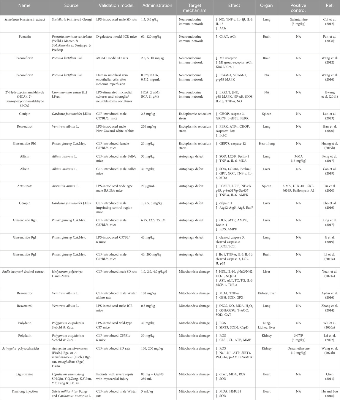
Table 4. Characteristics of TCM extracts by the modulation of neuroendocrine immune network, endoplasmic reticulum stress, autophagy defect, and mitochondria damage on sepsis.
The administration of S. baicalensis extract in rats with acute lung injury resulted in an increase in serum ACh levels and a decrease in serum TNF-α and NO levels, effectively inhibiting the progression of inflammatory response (Cui et al., 2012). However, there were no significant effects observed on choline acetyltransferase (ChAT) and acetylcholinesterase (AChE) activities related to ACh synthesis and decomposition. Therefore, it can be inferred that the cholinergic anti-inflammatory pathway modulated by S. baicalensis extract may be attributed to its ability to enhance ACh release, which subsequently binds to nicotinoid α7 receptors or M receptors on monocytes, leading to a significant inhibition of TNF-α through intracellular signal transduction pathways. This mechanism also suppresses the release of proinflammatory factors IL-1β, IL-6, and IL-18, thereby regulating the overall inflammatory response.
Puerarin was found to enhance choline acetyltransferase activity and increase ACh content in the cerebral cortex of D-galactose rats (Pan et al., 2008). However, it did not exert a significant effect on cholinesterase activity. These findings suggest that puerarin’s anti-inflammatory effect may be attributed to its ability to activate the vagus nerve. Experimental studies have demonstrated that paeoniflorin exerts a protective effect on inflammatory cells through the regulation of cholinergic M receptor signaling pathway, modulation of cholinergic M receptor activity, and activation of the M-receptor-G protein-KATP channel (Wang et al., 2012). Paeoniflorin was postulated to exert its inhibitory effects on adhesion factors and the p38MAPK signaling pathway through the regulation of the cholinergic anti-inflammatory pathway, as demonstrated in experimental models (Wang et al., 2016). This regulatory mechanism ultimately leads to an amelioration of inflammatory responses in endothelial cells, alleviation of tissue damage, and a protective role in HUVECs.
2′-hydroxycinnamaldehyde (HCA) and 2′-benzoyloxycinnamaldehyde (BCA) in LPS-stimulated microglia cultures and microglia/neuroblastoma cocultures were observed to have the potential anti-neuroinflammatory effects, which protected neuroblastoma cells from microglia-mediated cell death (Hwang et al., 2011). It is postulated that low density lipoprotein receptor-related protein 1 (LRP1) serves as a promising molecular target for HCA in modulating microglial responses. The findings demonstrated that treatment with HCA at a concentration of 2 μmol/L and BCA at a concentration of 1 μmol/L effectively suppressed the expression of iNOS, proinflammatory cytokines IL-1β and TNF-α, as well as NO production by inhibiting the activation of ERK1/2, JNK, p38 MAPK, and NF-κB signaling pathways. These results highlight the therapeutic potential of HCA and BCA in managing neuroinflammatory diseases.
In summary, TCM extracts may inhibit the release of pro-inflammatory cytokines through the cholinergic anti-inflammatory pathway, playing an anti-neuroinflammatory effect and ultimately reducing tissue damage in sepsis (show in Figure 2).
The endoplasmic reticulum is an intracellular organelle involved in the translocation, folding, post-translational modifications, and subsequent transport of intracellular organelles to the Golgi apparatus (Khan et al., 2015). In sepsis, accumulation of unfolded or misfolded proteins in the endoplasmic reticulum disrupts its homeostasis and leads to oxidative stress and severe calcium disturbances, resulting in endoplasmic reticulum stress (Khan et al., 2015). During endoplasmic reticulum stress, the unfolded protein response sensor may undergo a unique signal switch to induce cell death. The registration signaling mechanism involves several sequential steps: Firstly, the transcriptional activation of the C/EBP homologous protein (CHOP) gene is mediated by pkr-like endoplasmic reticulum kinase (PERK), inositol-requiring enzyme 1 (IRE1), and transcriptional activator 6 (ATF6). Secondly, there is an activation of the IRE-mediated JNK pathway, followed by TNF receptor-associated activator 2 and apoptosis signal-regulated kinase 1. Finally, activated caspase-12 associated with caspase-12 activation migrates from the endoplasmic reticulum to the cytoplasm, leading to cleavage of caspase-9 and ultimately activating caspase-3 (Li et al., 2014; García de la Cadena and Massieu, 2016; Zhang et al., 2018). In animal models of sepsis, increased markers of endoplasmic reticulum stress [e.g., glucose-regulated protein 94 (GRP94), CHOP, and caspase-12] have been observed in various organs including the heart and liver. Additionally, a direct correlation between these markers and the degree of organ dysfunction suggests that they may play a significant role in causing multi-organ failure in sepsis (Jiao et al., 2017). The induction of abnormal apoptosis due to endoplasmic reticulum stress in septic animals indicates that targeting endoplasmic reticulum stress-mediated apoptosis could be a promising avenue for research on clinical prevention and treatment strategies for sepsis. Traditional Chinese medicine has shown potential in attenuating lymphocyte apoptosis induced by sepsis through inhibition of the endoplasmic reticulum stress pathway (Xi et al., 2017). The characteristics of TCM extracts by the modulation of endoplasmic reticulum stres on sepsis are summarized in Table 4.
Genipin, derived from Gardenia jasminoides J.Ellis fruit, is a well-known traditional Chinese medicine with antipyretic and detoxifying properties. It has been demonstrated to possess anti-inflammatory, antioxidant, and bacteriostatic activities (Ishiguro et al., 1983; Luo et al., 2019). Recent studies have indicated that genipin can enhance the prognosis of sepsis while reducing sepsis-related liver damage and lung injury (Kim et al., 2012).
The impact of genipin on apoptosis induced by endoplasmic reticulum stress in spleen cells following CLP was further investigated (Luo et al., 2023). The findings demonstrated that genipin significantly attenuated the expression levels of splenic CHOP and caspase-3 proteins in CLP mice. Moreover, genipin markedly reduced the number of tunel-positive splenocytes. These results indicated that genipin possessed potential protective effects against sepsis. Furthermore, it was revealed that the underlying protective mechanism of genipin against sepsis involves inhibiting endoplasmic reticulum stress, downregulating CHOP protein expression, and mitigating splenocyte apoptosis.
Curcumin is a crucial active ingredient in Curcuma longa L. that exerts pharmacological effects. (Marchiani et al., 2014). Numerous animal experiments have consistently demonstrated that curcumin exhibits a broad spectrum of pharmacological activities including anti-inflammatory, anti-oxidative stress, anti-apoptotic, and immunomodulatory effects (Shehzad et al., 2010). Moreover, an extensive body of research has confirmed the protective effect of curcumin against sepsis and septic shock (Karimi et al., 2019; Wang S. et al., 2021; Vieira et al., 2023).
Resveratrol is a small non-flavonoidal polyphenolic compound present in various plants. Pharmacological studies have indicated that resveratrol exhibits diverse biological activities, such as anti-inflammatory, antioxidant properties, oxygen free radical scavenging abilities, and antitumor effects (Hiramatsu et al., 2015; Lu et al., 2018). Han et al. (2020) utilized the LPS method to induce the establishment of a rabbit model of acute lung injury (ALI). The rabbits were grouped and intervened with resveratrol and sodium chloride injection. The findings from this study demonstrated that resveratrol effectively reduced the protein level and mRNA expression of PERK, ATF4, CHOP, caspase-9, Bax while increasing the protein and mRNA expression level of Bcl-2 in ALI-induced lung tissues. Additionally, resveratrol attenuated histopathological injury induced by LPS in ALI rabbit lungs. These results suggest that resveratrol exerts a protective role against LPS-induced ALI in rabbits by mitigating endoplasmic reticulum stress and apoptosis.
The sepsis mouse model using the CLP method was successfully replicated by Huang et al. (2019b), and their experimental study demonstrated significant improvement in the overall condition of mice treated with ginsenoside Rb1 compared to those in the CLP group. Additionally, the alveolar tissues exhibited clear and intact structures with reduced infiltration of inflammatory cells, resulting in uniform-sized alveoli. Myocardial tissues showed decreased infiltration of inflammatory cells, visible transverse striations of the myocardium, and a reduction in necrotic cells. The expression levels of GRP78 and caspase-12 proteins in myocardial and lung tissues of the ginsenoside group exhibited a significant reduction, with highly statistically significant differences (p < 0.01). These findings suggest that ginsenoside Rb1 can attenuate the severity of infection and exert a protective effect on heart and lung tissue damage in septic mice. This mechanism may be attributed to the inhibition of excessive endoplasmic reticulum stress by ginsenoside Rb1.
Therefore, TCM extracts inhibit endoplasmic reticulum stress and reduce the expression of CHOP apoptotic protein, thereby alleviating the degree of cell apoptosis and achieving improvement in organ dysfunction caused by sepsis (show in Figure 2).
Autophagy is a natural process in which cytoplasmic material or pathogens are phagocytosed and subsequently degraded through fusion with lysosomes. It serves as a crucial defense mechanism employed by the host to combat external pathogens and danger signals, while also playing an integral role in inducing and regulating the inflammatory response of innate immune cells - a key determinant influencing sepsis development (Qiu et al., 2019). Autophagy plays a protective role in sepsis, potentially through the following mechanisms: pathogen elimination, neutralization of microbial toxins, regulation of cytokine release, reduction of apoptotic cancer targets, and promotion of antigen expression (Nakahira et al., 2011; Ryter et al., 2014; Maurer et al., 2015). Chinese medicines can prevent sepsis by modulating autophagy-related signaling pathways, primarily involving the regulation of the AMPK pathway and PINK1/Parkin pathway. Additionally, they inhibit autophagy-related signaling pathways such as the JNK pathway, mTOR pathway, NF-κB pathway, as well as apoptosis to achieve anti-inflammatory effects and mitigate organ damage (Chang et al., 2023). The characteristics of TCM extracts by the modulation of autophagy defect on sepsis are summarized in Table 4.
Recently, there is mounting evidence indicating that autophagy defects may contribute to the development of sepsis-induced immunosuppression (Ren et al., 2017; Venet and Monneret, 2018). In a study, the expression of autophagy marker proteins LC3B and Beclin-l in sepsis-associated acute lung injury (ALI) was examined, along with the impact of allicin treatment on these markers (Peng et al., 2017). The findings revealed an increase in the expression of LC3B and Beclin-1 in lung tissues of mice with sepsis ALI, suggesting an upregulation in autophagy levels during septic ALI. Furthermore, treatment with allicin further augmented the expression of LC3B and Beclin-1 in lung tissue, indicating its ability to enhance autophagy levels and exert a protective effect against septic ALI. Another study showed that allicin enhanced the expression of LC3-B and Beclin-1 in hepatic tissues of septic mice, with a direct correlation to the level of autophagy (Gao et al., 2019). This study suggested that allicin’s hepatoprotective effect might be attributed to its modulation of autophagic activity.
The administration of artesunate upregulated autophagy and reversed the tolerant state in LPS-tolerant mice (Liu et al., 2020). Their findings unveiled a novel immunopharmacological effect of artesunate in reversing LPS tolerance by restoring autophagy. It has been demonstrated that artesunate restores cytokine production and enhances bacterial clearance through the induction of autophagy.
The mice were induced sepsis in through CLP and observed a decrease in hepatic expression of autophagy-associated protein Atg3, which was attenuated by genipin. CLP impaired autophagic flux, resulting in increased expression of hepatic microtubule-associated protein-1 light chain 3-II and sequestoome-1/p62 proteins; however, genipin restored the impaired autophagic flux and attenuated the CLP-induced decrease in hepatic lysosome-associated membrane protein-2 and Rab7 proteins as well as elevated expression of calpain 1 protein. These findings suggest that genipin protects against septic injury by restoring impaired autophagic flux through inhibition of the calpain system (Cho et al., 2016).
Xing et al. (2017) conducted in vivo and in vitro models of sepsis using CLP rats and LPS-treated human primary hepatocytes, respectively. They discovered that ginsenoside Rg3 can alleviates mitochondrial autophagy by activating the AMPK signaling pathway, increasing the levels of autophagy-associated proteins LC3BI, LC3II, and Beclin-1. The role of mitochondrial autophagy was investigated in rat liver and human primary hepatocytes. The effect of LPS on autophagy in mouse lung epithelial cells was observed a gradual increase in the expression of LC3II/LC3I (an autophagosome membrane protein) from 0 to 16 h followed by a decrease at 24 h (Ji et al., 2019). This suggested that autophagy initially increased but then decreased during the process induced by LPS. In addition, this study revealed that ginsenoside Rg1 administration increased autophagy and mitigated LPS-induced lung epithelial cell modulation (modulation rate decreased from 9.33% to 6.14%) after 24 h of LPS exposure in mouse lung epithelial cells, thereby attenuating sepsis-induced lung injury. Ginsenoside Rg1 was demostrated to protect against sepsis-induced brain injury by reducing the levels of autophagy-associated proteins LC3-II and p62 in the hippocampal region, inhibiting accumulation, and suppressing atypical Beclin-independent autophagy to safeguard against sepsis-induced brain injury (Li Y. et al., 2017). These studies collectively highlight the significance of modulating autophagy using ginsenosides within specific temporal and spatial parameters for effective sepsis treatment.
Traditional Chinese medicine extracts effectively modulate autophagy levels to mitigate sepsis, suppress inflammation, and attenuate multipe organ damage (show in Figure 2).
Mitochondria are crucial microcellular organelles involved in energy production, protein synthesis, and catabolism (Vringer and Tait, 2023). However, sepsis-induced mitochondrial damage or dysfunction can result in cellular damage, metabolic disorders, inadequate energy production, and oxidative stress. This ultimately leads to apoptosis of organ cells and immune cells, thereby causing multiorgan immune disorders, multiorgan failure, and even death. During sepsis, the antioxidant system mechanisms are disrupted due to limited oxygen supply and incomplete oxidative reactions as well as hypoxia. Consequently, there is a significant increase in the production of free radicals (Rocha et al., 2012). When exposed to DAMP or PAMP, activated leukocytes release inflammatory cytokines that stimulate NADPH-expressing oxidases (Quoilin et al., 2014). These cytokines induce an excessive production of reactive nitrogen species (RNS), which inhibits NO through the promotion of iNOS activity. The combination of NO and ROS can lead to peroxidation, resulting in the formation of RNS, which irreversibly inhibits electron transport chain (ETC.) activity. Consequently, this dysfunction of the ETC contributes to the generation of additional ROS within mitochondria during sepsis, unfortunately leading to further mitochondrial damage such as endosomal impairment, inhibition of ETC activity, and mitochondrial DNA damage (Singer, 2014). Eventually, the mitochondrial matrix undergoes swelling, leading to rupture of the mitochondrial membrane and initiation of apoptosis. Traditional Chinese medicine has the ability to modulate apoptosis balance, stabilize mitochondrial membrane potential, and regulate other pathways in order to exert a protective effect against sepsis-induced mitochondrial damage. The characteristics of TCM extracts by the modulation of mitochondria damage on sepsis are summarized in Table 4.
Nuclear related factor 2 (Nrf2) is the most potent transcription factor that regulates the antioxidant stress response in the organism. The stimulation of cells by oxygen free radicals leads to the phosphorylation of Nrf2, its dissociation from specific receptors, translocation to the nucleus, and subsequent initiation of the expression of various downstream antioxidant genes including HO-1 and NQO-1 (Liu et al., 2016; Senthil et al., 2016). The proteins HO-1 and NQO-1 possess antioxidant, anti-apoptotic, anti-inflammatory, and microcirculation maintenance functions, which play a crucial role in the body’s defense against oxidative stress (Choi, 2016; Hafez et al., 2019). The levels of Nrf2 were significantly reduced in sepsis-induced liver-injured rats, leading to an imbalance in the body’s oxidative/antioxidant system. This imbalance results in the release of a substantial amount of oxygen free radicals and subsequently induces oxidative stress injury in liver tissues (Yang et al., 2018). The alcoholic extract derived from Hedysarum polybotrys Hand.-Mazz. (Radix hedysari) was discovered to significantly enhance the phosphorylation level of pNrf2/Nrf2 and the expression levels of HO-1 and NQO-1 in hepatic tissues of septic rats, indicating that R. hedysari alcohol extract could elevate antioxidant activity and eliminate excessive oxygen free radicals in liver tissues of septic rats, thereby mitigating inflammatory damage to hepatic tissues (Yuan C. et al., 2021).
Aydin et al. (2016); Zhang et al. (2014) demonstrated that resveratrol exhibited a reduction in iNOS and NO concentrations, an increase in the ratio of reduced glutathione to oxidized glutathione (GSH/GSSG), as well as an elevation in peroxidase activity (catalase, superoxide dismutase, and other antioxidant enzymes). Moreover, these compounds effectively mitigated organ damage in septic mice.
Polydatin could effectively activate SIRT3, leading to the deacetylation of SOD2, reduction in ROS production, stabilization of endothelial adherens junction (AJ), and ultimately improved prognosis in septic mice through the protection of endothelial permeability (Wu J. et al., 2020). Polydatin enhanced the expression of CLS1 in SAKI through SIRT3, thereby promoting CL synthesis and improving mitochondrial function (Lei et al., 2022). Astragalus polysaccharides could enhance the energy metabolism of renal epithelial cells, improved the mitochondrial structure of the renal cortex, and mitigated histopathological changes in septic acute kidney injury rats (Wang X. et al., 2021).
The amide alkaloid ligustrazine was obtained through isolation and purification from the traditional Chinese medicine Ligusticum chuanxiong S.H.Qiu, Y.Q.Zeng, K.Y.Pan, Y.C.Tang & J.M.Xu. It acts as a novel calcium antagonist, exhibiting a wide range of pharmacological effects including antioxidant, anti-inflammatory, and antifibrotic activities (Ran et al., 2011). The compound can scavenge ROS by reducing plasma malondialdehyde (MDA) levels and increasing superoxide dismutase (SOD) activity, thereby providing protection against biofilm damage, acting as an antioxidant, and effectively improving myocardial injury in sepsis (Chen, 2011). Early administration of ligustrazine can effectively scavenge ROS, inhibit lipid peroxidation, and have other beneficial effects that contribute to improved patient prognosis (Wang, 2012; Liu, 2014). The Danhong injection exhibits a certain efficacy in combating myocardial damage induced by sepsis, as it effectively suppresses the levels of MDA and HMGB1 in the cardiac tissue of CLP septic rats while simultaneously enhancing SOD activity (Hu and Lou, 2016).
In summary, TCM extracts can exert a protective effect on sepsis-induced mitochondrial damage by inhibiting oxidative stress reactions, regulating the balance of oxidation/antioxidation, and eliminating ROS (show in Figure 2).
As a significant form of complementary and integrated medicine, TCM possesses immense potential and advantages in the treatment of sepsis (Liang et al., 2015; Wang J. et al., 2022). The intervention of TCM can effectively leverage the role in addressing bacterial resistance during the diagnosis and treatment of sepsis patients, capitalizing on its distinct advantages across different stages. In the early stage of sepsis, the implementation of TCM as a complementary antibiotic therapy, particularly for patients with resistant bacterial infections, can effectively mitigate the emergence of drug-resistant bacteria, alter the resistance patterns exhibited by existing resistant or pan-resistant strains, and subsequently reduce the occurrence rate of multiple infections. The spleen and stomach play a crucial role in the development of sepsis by enhancing mental resilience and reducing the incidence of multiple organ dysfunction syndrome (MODS) through promoting clearance and minimizing turbidity. If MODS occurs, TCM can also be utilized to combat shock, dysfunction in blood coagulation, rectify the imbalance of inflammatory response, and mitigate organ damage. On the other hand, the exceptional merits of TCM are evident in its remarkable safety profile: according to a comprehensive investigation on drug-induced liver injury (DILI)-related adverse drug reactions (ADR) in China (L-ADR), Chinese medicine accounted for merely 4.5% of all reported ADR cases, while conventional drugs encompassing chemical and biological agents constituted 95.5%, with antibiotics including anti-tuberculosis medications representing the highest proportion (Wang J. et al., 2022).
In the treatment of sepsis and its complications, traditional Chinese medicine extracts and active ingredients have multiple effects such as anti-inflammatory properties, improvement of microcirculation, alleviation of gastrointestinal dysfunction, enhancement of the immune system, and prevention of organ damage. Traditional Chinese medicine extracts not only regulate the inflammation imbalance, improve impaired immune function, and enhance coagulation disorders to treat sepsis and improve patient prognosis. They also exert therapeutic effects on sepsis and multiple organ dysfunction by modulating the neuroendocrine-immune network, inhibiting endoplasmic reticulum stress, regulating autophagy defects, and improving mitochondrial damage. Given the complexity of chemical components in traditional Chinese medicine and the uncertainty of active ingredients, most studies have focused on isolating active compounds from prescriptions and single herbs for cellular or animal research. However, whether traditional Chinese medicine exacerbates multi-organ damage in sepsis patients due to potential side effects on relevant organs remains a crucial question that has not been thoroughly studied. Therefore, identifying and evaluating the potential harm of drugs on organ function is the main challenge faced by traditional Chinese medicine in terms of medication safety for sepsis treatment. In addition, there is currently no universally recognized standard for evaluating the efficacy of traditional Chinese medicine. Furthermore, the quality control of clinical trials for traditional Chinese medicine also needs improvement. In the future, a multidisciplinary collaborative effort will be necessary to elucidate the material basis of traditional Chinese medicine and provide important evidence for understanding the pathogenesis of sepsis and developing new drugs for treating sepsis. Therefore, it is imperative to introduce new systematic, standardized, and operationally strong guidelines in this research field to expedite the modernization and standardization process of traditional Chinese medicine.
This review classifies and summarizes the efficacious monomeric constituents of traditional Chinese medicine for the treatment of sepsis based on their mechanisms of action. The findings demonstrate that the majority of potent monomeric components exert their effects by directly attenuating inflammatory factors, while some possess inhibitory properties against the NF-κB pathway or exhibit direct antagonism towards LPS. Others enhance patient survival rates by mitigating disseminated intravascular coagulation through anticoagulant activity. Furthermore, polysaccharides and saponins rectify functional abnormalities in sepsis via immunoregulatory effects, whereas specific monomers and extracts regulate autophagy to prevent and manage sepsis. This review provides valuable insights into the diverse pathways and therapeutic targets of monotherapy with TCM for sepsis treatment, facilitating a comprehensive comprehension of its underlying mechanisms.
The current research primarily focuses on the comprehensive study of TCM. The clinical application of TCM compound, such as Qingwenbaidu decoction (Leng et al., 2012), Dachengqi decoction, and Xuebijing injection (Ma and Song, 2009), is prevalent to therapy spesis; however, there is limited research on the TCM monomer. However, the mechanisms of action and target sites of TCM extracts are still unclear, and issues such as the lack of standardized dosages for effective ingredients from these components remain unresolved. The chemical structure of medicine, either similar to or belonging to the genus of TCM itself, exhibits a certain correlation. By means of animal and cell experimental research, it is possible to expedite the identification of TCM monomer compositions and leverage modern high-tech advancements for better understanding important targets and advantages in sepsis treatment. Therefore, the researchers can further facilitate the discovery or synthesis of analogs or derivatives, expanding the scope of the new drug research and development of TCM. The potential targets of TCM extracts and active components in the treatment of sepsis and its complications have been identified through a network pharmacological prediction, molecular docking analysis, and visual analysis in the field of molecular biology research. The experimental study on the mechanism of TCM treatment for sepsis, combined with the aforementioned methods, offers a novel direction in the quest for more efficacious intervention strategies. The research field requires the introduction of new systematic, normative, and operable guidelines to gain international recognition for the therapeutic advantages of TCM in treating acute and critical diseases. Most traditional Chinese medicines are administered orally, thereby entering the digestive tract. Consequently, the gut microbiota plays a pivotal role in the metabolism of these medications. Moreover, numerous traditional Chinese medicines exert an influence on the equilibrium of gut microbiota. Henceforth, future research endeavors should consider exploring the correlation between sepsis and gut microbiota status, as well as investigating the regulatory effects of traditional Chinese medicines on gut microbiota.
C-FF: Writing–original draft, Conceptualization. J-LL: Writing–original draft, Conceptualization. J-WC: Writing–original draft, Project administration. HL: Writing–original draft, Visualization. W-RZ: Writing–original draft, Visualization. S-YH: Writing–original draft, Project administration. X-WM: Writing—review and editing, Funding acquisition. X-FY: Writing–review and editing. H-LW: Writing–review and editing.
The author(s) declare financial support was received for the research, authorship, and/or publication of this article. This work was supported by Heilongjiang Province Provincial Higher Education Institutions’ Basic Research Operating Expenses Scientific Research Programs (No. 2023-KYYWFMY-0011).
The authors would like to thank the reviewers and authors of all references.
The authors declare that the research was conducted in the absence of any commercial or financial relationships that could be construed as a potential conflict of interest.
All claims expressed in this article are solely those of the authors and do not necessarily represent those of their affiliated organizations, or those of the publisher, the editors and the reviewers. Any product that may be evaluated in this article, or claim that may be made by its manufacturer, is not guaranteed or endorsed by the publisher.
Abu Bakar, N. H., Sukari, M. A., Rahmani, M., Sharif, A. M., Khalid, K., and Yusuf, U. K. (2007). Chemical constituents from stem barks and roots of Murraya koenigii (Rutaceae). Malays. J. Anal. Sci. 11, 173–176.
Arefian, H., Heublein, S., Scherag, A., Brunkhorst, F. M., Younis, M. Z., Moerer, O., et al. (2017). Hospital-related cost of sepsis: a systematic review. J. Infect. 74, 107–117. doi:10.1016/j.jinf.2016.11.006
Aydın, S., Şahin, T. T., Bacanlı, M., Taner, G., Başaran, A. A., Aydın, M., et al. (2016). Resveratrol protects sepsis-induced oxidative DNA damage in liver and kidney of rats. Balk. Med. J. 33, 594–601. doi:10.5152/balkanmedj.2016.15516
Biemond, B. J., Levi, M., Ten Cate, H., Van Der Poll, T., Büller, H. R., Hack, C. E., et al. (1995). Plasminogen activator and plasminogen activator inhibitor I release during experimental endotoxaemia in chimpanzees: effect of interventions in the cytokine and coagulation cascades. Clin. Sci. 88, 587–594. doi:10.1042/cs0880587
Cecconi, M., Evans, L., Levy, M., and Rhodes, A. (2018). Sepsis and septic shock. Lancet 392, 75–87. doi:10.1016/S0140-6736(18)30696-2
Chang, J., Shang, T., Wang, X., Zhou, Z., Zhang, Z., and Feng, Y. (2023). Research progress on traditional Chinese medicine in treatment of sepsis by regulating autophagy signaling pathway. Chin. Tradit. Herb. Drugs 54, 2349–2356. doi:10.7501/j.issn.0253-2670.2023.07.035
Chang, Y., Chou, Y.-T., Kan, W., and Shiao, C. (2022). Sepsis and acute kidney injury: a review focusing on the bidirectional interplay. Int. J. Mol. Sci. 23, 9159. doi:10.3390/ijms23169159
Chen, C. (2011). The clinical study of severe sepsis myocardial damage treated with tetramethylpyrazine. Grass-Roots Med. Forum 15, 1097–1099.
Cho, H.-I., Kim, S.-J., Choi, J.-W., and Lee, S.-M. (2016). Genipin alleviates sepsis-induced liver injury by restoring autophagy. Br. J. Pharmacol. 173, 980–991. doi:10.1111/bph.13397
Choi, Y. H. (2016). The cytoprotective effects of ethanol extract of Ecklonia cava against oxidative stress are associated with upregulation of Nrf2-mediated HO-1 and NQO-1 expression through activation of the MAPK pathway. Gen. Physiol. Biophys. 35, 45–53. doi:10.4149/gpb_2015029
Cui, R., Meng, X., Wang, P., Li, H., and Yu, Y. (2012). Correlation between protection of Scutellaria baicalensis extract on rats’ acute lung injury and cholinergic anti-inflammatory pathway. Chin. Tradit. Herb. Drugs 43, 321–326.
Deng, X., Huang, J., and Dong, J. (2017). Cholinergic anti-inflammatory pathway and its progress on anti-inflammatory research of traditional Chinese medicine. Chin. J. Tradit. Chin. Med. 32, 1680–1683.
Fan, T., Cheng, B., Fang, X., Chen, Y.-C., and Su, F. (2020). Application of Chinese medicine in the management of critical conditions: a review on sepsis. Am. J. Chin. Med. 48, 1315–1330. doi:10.1142/S0192415X20500640
Fitridge, R., and Thompson, M. (2007) Mechanisms of vascular disease: a textbook for vascular surgeons. Cambridge, New York: Cambridge University Press. Available at: https://cir.nii.ac.jp/crid/1130282269895309952 (Accessed January 2, 2024).
Fujii, T., Mashimo, M., Moriwaki, Y., Misawa, H., Ono, S., Horiguchi, K., et al. (2017). Expression and function of the cholinergic system in immune cells. Front. Immunol. 8, 1085. doi:10.3389/fimmu.2017.01085
Gao, M., Peng, Y., Yang, M., Jiang, Y., and Yang, B. (2019). Effect of allicin on sepsis-induced acute liver injury in mice. Chin. J. Clin. Pharmacol. 35, 3213–3216. doi:10.13699/j.cnki.1001-6821.2019.24.010
Gao, S., Zhang, Y., and Niu, X. (2018). Effect of Astragalus injection on T lymphocyte subsets in peripheral blood of patients with traumatic splenic rupture undergoing splenectomy. Mod. J. Integr. Tradit. Chin. West. Med. 27, 409–411. doi:10.3969/j.issn.1008-8849.2018.04.021
García de la Cadena, S., and Massieu, L. (2016). Caspases and their role in inflammation and ischemic neuronal death. Focus on caspase-12. Apoptosis 21, 763–777. doi:10.1007/s10495-016-1247-0
Groote, T. V., and Meersch-Dini, M. (2022). Biomarkers for the prediction and judgement of sepsis and sepsis complications: a step towards precision medicine? J. Clin. Med. 11, 5782. doi:10.3390/jcm11195782
Guo, Q., Zhu, M., Zhang, J., Xu, J., and Guo, Y. (2022). Salidroside regulates pulmonary fibrosis and JAK2/STAT3 activation in CLP-induced sepsis mice. Chin. J. Immunol. 38, 1196–1200. doi:10.3969/j.issn.1000-484X.2022.10.008
Gupta, S., George, M., Singhal, M., Sharma, G. N., and Garg, V. (2010). Leaves extract of Murraya koenigii Linn for anti-inflammatory and analgesic activity in animal models. J. Adv. Pharm. Technol. Res. 1, 68–77. doi:10.4103/2231-4040.70525
Gupta, S., Khajuria, V., Wani, A., Nalli, Y., Bhagat, A., Ali, A., et al. (2019). Murrayanine attenuates lipopolysaccharide-induced inflammation and protects mice from sepsis-associated organ failure. Basic Clin. Pharmacol. Toxicol. 124, 351–359. doi:10.1111/bcpt.13032
Hafez, H. M., Ibrahim, M. A., Zedan, M. Z., Hassan, M., and Hassanein, H. (2019). Nephroprotective effect of cilostazol and verapamil against thioacetamide-induced toxicity in rats may involve Nrf2/HO-1/NQO-1 signaling pathway. Toxicol. Mech. Methods 29, 146–152. doi:10.1080/15376516.2018.1528648
Han, H., and Wu, W. (2010). Research progress on anti-inflammatory effect of Salvia miltiorrhiza. Med. J. Gentral Equip. Hqrs. 12, 118–119.
Han, X., Sun, J., Zhang, M., Yu, H., and Wang, J. (2020). Effect of resveratrol on endoplasmic reticulum stress and apoptosis in rabbit with acute lung injury. Acad. J. Chin. PLA Med. Sch. 41, 364–368. doi:10.3969/j.issn.2095-5227.2020.04.012
Hiramatsu, N., Chiang, W.-C., Kurt, T. D., Sigurdson, C. J., and Lin, J. H. (2015). Multiple mechanisms of unfolded protein response–induced cell death. Am. J. Pathol. 185, 1800–1808. doi:10.1016/j.ajpath.2015.03.009
Honma, K., Udono, H., Kohno, T., Yamamoto, K., Ogawa, A., Takemori, T., et al. (2005). Interferon regulatory factor 4 negatively regulates the production of proinflammatory cytokines by macrophages in response to LPS. Proc. Natl. Acad. Sci. 102, 16001–16006. doi:10.1073/pnas.0504226102
Hu, D., and Lou, L. (2016). Expressions of oxygen-free radicals and HMGB1 in heart tissue of rats with sepsis and effect of danhong injection. Chin. Arch. Tradit. Chin. Med. 34, 163–166. doi:10.13193/j.issn.1673-7717.2016.01.045
Huang, M., Cai, S., and Su, J. (2019a). The pathogenesis of sepsis and potential therapeutic targets. Int. J. Mol. Sci. 20, 5376. doi:10.3390/ijms20215376
Huang, M., Tao, X., Jia, B., Yang, S., and Xu, C. (2019b). Protective effect of ginsenoside Rb1 on regulating endoplasmic reticulum stress in important organs of septic mice. China Med. Her. 16, 17–20+25.
Huynh, D. T. N., Baek, N., Sim, S., Myung, C.-S., and Heo, K.-S. (2020). Minor ginsenoside Rg2 and Rh1 attenuates LPS-induced acute liver and kidney damages via downregulating activation of TLR4-STAT1 and inflammatory cytokine production in macrophages. Int. J. Mol. Sci. 21, 6656. doi:10.3390/ijms21186656
Hwang, H., Jeon, H., Ock, J., Hong, S. H., Han, Y.-M., Kwon, B.-M., et al. (2011). 2′-Hydroxycinnamaldehyde targets low-density lipoprotein receptor-related protein-1 to inhibit lipopolysaccharide-induced microglial activation. J. Neuroimmunol. 230, 52–64. doi:10.1016/j.jneuroim.2010.08.021
Ishiguro, K., Yamaki, M., and Takagi, S. (1983). Studies on iridoid-related compounds, II. The structure and antimicrobial activity of aglucones of galioside and gardenoside. J. Nat. Prod. 46, 532–536. doi:10.1021/np50028a018
Ji, Q., Sun, Z., Yang, Z., Zhang, W., Ren, Y., Cao, L., et al. (2019). Effects of Rg_1 on LPS-induced apoptosis and autophagy of lung epithelial cells. China J. Chin. Mat. 44, 1648–1653. doi:10.19540/j.cnki.cjcmm.20190111.002
Jiang, D., Liu, A., and Ling, W. (2022). Action mechanism of curcumin on relieving lung injury in sepsis rats. Anhui Med. Pharm. J. 26, 1706–1710+1909. doi:10.3969/j.issn.1009-6469.2022.09.003
Jiao, G., Hao, L., Wang, M., Zhong, B., Yu, M., Zhao, S., et al. (2017). Upregulation of endoplasmic reticulum stress is associated with diaphragm contractile dysfunction in a rat model of sepsis. Mol. Med. Rep. 15, 366–374. doi:10.3892/mmr.2016.6014
Jin, F., Li, W., Zheng, Y., and Wang, D. (2022). Research progress on the mechanism of treating sepsis related coagulation dysfunction by Fuzheng Huayu Method. China Mod. Medcine 29, 37–40+49.
Jing, Y., Liu, L., Zhou, X., Zhao, G., Li, B., Zhang, H., et al. (2022). Effect of Shenfu injection on immune function of septic shock patients: a meta-analysis. World J. Integr. Tradit. West. Med. 17, 2367–2373. doi:10.13935/j.cnki.sjzx.221206
Jones, J. E., Le Sage, V., and Lakdawala, S. S. (2021). Viral and host heterogeneity and their effects on the viral life cycle. Nat. Rev. Microbiol. 19, 272–282. doi:10.1038/s41579-020-00449-9
Kanczkowski, W., Sue, M., Zacharowski, K., Reincke, M., and Bornstein, S. R. (2015). The role of adrenal gland microenvironment in the HPA axis function and dysfunction during sepsis. Mol. Cell. Endocrinol. 408, 241–248. doi:10.1016/j.mce.2014.12.019
Karimi, A., Ghodsi, R., Kooshki, F., Karimi, M., Asghariazar, V., and Tarighat-Esfanjani, A. (2019). Therapeutic effects of curcumin on sepsis and mechanisms of action: a systematic review of preclinical studies. Phytother. Res. 33, 2798–2820. doi:10.1002/ptr.6467
Khan, M. M., Yang, W., and Wang, P. (2015). Endoplasmic reticulum stress in sepsis. Shock Augusta Ga 44, 294–304. doi:10.1097/SHK.0000000000000425
Kim, M. K., Kang, H., Baek, C. W., Jung, Y. H., Woo, Y. C., Choi, G. J., et al. (2018). Antinociceptive and anti-inflammatory effects of ginsenoside Rf in a rat model of incisional pain. J. Ginseng Res. 42, 183–191. doi:10.1016/j.jgr.2017.02.005
Kim, T.-H., Yoon, S.-J., and Lee, S.-M. (2012). Genipin attenuates sepsis by inhibiting Toll-like receptor signaling. Mol. Med. 18, 455–465. doi:10.2119/molmed.2011.00308
Lee, W., Ku, S.-K., and Bae, J.-S. (2013). Emodin-6-O-β-D-glucoside down-regulates endothelial protein C receptor shedding. Arch. Pharm. Res. 36, 1160–1165. doi:10.1007/s12272-013-0114-6
Lei, X., Ding, Q., Li, T., and Deng, J. (2022). The effect and mechanism of polydatin on the cardiolipin synthesis of the septic acute kidney injury. J. Trop. Med. 22, 906–909+1033. doi:10.3969/j.issn.1672-3619.2022.07.003
Lei, X., and Li, T. (2021). Polydatin could mitigate the inflammation, oxidative stress and apoptosis via SIRT3 in the septic acute kidney injury of the mice. J. Trop. Med. 21, 831–834+808.
Leng, J., Luo, Y., and Guo, X. (2012). Effect of Qingwenbaiduyin on clinical effects of sepsis and immunological parameters in serum. China J. Tradit. Chin. Med. Pharm. 27, 758–760.
Levi, M., and Poll, T. van der (2015). Coagulation in patients with severe sepsis. Semin. Thromb. Hemost. 41, 9–15. doi:10.1055/s-0034-1398376
Li, F., Wang, H., Lu, D., Wang, Y., Qi, R., Fu, Y., et al. (2006). Neutral sulfate berberine modulates cytokine secretion and increases survival in endotoxemic mice. Acta Pharmacol. Sin. 27, 1199–1205. doi:10.1111/j.1745-7254.2006.00368.x
Li, L., Li, L., Li, G., and Ge, D. (2015). Different ratios and dosages of Huangqi (Astragalus) and Danggui (Angelica) for pulmonary fibrosis in mice lung morphology and hydroxyproline content. J. Liaoning Univ. TCM 17, 30–33. doi:10.13194/j.issn.1673-842x.2015.03.010
Li, Q., and Jin, X. (2016). Effects of safflower yellow A on the regulation of serum TNF-α, TF and TFPI and blood coagulation in septic rats. J. Baotou Med. Coll. 32, 3–4. doi:10.16833/j.cnki.jbmc.2016.02.002
Li, R., Guo, Y., Zhang, Y., Zhang, X., Zhu, L., and Yan, T. (2019). Salidroside ameliorates renal interstitial fibrosis by inhibiting the TLR4/NF-κB and MAPK signaling pathways. Int. J. Mol. Sci. 20, 1103. doi:10.3390/ijms20051103
Li, Y., Guo, Y., Tang, J., Jiang, J., and Chen, Z. (2014). New insights into the roles of CHOP-induced apoptosis in ER stress. Acta Biochim. Biophys. Sin. 46, 629–640. doi:10.1093/abbs/gmu048
Li, Y., Wang, F., and Luo, Y. (2017a). Ginsenoside Rg1 protects against sepsis-associated encephalopathy through beclin 1-independent autophagy in mice. J. Surg. Res. 207, 181–189. doi:10.1016/j.jss.2016.08.080
Li, Y., Zhou, F., Shan, Q., Li, Z., Yu, B., and Hou, W. (2013). Research progress in active constituents of Chinese materia medica for treating sepsis. Drugs Clin. 28, 242–246. doi:10.7501/j.issn.1674-5515.2013.02.036
Li, Z., Cao, X., Chen, Q., Wang, G., and Yang, X. (2017b). The protective effect of breviscapine on BMECs with OGD injury and mechanism study. Chin. Pharmacol. Bull. 33, 295–296.
Li, Z., and Wang, B. (2017). Applying “four patterns and four methods” for the differentiation and treatment of Septic Shock. J. Pract. Shock 1, 73–75.
Li, Z., and Xu, C. (2011). The fundamental theory of traditional Chinese medicine and the consideration in its research strategy. Front. Med. 5, 208–211. doi:10.1007/s11684-011-0126-x
Liang, X., Zhou, M., Ge, X., Li, C., Fang, S., Tang, L., et al. (2015). Efficacy of traditional Chinese medicine on sepsis: a systematic review and meta-analysis. Int. J. Clin. Exp. Med. 8, 20024–20034.
Lin, B., Qian, F., Qian, Y., Zhao, L., Sun, Q., Chu, M., et al. (2022). Research progress of sepsis complicated with capillary leakage syndrome. Basic Clin. Med. 42, 1144–1147. doi:10.16352/j.issn.1001-6325.2022.07.1144
Lin, Q., Song, J., Zeng, Q., Zhong, L., Song, X., and Zhang, X. (2018). Effect of emodin on coagulation disorders of septic rats. Mil. Med. J. Southeast China 20, 464–470. doi:10.3969/j.issn.1672-271X.2018.05.004
Ling, Z., Chen, L., Xing, X., and Wanf, Y. (2023). Research progress of anti-sepsis treatment using traditional Chinese medicine. J. Pharm. Pract. Serv. 41, 70–73. doi:10.12206/j.issn.2097-2024.202204063
Liu, C., Vojnovic, D., Kochevar, I. E., and Jurkunas, U. V. (2016). UV-A irradiation activates Nrf2-regulated antioxidant defense and induces p53/caspase3-dependent apoptosis in corneal endothelial cells. Invest. Ophthalmol. Vis. Sci. 57, 2319–2327. doi:10.1167/iovs.16-19097
Liu, G. (2014). Protective effect of Ligustrazine on brain injury after cardiopulmonary resuscitation and its mechanism. Chin. Foreign Med. Res. 12, 30–31. doi:10.14033/j.cnki.cfmr.2014.08.056
Liu, X., Wu, J., Wang, N., Xia, L., Fan, S., Lu, Y., et al. (2020). Artesunate reverses LPS tolerance by promoting ULK1-mediated autophagy through interference with the CaMKII-IP3R-CaMKKβ pathway. Int. Immunopharmacol. 87, 106863. doi:10.1016/j.intimp.2020.106863
Liu, X., Zheng, X., Wang, N., Cao, H., Lu, Y., Long, Y., et al. (2011). Kukoamine B, a novel dual inhibitor of LPS and CpG DNA, is a potential candidate for sepsis treatment. Br. J. Pharmacol. 162, 1274–1290. doi:10.1111/j.1476-5381.2010.01114.x
Liu, Y., Wu, C., and Li, C. (2022). Some thoughts on treating sepsis with integrated traditional Chinese and Western medicine. J. Chin. Physician 24, 481–485. doi:10.3760/cma.j.cn431274-20220214-00092
Lu, D., Wang, D., and Han, K. (2007). Effect of Ligustrazine on IL-8 and TNF-α during cardiopulmonary bypass. Shandong Med., 108–109.
Lu, J., Zhang, Q., Li, X., Liu, G., Liu, Y., and Gu, Z. (2018). Role of HMGB1 in post-traumatic endoplasmic reticulum stress in rat lung tissues. J. Forensic Med. 34, 347–351. doi:10.12116/j.issn.1004-5619.2018.04.001
Luo, N., Chen, G., Zhang, T., Zhao, J., Fu, J., Lu, N., et al. (2023). Genipin attenuates sepsis-induced splenocyte apoptosis via the inhibition of endoplasmic reticulum stress. Biol. Pharm. Bull. 46, 187–193. doi:10.1248/bpb.b22-00563
Luo, X., Lin, B., Gao, Y., Lei, X., Wang, X., Li, Y., et al. (2019). Genipin attenuates mitochondrial-dependent apoptosis, endoplasmic reticulum stress, and inflammation via the PI3K/AKT pathway in acute lung injury. Int. Immunopharmacol. 76, 105842. doi:10.1016/j.intimp.2019.105842
Lyu, M., Fan, G., Xiao, G., Wang, T., Xu, D., Gao, J., et al. (2021). Traditional Chinese medicine in COVID-19. Acta Pharm. Sin. B 11, 3337–3363. doi:10.1016/j.apsb.2021.09.008
Ma, J., and Song, Z. (2009). Research progress of Xuebijing injection in the treatment of sepsis and multiple organ dysfunction syndrome. Chin. J. Mult. Organ Elder. 8, 84–87.
Ma, Y., Zhou, Y., Wu, F., Ji, W., Zhang, J., and Wang, X. (2019). The bidirectional interactions between inflammation and coagulation in fracture hematoma. Tissue Eng. Part B Rev. 25, 46–54. doi:10.1089/ten.teb.2018.0157
Magani, S. K. J., Mupparthi, S. D., Gollapalli, B. P., Shukla, D., Tiwari, A., Gorantala, J., et al. (2020). Salidroside - can it be a multifunctional drug? Curr. Drug Metab. 21, 512–524. doi:10.2174/1389200221666200610172105
Marchiani, A., Rozzo, C., Fadda, A., Delogu, G., and Ruzza, P. (2014). Curcumin and curcumin-like molecules: from spice to drugs. Curr. Med. Chem. 21, 204–222. doi:10.2174/092986732102131206115810
Marik, P. E. (2018). The role of glucocorticoids as adjunctive treatment for sepsis in the modern era. Lancet Respir. Med. 6, 793–800. doi:10.1016/S2213-2600(18)30265-0
Maurer, K., Reyes-Robles, T., Alonzo, F., Durbin, J., Torres, V. J., and Cadwell, K. (2015). Autophagy mediates tolerance to staphylococcus aureus alpha-toxin. Cell Host Microbe 17, 429–440. doi:10.1016/j.chom.2015.03.001
Mukherjee, S., Huda, S., and Sinha Babu, S. P. (2019). Toll-like receptor polymorphism in host immune response to infectious diseases: a review. Scand. J. Immunol. 90, e12771. doi:10.1111/sji.12771
Muscatell, K. A., Dedovic, K., Slavich, G. M., Jarcho, M. R., Breen, E. C., Bower, J. E., et al. (2015). Greater amygdala activity and dorsomedial prefrontal–amygdala coupling are associated with enhanced inflammatory responses to stress. Brain. Behav. Immun. 43, 46–53. doi:10.1016/j.bbi.2014.06.201
Nakahira, K., Haspel, J. A., Rathinam, V. A. K., Lee, S.-J., Dolinay, T., Lam, H. C., et al. (2011). Autophagy proteins regulate innate immune responses by inhibiting the release of mitochondrial DNA mediated by the NALP3 inflammasome. Nat. Immunol. 12, 222–230. doi:10.1038/ni.1980
Negishi, H., Ohba, Y., Yanai, H., Takaoka, A., Honma, K., Yui, K., et al. (2005). Negative regulation of Toll-like-receptor signaling by IRF-4. Proc. Natl. Acad. Sci. 102, 15989–15994. doi:10.1073/pnas.0508327102
Nemmar, A., Al Dhaheri, R., Alamiri, J., Al Hefeiti, S., Al Saedi, H., Beegam, S., et al. (2015). Diesel exhaust particles induce impairment of vascular and cardiac homeostasis in mice: ameliorative effect of emodin. Cell. Physiol. biochem. 36, 1517–1526. doi:10.1159/000430315
Olinder, J., Börjesson, A., Norrman, J., West, T., Carlström, J., Gustafsson, A., et al. (2022). Hepcidin discriminates sepsis from other critical illness at admission to intensive care. Sci. Rep. 12, 14857. doi:10.1038/s41598-022-18826-0
Pan, M., Zhao, J., Song, Y., Lu, X., Zhang, Y., and Hu, B. (2022). Effect of salidroside on immunologic function of spleen in sepsis mice. Fudan Univ. J. Med. Sci. 49, 690–696. doi:10.3969/j.issn.1672-8467.2022.05.008
Pan, Z., Wang, X., Wang, Z., Wang, F., and Shen, Y. (2008). The effects of Puerarin on learning memory ability and cerebral cortex cholinergic system in D-galactose model mice. Chin. J. Gerontol. 28, 2308–2309.
Peng, Y., Jiang, Y., Ou, H., Xing, W., Yang, M., and Gao, M. (2017). Role of autophagy in ameliorating sepsis-induced acute lung injury by allicinin in mice. J. Cent. South Univ. Med. Sci. 42, 899–905. doi:10.11817/j.issn.1672-7347.2017.08.005
Pepin, D., Godeny, M., Russell, D., Mehta, P., and Lie, W. (2018). Profiling of soluble immune checkpoint proteins as potential non-invasive biomarkers in colorectal cancer and sepsis. J. Immunol. 200, 174.43–43. doi:10.4049/jimmunol.200.Supp.174.43
Petäjä, J. (2011). Inflammation and coagulation. An overview. Thromb. Res. 127, S34–S37. doi:10.1016/S0049-3848(10)70153-5
Qiu, P., Liu, Y., and Zhang, J. (2019). Review: the role and mechanisms of macrophage autophagy in sepsis. Inflammation 42, 6–19. doi:10.1007/s10753-018-0890-8
Quoilin, C., Mouithys-Mickalad, A., Lécart, S., Fontaine-Aupart, M.-P., and Hoebeke, M. (2014). Evidence of oxidative stress and mitochondrial respiratory chain dysfunction in an in vitro model of sepsis-induced kidney injury. Biochim. Biophys. Acta BBA - Bioenerg. 1837, 1790–1800. doi:10.1016/j.bbabio.2014.07.005
Ran, X., Ma, L., Peng, C., Zhang, H., and Qin, L. (2011). Ligusticum chuanxiong Hort: a review of chemistry and pharmacology. Pharm. Biol. 49, 1180–1189. doi:10.3109/13880209.2011.576346
Raymond, S. L., Holden, D. C., Mira, J. C., Stortz, J. A., Loftus, T. J., Mohr, A. M., et al. (2017). Microbial recognition and danger signals in sepsis and trauma. Biochim. Biophys. Acta BBA - Mol. Basis Dis. 1863, 2564–2573. doi:10.1016/j.bbadis.2017.01.013
Ren, C., Zhang, H., Wu, T., and Yao, Y. (2017). Autophagy: a potential therapeutic target for reversing sepsis-induced immunosuppression. Front. Immunol. 8, 1832. doi:10.3389/fimmu.2017.01832
Rocha, M., Herance, R., Rovira, S., Hernandez-Mijares, A., and M. Victor, V. (2012). Mitochondrial dysfunction and antioxidant therapy in sepsis. Infect. Disord. - Drug TargetsDisorders 12, 161–178. doi:10.2174/187152612800100189
Ru, W., Wang, D., Xu, Y., He, X., Sun, Y., Qian, L., et al. (2015). Chemical constituents and bioactivities of Panax ginseng (C. A. Mey.). Drug Discov. Ther. 9, 23–32. doi:10.5582/ddt.2015.01004
Ryter, S. W., Mizumura, K., and Choi, A. M. K. (2014). The impact of autophagy on cell death modalities. Int. J. Cell Biol. 2014, e502676. doi:10.1155/2014/502676
Sekino, N., Selim, M., and Shehadah, A. (2022). Sepsis-associated brain injury: underlying mechanisms and potential therapeutic strategies for acute and long-term cognitive impairments. J. Neuroinflammation 19, 101. doi:10.1186/s12974-022-02464-4
Senthil, K. K. J., Gokila, V. M., Mau, J., Lin, C., Chu, F., Wei, C., et al. (2016). A steroid like phytochemical Antcin M is an anti-aging reagent that eliminates hyperglycemia-accelerated premature senescence in dermal fibroblasts by direct activation of Nrf2 and SIRT-1. Oncotarget 7, 62836–62861. doi:10.18632/oncotarget.11229
Shah, A. S., Wakade, A. S., and Juvekar, A. R. (2008). Immunomodulatory activity of methanolic extract of Murraya koenigii (L) Spreng. leaves. Indian J. Exp. Biol. 46, 505–509. Available at: http://nopr.niscpr.res.in/handle/123456789/4537 (Accessed January 3, 2024).
Shehzad, A., Wahid, F., and Lee, Y. S. (2010). Curcumin in cancer chemoprevention: molecular targets, pharmacokinetics, bioavailability, and clinical trials. Arch. Pharm. Weinh. 343, 489–499. doi:10.1002/ardp.200900319
Shen, X., Cao, K., Jiang, J., Guan, W., and Du, J. (2017). Neutrophil dysregulation during sepsis: an overview and update. J. Cell. Mol. Med. 21, 1687–1697. doi:10.1111/jcmm.13112
Sheth, M., Benedum, C. M., Celi, L. A., Mark, R. G., and Markuzon, N. (2019). The association between autoimmune disease and 30-day mortality among sepsis ICU patients: a cohort study. Crit. Care 23, 93. doi:10.1186/s13054-019-2357-1
Shin, W.-Y., Shim, D.-W., Kim, M.-K., Sun, X., Koppula, S., Yu, S., et al. (2017). Protective effects of Cinnamomum cassia (Lamaceae) against gout and septic responses via attenuation of inflammasome activation in experimental models. J. Ethnopharmacol. 205, 173–177. doi:10.1016/j.jep.2017.03.043
Singer, M. (2014). The role of mitochondrial dysfunction in sepsis-induced multi-organ failure. Virulence 5, 66–72. doi:10.4161/viru.26907
Song, Y., Lin, W., and Zhu, W. (2023). Traditional Chinese medicine for treatment of sepsis and related multi-organ injury. Front. Pharmacol. 14, 1003658. doi:10.3389/fphar.2023.1003658
Sonneville, R., Verdonk, F., Rauturier, C., Klein, I. F., Wolff, M., Annane, D., et al. (2013). Understanding brain dysfunction in sepsis. Ann. Intensive Care 3, 15. doi:10.1186/2110-5820-3-15
Su, F., Xue, Y., Wang, Y., Zhang, L., Chen, W., and Hu, S. (2015). Protective effect of Ginsenosides Rg1 and Re on lipopolysaccharide-induced sepsis by competitive binding to Toll-like receptor 4. Antimicrob. Agents Chemother. 59, 5654–5663. doi:10.1128/aac.01381-15
Sun, J., Zhang, J., Wang, X., Ji, F., Ronco, C., Tian, J., et al. (2020). Gut-liver crosstalk in sepsis-induced liver injury. Crit. Care 24, 614. doi:10.1186/s13054-020-03327-1
Sygitowicz, G., and Sitkiewicz, D. (2021). Molecular mechanisms of organ damage in sepsis: an overview. Braz. J. Infect. Dis. 24, 552–560. doi:10.1016/j.bjid.2020.09.004
Tang, Y., Luo, H., Xiao, Q., Li, L., Zhong, X., Zhang, J., et al. (2021). Isoliquiritigenin attenuates septic acute kidney injury by regulating ferritinophagy-mediated ferroptosis. Ren. Fail. 43, 1551–1560. doi:10.1080/0886022X.2021.2003208
Tokuda, R., Nakamura, K., Takatani, Y., Tanaka, C., Kondo, Y., Ohbe, H., et al. (2023). Sepsis-associated delirium: a narrative review. J. Clin. Med. 12, 1273. doi:10.3390/jcm12041273
Tynan, R. J., Weidenhofer, J., Hinwood, M., Cairns, M. J., Day, T. A., and Walker, F. R. (2012). A comparative examination of the anti-inflammatory effects of SSRI and SNRI antidepressants on LPS stimulated microglia. Brain Behav. Immun. 26, 469–479. doi:10.1016/j.bbi.2011.12.011
Usmani, J., Khan, T., Ahmad, R., and Sharma, M. (2021). Potential role of herbal medicines as a novel approach in sepsis treatment. Biomed. Pharmacother. 144, 112337. doi:10.1016/j.biopha.2021.112337
Venet, F., and Monneret, G. (2018). Advances in the understanding and treatment of sepsis-induced immunosuppression. Nat. Rev. Nephrol. 14, 121–137. doi:10.1038/nrneph.2017.165
Vieira, B. M., Caetano, M. A. F., De Carvalho, M. T., Dos Santos Arruda, F., Tomé, F. D., De Oliveira, J. F., et al. (2023). Impacts of curcumin treatment on experimental sepsis: a systematic review. Oxid. Med. Cell. Longev. 2023, 2252213–2252215. doi:10.1155/2023/2252213
Vincent, J.-L., Jones, G., David, S., Olariu, E., and Cadwell, K. K. (2019). Frequency and mortality of septic shock in Europe and North America: a systematic review and meta-analysis. Crit. Care 23, 196. doi:10.1186/s13054-019-2478-6
Vringer, E., and Tait, S. W. G. (2023). Mitochondria and cell death-associated inflammation. Cell Death Differ. 30, 304–312. doi:10.1038/s41418-022-01094-w
Wang, B., Bellot, G. L., Iskandar, K., Chong, T. W., Goh, F. Y., Tai, J. J., et al. (2020a). Resveratrol attenuates TLR-4 mediated inflammation and elicits therapeutic potential in models of sepsis. Sci. Rep. 10, 18837. doi:10.1038/s41598-020-74578-9
Wang, C., Wang, Q., Lou, Y., Xu, J., Feng, Z., Chen, Y., et al. (2018). Salidroside attenuates neuroinflammation and improves functional recovery after spinal cord injury through microglia polarization regulation. J. Cell. Mol. Med. 22, 1148–1166. doi:10.1111/jcmm.13368
Wang, G., Yin, L., Zhao, X., and Chen, D. (2012). The role of cholinergic muscarinic receptor signal pathway in the neuroprotection of paeoniflorin against cerebral ischemia. Chin. Pharmacol. Bull. 28, 311–316.
Wang, J., Dong, Z., Lu, B., and Fu, D. (2019a). Research progress of active components and preparations of Salvia miltiorrhiza in cardiovascular diseases. Lishizhen Med. Mater Med. Res. 30, 1453–1456. doi:10.3969/j.issn.1008-0805.2019.06.062
Wang, J., Song, H., Ge, F., Xiong, P., Jing, J., He, T., et al. (2022a). Landscape of DILI-related adverse drug reaction in China Mainland. Acta Pharm. Sin. B 12, 4424–4431. doi:10.1016/j.apsb.2022.04.019
Wang, Q. (2012). Clinical study of ligustrazine on cerebral ischemia-reperfusion injury after cardiopulmonary resuscitation. China Pr. Med. 7, 157. doi:10.14163/j.cnki.11-5547/r.2012.17.214
Wang, S., Li, L., Xie, F., Zeng, Z., Zhang, W., and Deng, H. (2019b). Effect and mechanism of puerarin on vascular endothelial injury in mice with endotoxemia. J. Pract. Shock 3, 37–40.
Wang, S., Zhao, P., Zhang, Y., Zhu, L., Zhu, J., Luo, Y., et al. (2021a). The therapeutic effects of curcumin in early septic acute kidney injury: an experimental study. Drug Des. devel. Ther. 15, 4243–4255. doi:10.2147/DDDT.S332623
Wang, W., Zhang, Y., Liu, C., Meng, C., and Wang, B. (2016). Effects of Paeoniflorin on cholinergic anti-inflammatory pathway and related factors in human umbilical vein endothelial cells after ischemia reperfusion. Chin. J. Tradit. Chin. Med. 31, 1106–1108.
Wang, X., Zhao, Y., Shi, W., Zhao, Y., Liu, Y., Liu, Y., et al. (2021b). Effects of Astragalus polysaccharides on energy metabolism of renal epithelial cells mediated by AMPK/SIRT1 signaling pathway in rats with septic acute kidney injury. Chin. J. Hosp. Pharm. 41, 2181–2185. doi:10.13286/j.1001-5213.2021.21.05
Wang, X.-H., Xu, D.-Q., Chen, Y.-Y., Yue, S.-J., Fu, R.-J., Huang, L., et al. (2022b). Traditional Chinese Medicine: a promising strategy to regulate inflammation, intestinal disorders and impaired immune function due to sepsis. Front. Pharmacol. 13, 952938. doi:10.3389/fphar.2022.952938
Wang, Y., and Chen, W. (2017). Progress of TCM treatment of sepsis from “toxin”, “blood stasis”, and “deficiency” in traditional Chinese medicine. Emerg. Tradit. Chin. Med. 26, 1609–1612. doi:10.3969/j.issn.1004-745X.2017.09.032
Wang, Y., Du, P., and Jiang, D. (2020b). Berberine functions as a negative regulator in lipopolysaccharide-induced sepsis by suppressing NF-κB and IL-6 mediated STAT3 activation. Pathog. Dis. 78, ftaa047. doi:10.1093/femspd/ftaa047
Wei, G., Xiao, Y., Wang, L., Wang, Q., and Liu, C. (2021). Effect of Danshensu on inflammatory liver injury in rat sepsis model. J. Clin. Emerg. Call. 22, 76–80. doi:10.13201/j.issn.1009-5918.2021.01.017
Wei, S., Chen, Y., and Xu, X. (2014). Progress on the pharmacological research of puerarin: a review. Chin. J. Nat. Med. 12, 407–414. doi:10.1016/S1875-5364(14)60064-9
Wu, J., Deng, Z., Sun, M., Zhang, W., Yang, Y., Zeng, Z., et al. (2020a). Polydatin protects against lipopolysaccharide-induced endothelial barrier disruption via SIRT3 activation. Lab. Invest. 100, 643–656. doi:10.1038/s41374-019-0332-8
Wu, S., Hou, R., Li, Q., Kang, S., Sun, Z., Xu, T., et al. (2020b). Anti-inflammatory activity of salvianolic acid A from the ground part of Salvia miltiorrhiza and its effect on p38MAPK pathway. Heilongjiang Anim. Sci. Vet. Med., 119–123+150. doi:10.13881/j.cnki.hljxmsy.2019.03.0305
Xi, X., Liu, Y., Zeng, R., Li, J., Li, S., and Yu, J. (2017). Effect of supplementing Qi and warming Yang on lymphocyte apoptosis in the endoplasmic reticulum stress pathway of sepsis. J. Jinan Univ. Nat. Sci. Med. Ed. 38, 475–480. doi:10.11778/j.jdxb.2017.06.002
Xing, W., Yang, L., Peng, Y., Wang, Q., Gao, M., Yang, M., et al. (2017). Ginsenoside Rg3 attenuates sepsis-induced injury and mitochondrial dysfunction in liver via AMPK-mediated autophagy flux. Biosci. Rep. 37, BSR20170934. doi:10.1042/BSR20170934
Xu, H., Chen, G., Wu, Y., Xie, L., Tan, Z., Liu, B., et al. (2022a). Ginsenoside Ro, an oleanolic saponin of Panax ginseng, exerts anti-inflammatory effect by direct inhibiting toll like receptor 4 signaling pathway. J. Ginseng Res. 46, 156–166. doi:10.1016/j.jgr.2021.05.011
Xu, L., Wang, H., Wang, Q., Jie, F., Deng, S., and Dong, F. (2022b). Effects of Baicalin on TLR4/NF-κB pathway and Treg/Th17 balance in mice with sepsis-induced acute lung injury. Chin. J. Immunol. 38, 1813–1818+1823. doi:10.3969/j.issn.1000-484X.2022.15.005
Xu, M., Shen, Y., Mi, Y., Wang, C., and Zhou, H. (2023). Astragalus polysaccharides protect pulmonary vascular endothelial cells in sepsis rats through TLR4/NF-κB pathway. Chin. J. Microcirc. 33, 6–11. doi:10.3969/j.issn.1005-1740.2023.03.002
Xu, P., and Lu, S. (2018). Research progress of TCM syndrome differentiation and cluster treatment of sepsis. J. Emerg. Tradit. Chin. Med. 27, 1300–1303. doi:10.3969/j.issn.1004-745X.2018.07.054
Xu, X., Chen, J., Zhong, X., and Wu, G. (2022c). Effects of different doses of Astragalus on peripheral blood T lymphocyte subsets, NK cells, serum PAF level and short-term prognosis in patients with sepsis. Chin. J. Nosocomiol 32, 481–485. doi:10.11816/cn.ni.2022-211183
Xu, Y. (2022). Effects of salvianolic acid on blood gas analysis, inflammation, and stress mediators in children with sepsis combined with shock. Matern. Child. Health Care China 37, 1251–1254. doi:10.19829/j.zgfybj.issn.1001-4411.2022.07.026
Xu, Z., and Liu, H. (2022). Effect of Astragalus polysaccharide on inflammatory cytokines and cellular immunity in lung tissue of mice with sepsis. World J. Integr. Tradit. West. Med. 17, 2180–2184+2189. doi:10.13935/j.cnki.sjzx.221110
Yang, J., and Fan, Z. (2021). Regulating effect of traditional Chinese medicine on the body’s immune function. China Pharm. 30, 125–127. doi:10.3969/j.issn.1006-4931.2021.22.034
Yang, L., Chen, W., Hua, L., and Lv, X. (2022). Therapeutic effects and mechanisms of Baicalin for septic rats. J. Guangzhou Univ. Tradit. Chin. Med. 39, 1637–1643. doi:10.13359/j.cnki.gzxbtcm.2022.07.030
Yang, Z.-B., Chen, W.-W., Chen, H.-P., Cai, S.-X., Lin, J.-D., and Qiu, L.-Z. (2018). MiR-155 aggravated septic liver injury by oxidative stress-mediated ER stress and mitochondrial dysfunction via targeting Nrf-2. Exp. Mol. Pathol. 105, 387–394. doi:10.1016/j.yexmp.2018.09.003
Yi, M., Xu, J., Hao, E., and Deng, J. (2016). Antithrombotic mechanism of persicae semen extract in blood stasis rats. Chin. J. Exp. Tradit. Med. Formulae 22, 125–128. doi:10.13422/j.cnki.syfjx.2016010125
Yin, C., and Guo, H. (2015). Effect of gavage of rhubarb preparation on immune function in patients with sepsis. Med. J. Chin. PLA 40, 1011–1014. doi:10.11855/j.issn.0577-7402.2015.12.15
Yuan, C., You, S., Li, K., Xu, Y., and Jin, N. (2021a). Effects of Radix Hedysari alcohol extract on liver damage in spsis rats. J. Guangzhou Univ. Tradit. Chin. Med. 38, 1447–1453. doi:10.13359/j.cnki.gzxbtcm.2021.07.026
Yuan, J., Du, H., Wan, M., Li, Z., Zhang, Y., Li, D., et al. (2021b). Research progress on anti-inflammatory pharmacological action of components of Salvia miltiorrhiza and its preparations. Drug Eval. Res. 44, 2322–2332. doi:10.7501/j.issn.1674-6376.2021.11.005
Yuan, Y., Hua, L., Zhou, J., Liu, D., Ouyang, F., Chen, X., et al. (2023). The effect of artesunate to reverse CLP-induced sepsis immunosuppression mice with secondary infection is tightly related to reducing the apoptosis of T cells via decreasing the inhibiting receptors and activating MAPK/ERK pathway. Int. Immunopharmacol. 124, 110917. doi:10.1016/j.intimp.2023.110917
Zeng, T. (2010). Effects of Ligustrazine on inflammatory lung injury in septic mice. J. Jingchu Univ. Technol. 25, 16–19.
Zhang, D., Qi, B., Li, D., Feng, J., Huang, X., Ma, X., et al. (2020). Phillyrin relieves lipopolysaccharide-induced AKI by protecting against glycocalyx damage and inhibiting inflammatory responses. Inflammation 43, 540–551. doi:10.1007/s10753-019-01136-5
Zhang, H.-X., Duan, G.-L., Wang, C.-N., Zhang, Y.-Q., Zhu, X.-Y., and Liu, Y.-J. (2014). Protective effect of resveratrol against endotoxemia-induced lung injury involves the reduction of oxidative/nitrative stress. Pulm. Pharmacol. Ther. 27, 150–155. doi:10.1016/j.pupt.2013.07.007
Zhang, J., Zhang, X., Cui, Y., Cui, L., and Zhao, P. (2018). Macrophage migration inhibitory factor knockout attenuates endotoxin-induced cardiac dysfunction in mice. Pol. Heart J. Kardiologia Pol. 76, 871–880. doi:10.5603/KP.a2018.0032
Zhang, Y., Zhan, D., Chen, H., Lv, Q., and Zhu, W. (2017). The intervention effect of tanshinone IIA on cerebral blood flow velocity and brain tissue inflammatory mediators in septic mice. Chin. J. Microcirc. 27, 1–4. doi:10.3969/j.issn.1005-1740.2017.03.001
Zhang, Z., Zhang, L., and Xu, H. (2019). Effect of Astragalus polysaccharide in treatment of diabetes mellitus: a narrative review. J. Tradit. Chin. Med. 39, 133–138. doi:10.19852/j.cnki.jtcm.2019.01.017
Zhao, G., Guo, Y., Li, B., Hu, J., Chen, T., Di, H., et al. (2017). Research progress of traditional Chinese medicine in prevention and treatment of sepsis. China J. Chin. Mat. Medica 42, 1423–1429. doi:10.19540/j.cnki.cjcmm.2017.0038
Zhao, H., Wang, Y., and Zhu, X. (2022). Chrysophanol exerts a protective effect against sepsis-induced acute myocardial injury through modulating the microRNA-27b-3p/Peroxisomal proliferating-activated receptor gamma axis. Bioengineered 13, 12673–12690. doi:10.1080/21655979.2022.2063560
Zheng, T., Yang, X., Li, W., Wang, Q., Chen, L., Wu, D., et al. (2018). Salidroside attenuates high-fat diet-induced nonalcoholic fatty liver disease via AMPK-dependent TXNIP/NLRP3 pathway. Oxid. Med. Cell. Longev. 2018, e8597897. doi:10.1155/2018/8597897
Zheng, Z., Song, X., Shi, Y., Long, X., Li, J., and Zhang, M. (2023). Recent advances in biologically active ingredients from natural drugs for sepsis treatment. Comb. Chem. High. Throughput Screen. 27, 688–700. doi:10.2174/1386207326666230529101918
Zhong, S., and Yin, Y. (2023). Regulatory role of the programmed cell death 1 signaling pathway in sepsis induced immunosuppression. Front. Immunol. 14, 1183542. doi:10.3389/fimmu.2023.1183542
Zhou, X., and Liao, Y. (2021). Gut-lung crosstalk in sepsis-induced acute lung injury. Front. Microbiol. 12, 779620. doi:10.3389/fmicb.2021.779620
Zhou, X., Liu, Z., Long, T., Zhou, L., and Bao, Y. (2018). Immunomodulatory effects of herbal formula of astragalus polysaccharide (APS) and polysaccharopeptide (PSP) in mice with lung cancer. Int. J. Biol. Macromol. 106, 596–601. doi:10.1016/j.ijbiomac.2017.08.054
Zhu, X., Liu, D., Wang, Y., and Dong, M. (2020). Salidroside suppresses nonsmall cell lung cancer cells proliferation and migration via microRNA-103-3p/Mzb1. Anticancer. Drugs 31, 663–671. doi:10.1097/CAD.0000000000000926
Zou, Y., Tao, T., Tian, Y., Zhu, J., Cao, L., Deng, X., et al. (2013). Ginsenoside Rg1 improves survival in a murine model of polymicrobial sepsis by suppressing the inflammatory response and apoptosis of lymphocytes. J. Surg. Res. 183, 760–766. doi:10.1016/j.jss.2013.01.068
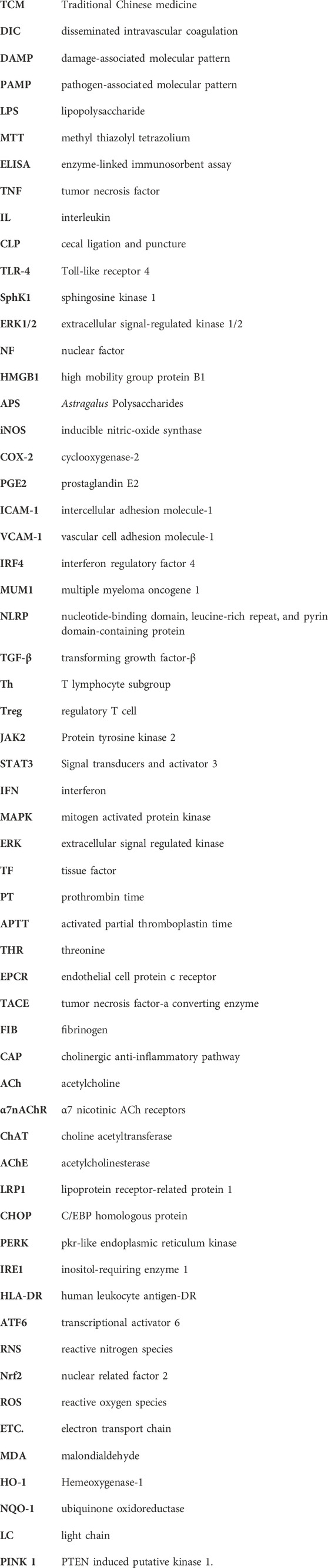
Keywords: sepsis, traditional Chinese medicine, extracts, pharmacological action, mechanism
Citation: Fu C-F, Li J-L, Chen J-W, Liang H, Zhao W-R, He S-Y, Ma X-W, Yang X-F and Wang H-L (2024) Mechanism and therapeutic potential of traditional Chinese medicine extracts in sepsis. Front. Pharmacol. 15:1365639. doi: 10.3389/fphar.2024.1365639
Received: 04 January 2024; Accepted: 03 June 2024;
Published: 03 July 2024.
Edited by:
Irina Ielciu, University of Medicine and Pharmacy Iuliu Hatieganu, RomaniaReviewed by:
Priyanka Choudhury, Medical College of Wisconsin, United StatesCopyright © 2024 Fu, Li, Chen, Liang, Zhao, He, Ma, Yang and Wang. This is an open-access article distributed under the terms of the Creative Commons Attribution License (CC BY). The use, distribution or reproduction in other forums is permitted, provided the original author(s) and the copyright owner(s) are credited and that the original publication in this journal is cited, in accordance with accepted academic practice. No use, distribution or reproduction is permitted which does not comply with these terms.
*Correspondence: Xiao-Wei Ma, NjA1NEAxNjMuY29t">xiaoweiNjA1NEAxNjMuY29t; Xiao-Fan Yang, MzcyODk1MTBAcXEuY29t; He-Lin Wang, NDI5NDE1NTRAcXEuY29t
†These authors have contributed equally to this work and share first authorship
Disclaimer: All claims expressed in this article are solely those of the authors and do not necessarily represent those of their affiliated organizations, or those of the publisher, the editors and the reviewers. Any product that may be evaluated in this article or claim that may be made by its manufacturer is not guaranteed or endorsed by the publisher.
Research integrity at Frontiers
Learn more about the work of our research integrity team to safeguard the quality of each article we publish.