- 1Department of Hematology Laboratory, Shengjing Hospital of China Medical University, Shenyang, China
- 2Department of Hematology, Shengjing Hospital of China Medical University, Shenyang, China
Acute promyelocytic leukemia (APL) is currently considered a disease with a higher cure rate. And cases of secondary malignant tumors following successful APL treatment are rare. Here we described a rare case of a 29-year-old man who was treated for APL in 2019 and developed BCR-ABL1-positive acute lymphoblastic leukemia 2 years later. The patient responded well to tyrosine kinase inhibitors and chemotherapy, and achieved a molecular remission. Although APL usually has a good prognosis, the prognosis of its secondary malignancies is uncertain. There are no effective measures to prevent the occurrence of secondary tumors. Continuing to increase the monitoring frequency of laboratory tests, especially the molecular biomarkers, is essential for the diagnosis and treatment of secondary malignancies after the patients achieving complete remission.
Introduction
Acute promyelocytic leukemia (APL) is a distinct type of acute myeloid leukemia (AML), which is designated as M3 subtype by French-American-British classification (Krause, 2000). It is characterized by a large number of abnormal promyelocytic cells in the bone marrow, accompanied with the typical chromosomal translocation t(15; 17) (q22; q12-21). As a result, a fusion gene of the promyelocytic leukemia (PML) gene and the retinoic acid receptor alpha (RARα) gene was formed by this chromosomal abnormality (Tan et al., 2021; Yilmaz et al., 2021). The application of target specific agents, all-trans retinoic acid (ATRA) and arsenic trioxide (ATO) with or without chemotherapy, is an internationally recognized standard therapy regime for APL patients (Ferrara et al., 2022; Jamy et al., 2022). Despite the success of ATRA and ATO in the treatment of patients with APL, secondary malignant tumors after complete remission (CR) of patients are worthy of attention. BCR-ABL1 fusion protein has two major isoforms, BCR-ABL1 (p190) and BCR-ABL1 (p210). And the BCR-ABL1 (p190) mainly occurs in acute lymphoblastic leukemia (ALL) patients (El Fakih et al., 2018; Adnan-Awad et al., 2021). Here, we reported a rare patient who developed secondary BCR-ABL1 (p190)-positive ALL following successful treatment of APL, and provide a literature review to summarize the characteristics of this subset of patients.
Case report
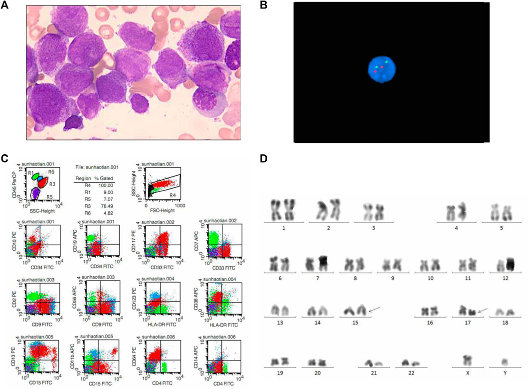
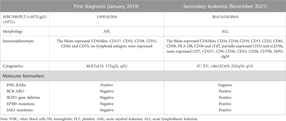
A 29-year-old man was admitted to our hospital for pancytopenia and clustered petechiae and ecchymoses on his left arm in January 2019. The laboratory features of the patient were shown in Table 1. Complete blood count at diagnosis showed white blood cell (WBC) counts 1.0 × 10⁹/L, hemoglobin level 95.0 g/L, platelet counts 20.0 × 10⁹/L. The bone marrow (BM) morphologic evaluation revealed 80.4% typical promyelocytes (Figure 1A). Bone marrow pathology analysis showed a large number of blasts in the hematopoietic tissue (Figure 1B). Immunophenotyping studies of BM cells revealed that the blasts expressed CD45dim, CD117, CD33, CD38, CD13, CD64 and CD15; no lymphoid antigens were expressed (Figure 1C). Cytogenetic analysis result showed an abnormal karyotype of t(15; 17) (q22; q21) (Figure 1D), and PML-RARa fusion gene was also detected in the patient’s bone marrow cells. BCR-ABL1 fusion gene was not detected (Table 1). Normalized PML-RARα/ABL1 by qRT-PCR was 100% (Figure 2E). The patient was diagnosed with APL, and evaluated as low risk, therefore he was treated by ATRA and ATO without chemotherapy for three courses. Then he treated with ATO alone for five courses due to ATRA intolerance. He achieved molecular remission (MR) 3 months after diagnosis and remained MR status.

FIGURE 1. Laboratory results of the patient at diagnosis. (A) Morphologic evaluation of bone marrow (Wright–Giemsa stain, × 1000). (B) FISH results of BCR-ABL1 in bone marrow. (C) Flow cytometry results of bone marrow. R1, lymphocytes; R3, promyeloblasts; R4, total cells; R5, erythrocytoblasts; R6, monocytes. (D) Karyotype analysis results of bone marrow.

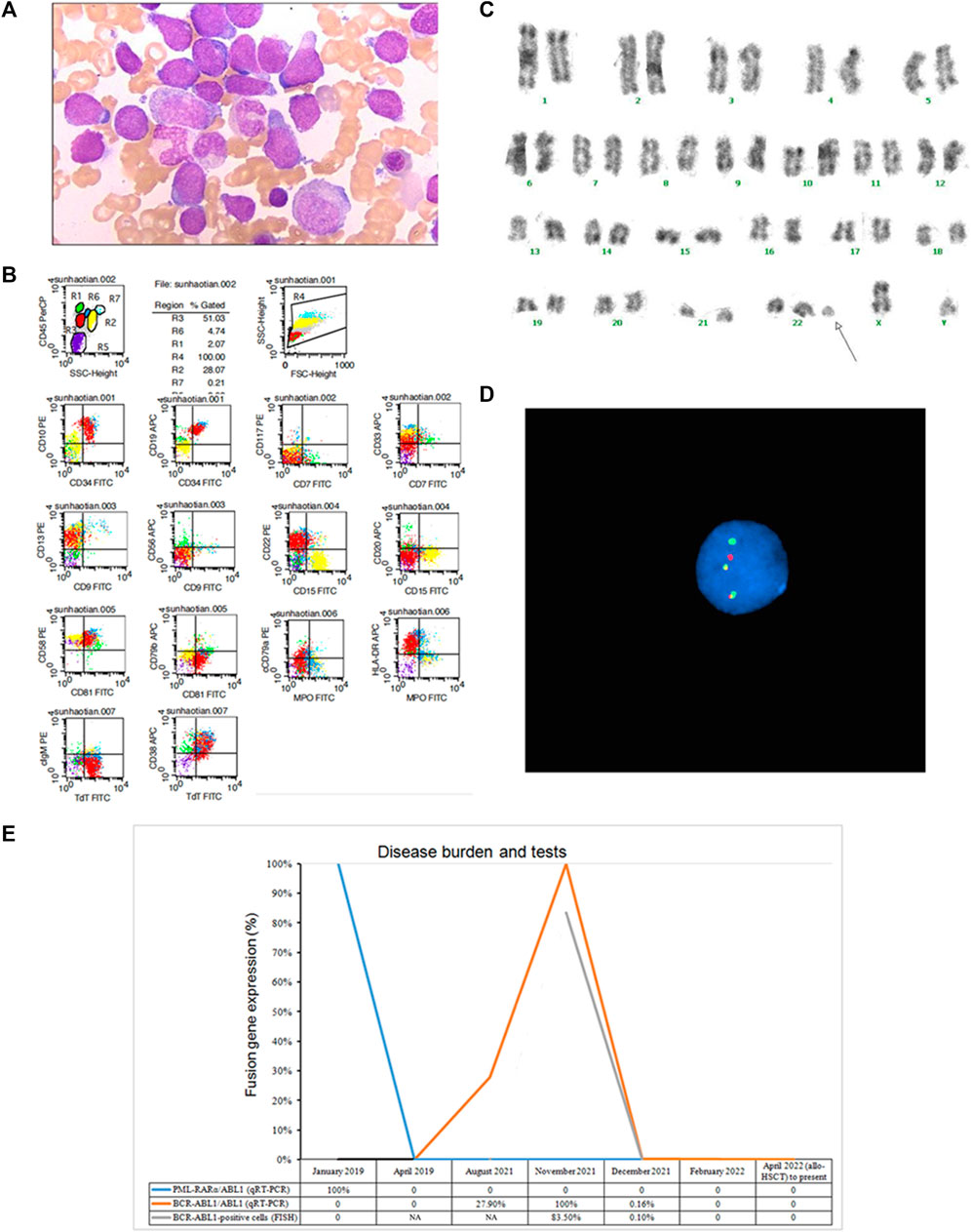
FIGURE 2. Laboratory results of the patient at secondary ALL and gene expression curve. (A) Morphologic evaluation of bone marrow (Wright–Giemsa stain, × 1000). (B) Flow cytometry results of bone marrow. R1, lymphocytes; R2, neutrophils; R3, prolymphocytes; R4, total cells; R5, erythrocytoblasts; R6, monocytes; R7, eosinophils. (C) Karyotype analysis results of bone marrow. (D) FISH results of bone marrow. (E) Gene expression curve of the patient by RT-PCR and FISH.
In November 2021, the patient’s laboratory examinations excluded relapse of APL but showed B-ALL (Figure 2A; Table 1). The marrow aspirate showed 51% blasts which expressed CD45dim, CD34, CD10, CD19, CD13, CD22, CD81, CD58, HLA-DR, CD38 and cTdT, partially expressed CD33 and cCD79a, none expressed CD7, CD117, CD9, CD56, CD15, CD20, CD79b, MPO, clgM (Figure 2B). Cytogenetic studies of BM revealed a 47, XY, +der (22) t (9; 22) (q34; q11) karyotype. Fluorescence in situ hybridization (FISH) analysis (GP Medical, Beijing, China) showed that 83.5% of examined cells possessed BCR (green) and ABL1 (red) fusion signals (yellow) (Figures 2D, E). Quantitative RT-PCR can also detect BCR-ABL1 p190 fusion gene (Figure 2E). RNA-sequencing results demonstrated the patient had IKZF1 gene deletion (IK6, exon4-exon7 del), EP300 (exon31:c.C5957T: p.P1986L) and JAK3 (exon11:c.G1503T:p.Q501H) mutations. Then the patient was treated by chemotherapy with tyrosine kinase inhibitors (TKI) dasatinib (100 mg/day). The induction chemotherapy regime was VICD (Vincristine, Demethoxydaunomycin, Cyclophosphamide and Dexamethasone). After the induction chemotherapy, the patient achieved a complete hematologic response, while FISH showed that 0.1% of examined cells still expressed the BCR-ABL1 fusion signals, and molecular biology showed a considerable reduction in BCR-ABL1 (p190)/ABL1 from 100% to 0.16% (Figure 2E). The patient’s condition was improved, and he was released from the hospital. After that, the patient received further consolidation chemotherapy of CAM (Cyclophosphamide, Cytarabine and Mercaptopurine) and HD-MTX (high dose methotrexate) with continued usage of dasatinib at 100 mg/day. In April 2022, he underwent allogeneic hematopoietic stem cell transplantation (mother to son, HLA 5/10). During the follow-up, the patient survived and continued to obtain complete molecular biological remission.
In order to determine the specific time of the patient’s secondary leukemia, we retrospectively tested his BM samples before November 2021, and the results showed that the positive expression of the BCR-ABL1 (p190) gene could be detected as early as August 2021. However, the patient’s blood counts and bone marrow characteristics was normal at that time, and the PML-RARα gene was also negative, so the chemotherapy regimen was not changed.
Discussion
Since the combination of ATRA and ATO with or without chemotherapy, the survival rates of APL patients have been dramatically improved, exceeding 80%–95% (Kantarjian et al., 2021; Ferrara et al., 2022). However, some patients developed therapy-relate myeloid neoplasms (t-MN), such as therapy-related AML or myelodysplastic syndrome (t-AML/MDS), with an incidence of 1%–9.8% (Gaut et al., 2018). And others developed secondary solid tumors (Huang et al., 2001; Au et al., 2007; Eghtedar et al., 2015). Secondary lymphoid neoplasms following treatment for APL were rare, and only four cases have been reported. One case developed precursor T-lymphocytic lymphoma (Szotkowski et al., 2009), two cases developed T-ALL (Liso et al., 1998; Bee et al., 2004), and one case developed early pre-B ALL with MLL/AF-1p fusion gene (Tsujioka et al., 2003). To our knowledge, the case we reported here was the first one who developed a secondary ALL with BCR-ABL1 fusion gene. The BCR-ABL1 fusion protein is sensitive to TKI, such as dasatinib (Shen et al., 2020), and the patient we reported achieved MR after 3 months of TKI and chemotherapy.
There are many hypotheses regarding the mechanism of secondary malignant neoplasms after successful treatment of APL. One of the hypotheses recognized by the majority of people is that APL induces therapy-related malignancies due to exposure to cytotoxic drugs during treatment (Wang Z. et al., 2019). While it is still unclear which drug may lead to it. In our case, the patient only received ATRO and ATO, without any other chemotherapy. ATRO and ATO maybe the possible reason. The underling possible mechanism is clonal selection. After exposure to APL treatment, the preexisting somatic mutation in hematopoietic stem cell may development to secondary neoplasms. The preexisting somatic mutations mainly tend to DNA damage, such as TP53 or PPM1D mutations (Martin et al., 2020). Moreover, there were also some cases about co-expression of t(15; 17) and other chromosome translocations, such as t(8; 21) (Uz et al., 2013). While the incidence of chromosomal rearrangements in addition to t(15; 17) was rare, and the role of additional translocations was still unclear. The current literature reviews tended to similar prognosis between additional abnormality and t(15; 17) alone. We detected the next-generation sequencing and chromosomal examination of this case before treatment, but no additional rearrangements or mutations were positive. The other possible reason is lineage switch. However, “Lineage switch” is a term used to describe the phenomenon of acute leukemias that meet standard criteria for a specific lineage (either lymphoid or myeloid) at the time of initial diagnosis, but later switch to another lineage upon relapse, including changes in cell morphology, histology, and immunotype (Rossi et al., 2012). While in our case, we detected the fusion gene of BCR/ABL at initial diagnosis which was negative, so the possibility of lineage switch was small. Furthermore, the PML-RARα fusion gene in our case remained continuous negative, and neither the clinical symptoms nor laboratory data exhibited typical features of APL. We supposed that this secondary ALL might be related to therapy.
To analyze the characteristics of secondary malignancies after APL treatment, we conducted a literature search on PubMed with the keywords “secondary” or “therapy-related”, combined with “after acute promyelocytic leukemia”, “following acute promyelocytic leukemia” to gather related case reports (Eghtedar et al., 2015; Huang et al., 2001; Au et al., 2007; Szotkowski et al., 2009; Liso et al., 1998; Bee et al., 2004; Tsujioka et al., 2003; Wang Z. et al., 2019; Gong et al., 2021; Vicente-Ayuso et al., 2017; Imagawa et al., 2010; Garcia-Manero et al., 2002; Renneville et al., 2018; Athanasiadou et al., 2002; Tang et al., 2016; Dang et al., 2014; Drake et al., 2003; Lee et al., 2005; Bao et al., 2009; Kelemen et al., 2012; Pawarode et al., 2006; Bseiso et al., 1997; Panizo et al., 2003; Snijder et al., 2008; Stavroyianni et al., 2000; Chen et al., 2012; Jubashi et al., 1993; Au et al., 2001; Todisco et al., 1995; Hatzis et al., 1995; Ojeda-Uribe et al., 2012; Meloni et al., 1997; Sawada et al., 1999; Park et al., 2014; Zompi et al., 2000; Liu et al., 2017; Latagliata et al., 2002; Sahoo et al., 2013; Batzios et al., 2009; Lobe et al., 2003; Wang T. et al., 2019; Annunziata et al., 2003; Park et al., 2008; Asou et al., 2010; Felice et al., 1999; Kim et al., 2014; Miyazaki et al., 1994; Takeshita et al., 2004), and combined with our case. A total of 100 cases were included in this literature review. The patients were divided into five groups according to the types of secondary malignancies. Details of age, gender, duration from APL to secondary malignancies, karyotype, and survival time of the patients are shown in Table 2. Among these cases, 46 cases were male, 45 cases were female, and 9 cases were unknown. The age of the patients ranged from 15 to 76 years old (median 48 years old). The median duration from APL to secondary malignancies was 23 (12–168) months. Most of the patients have karyotype changes after secondary disease and often have poor prognosis. Therefore, multiple laboratory testing methods should be combined for early detection of secondary malignancies.
In conclusion, we reported a rare case of secondary BCR-ABL1 positive ALL after successful APL treatment. The patient responded well to TKI and chemotherapy and achieved a MR. Although APL usually has a good prognosis, the therapeutic effects of secondary malignant tumors are different. At present, there are no effective measures to prevent the occurrence of secondary tumors. For this subset of patients, the monitoring frequency of molecular biomarkers (not only PML-RARα) should be increased after receiving CR, in order to achieve the purpose of early detection and early treatment.
Data availability statement
The original contributions presented in the study are included in the article/Supplementary Material, further inquiries can be directed to the corresponding author.
Ethics statement
Written informed consent was obtained from the participants/patient for the publication of this case report, including the images and data in this article.
Author contributions
SF and HW performed the study concept and design. SF developed the methodology and wrote the paper. SF and ML acquired, analyzed and interpreted the data and performed the statistical analysis. HW reviewed and revised the paper. All authors contributed to the article and approved the submitted version.
Funding
This work was supported by the National Natural Science Foundation of China (NSFC) [grant number: 82070165 and 81600115], the Applied Basic Research Program of Science and Technology Department of Liaoning Province [grant number: 2022020495-JH2/1015], and 345 Talent Project of Shengjing Hospital [grant number: M0957 and M0726].
Conflict of interest
The authors declare that the research was conducted in the absence of any commercial or financial relationships that could be construed as a potential conflict of interest.
Publisher’s note
All claims expressed in this article are solely those of the authors and do not necessarily represent those of their affiliated organizations, or those of the publisher, the editors and the reviewers. Any product that may be evaluated in this article, or claim that may be made by its manufacturer, is not guaranteed or endorsed by the publisher.
References
Adnan-Awad, S., Kim, D., Hohtari, H., Javarappa, K. K., Brandstoetter, T., Mayer, I., et al. (2021). Characterization of p190-Bcr-Abl chronic myeloid leukemia reveals specific signaling pathways and therapeutic targets. Leukemia 35 (7), 1964–1975. doi:10.1038/s41375-020-01082-4
Annunziata, M., Palmieri, S., Pocali, B., De Simone, M., Del Vecchio, L., Vicari, L., et al. (2003). Therapy-related acute myeloid leukemia with t(9;11)(p12;q23) in a patient treated for acute promyelocytic leukemia. Hematol. J. 4 (4), 289–291. doi:10.1038/sj.thj.6200256
Asou, N., Iwanaga, E., Nanri, T., and Mitsuya, H. (2010). Successful treatment with low-dose imatinib mesylate of therapy-related myeloid neoplasm harboring TEL-PDGFRB in a patient with acute promyelocytic leukemia. Haematologica 95 (9), e1. doi:10.3324/haematol.2010.27656
Athanasiadou, A., Saloum, R., Zorbas, I., Tsompanakou, A., Batsis, I., Fassas, A., et al. (2002). Therapy-related myelodysplastic syndrome with monosomy 5 and 7 following successful therapy for acute promyelocytic leukemia with anthracyclines. Leuk. Lymphoma 43 (12), 2409–2411. doi:10.1080/1042819021000040143
Au, W. Y., Kumana, C. R., Lam, C. W., Cheng, V. C., Shek, T. W., Chan, E. Y., et al. (2007). Solid tumors subsequent to arsenic trioxide treatment for acute promyelocytic leukemia. Leuk. Res. 31 (1), 105–108. doi:10.1016/j.leukres.2006.03.018
Au, W. Y., Lam, C. C., Ma, E. S., Man, C., Wan, T., and Kwong, Y. L. (2001). Therapy-related myelodysplastic syndrome after eradication of acute promyelocytic leukemia: Cytogenetic and molecular features. Hum. Pathol. 32 (1), 126–129. doi:10.1053/hupa.2001.21128
Bao, L., Lu, X., Lai, Y., Zhang, X., Zhu, H., Liu, Y., et al. (2009). Therapy-related acute myeloid leukemia after successful therapy for acute promyelocytic leukemia with t(15;17): A case report and literature review. Leuk. Res. 33 (7), e64–e68. doi:10.1016/j.leukres.2009.01.043
Batzios, C., Hayes, L. A., He, S. Z., Quach, H., McQuilten, Z. K., Wall, M., et al. (2009). Secondary clonal cytogenetic abnormalities following successful treatment of acute promyelocytic leukemia. Am. J. Hematol. 84 (11), 715–719. doi:10.1002/ajh.21528
Bee, P. C., Gan, G. G., Sangkar, J. V., Teh, A., and Goh, K. Y. (2004). A case of T-cell acute lymphoblastic leukemia after treatment of acute promyelocytic leukemia. Int. J. Hematol. 79 (4), 358–360. doi:10.1532/ijh97.a20304
Bseiso, A. W., Kantarjian, H., and Estey, E. (1997). Myelodysplastic syndrome following successful therapy of acute promyelocytic leukemia. Leukemia 11 (1), 168–169. doi:10.1038/sj.leu.2400539
Chen, J., Zheng, Z., Shen, J., Peng, L., Zhuang, H., Liu, W., et al. (2012). Secondary acute myeloid leukemia occurring after successful treatment of acute promyelocytic leukemia. Int. J. Hematol. 95 (3), 327–328. doi:10.1007/s12185-012-1019-8
Dang, D. N., Morris, H. D., Feusner, J. H., Koduru, P., Wilson, K., Timmons, C. F., et al. (2014). Therapy-induced secondary acute myeloid leukemia with t(11;19)(q23;p13.1) in a pediatric patient with relapsed acute promyelocytic leukemia. J. Pediatr. Hematol. Oncol. 36 (8), e546–e548. doi:10.1097/MPH.0000000000000183
Drake, M., Humphreys, M. W., Alexander, H. D., and Morris, T. C. (2003). Early second leukaemia in a patient with successfully treated acute promyelocytic leukaemia. Leuk. Lymphoma 44 (5), 895–896. doi:10.1080/1042819021000029975
Eghtedar, A., Rodriguez, I., Kantarjian, H., O'Brien, S., Daver, N., Garcia-Manero, G., et al. (2015). Incidence of secondary neoplasms in patients with acute promyelocytic leukemia treated with all-trans retinoic acid plus chemotherapy or with all-trans retinoic acid plus arsenic trioxide. Leuk. Lymphoma 56 (5), 1342–1345. doi:10.3109/10428194.2014.953143
El Fakih, R., Jabbour, E., Ravandi, F., Hassanein, M., Anjum, F., Ahmed, S., et al. (2018). Current paradigms in the management of Philadelphia chromosome positive acute lymphoblastic leukemia in adults. Am. J. Hematol. 93 (2), 286–295. doi:10.1002/ajh.24926
Felice, M. S., Rossi, J., Gallego, M., Zubizarreta, P. A., Cygler, A. M., Alfaro, E., et al. (1999). Acute trilineage leukemia with monosomy of chromosome 7 following an acute promyelocytic leukemia. Leuk. Lymphoma 34 (3-4), 409–413. doi:10.3109/10428199909050968
Ferrara, F., Molica, M., and Bernardi, M. (2022). Drug treatment options for acute promyelocytic leukemia. Expert Opin. Pharmacother. 23 (1), 117–127. doi:10.1080/14656566.2021.1961744
Garcia-Manero, G., Kantarjian, H. M., Kornblau, S., and Estey, E. (2002). Therapy-related myelodysplastic syndrome or acute myelogenous leukemia in patients with acute promyelocytic leukemia (APL). Leukemia 16 (9), 1888. doi:10.1038/sj.leu.2402616
Gaut, D., Sasine, J., and Schiller, G. (2018). Secondary clonal hematologic neoplasia following successful therapy for acute promyelocytic leukemia (APL): A report of two cases and review of the literature. Leuk. Res. Rep. 9, 65–71. doi:10.1016/j.lrr.2018.04.005
Gong, Y., Wang, M., Shen, H., Chen, Y., Cen, J., Yin, X., et al. (2021). Novel MLL/KMT2A-Mon2 fusion in a child with therapy-related acute myeloid leukemia after treatment for acute promyelocytic leukemia. Mol. Carcinog. 60 (11), 721–725. doi:10.1002/mc.23333
Hatzis, T., Standen, G. R., Howell, R. T., Savill, C., Wagstaff, M., and Scott, G. L. (1995). Acute promyelocytic leukaemia (M3): Relapse with acute myeloblastic leukaemia (M2) and dic(5;17) (q11;p11). Am. J. Hematol. 48 (1), 40–44. doi:10.1002/ajh.2830480108
Huang, F. S., Zwerdling, T., Stern, L. E., Ballard, E. T., and Warner, B. W. (2001). Renal cell carcinoma as a secondary malignancy after treatment of acute promyelocytic leukemia. J. Pediatr. Hematol. Oncol. 23 (9), 609–611. doi:10.1097/00043426-200112000-00011
Imagawa, J., Harada, Y., Shimomura, T., Tanaka, H., Okikawa, Y., Hyodo, H., et al. (2010). Clinical and genetic features of therapy-related myeloid neoplasms after chemotherapy for acute promyelocytic leukemia. Blood 116 (26), 6018–6022. doi:10.1182/blood-2010-06-289389
Jamy, O. H., Dhir, A., Costa, L. J., and Xavier, A. C. (2022). Impact of sociodemographic factors on early mortality in acute promyelocytic leukemia in the United States: A time-trend analysis. Cancer 128 (2), 292–298. doi:10.1002/cncr.33914
Jubashi, T., Nagai, K., Miyazaki, Y., Nakamura, H., Matsuo, T., Kuriyama, K., et al. (1993). A unique case of t(15;17) acute promyelocytic leukaemia (M3) developing into acute myeloblastic leukaemia (M1) with t(7;21) at relapse. Br. J. Haematol. 83 (4), 665–668. doi:10.1111/j.1365-2141.1993.tb04709.x
Kantarjian, H. M., Kadia, T. M., DiNardo, C. D., Welch, M. A., and Ravandi, F. (2021). Acute myeloid leukemia: Treatment and research outlook for 2021 and the MD Anderson approach. Cancer 127 (8), 1186–1207. doi:10.1002/cncr.33477
Kelemen, K., Kovacsovics, T., Braziel, R., Corless, C., Beadling, C., and Fan, G. (2012). RAS mutations in therapy-related acute myeloid leukemia after successful treatment of acute promyelocytic leukemia. Leuk. Lymphoma 53 (5), 999–1002. doi:10.3109/10428194.2011.634047
Kim, H. G., Jang, J. H., and Koh, E. H. (2014). TRIP11-PDGFRB fusion in a patient with a therapy-related myeloid neoplasm with t(5;14)(q33;q32) after treatment for acute promyelocytic leukemia. Mol. Cytogenet 7 (1), 103. doi:10.1186/s13039-014-0103-6
Krause, J. R. (2000). Morphology and classification of acute myeloid leukemias. Clin. Lab. Med. 20 (1), 1–16. doi:10.1016/s0272-2712(18)30072-6
Latagliata, R., Petti, M. C., Fenu, S., Mancini, M., Spiriti, M. A., Breccia, M., et al. (2002). Therapy-related myelodysplastic syndrome-acute myelogenous leukemia in patients treated for acute promyelocytic leukemia: An emerging problem. Blood 99 (3), 822–824. doi:10.1182/blood.v99.3.822
Lee, G. Y., Christina, S., Tien, S. L., Ghafar, A. B., Hwang, W., Lim, L. C., et al. (2005). Acute promyelocytic leukemia with PML-RARA fusion on i(17q) and therapy-related acute myeloid leukemia. Cancer Genet. Cytogenet 159 (2), 129–136. doi:10.1016/j.cancergencyto.2004.09.019
Liso, V., Specchia, G., Pannunzio, A., Mestice, A., Palumbo, G., and Biondi, A. (1998). T-cell acute lymphoblastic leukemia occurring in a patient with acute promyelocytic leukemia. Haematologica 83 (5), 471–473.
Liu, J. X., Ren, J. H., Guo, X. L., and Cai, S. X. (2017). A case report of acute promyelocytic leukemia transforming into acute myeloid leukemia M(4). Zhonghua Xue Ye Xue Za Zhi 38 (12), 1052. Chinese. doi:10.3760/cma.j.issn.0253-2727.2017.12.009
Lobe, I., Rigal-Huguet, F., Vekhoff, A., Desablens, B., Bordessoule, D., Mounier, C., et al. (2003). Myelodysplastic syndrome after acute promyelocytic leukemia: The European APL group experience. Leukemia 17 (8), 1600–1604. doi:10.1038/sj.leu.2403034
Martin, J. E., Khalife-Hachem, S., Grinda, T., Kfoury, M., Garciaz, S., Pasquier, F., et al. (2020). Therapy related myeloid neoplasm post PARP inhibitors: Potential clonal selection. Blood 136 (1), 14–15. doi:10.1182/blood-2020-139971
Meloni, G., Diverio, D., Vignetti, M., Avvisati, G., Capria, S., Petti, M. C., et al. (1997). Autologous bone marrow transplantation for acute promyelocytic leukemia in second remission: Prognostic relevance of pretransplant minimal residual disease assessment by reverse-transcription polymerase chain reaction of the PML/RAR alpha fusion gene. Blood 90 (3), 1321–1325. doi:10.1182/blood.v90.3.1321.1321_1321_1325
Miyazaki, H., Ino, T., Sobue, R., Kojima, H., Wakita, M., Nomura, T., et al. (1994). Translocation (3;21)(q26;q22) in treatment-related acute leukemia secondary to acute promyelocytic leukemia. Cancer Genet. Cytogenet 74 (2), 84–86. doi:10.1016/0165-4608(94)90002-7
Ojeda-Uribe, M., Schneider, A., Luquet, I., Berceanu, A., Cornillet-Lefebvre, P., Jeandidier, E., et al. (2012). Therapy-related acute myeloid leukemia (t-AML) with poor-risk cytogenetics in two patients with persistent molecular complete remission of acute promyelocytic leukemia. Eur. J. Haematol. 89 (3), 267–272. doi:10.1111/j.1600-0609.2012.01805.x
Panizo, C., Patiño, A., Lecumberri, R., Calasanz, M. J., Odero, M. D., Bendandi, M., et al. (2003). Secondary myelodysplastic syndrome after treatment for promyelocytic leukemia: Clinical and genetic features of two cases. Cancer Genet. Cytogenet 143 (2), 178–181. doi:10.1016/s0165-4608(02)00859-2
Park, S. H., Chi, H. S., Cho, Y. U., Jang, S., Park, C. J., and Lee, J. H. (2014). A case of therapy-related acute myeloid leukemia with a normal karyotype after sustained molecular complete remission of acute promyelocytic leukemia. Ann. Lab. Med. 34 (1), 68–70. doi:10.3343/alm.2014.34.1.68
Park, T. S., Choi, J. R., Yoon, S. H., Song, J., Kim, J., Kim, S. J., et al. (2008). Acute promyelocytic leukemia relapsing as secondary acute myelogenous leukemia with translocation t(3;21)(q26;q22) and RUNX1-MDS1-EVI1 fusion transcript. Cancer Genet. Cytogenet 187 (2), 61–73. doi:10.1016/j.cancergencyto.2008.06.015
Pawarode, A., Finlay, E., Sait, S. N., Barcos, M., and Baer, M. R. (2006). Isochromosome 1q in a myelodysplastic syndrome after treatment for acute promyelocytic leukemia. Cancer Genet. Cytogenet 167 (2), 155–160. doi:10.1016/j.cancergencyto.2005.11.013
Renneville, A., Attias, P., Thomas, X., Bally, C., Hayette, S., Farhat, H., et al. (2018). Genetic analysis of therapy-related myeloid neoplasms occurring after intensive treatment for acute promyelocytic leukemia. Leukemia 32 (9), 2066–2069. doi:10.1038/s41375-018-0137-6
Rossi, J. G., Bernasconi, A. R., Alonso, C. N., Rubio, P. L., Gallego, M. S., Carrara, C. A., et al. (2012). Lineage switch in childhoodacute leukemia: An unusual event with poor outcome. Am. J. Hematol. 87 (9), 890–897. doi:10.1002/ajh.23266
Sahoo, R. K., Kumar, L., Kumar, R., and Sharma, A. (2013). Acute promyelocytic leukemia relapsing into acute myeloid leukemia-M2 with normal cytogenetics. Indian J. Med. Paediatr. Oncol. 34 (4), 327–329. doi:10.4103/0971-5851.125261
Sawada, H., Morimoto, H., Wake, A., Yamasaki, Y., and Izumi, Y. (1999). Therapy-related acute myeloid leukemia with t(10;11)(q23;p15) following successful chemotherapy for acute promyelocytic leukemia with t(15;17)(q22;q21). Int. J. Hematol. 69 (4), 270–271.
Shen, S., Chen, X., Cai, J., Yu, J., Gao, J., Hu, S., et al. (2020). Effect of dasatinib vs imatinib in the treatment of pediatric philadelphia chromosome-positive acute lymphoblastic leukemia: A randomized clinical trial. JAMA Oncol. 6 (3), 358–366. doi:10.1001/jamaoncol.2019.5868
Snijder, S., Mellink, C. H., and van der Lelie, H. (2008). Translocation (2;11)(q37;q23) in therapy-related myelodysplastic syndrome after treatment for acute promyelocytic leukemia. Cancer Genet. Cytogenet 180 (2), 149–152. doi:10.1016/j.cancergencyto.2007.10.003
Stavroyianni, N., Yataganas, X., Abazis, D., Pangalos, C., and Meletis, J. (2000). Acute promyelocytic leukemia relapsing into FAB-M2 acute myeloid leukemia with trisomy 8. Cancer Genet. Cytogenet 117 (1), 82–83. doi:10.1016/s0165-4608(99)00132-6
Szotkowski, T., Jarosova, M., Faber, E., Hubacek, J., Hlusi, A., Papajik, T., et al. (2009). Precursor T-lymphoblastic lymphoma as a secondary malignancy in a young patient after successful treatment of acute promyelocytic leukemia. Onkologie 32 (8-9), 513–515. doi:10.1159/000226584
Takeshita, A., Naito, K., Shinjo, K., Sahara, N., Matsui, H., Ohnishi, K., et al. (2004). Deletion 6p23 and add(11)(p15) leading to NUP98 translocation in a case of therapy-related atypical chronic myelocytic leukemia transforming to acute myelocytic leukemia. Cancer Genet. Cytogenet 152 (1), 56–60. doi:10.1016/j.cancergencyto.2003.10.002
Tan, Y., Wang, X., Song, H., Zhang, Y., Zhang, R., Li, S., et al. (2021). A PML/RARα direct target atlas redefines transcriptional deregulation in acute promyelocytic leukemia. Blood 137 (11), 1503–1516. doi:10.1182/blood.2020005698
Tang, Y. L., Chia, W. K., Yap, E. C., Julia, M. I., Leong, C. F., Salwati, S., et al. (2016). Dismal outcome of therapy-related myeloid neoplasm associated with complex aberrant karyotypes and monosomal karyotype: A case report. Malays J. Pathol. 38 (3), 315–319.
Todisco, E., Testi, A. M., Avvisati, G., Moleti, M. L., Cedrone, M., Cimino, G., et al. (1995). Therapy-related acute myelomonocytic leukemia following successful treatment for acute promyelocytic leukemia. Leukemia 9 (9), 1583–1585.
Tsujioka, T., Wada, H., Yamamori, S., Otsuki, T., Suemori, S., Kondo, T., et al. (2003). MLL/AF-1p fusion in therapy-related early pre-B acute lymphoblastic leukemia with t (1;11) (p32; q23) translocation developing in the relapse phase of acute promyelocytic leukemia. Int. J. Hematol. 78 (5), 439–442. doi:10.1007/BF02983817
Uz, B., Eliacik, E., Isik, A., Aksu, S., Buyukasik, Y., Haznedaroglu, I. C., et al. (2013). Co-Expression of t(15;17) and t(8;21) in a case of acute promyelocytic leukemia: Review of the literature. Turk J. Haematol. 30 (4), 400–404. doi:10.4274/Tjh.2012.0180
Vicente-Ayuso, M. D. C., García-Roa, M., González-Fernández, A., Álvarez-Carmona, A. M., Benavente-Cuesta, C., Mateo-Morales, M., et al. (2017). Therapy-related myeloid neoplasms as a concerning complication in acute promyelocytic leukemia. Hematol. Rep. 9 (3), 7204. doi:10.4081/hr.2017.7204
Wang, T., Jacoby, M. A., Duncavage, E. J., Miller, C. A., Heath, S., Rahme, R., et al. (2019b). Exome analysis of treatment-related AML after APL suggests secondary evolution. Br. J. Haematol. 185 (5), 984–987. doi:10.1111/bjh.15681
Wang, Z., Xu, M. Z., Chen, Y. F., Xue, F., Zhang, L., Hu, Y. M., et al. (2019a). Therapy-related myeloid neoplasms after successful treatment for acute promyelocytic leukemia: A report of four cases and literature review. Zhonghua Xue Ye Xue Za Zhi 40 (12), 1008–1014. doi:10.3760/cma.j.issn.0253-2727.2019.12.007
Yilmaz, M., Kantarjian, H., and Ravandi, F. (2021). Acute promyelocytic leukemia current treatment algorithms. Blood Cancer J. 11 (6), 123. doi:10.1038/s41408-021-00514-3
Zompi, S., Legrand, O., Bouscary, D., Blanc, C. M., Picard, F., Casadevall, N., et al. (2000). Therapy-related acute myeloid leukaemia after successful therapy for acute promyelocytic leukaemia with t(15;17): A report of two cases and a review of the literature. Br. J. Haematol. 110 (3), 610–613. doi:10.1046/j.1365-2141.2000.02240.x
Keywords: acute promyelocytic leukemia, acute lymphoblastic leukemia, PML-RARα, BCR-ABL1, secondary malignancy
Citation: Fu S, Li M and Wang H (2023) BCR-ABL1-positive acute lymphoblastic leukemia following successful treatment of acute promyelocytic leukemia: case report. Front. Pharmacol. 14:1141311. doi: 10.3389/fphar.2023.1141311
Received: 10 January 2023; Accepted: 05 June 2023;
Published: 16 June 2023.
Edited by:
Anna Maria Testi, Sapienza University of Rome, ItalyReviewed by:
Ibrahim C. Haznedaroglu, Hacettepe University Hospital, TürkiyeGertjan Kaspers, Princess Maxima Center for Pediatric Oncology, Netherlands
Copyright © 2023 Fu, Li and Wang. This is an open-access article distributed under the terms of the Creative Commons Attribution License (CC BY). The use, distribution or reproduction in other forums is permitted, provided the original author(s) and the copyright owner(s) are credited and that the original publication in this journal is cited, in accordance with accepted academic practice. No use, distribution or reproduction is permitted which does not comply with these terms.
*Correspondence: Hongtao Wang, d2FuZ2h0QHNqLWhvc3BpdGFsLm9yZw==
 Shuang Fu
 Mengqi Li
 Hongtao Wang
