 Man Yao1†
 Shufang Liang
 Binbin Cheng
94% of researchers rate our articles as excellent or good
Learn more about the work of our research integrity team to safeguard the quality of each article we publish.
Find out more
REVIEW article
Front. Pharmacol. , 17 January 2023
Sec. Pharmacology of Anti-Cancer Drugs
Volume 14 - 2023 | https://doi.org/10.3389/fphar.2023.1110922
This article is part of the Research Topic Exosomes in Tumor Microenvironment and the Role of Medicinal Plants View all articles
Hepatocellular carcinoma (HCC) usually occurs on the basis of chronic liver inflammatory diseases and cirrhosis. The liver microenvironment plays a vital role in the tumor initiation and progression. Exosomes, which are nanometer-sized membrane vesicles are secreted by a number of cell types. Exosomes carry multiple proteins, DNAs and various forms of RNA, and are mediators of cell-cell communication and regulate the tumor microenvironment. In the recent decade, many studies have demonstrated that exosomes are involved in the communication between HCC cells and the stromal cells, including endothelial cells, macrophages, hepatic stellate cells and the immune cells, and serve as a regulator in the tumor proliferation and metastasis, immune evasion and immunotherapy. In addition, exosomes can also be used for the diagnosis and treatment HCC. They can potentially serve as specific biomarkers for early diagnosis and drug delivery vehicles of HCC. Chinese herbal medicine, which is widely used in the prevention and treatment of HCC in China, may regulate the release of exosomes and exosomes-mediated intercellular communication. In this review, we summarized the latest progresses on the role of the exosomes in the initiation, progression and treatment of HCC and the potential value of Traditional Chinese medicine in exosomes-mediated biological behaviors of HCC.
Hepatocellular carcinoma (HCC) is the most common type of primary liver cancer. HCC usually occurs on the basis of chronic liver diseases, including viral hepatitis, alcoholic and non-alcoholic fatty liver diseases, and cirrhosis. Chronic liver inflammation plays a central role in the hepatocarcinogenesis through multiple mechanisms. Interactions between hepatocytes, hepatic stroma cells, inflammatory and immune cells lead to the production of pro-inflammatory cytokines and chemokines, such as TGF-β, and activation of many key signaling pathways, such as NF-κB and MAPKs pathways. After the tumor developed, cell-to-cell interactions within the tumor microenvironment (TME) induce an inhibitory immune microenvironment and promote the cancer progression and metastasis.
Exosomes, which are small extracellular vesicles with a diameter of 30 nm–150 nm, carry many active components (such as proteins, lipid, RNA and microRNAs), and play an important role in the cell-to-cell communication in the TME. The characteristics of exosomes are usually based on their size or the expression of surface markers, such as CD9, CD37, CD53, CD63, CD81, CD82, TSG101, Alix and Hsp70, etc that could be used in the identification of exosomes (Huang et al., 2018). Currently, many studies have shown that exosomes can be secreted by different types of cells and released into the TME, promoting the proliferation, angiogenesis, migration, invasion, metastasis, and drug resistance of tumor cells (Hao et al., 2022). In addition, the exosomes are also closely related to the occurrence of HCC (Han et al., 2019). This review mainly focused on the role of exosomes in the tumorigenesis, development, diagnosis and treatment of HCC. We also further expounded the value of exosomes for Traditional Chinese Medicine (TCM) in the treatment of HCC.
Due to the absent of early signs, most HCC patients are diagnosed at the intermediate and late stages. Delayed diagnosis of HCC often deprives patients of the opportunity for radical resection. In addition, the lack of sensitive biomarkers or diagnostic methods for cancer recurrence and metastasis also restrict the execution of local and systemic therapies, such as radiotherapy, chemotherapy and immunotherapy. Hence, predicting tumors at a relative earlier stage may improve therapeutic approaches, treatment outcomes and the prognosis.
At present, the most commonly used classical biomarkers for the diagnosis of HCC are alpha fetoprotein (AFP), AFP-L3 and prothrombin induced by vitamin K absence-II (PIVKA-II) (Wang X et al., 2022). However, the AFP level is also increased under pregnancy, hepatitis, cirrhosis and other benign diseases (Wong et al., 2015). In addition, the serum AFP is always at a low level regardless of disease occurrence and progression in about 35%–40% HCC patients. The serum PIVKA-II level is influenced by many factors, such as abnormal coagulation function, cirrhosis, etc., which can cause its level to rise. In recent years, despite new breakthroughs in the diagnosis and treatment methods, the early diagnosis of HCC is still very difficult, and the 5-year survival rate of HCC is no more than 20%. Therefore, there is a clear need for the development of novel, accurate and less invasive methods for early diagnosis of HCC.
Many studies have shown that exosomes are the ideal biomarkers for the early diagnosis of HCC, prediction of tumor recurrence and therapeutic effects. Exosomes have got widely attentions due to their critical role in regulating genes of tumor cells and altering the cell-to-cell communication pathways. Recently, exosomal non-coding RNAs and other cargo molecules have become of interest as candidate biomarkers of HCC diagnosis, recurrence and prognosis (Table 1) (Xue et al., 2019; Wei et al., 2022). Wang et al. (Wang et al., 2018) screened the differentially expressed exosomal miRNAs between HCC and cirrhosis patients and found the combination of miR-122, miR-148a, and AFP could be applied for distinguishing early HCC from liver cirrhosis. Ghosh et al. (Ghosh et al., 2020) showed the combination of exosomal miR-10b-5p, miR-221-3p and miR-223-3p could be taken as a sensitive diagnostic marker for HCC with low AFP. Yao et al. (Yao et al., 2020) identified differentially expressed serum exosomal lncRNAs lnc-FAM72D-3 and lnc-EPC1-4 in HCC and suggested they may be taken as potential candidate biomarkers for HCC diagnosis. Zuo et al. (Zuo et al., 2022) analyzed the differentially expressed liver exosome-related genes and constructed an exosome-related prognostic model, indicating the higher risk score is associated with higher expression of immune checkpoint molecules, including programmed death ligand 1 (PD-L1), programmed death ligand 2 (PD-L2), T-cell Ig and ITIM domain (TIGIT), and indoleamine-2,3-dioxygenase 1 (IDO1).

TABLE 1. Potential exosomal biomarkers for HCC diagnosis, recurrence and prognosis.
Interestingly, the plasma exosomal miRNAs could also serve as potential metastasis-related biomarkers for HCC (Huang et al., 2021; Kim et al., 2021). Huang et al. (Huang et al., 2021) showed a total of 32 differentially expressed miRNAs in the plasma exosomes of patients with metastatic HCC compared with those without metastasis and the combination of three miRNAs (miR-18a, miR-27a and miR-20b) could discriminate metastatic HCC from non-metastatic HCC. Exosomal miR-125b exerts an anti-metastatic effect through interfering TGF-β1-induced epithelial-mesenchymal transition (EMT) and therefore drastically downregulated miR-125b predicts early metastasis of HCC (Kim et al., 2021). Circulating exosomal miR-1307-5p promotes metastasis and helps predict metastasis in HCC through targeting the tumor suppressor genes SEC14L2 and ENG to promote EMT (Eun et al., 2020). In addition, the exosomes may also predict the risk of HCC recurrence. Circulating exosomal circAKT3 is positively correlated with the recurrence rates and higher mortality in HCC patients undergoing surgical treatment (Luo et al., 2020). The circulating exosomal miR-92b may downregulate CD69 and NK cell-mediated cytotoxicity and predicts the risk of posttransplant HCC recurrence (Nakano et al., 2019).
Tyrosine kinase inhibitors (TKIs), including sorafenib, lenvatinib and donafenib are still the first line drugs for advanced HCC, especially for those patients that could not benefit from atezolizumab plus bevacizumab treatment (Bruix et al., 2021; Reig et al., 2022). However, there are still no considerable biomarkers to predict the treatment efficacy. It has been shown that low miR-10b-3p level is associated with sorafenib resistance in HCC (Shao et al., 2022). Exosomal miR-30d is highly expressed in the sorafenib effective patients and thereby maybe serve as a predictive biomarker for the efficacy of sorafenib in HCC (Kohno et al., 2020) . MiR-200c-3p, miR-222-5p and miR-512-3p constitute a biomarker signature of sorafenib effectiveness in advanced HCC (de la Cruz-Ojeda et al., 2022). The baseline miR-200c-3p level is correlated with increased survival, whereas increased miR-222-5p and miR-512-3p levels at 1 month after sorafenib treatment are related to poor prognosis.
Many studies have demonstrated that exosomes are correlated with the development and progression of HCC. In the TME, exosomes transfer bioactive molecules between tumor-tumor cells, tumor-stromal cells and stromal-stromal cells to promote tumor growth, proliferation, and metastasis and create a suitable microenvironment for cancer cells invasion and colonization (Figure 1).
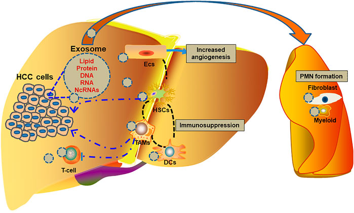
FIGURE 1. Role of exosomes in the progression of HCC.
It has been well recognized that exosomes could represent a contributory factor in the hepatic tumorigenesis, growth and metastasis by regulating the intercellular signal transduction between tumor and normal cells or tumor and tumor cells. Chronic exposure to arsenite contributes to the malignant transformation of hepatocyte. CircRNA 100284, which is involved in the malignant transformation of hepatocyte after arsenite exposure is embodied in the exosomes released by transformed cells and transferred into normal cells (Dai et al., 2018). Exosomal circRNA 100284 accelerates cell cycle and proliferation of normal liver cells by targeting miR-217 to promote the malignant transformation (Dai et al., 2018).
LOXL4, a member of the lysyl oxidase (LOX) family, is commonly upregulated in HCC tissues and predicts a poor prognosis (Li et al., 2019). Overexpression of LOXL4 promotes the migration and invasion of HCC in vitro, and intrahepatic and pulmonary metastases in vivo. HCC-derived exosomes transfer LOXL4 between HCC cells, and thereby promotes HCC cells invasion and metastasis (Li et al., 2019). In addition, HCC-derived exosomal LOXL4 could also activate endothelial cells and promote angiogenesis (Li et al., 2019). Golgi membrane protein 1 (GOLM1/GP73), a serum marker for HCC, is able to promote the tumorigenesis of HCC (Chen et al., 2015). GOLM1-enriched exosomes promote cell proliferation and migration of HCC through activating the GSK-3β/MMP signaling pathway in recipient cells (Gai et al., 2019). Alpha-enolase (ENO1), a key regulatory enzyme in glycolysis, promotes glucose uptake and lactic acid production by tumor cells (Diaz-Ramos et al., 2012; Capello et al., 2016). ENO1 is also enriched in highly metastatic HCC cells and exosomes derived from these cells. The exosomal ENO1 promotes growth and metastasis of HCC with low metastatic potential by regulating integrin α6β4 and the activation of FAK/Src-p38MAPK pathway, suggesting that ENO1 can be transferred to other HCC cells via exosome-mediated crosstalk and further promotes HCC growth and metastasis (Jiang et al., 2020). Angiopoietin-2 (ANGPT2) has been verified as a context-dependent antagonist through destroying vascular stability. HCC-derived exosomes carrying ANGPT2 increase the angiogenesis by a Tie2-independent pathway (Xie Y et al., 2020).
The deregulation of miRNAs plays an important role in human hepatocarcinogenesis. HCC-derived exosomes containing a cluster of differential expressed miRNAs can modulate TAK1 expression and enhance transformed cell growth through shuttling miRNAs to recipient cells (Kogure et al., 2011; Wei et al., 2015). PTEN, an important anti-oncogene, is generally decreased in HCC. Cao et al. (Cao et al., 2019) showed that uptake of exosomal miR-21 derived from HCC cells by other HCC cells could affect cell growth and promote HCC cell proliferation via downregulating PTENp1 expression. Liu et al. (Liu et al., 2018) reported that HCC-derived exosomes promoted tumor self-seeding by enhancing the invasive and migration capability of recipient HCC cells through transferring miR-25–5p. Exosomal miR-25 released from cancer cells enhances cell malignant phenotype in vitro and tumor growth and metastases in vivo through targeting SIK1 (Fu et al., 2022). In acidic conditions, HCC cells release exosomes containing high levels of miR-21 and miR-10b, which promote cancer cell proliferation and metastasis (Tian et al., 2019). In addition, miR-1290 which is overexpressed in HCC patients serum-derived exosomes is able to promote the angiogenesis via targeting SMEK1 after delivering into human endothelial cells (Wang S. S et al., 2021).
Circular RNAs (circRNAs) are a type of naturally occurring RNAs which are synthesized by “head to tail” splicing of coding or non-coding RNAs (ncRNAs) (Liu and Chen, 2022). In recent years, the functions of circRNAs on HCC have been deeply investigated. Up to date, more and more circRNAs which could accelerate the progression of HCC have been reported (Wu et al., 2021; Liu Y et al., 2022; Lu Y et al., 2022; Wu J et al., 2022; Shen et al., 2022). The exosomal circCMTM3 is elevated in the serum of HCC patients and HCC cells. CircCMTM3, as a miR-3619–5p sponge, promotes the tumorigenesis of HCC through miR-3619-5p-SOX9 pathway (Hu et al., 2021).The exosomal circRNA-100,338 is highly expressed in metastatic HCC exosomes and promotes the metastasis of HCC via enhancing invasiveness and angiogenesis (Huang et al., 2020a). CircFBLIM1 is highly expressed in the serum exosomes of HCC patients and HCC cells, and promotes HCC progression and glycolysis by regulating the miR-338/LRP6 Axis (Lai et al., 2020).
Long non-coding RNAs (LncRNAs) have also been demonstrated to play crucial roles in the growth and metastasis of HCC (Wu W et al., 2022). LncRNAs regulate gene expression through sponging miRNA or competing circRNAs. A numbers of lncRNAs have been verified to be elevated in exosomes derived from the tumor cells and/or serum of HCC patients. After transferred into the receipt HCC cells, the exosomal lncRNAs promote the progression of HCC through accelerating the proliferation and metastasis of HCC cells, and suppressing tumor apoptosis (Li B et al., 2018; Wang et al., 2020). Furthermore, exosomal lncRNAs could also promote the angiogenesis of HCC (Li W et al., 2021; You et al., 2021). Insufficient radiofrequency ablation (RFA) may lead to recurrence and metastasis of residual HCC tumors (Su et al., 2018). Ma et al. (Ma et al., 2020) reported that ASMTL-AS1 was upregulated in the tumor after insufficient RFA and could be wrapped by exosomes and then convey malignancy through NLK/YAP axis between cells in residual HCC. In addition, some anti-tumor lncRNAs are decreased in HCC tissues and cell lines. For example, lnc-FAM138B is reduced in HCC tissues and cell lines, and correlated with poor prognosis (Zhuo et al., 2020). Whereas force expressed exosomal lnc-FAM138B may alleviate HCC (Zhuo et al., 2020).
As an important mediator of cell-cell communication, exosomes transport multiple proteins, miRNAs, IncRNAs and circRNAs from HCC cells to normal hepatocytes and tumor cells, and thereby promote the tumorigenesis and progression of HCC through enhancing the proliferation, migration, invasiveness, angiogenesis, et al.
Extrahepatic metastasis is a main reason for the poor prognosis of HCC and the lungs and bone are the main target organs of HCC metastasis (Zhang et al., 2021; Woo et al., 2022). According to the “seed and soil” theory, the metastatic target organ provides a favorable microenvironment for the extravasation, colonization and growth of the circulating tumor cells, including increase of vascular permeability, local inflammation and immunosuppression (de Groot et al., 2017). Accumulating evidence shows that primary tumors-released soluble factors or exosomes create a favorable microenvironment in the secondary organ for circulating tumor cells arrival and colonization, namely, pre-metastatic niche (PMN) (Peinado et al., 2011; Liu and Cao, 2016; Dong et al., 2021). Tumor-derived exosomes are the key players that mediate the formation of pre-metastatic niche (Morrissey et al., 2021). HCC-derived Nidogen 1-enriched exosomes activate lung fibroblasts and enhance angiogenesis and pulmonary endothelial permeability to facilitate tumor extrahepatic metastasis (Mao et al., 2020). Fang et al. (Fang et al., 2018) showed that high-metastatic HCC cells-derived exosomal miR-1247-3p induced lung fibroblast activation to foster lung metastasis of liver cancer through targeting B4GALT3 and thereby activating β1-integrin-NFκB signaling in fibroblasts. The upregulation of lncRNA HANR in HCC promotes the occurrence of lymphangiogenesis via binding to miR-296 and inhibiting the EAG1/VEGF-A pathway in HLECs (Shi et al., 2019). Vascular endothelial cells (ECs) are crucial for vascular remodeling. Huh-7M-derived exosomal miRNAs (miR-638, miR-663a, miR-3648, and miR-4258) may increase the vascular permeability through suppressing the expression of ZO-1 and vascular endothelial-cadherin (VE-cadherin) in ECs and thereby promote intrahepatic tumorigenesis (Yokota et al., 2021). Furthermore, exosomes could also recruit some immune cells into the PMN. Natural killer (NK) cells, the key mediator of the innate immune response, can inhibit tumor initiation and regulate metastatic dissemination. Chen et al. (Chen C et al., 2019) suggested that high-metastatic-potential HCC cells overexpressed miR-561-5p promoted pulmonary metastasis via targeting CX3CL1-dependent regulation of CX3CR1+ NK cells infiltration.
EMT causes epithelial cells to lose their polarity and adhesion property and acquire the ability to disseminate and invade. EMT plays a crucial role in the cancer cells invasion and metastasis, generation of circulating tumor cells and cancer stem cells, and resistance to chemo- and radiation therapies. Exosomes from highly metastatic MHCC97H cells promotes the migration, chemotaxis and invasion of low metastatic HCC cells by inducing EMT via MAPK/ERK signaling (Chen et al., 2018). It has been reported that a variety of miRNAs enriched in the exosomes of HCC cells promote liver cancer EMT and metastasis. Upregulated miR-374a-5p in exosomes of EMT-HCC cells promotes the migration and invasion of Hep3B and 7,721 cells. High-metastatic cancer cells-derived exosomal miR92a-3p promotes EMT and metastasis of low-metastatic cancer cells by regulating PTEN/Akt pathway in HCC (Yang et al., 2020). In the exosomes derived from TGF-β1-treated HCC cells and the serum of HCC patients, the expression of miR-4800-3p expression is upregulated (Lin H et al., 2022). Exosomal miR-4800-3p promotes the EMT and stemness of low-metastatic HCC cells through targeting STK25-mediated Hippo signaling pathway (Lin J et al., 2022). Local hypoxia induces the production of exosomal miR-123f by HCC cells, and miR-123f, in turn, enhances the EMT of HCC cells by targeting LHX6 through inhibiting the Wnt/β-catenin pathway (Yu et al., 2019). Other non-coding RNAs, such as circRNA could also induce EMT in HCC (Liu W et al., 2022). Overexpression of HCC exosomal circ-0004277 enhances the proliferation, migration, and EMT of HCC cells via inhibition of ZO-1 (Zhu et al., 2020). In addition, other cargos in HCC-derived exosomes also induce EMT and promote tumorigenesis through multiple signaling pathways, such as Hedgehog pathway (Li L et al., 2021).
More than 70% of patients with HCC are diagnosed at mid- or advanced stages and thereby lose the opportunity for radical hepatectomy. Systemic therapies, including chemotherapy, targeted therapy and immunotherapy are still the main methods for advanced HCC. However, the severe adverse events induced by these drugs are a major obstacle for the treatment. Exosomes are widely involved in the transport of biomolecules, including proteins, amino acids, lipids, non-coding RNAs and genetic components to the recipient cells (Tai et al., 2018). Since exosomes have many advantages, such as small size, natural molecular transport properties and good biocompatibility, it could be potentially utilized as a carrier of drugs (Guo and Jiang, 2020; Lin et al., 2020). In vivo studies have shown that exosomes can directly deliver mRNA into recipient cells. Furthermore, conventional anticancer drugs may influence the composition and abundance of the cargos. Considering these findings, the exosome-based cancer therapeutics have been extensively explored.
Exosomes provide a potential carrier of many types of molecules such as small molecules, biologics, and other therapeutic agents. Chen et al. (Chen J et al., 2021) encapsulated Asiatic acid (AA) into exosomes and investigated the effect of AA-loaded exosomes on HCC cells. AA-loaded exosomes significantly reduced cell vitality, migration, invasion and EMT of HCC compared with free AA, indicating exosomes are potential drug delivery vehicles in HCC treatment (Chen Tet al., 202a). MiR-122 may enhance the chemosensitivity of HCC cells. Adipose tissue-derived MSC (AMSC) exosomes mediated miR-122 transport to HCC cells increases the antitumor efficacy of chemotherapeutic agents and sorafenib on HCC (Lou et al., 2015). Circ-0051443 is lower in the plasma exosomes and tissues of HCC. Exosome-transmitted circ-0051443 induces HCC cells apoptosis and cell cycle arrest and inhibits the xenograft tumors growth via the upregulation of BAK1 through miR-331–3p (Chen et al., 2020). Wan et al. (Wan et al., 2022) developed an exosomeRNP platform for precise and tissue-specific gene therapies of liver diseases via delivery Cas9 ribonucleoprotein complexes. Mo et al. (Mo et al., 2022) constructed exosomes-encapsulated mPEG-PLGA polymer drug-loaded particles and tested the anti-liver cancer effect, targeting ability and biosafety. The data suggest that exosomes could transport the drug to the tumor site with high biosafety and have an effective anti-tumor effect (Scharping et al., 2021). Li et al. (Li X et al., 2022) designed a new targeted delivery exosomes containing a multiplex siRNA (multi-siRNA) capable of simultaneously silencing GPX4 and DHODH. The exosomes with high multi-siRNA loading enhance sorafenib-induced ferroptosis and overcome sorafenib resistance. NK cells-derived exosomes (NK-exo) contain cytotoxic proteins, which may kill various types of tumor cells (Zhu et al., 2017; Di Pace et al., 2020). In HCC subcutaneous and orthotopic animal models, NK-exo significantly inhibits the growth of implanted tumor and exhibits strong tumor-targeting efficacy (Kim et al., 2022). Exosomes, as drug delivery vehicles, have shown huge advantages and encouraging results.
Chemotherapy is a common treatment for malignant tumors. However, HCC is not sensitive to traditional chemotherapy drugs and the effect is not satisfactory. Drug resistance is the main cause of failure of chemotherapy in HCC patients, especially in advanced HCC or metastasis (Tang et al., 2021). Additionally, the exosomes can further reduce the efficacy of chemotherapy for HCC (Giallombardo et al., 2016). Evidences show that exosome-mediated cellular communication is involved in drug resistance (Li Y et al., 2021; Lu et al., 2021; Zhou J et al., 2022). It was reported that circZFR was highly expressed in CAFs and cisplatin-resistant HCC cells (Zhou Y et al., 2022). CAFs-derived exosomes deliver circZFR into HCC, inhibit the STAT3/NF-κB pathway, enhance cisplatin resistance, and promote the tumor progression (Kudo et al., 2018). MiR-27a-3p is upregulated in the exosomes secreted by M2 tumor-associated macrophages (TAMs) and induces cancer stemness and 5-fluorouracil (5-Fu)-resistance of HCC via targeting TXNIP (Li et al., 2021). RAB27B, a Rab family small guanosine triphosphate-binding protein, is upregulated in the 5-Fu-resistant Bel-7402 (Bel/5Fu) cells and thereby promotes exosome-mediated drug efflux and causes drug resistance (Hussein et al., 2019). Exosomes from Bel/5-FU cells are also able to deliver miR-32-5p into sensitive Bel-7402 cells, thus inducing angiogenesis, EMT and drug resistance through PI3K/AKT pathway (Fu et al., 2018). In addition, miR-199a-3p that is downregulated in HCC can enhance the chemosensitivity of HCC (Callegari et al., 2018). Exosomes from adipose tissue-derived MSCs (AMSCs) mediated miR-199a-3p delivery to HCC cells sensitizes HCC cells to doxorubicin through mTOR pathway (Lou et al., 2020). Exosomal miR-451a acts as a tumor suppressor and is downregulated in HCC (Zhao et al., 2019). Human umbilical cord mesenchymal stem cells (hucMSCs)-derived exosomal miR-451a suppresses the paclitaxel resistance, cell proliferation, migration and invasion, and promotes apoptosis of HCC cells (Xu et al., 2021).
Sorafenib is the first approved molecular targeted drug for the treatment of unresectable HCC (Llovet et al., 2008). Up to date, sorafenib is also recommended as the first-line treatment for patients with advanced HCC. However, more and more evidences suggest HCC or stroma-cells derived exosomes may induce drug resistance and many patients develop resistance after months of treatment (Tian D et al., 2022). It has been shown that exosomes from HCC cells could induce sorafenib resistance through activating the HGF/c-Met/Akt signaling pathway and inhibiting sorafenib-induced apoptosis (Qu et al., 2016). Hepatitis B core antigen (HBc) increases the expression of exosomal miR-135a-5p which may induce apoptosis protection and chemotherapy resistance in HCC through targeting vesicle-associated membrane protein 2 (Wei et al., 2021). There are also some downregulated miRNAs in HCC tissues and exosomes of sorafenib-resistant liver cancer cells (Wang et al., 2019). Treatment with exosomal miR-744 could reduce sorafenib resistance targeting PAX2 (Wang et al., 2019).
In recent years, immunotherapy has gained more and more attention, and a variety of drugs, such as pembrolizumab, sintilimab and atezolizumab, have been approved for the clinical application in HCC patients (Finn et al., 2019; Wen et al., 2021; Zhang X et al., 2022). However, the efficacy of immune checkpoint inhibitors (ICIs) in the treatment of HCC is still limited (Rallis et al., 2022). Since HCC develops on the basis of fibrosis/cirrhosis and chronic inflammation, the tumor inflammatory environment is a key player in the initiation, development and metastasis of HCC. Chronic inflammation-induced immunosuppression and immune tolerance are pivotal for the immunotherapeutic resistance of HCC. Exosomes serve important roles in the communication between HCC cells and immune cells, thereby mediating immune evasion and immunotherapeutic resistance (Xie et al., 2018).
Immune cells, including NK cells, dendritic cells (DCs), monocytes, macrophages, myeloid-derived suppressor cells (MDSCs), T-cell, et al., participate in the tumor immunity either by killing tumor cells or by triggering immune evasion (Zhang and Zhang, 2020). In the TME, tumor cells activate various ways to escape the immune system, including inducing immunosuppressive factors, reducing the expression of tumor antigens and promoting immune tolerance (Vinay et al., 2015). The immune cells infiltrated in the TME are the key to mediating antitumor immunity and improving responsive rates of immunotherapy. In HCC, tumor-derived exosomal HMGB1 activates B-cell and promotes T-cell immunoglobulin domain and mucin domain protein (TIM)-1 (+) regulatory B (Breg) cells expansion via TLR2/4-MAPK pathway (Ye et al., 2018). TIM-1 (+) Breg cells express high levels of IL-10 and exhibit suppressive activity against CD8+ T-cell resulting in HCC immune evasion (Ye et al., 2018). Tan et al. (Tan et al., 2021) and Liu et al. (Liu et al., 2019a) found that HCC-derived exosomes encapsulated and transferred LOXL4 and miR-23a-3p into macrophages, thereby activated the expression of PD-L1 in TAMs to promote apoptosis or inhibit CD8+ T-cell function. GOLM1, which is positively correlated with infiltrating TAMs with high PD-L1 expression in HCC tissues, exacerbates CD8+ T-cell suppression by transporting PD-L1 into TAMs through exosomes-dependent pathway (Chen J et al., 2021). TIM-3 could inhibit the antitumor immunity after binding with its ligand in a variety of cancers, including HCC. NK cells play a critical role in the innate antitumor immune response. TIM-3 is one of the major inhibitory receptors on NK cells. Overexpressed TIM-3 reduces the antitumor immunity of NK cells and blockade of TIM-3 may be a novel strategy to increase NK function in cancer patients (Ju et al., 2010; Sanchez-Correa et al., 2019). Lu et al. (Lu et al., 2021) found that circTMEM181 was increased in anti-PD1 antibody-resistant HCC patients. Exosomal circTMEM181 sponges miR-488–3p and upregulates CD39 expression in macrophages, thereby induces an immunosuppressive microenvironment and anti-PD1 resistance in HCC (Lu et al., 2021). A higher density of tumoral NK cells is associated with the response to anti-PD1 therapy in tumors (Barry et al., 2018; Lee Y. R et al., 2019). Cancer cell-derived exosomal circUHRF1 induces NK cells exhaustion and causes resistance to anti-PD1 therapy in HCC (Zhang et al., 2020). HCC-derived exosomal circUHRF1 upregulates the expression of TIM-3 in NK cells by degrading miR-449c-5p, and thereby promotes immune evasion and resistance to anti-PD1 immunotherapy in HCC (Zhang et al., 2020). Thus, circUHRF1 might act as a promising therapeutic target in HCC patients. Hang et al. (Huang et al., 2020b) showed that circMET was overexpressed in HCC and promoted HCC development by inducing EMT and immunosuppressive TME via the Snail/DPP4/CXCL10 axis.
In contrast, some studies have suggested that exosomes in TME may trigger anti-tumor immunity. In HCC, DCs-derived exosomes containing tumor antigens can stimulate immature T-cell to differentiate into CD8+T-cell and activate their killing ability by increasing the secretion of IFN-γ (Li J et al., 2018b). Rao et al. (Rao et al., 2016) showed that HCC-derived exosomes triggered a DCs-mediated immune response and thereby suppressed ectopic and orthotopic tumor growth in mice. Exosomes derived from AFP-expressing DCs (DEXAFP) increase IFN-γ-expressing CD8+ T lymphocytes, elevate IFN-γ and IL-2, and downregulate Treg cells, IL-10 and TGF-β in the TME, implying DEXAFP triggers antigen-specific antitumor immune responses in mice with HCC tumors (Lu et al., 2017).
Up to date, a lot of papers have identified direct cell-to-cell contacts in the HCC TME, including soluble cytokines, extracellular matrix (ECM) and metabolic products (Colegio et al., 2014; Tripathi et al., 2014; Zhang R et al., 2018; Xu et al., 2019). Increasing evidences indicate that exosomes mediate the complex cross talk between stroma and cancer cells (Wan et al., 2019; Pascut et al., 2020).
In the TEM, the M2 TAMs are the major cell types of macrophages and associate with a poor clinical outcome in HCC. It is well known that M2 TAMs usually promote tumor growth, malignance, and metastasis (Deng et al., 2021; Zhou J et al., 2022). In the TME of HCC, tumor-derived exosomes can promote the M2 polarization of macrophages (Kwon et al., 2020). HCC-derived exosomes could deliver lncRNAs and miRNAs, such as lncRNA TUC339, miR-452-5p and miR-146-5p into macrophages and induce M2 polarization through multiple pathways (Li X et al., 2018; Yin et al., 2019; Zongqiang et al., 2022). In hypoxic condition, HIF-1α increases the number of exosomes carrying HMMR-AS1, which may promote the M2 polarization of macrophages mediated by miR-147a-ARID3A pathway (Wang M. D et al., 2022). HCC cells also modulate the metabolic reprogramming of TAMs through exosomes. HCC-derived exosomal PKM2 induces metabolic reprogramming in monocytes, phosphorylates STAT3 and M2 polarization of macrophages (Hou et al., 2020).
In turn, M2 TAMs-derived exosomes facilitate HCC malignance and eventually accelerate tumor progression and metastasis (Zhang Z et al., 2022). The miR-92a-2-5p in exosomes from macrophages increases liver cancer cells invasion via altering the AR/PHLPP/p-AKT/β-catenin signaling (Liu et al., 2020a). Tian et al. (Tian et al., 2021) reported that miR-660-5p-loaded M2 macrophages-derived exosomes augmented HCC development through regulating KLF3. TAMs-derived exosomal lncMMPA facilitates HCC malignance by interacting with miR-548s and increasing ALDH1A3 to promote the glucose metabolism in tumor cells (Xu et al., 2022). M2 macrophage-derived extracellular vesicles facilitate CD8+T-cell exhaustion in HCC via the miR-21–5p/YOD1/YAP/β-catenin pathway (Pu et al., 2021). Considering the important role of M2 TAMs in the TME and HCC progression, TAMs have attracted more and more attention for developing new therapeutic strategies for patients with HCC (Cheng et al., 2022).
HCC usually occurs on the basis of chronic liver diseases and advanced fibrosis. As the main source of ECM, hepatic stellate cells (HSCs) play a central role in the hepatic fibrosis/cirrhosis and therefore directly influence HCC formation and progression via affecting the TME. It has been shown that activated HSCs infiltrate the stroma of HCC and are associated with tumor progression (Hellerbrand, 2013). Activation of HSC stimulates the release of cytokines, chemokines and growth factors (such as TGF-β, HGF, FGF, EGF, VEGF, et al.), which may aggravate liver inflammation and promote HCC progression (Thompson et al., 2015).
HSCs are also a source of exosomes in the TME. HSCs exosomes-derived circWDR25 regulates ALOX15 expression by sponging miR-4474-3p and ultimately induces EMT in HCC cells (Liu L. H et al., 2022). HSCs exosomes-derived circWDR25 also promotes the expression of CTLA-4 in HSCs and PD-L1 in HCC cells (Liu L. H et al., 2022). Zhang et al. (Zhang W et al., 2022) showed that miR-148a-3p was downregulated in HSCs and HSCs-derived exosomes, and exosomes-depleted miR-148a-3p accelerated HCC progression through ITGA5/PI3K/Akt axis. In addition, HCC-derived exosomes further activate HSCs. Xia et al. (Xia et al., 2021) found smoothened (SMO), the key signal transducer for Hedgehog pathway was low-expressed in quiescent HSCs. HCC cells-derived exosomes transmit SMO to HSCs and activate HSCs through hedgehog pathway. More interestingly, tumor-derived exosome-educated HSCs could also induce drug resistance of hypoxic colorectal tumor cells through regulating lactate metabolism (Li Y et al., 2020).
Cancer-associated fibroblasts (CAFs) are also an important component in the TME of HCC and promote the progression and metastasis (Leonardi et al., 2012). In HCC, CAFs are widely believed to be derived from HSCs (Wang Q et al., 2021). Activated HSCs can be stimulated by cancer cells and then become CAFs (Kubo et al., 2016). Zhou et al. (Zhou et al., 2018) showed that HCC-derived exosomal miR-21 converted HSCs to CAFs by targeting PTEN/PDK1/AKT signaling in HSCs. CAFs promote the malignant biological behavior of HCC by producing various cytokines, non-coding RNAs, ECM and exosomes. Exosomal Gremlin-1 derived from CAFs promotes EMT of hepatoma cells (Qin et al., 2022). CAFs-derived exosomal lncRNA TUG1 promotes liver cancer cells migration, invasion, and glycolysis by regulating the miR-524–5p/SIX1 axis (Lu L et al., 2022). CAFs-derived exosomes also promote the progression of HCC through loss of some tumor suppressors, such as miR-150–3p and miR-320a (Zhang C et al., 2017; Yugawa et al., 2021). Exosomal transfer of anti-tumor miR-29b from CAFs inhibits the migration and invasion of HCC cells (Liu et al., 2020b).
Tumor angiogenesis has been recognized as the leading cause of rapid tumor growth, early metastasis and poor survival. In the TME, endothelial cells (ECs) not only participate in tumor angiogenesis, which is a fundamental process of tumor occurrence, growth and metastasis, also contribute to the immunosuppressive TME through crosstalk with immune cells, such as CD8+ T-cell (Sakano et al., 2022). Tumor-derived exosomes can regulate the biological fate of ECs and promote tumor angiogenesis. Huang et al. (Huang et al., 2015) demonstrated that HepG2-derived vasorin could be effectively delivered into human umbilical vein endothelial cells (HUVECs) via receptor-dependent endocytosis of exosomes, which is able to promote the migration of HUVECs. SNHG16 and circRNA-100,338, which are upregulated in HCC cells and their secreted exosomes, enhance the proliferative, migratory, and angiogenic abilities of HUVECs after exosomes-mediated transfer from HCC cells (Huang et al., 2020a; Li S et al., 2021). HCC-secreted exosomal miR-210 promotes the tube formation of ECs in vitro and enhances angiogenesis by inhibiting the expression of SMAD4 and STAT6 in ECs (Lin et al., 2018). Hypoxia is a common feature of the HCC TME. Exosomes of HCC under hypoxia enhance tube formation by upregulating miR-155 (Matsuura et al., 2019).
Furthermore, exosomes are involved in the production of pro-angiogenic cytokines. Conigliaro et al. (Conigliaro et al., 2015) found that CD90+ liver cancer cells-derived exosomes could increase VEGF release and the production of VEGF-R1, hence stimulating angiogenesis through transferring lncRNA H19. In addition, exosomes also regulate angiogenesis-related signaling pathways in HCC. In HCC-derived exosomes, high levels of miR-221 and miR-21 can stimulate the activation of the SAND/NF-κB and STAT3/VEGF signaling pathways, respectively (Liu et al., 2010; Santhekadur et al., 2012).
Anti-angiogenic therapy is a commonly used method in the treatment of liver cancer, such as bevacizumab (D'Alessio et al., 2022). However, the efficacy of anti-angiogenesis is limited by subsequent tumor vasculogenesis and progression (Kuczynski et al., 2016). Zeng et al. (Zeng et al., 2019) reported that the anti-angiogenic therapy triggered VEGF-enriched exosomes release from ECs to promote tumor vasculogenesis. Angiopoietin-2 (ANGPT2), an attractive target for antiangiogenic therapy, induces angiogenesis via exosomes derived from HCC cells by a Tie2-independent pathway. Yes-associated protein 1 (YAP1), a core transcriptional regulator of Hippo pathway, is increased in HCC and participates in the tumor angiogenesis (Zhang X et al., 2018). However, YAP1 depletion or inhibition in ECs leads to increased release of exosomes containing the lncRNA MALAT1 into the TME, leading to increased invasion and migration of HCC cells (Li W et al., 2020). These observations suggest exosomes play important roles in remodeling the TME and promoting tumor angiogenesis of HCC.
TCM has been widely used in the prevention and treatment of HCC and has gained a great deal of attention in recent years (Ling et al., 2018; Gou et al., 2021; Chen S et al., 2022; Chen W et al., 2022). More and more studies showed that TCM formulas, Chinese medicine monomers, or compounds isolated from TCM exert anti-cancer effects by interfering with the TME and influencing the secretion and function of exosomes (Wei et al., 2016; Wu Y et al., 2022). Jianpi Huayu decoction (JHD) has been implicated to be an effective prescription for the treatment of HCC through regulating EMT, immunity, cell cycle and apoptosis (Zhong et al., 2017; Xie J. Y et al., 2020; Liu L et al., 2022). Further study showed that JHD inhibited EMT, migration and invasion of HCC suppressing exosomal miR-23a-3p/Smad signaling (Xie et al., 2022). Jianpi Yangzheng decoction, an effective recipe for advanced gastric cancer, decreases the abundance of serum exosomal PKM2, reduces the delivery of exosomal PKM2 from tumor cells to macrophages, and alleviates exosomal PKM2-induced differentiation of M2-TAMs (Wu W et al., 2022). Dahuang Zhechong Pill, another classical formula, suppresses the metastasis of colorectal cancer through inhibiting exosomal CCL2-induced pre-metastatic niche in liver and M2 polarization of macrophages (Chen E. B et al., 2019). Fei-Liu-Ping ointment downregulates the VEGF, PDGF, IL-6, IL-1β, and TNF-α levels in the serum exosomes of Lewis xenograft model to exert antiangiogenesis roles (Zheng et al., 2020). Coptisine, which is extracted from rhizoma coptidis, blocks the secretion of exosomal circCCT3 from CAFs to reprogram glucose metabolism in HCC (Lv et al., 2020). Shikonin may also reduce the extracellular secretion of exosomal PKM2 and thereby sensitize cisplatin treatment in non-small cell lung cancer (Dai et al., 2022). Astragaloside IV, a major effective component of Astragalus membranaceus, increases the secretion of exosomal miR-126-3p and miR-126-5p from human endothelial progenitor cells and promotes angiogenesis (Xiong et al., 2020). Tanshinone IIA, the main pharmacological component of Radix Salvia miltiorrhiza, protects against chronic obstructive pulmonary disease via exosome-shuttled miR-486-5p (Tian X et al., 2022).
Additionally, exosomes may also be the potential delivery carriers for compounds isolated from TCM (Gutierrez-Millan et al., 2021). Norcantharidin (NCTD), a derivative of Cantharidin isolated from the dried body of Mylabris phalerata Pallas, has multi-target anticancer activities, including HCC (Zhou et al., 2020). Bone mesenchymal stem cell-derived exosomes (BMSC-Exos) carrying NCTD (BMSC-Exos-NCTD) significantly facilitates cellular uptake, inhibits tumor cells proliferation, migration and invasion, and induces cell cycle arrest and apoptosis in HepG2 cells (Liang et al., 2021). In addition, BMSC-Exos-NCTD also suppresses tumor growth in vivo than NCTD alone, indicating BMSC-Exos can be used as safe and effective drug-delivery carriers for HCC therapy (Liang et al., 2021). Liu et al. (Liu et al., 2019b) packaged triptolide into exosomes derived from human ovarian cancer SKOV3 cells to construct a triptolide-loaded exosomes (Exos-TP) delivery system. The effects of Exos-TP on proliferation and apoptosis of cells have been confirmed in vitro and in vivo. Furthermore, Curcumin, a polyphenol compound extracted from Curcuma longa L., has broad-spectrum pharmacological effects, including anti-tumor, anti-inflammatory, and antioxidative activities (Zeng et al., 2018; Shao et al., 2019; Chen Y et al., 2022; Pi et al., 2022). Recently, Abbasifarid et al. (Abbasifarid et al., 2021) reported that HEK-293T cells-exosomes loaded with curcumin eradicate tumor cells through induction of T-cell immune responses in mice. In summary, TCM formulas and compounds isolated from TCM could exert anti-tumor effects through modulating exosomes. Exosomes also play important roles in delivering compounds isolated from TCM to target cells or tissues.
Up to date, it is still difficult for the early diagnosis and radical treatment of HCC. Exosomes mediate the communication between HCC and non-HCC cells, and thereby act as pro-tumorigenesis factors or tumor suppressors. In this review, we summarized the roles of exosomes in HCC diagnosis, progression, treatment and regulation of TME. Additionally, we also introduced the effect of TCM on the regulation of exosomes in the treatment of HCC. Due to space limitation, many excellent studies are not included in this review. Despite all these achievements, there is still much unknown about the roles of exosomes in HCC. The mechanisms of TCM in the treatment of HCC through regulating exosomes have also not been deeply clarified. Therefore, further investigations are still needed to completely evaluate the clinical application of exosomes in the diagnosis and treatment of HCC and the role of TCM.
MY and SL wrote the manuscript. BC revised the manuscript.
The present study was supported by grants from the National Natural Science Foundation of China (No. 82074203 and No. 82030117).
The authors declare that the research was conducted in the absence of any commercial or financial relationships that could be construed as a potential conflict of interest.
All claims expressed in this article are solely those of the authors and do not necessarily represent those of their affiliated organizations, or those of the publisher, the editors and the reviewers. Any product that may be evaluated in this article, or claim that may be made by its manufacturer, is not guaranteed or endorsed by the publisher.
Abbasifarid, E., Bolhassani, A., Irani, S., and Sotoodehnejadnematalahi, F. (2021). Synergistic effects of exosomal crocin or curcumin compounds and HPV L1-E7 polypeptide vaccine construct on tumor eradication in C57BL/6 mouse model. PLoS One 16 (10), e0258599. doi:10.1371/journal.pone.0258599
Abd El Gwad, A., Matboli, M., El-Tawdi, A., Habib, E. K., Shehata, H., Ibrahim, D., et al. (2018). Role of exosomal competing endogenous RNA in patients with hepatocellular carcinoma. J. Cell Biochem. 119 (10), 8600–8610. doi:10.1002/jcb.27109
Barry, K. C., Hsu, J., Broz, M. L., Cueto, F. J., Binnewies, M., Combes, A. J., et al. (2018). A natural killer-dendritic cell axis defines checkpoint therapy-responsive tumor microenvironments. Nat. Med. 24 (8), 1178–1191. doi:10.1038/s41591-018-0085-8
Bruix, J., Chan, S. L., Galle, P. R., Rimassa, L., and Sangro, B. (2021). Systemic treatment of hepatocellular carcinoma: An EASL position paper. J. Hepatol. 75 (4), 960–974. doi:10.1016/j.jhep.2021.07.004
Callegari, E., D'Abundo, L., Guerriero, P., Simioni, C., Elamin, B. K., Russo, M., et al. (2018). miR-199a-3p modulates MTOR and PAK4 pathways and inhibits tumor growth in a hepatocellular carcinoma transgenic mouse model. Mol. Ther. Nucleic Acids 11, 485–493. doi:10.1016/j.omtn.2018.04.002
Cao, L. Q., Yang, X. W., Chen, Y. B., Zhang, D. W., Jiang, X. F., and Xue, P. (2019). Exosomal miR-21 regulates the TETs/PTENp1/PTEN pathway to promote hepatocellular carcinoma growth. Mol. Cancer 18 (1), 148. doi:10.1186/s12943-019-1075-2
Capello, M., Ferri-Borgogno, S., Riganti, C., Chattaragada, M. S., Principe, M., Roux, C., et al. (2016). Targeting the Warburg effect in cancer cells through ENO1 knockdown rescues oxidative phosphorylation and induces growth arrest. Oncotarget 7 (5), 5598–5612. doi:10.18632/oncotarget.6798
Chen, C, C., Yao, X., Xu, Y., Zhang, Q., Wang, H., Zhao, L., et al. (2019). Dahuang Zhechong Pill suppresses colorectal cancer liver metastasis via ameliorating exosomal CCL2 primed pre-metastatic niche. J. Ethnopharmacol. 238, 111878. doi:10.1016/j.jep.2019.111878
Chen, E. B, E. B., Zhou, Z. J., Xiao, K., Zhu, G. Q., Yang, Y., Wang, B., et al. (2019). The miR-561-5p/cx3cl1 signaling Axis regulates pulmonary metastasis in hepatocellular carcinoma involving CX3CR1(+) natural killer cells infiltration. Theranostics 9 (16), 4779–4794. doi:10.7150/thno.32543
Chen, J, J., Lin, Z., Liu, L., Zhang, R., Geng, Y., Fan, M., et al. (2021). GOLM1 exacerbates CD8(+) T cell suppression in hepatocellular carcinoma by promoting exosomal PD-L1 transport into tumor-associated macrophages. Signal Transduct. Target Ther. 6 (1), 397. doi:10.1038/s41392-021-00784-0
Chen, L., Guo, P., He, Y., Chen, Z., Luo, Y., Qi, L., et al. (2018). HCC-derived exosomes elicit HCC progression and recurrence by epithelial-mesenchymal transition through MAPK/ERK signalling pathway. Cell Death Dis. 9 (5), 513. doi:10.1038/s41419-018-0534-9
Chen, S, S., Mao, Y., Chen, W., Liu, C., Wu, H., Zhang, J., et al. (2022). Serum exosomal miR-34a as a potential biomarker for the diagnosis and prognostic of hepatocellular carcinoma. J. Cancer 13 (5), 1410–1417. doi:10.7150/jca.57205
Chen, S. L, S. L., Ho, C. Y., Lin, W. C., Lee, C. W., Chen, Y. C., Chen, J. L., et al. (2022). The characteristics and mortality of Chinese herbal medicine users among newly diagnosed inoperable huge hepatocellular carcinoma (≥10 cm) patients: A retrospective cohort study with exploration of core herbs. Int. J. Environ. Res. Public Health 19 (19), 12480. doi:10.3390/ijerph191912480
Chen, T, T., Zhu, C., Wang, X., Pan, Y., and Huang, B. (2021). Asiatic acid encapsulated exosomes of hepatocellular carcinoma inhibit epithelial-mesenchymal transition through transforming growth factor beta/smad signaling pathway. J. Biomed. Nanotechnol. 17 (12), 2338–2350. doi:10.1166/jbn.2021.3208
Chen, W., Quan, Y., Fan, S., Wang, H., Liang, J., Huang, L., et al. (2020). Exosome-transmitted circular RNA hsa_circ_0051443 suppresses hepatocellular carcinoma progression. Cancer Lett. 475, 119–128. doi:10.1016/j.canlet.2020.01.022
Chen, W, W., Yang, X., Sun, J., Chen, Y., Zhao, W., He, C., et al. (2022). Biejiajian pill inhibits progression of hepatocellular carcinoma by downregulating PDGFRβ signaling in cancer-associated fibroblasts. J. Ethnopharmacol. 301, 115825. doi:10.1016/j.jep.2022.115825
Chen, X., Wang, Y., Tao, J., Shi, Y., Gai, X., Huang, F., et al. (2015). mTORC1 up-regulates GP73 to promote proliferation and migration of hepatocellular carcinoma cells and growth of xenograft tumors in mice. Gastroenterology 149 (3), 741–752. doi:10.1053/j.gastro.2015.05.005
Chen, Y, Y., Zhang, L., Yang, Z., and Yu, J. (2022). Curcumin inhibits cerebral ischaemia-reperfusion injury and cell apoptosis in rats through the ERK-CHOP-caspase-11 pathway. Pharm. Biol. 60 (1), 854–861. doi:10.1080/13880209.2022.2069271
Cheng, K., Cai, N., Zhu, J., Yang, X., Liang, H., and Zhang, W. (2022). Tumor-associated macrophages in liver cancer: From mechanisms to therapy. Cancer Commun. (Lond) 42, 1112–1140. doi:10.1002/cac2.12345
Cho, H. J., Baek, G. O., Seo, C. W., Ahn, H. R., Sung, S., Son, J. A., et al. (2020). Exosomal microRNA-4661-5p-based serum panel as a potential diagnostic biomarker for early-stage hepatocellular carcinoma. Cancer Med. 9 (15), 5459–5472. doi:10.1002/cam4.3230
Cho, H. J., Eun, J. W., Baek, G. O., Seo, C. W., Ahn, H. R., Kim, S. S., et al. (2020). Serum exosomal MicroRNA, miR-10b-5p, as a potential diagnostic biomarker for early-stage hepatocellular carcinoma. J. Clin. Med. 9 (1), 281. doi:10.3390/jcm9010281
Colegio, O. R., Chu, N. Q., Szabo, A. L., Chu, T., Rhebergen, A. M., Jairam, V., et al. (2014). Functional polarization of tumour-associated macrophages by tumour-derived lactic acid. Nature 513 (7519), 559–563. doi:10.1038/nature13490
Conigliaro, A., Costa, V., Lo Dico, A., Saieva, L., Buccheri, S., Dieli, F., et al. (2015). CD90+ liver cancer cells modulate endothelial cell phenotype through the release of exosomes containing H19 lncRNA. Mol. Cancer 14, 155. doi:10.1186/s12943-015-0426-x
Cui, Y., Xu, H. F., Liu, M. Y., Xu, Y. J., He, J. C., Zhou, Y., et al. (2019). Mechanism of exosomal microRNA-224 in development of hepatocellular carcinoma and its diagnostic and prognostic value. World J. Gastroenterol. 25 (15), 1890–1898. doi:10.3748/wjg.v25.i15.1890
D'Alessio, A., Fulgenzi, C. A. M., Nishida, N., Schonlein, M., von Felden, J., Schulze, K., et al. (2022). Preliminary evidence of safety and tolerability of atezolizumab plus bevacizumab in patients with hepatocellular carcinoma and child-pugh A and B cirrhosis: A real-world study. Hepatology 76 (4), 1000–1012. doi:10.1002/hep.32468
Dai, X., Chen, C., Yang, Q., Xue, J., Chen, X., Sun, B., et al. (2018). Exosomal circRNA_100284 from arsenite-transformed cells, via microRNA-217 regulation of EZH2, is involved in the malignant transformation of human hepatic cells by accelerating the cell cycle and promoting cell proliferation. Cell Death Dis. 9 (5), 454. doi:10.1038/s41419-018-0485-1
Dai, Y., Liu, Y., Li, J., Jin, M., Yang, H., and Huang, G. (2022). Shikonin inhibited glycolysis and sensitized cisplatin treatment in non-small cell lung cancer cells via the exosomal pyruvate kinase M2 pathway. Bioengineered 13 (5), 13906–13918. doi:10.1080/21655979.2022.2086378
de Groot, A. E., Roy, S., Brown, J. S., Pienta, K. J., and Amend, S. R. (2017). Revisiting seed and soil: Examining the primary tumor and cancer cell foraging in metastasis. Mol. Cancer Res. 15 (4), 361–370. doi:10.1158/1541-7786.MCR-16-0436
de la Cruz-Ojeda, P., Schmid, T., Boix, L., Moreno, M., Sapena, V., Praena-Fernandez, J. M., et al. (2022). miR-200c-3p, miR-222-5p, and miR-512-3p constitute a biomarker signature of sorafenib effectiveness in advanced hepatocellular carcinoma. Cells 11 (17), 2673. doi:10.3390/cells11172673
Deng, L., He, K., Pan, Y., Wang, H., Luo, Y., and Xia, Q. (2021). The role of tumor-associated macrophages in primary hepatocellular carcinoma and its related targeting therapy. Int. J. Med. Sci. 18 (10), 2109–2116. doi:10.7150/ijms.56003
Deng, P., Li, M., and Wu, Y. (2022). The predictive efficacy of serum exosomal microRNA-122 and microRNA-148a for hepatocellular carcinoma based on smart healthcare. J. Healthc. Eng. 2022, 5914541. doi:10.1155/2022/5914541
Di Pace, A. L., Tumino, N., Besi, F., Alicata, C., Conti, L. A., Munari, E., et al. (2020). Characterization of human NK cell-derived exosomes: Role of DNAM1 receptor in exosome-mediated cytotoxicity against tumor. Cancers (Basel) 12 (3), 661. doi:10.3390/cancers12030661
Diaz-Ramos, A., Roig-Borrellas, A., Garcia-Melero, A., and Lopez-Alemany, R. (2012). α-Enolase, a multifunctional protein: Its role on pathophysiological situations. J. Biomed. Biotechnol. 2012, 156795. doi:10.1155/2012/156795
Dong, Q., Liu, X., Cheng, K., Sheng, J., Kong, J., and Liu, T. (2021). Pre-metastatic niche formation in different organs induced by tumor extracellular vesicles. Front. Cell Dev. Biol. 9, 733627. doi:10.3389/fcell.2021.733627
Eun, J. W., Seo, C. W., Baek, G. O., Yoon, M. G., Ahn, H. R., Son, J. A., et al. (2020). Circulating exosomal MicroRNA-1307-5p as a predictor for metastasis in patients with hepatocellular carcinoma. Cancers (Basel) 12 (12), 3819. doi:10.3390/cancers12123819
Fang, T., Lv, H., Lv, G., Li, T., Wang, C., Han, Q., et al. (2018). Tumor-derived exosomal miR-1247-3p induces cancer-associated fibroblast activation to foster lung metastasis of liver cancer. Nat. Commun. 9 (1), 191. doi:10.1038/s41467-017-02583-0
Finn, R. S., Ryoo, B. Y., Merle, P., Kudo, M., Bouattour, M., Lim, H. Y., et al. (2019). Pembrolizumab (pembro) therapy vs best supportive care (BSC) in advanced hepatocellular carcinoma (HCC): KEYNOTE-240. Ann. Oncol. 30 (4), iv135–iv136. doi:10.1093/annonc/mdz154.026
Fu, X., Liu, M., Qu, S., Ma, J., Zhang, Y., Shi, T., et al. (2018). Exosomal microRNA-32-5p induces multidrug resistance in hepatocellular carcinoma via the PI3K/Akt pathway. J. Exp. Clin. Cancer Res. 37 (1), 52. doi:10.1186/s13046-018-0677-7
Fu, X., Tang, Y., Wu, W., Ouyang, Y., Tan, D., and Huang, Y. (2022). Exosomal microRNA-25 released from cancer cells targets SIK1 to promote hepatocellular carcinoma tumorigenesis. Dig. Liver Dis. 54 (7), 954–963. doi:10.1016/j.dld.2021.07.017
Gai, X., Tang, B., Liu, F., Wu, Y., Wang, F., Jing, Y., et al. (2019). mTOR/miR-145-regulated exosomal GOLM1 promotes hepatocellular carcinoma through augmented GSK-3β/MMPs. J. Genet. Genomics 46 (5), 235–245. doi:10.1016/j.jgg.2019.03.013
Ghosh, S., Bhowmik, S., Majumdar, S., Goswami, A., Chakraborty, J., Gupta, S., et al. (2020). The exosome encapsulated microRNAs as circulating diagnostic marker for hepatocellular carcinoma with low alpha-fetoprotein. Int. J. Cancer 147 (10), 2934–2947. doi:10.1002/ijc.33111
Giallombardo, M., Taverna, S., Alessandro, R., Hong, D., and Rolfo, C. (2016). Exosome-mediated drug resistance in cancer: The near future is here. Ther. Adv. Med. Oncol. 8 (5), 320–322. doi:10.1177/1758834016648276
Gou, H., Huang, R. C., Zhang, F. L., and Su, Y. H. (2021). Design of dual targeting immunomicelles loaded with bufalin and study of their anti-tumor effect on liver cancer. J. Integr. Med. 19 (5), 408–417. doi:10.1016/j.joim.2021.05.001
Guo, Q., and Jiang, C. (2020). Delivery strategies for macromolecular drugs in cancer therapy. Acta Pharm. Sin. B 10 (6), 979–986. doi:10.1016/j.apsb.2020.01.009
Gutierrez-Millan, C., Calvo Diaz, C., Lanao, J. M., and Colino, C. I. (2021). Advances in exosomes-based drug delivery systems. Macromol. Biosci. 21 (1), e2000269. doi:10.1002/mabi.202000269
Han, Q., Zhao, H., Jiang, Y., Yin, C., and Zhang, J. (2019). HCC-derived exosomes: Critical player and target for cancer immune escape. Cells 8 (6), 558. doi:10.3390/cells8060558
Hao, Q., Wu, Y., Wang, P., and Vadgama, J. V. (2022). Tumor-derived exosomes in tumor-induced immune suppression. Int. J. Mol. Sci. 23 (3), 1461. doi:10.3390/ijms23031461
Hellerbrand, C. (2013). Hepatic stellate cells-the pericytes in the liver. Pflugers Arch. 465 (6), 775–778. doi:10.1007/s00424-012-1209-5
Hou, P. P., Luo, L. J., Chen, H. Z., Chen, Q. T., Bian, X. L., Wu, S. F., et al. (2020). Ectosomal PKM2 promotes HCC by inducing macrophage differentiation and remodeling the tumor microenvironment. Mol. Cell 78 (6), 1192–1206. doi:10.1016/j.molcel.2020.05.004
Hu, K., Li, N. F., Li, J. R., Chen, Z. G., Wang, J. H., and Sheng, L. Q. (2021). Exosome circCMTM3 promotes angiogenesis and tumorigenesis of hepatocellular carcinoma through miR-3619-5p/SOX9. Hepatol. Res. 51 (11), 1139–1152. doi:10.1111/hepr.13692
Huang, A., Dong, J., Li, S., Wang, C., Ding, H., Li, H., et al. (2015). Exosomal transfer of vasorin expressed in hepatocellular carcinoma cells promotes migration of human umbilical vein endothelial cells. Int. J. Biol. Sci. 11 (8), 961–969. doi:10.7150/ijbs.11943
Huang, C., Tang, S., Shen, D., Li, X., Liang, L., Ding, Y., et al. (2021). Circulating plasma exosomal miRNA profiles serve as potential metastasis-related biomarkers for hepatocellular carcinoma. Oncol. Lett. 21 (2), 168. doi:10.3892/ol.2021.12429
Huang, X. L., and Zhang, G. M. (2022). Serum exosomal long noncoding RNA CRNDE level for hepatocellular carcinoma diagnosis. J. Clin. Lab. Anal. 36 (1), e24144. doi:10.1002/jcla.24144
Huang, X. Y., Huang, Z. L., Huang, J., Xu, B., Xu, Y. H., Zhou, J., et al. (2020). Exosomal circRNA-100338 promotes hepatocellular carcinoma metastasis via enhancing invasiveness and angiogenesis. J. Exp. Clin. Cancer Res. 39 (1), 20. doi:10.1186/s13046-020-1529-9
Huang, X. Y., Zhang, P. F., Wei, C. Y., Peng, R., Lu, J. C., Gao, C., et al. (2020). Circular RNA circMET drives immunosuppression and anti-PD1 therapy resistance in hepatocellular carcinoma via the miR-30-5p/snail/DPP4 axis. Mol. Cancer 19 (1), 92. doi:10.1186/s12943-020-01213-6
Huang, Z., Yang, M., Li, Y., Yang, F., and Feng, Y. (2018). Exosomes derived from hypoxic colorectal cancer cells transfer Wnt4 to normoxic cells to elicit a prometastatic phenotype. Int. J. Biol. Sci. 14 (14), 2094–2102. doi:10.7150/ijbs.28288
Hussein, N. S., Sherif, N. M., Ghanem, H. Z., El Gendy, A. N. G., et al. (2019). Lipidomic analysis reveals the efficiency of Eclipta prostrata on diet-induced nonalcoholic fatty liver disease in rats. J. Pharm. Biomed. Anal. 165, 224–232. doi:10.1016/j.jpba.2018.11.060
Jiang, K., Dong, C., Yin, Z., Li, R., Mao, J., Wang, C., et al. (2020). Exosome-derived ENO1 regulates integrin α6β4 expression and promotes hepatocellular carcinoma growth and metastasis. Cell Death Dis. 11 (11), 972. doi:10.1038/s41419-020-03179-1
Ju, Y., Hou, N., Meng, J., Wang, X., Zhang, X., Zhao, D., et al. (2010). T cell immunoglobulin- and mucin-domain-containing molecule-3 (Tim-3) mediates natural killer cell suppression in chronic Hepatitis B. J. Hepatol. 52 (3), 322–329. doi:10.1016/j.jhep.2009.12.005
Kim, H. S., Kim, J. S., Park, N. R., Nam, H., Sung, P. S., Bae, S. H., et al. (2021). Exosomal miR-125b exerts anti-metastatic properties and predicts early metastasis of hepatocellular carcinoma. Front. Oncol. 11, 637247. doi:10.3389/fonc.2021.637247
Kim, H. Y., Min, H. K., Song, H. W., Yoo, A., Lee, S., Kim, K. P., et al. (2022). Delivery of human natural killer cell-derived exosomes for liver cancer therapy: An in vivo study in subcutaneous and orthotopic animal models. Drug Deliv. 29 (1), 2897–2911. doi:10.1080/10717544.2022.2118898
Kogure, T., Lin, W. L., Yan, I. K., Braconi, C., and Patel, T. (2011). Intercellular nanovesicle-mediated microRNA transfer: A mechanism of environmental modulation of hepatocellular cancer cell growth. Hepatology 54 (4), 1237–1248. doi:10.1002/hep.24504
Kohno, T., Morishita, A., Iwama, H., Fujita, K., Tani, J., Takuma, K., et al. (2020). Comprehensive analysis of circulating microRNAs as predictive biomarkers for sorafenib therapy outcome in hepatocellular carcinoma. Oncol. Lett. 20 (2), 1727–1733. doi:10.3892/ol.2020.11696
Kubo, N., Araki, K., Kuwano, H., and Shirabe, K. (2016). Cancer-associated fibroblasts in hepatocellular carcinoma. World J. Gastroenterol. 22 (30), 6841–6850. doi:10.3748/wjg.v22.i30.6841
Kuczynski, E. A., Yin, M., Bar-Zion, A., Lee, C. R., Butz, H., Man, S., et al. (2016). Co-Option of liver vessels and not sprouting angiogenesis drives acquired sorafenib resistance in hepatocellular carcinoma. J. Natl. Cancer Inst. 108 (8), djw030. doi:10.1093/jnci/djw030
Kudo, M., Finn, R. S., Qin, S., Han, K. H., Ikeda, K., Piscaglia, F., et al. (2018). Lenvatinib versus sorafenib in first-line treatment of patients with unresectable hepatocellular carcinoma: A randomised phase 3 non-inferiority trial. Lancet 391 (10126), 1163–1173. doi:10.1016/S0140-6736(18)30207-1
Kwon, Y., Kim, M., Kim, Y., Jung, H. S., and Jeoung, D. (2020). Exosomal MicroRNAs as mediators of cellular interactions between cancer cells and macrophages. Front. Immunol. 11, 1167. doi:10.3389/fimmu.2020.01167
Lai, Z., Wei, T., Li, Q., Wang, X., Zhang, Y., and Zhang, S. (2020). Exosomal circFBLIM1 promotes hepatocellular carcinoma progression and glycolysis by regulating the miR-338/LRP6 Axis. Cancer Biother Radiopharm. 9. doi:10.1089/cbr.2020.3564
Lee, H, H., Quek, C., Silva, I., Tasker, A., Batten, M., Rizos, H., et al. (2019). Integrated molecular and immunophenotypic analysis of NK cells in anti-PD-1 treated metastatic melanoma patients. Oncoimmunology 8 (2), e1537581. doi:10.1080/2162402X.2018.1537581
Lee, Y. R, Y. R., Kim, G., Tak, W. Y., Jang, S. Y., Kweon, Y. O., Park, J. G., et al. (2019). Circulating exosomal noncoding RNAs as prognostic biomarkers in human hepatocellular carcinoma. Int. J. Cancer 144 (6), 1444–1452. doi:10.1002/ijc.31931
Leonardi, G. C., Candido, S., Cervello, M., Nicolosi, D., Raiti, F., Travali, S., et al. (2012). The tumor microenvironment in hepatocellular carcinoma (review). Int. J. Oncol. 40 (6), 1733–1747. doi:10.3892/ijo.2012.1408
Li, B, B., Mao, R., Liu, C., Zhang, W., Tang, Y., and Guo, Z. (2018). LncRNA FAL1 promotes cell proliferation and migration by acting as a CeRNA of miR-1236 in hepatocellular carcinoma cells. Life Sci. 197, 122–129. doi:10.1016/j.lfs.2018.02.006
Li, F, F., Zhan, L., Dong, Q., Wang, Q., Wang, Y., Li, X., et al. (2020). Tumor-derived exosome-educated hepatic stellate cells regulate lactate metabolism of hypoxic colorectal tumor cells via the IL-6/STAT3 pathway to confer drug resistance. Onco Targets Ther. 13, 7851–7864. doi:10.2147/OTT.S253485
Li, J, J., Huang, S., Zhou, Z., Lin, W., Chen, S., Chen, M., et al. (2018). Exosomes derived from rAAV/AFP-transfected dendritic cells elicit specific T cell-mediated immune responses against hepatocellular carcinoma. Cancer Manag. Res. 10, 4945–4957. doi:10.2147/CMAR.S178326
Li, L, L., Zhao, J., Zhang, Q., Tao, Y., Shen, C., Li, R., et al. (2021). Cancer cell-derived exosomes promote HCC tumorigenesis through hedgehog pathway. Front. Oncol. 11, 756205. doi:10.3389/fonc.2021.756205
Li, R., Wang, Y., Zhang, X., Feng, M., Ma, J., Li, J., et al. (2019). Exosome-mediated secretion of LOXL4 promotes hepatocellular carcinoma cell invasion and metastasis. Mol. Cancer 18 (1), 18. doi:10.1186/s12943-019-0948-8
Li, S., Qi, Y., Huang, Y., Guo, Y., Huang, T., and Jia, L. (2021). Exosome-derived SNHG16 sponging miR-4500 activates HUVEC angiogenesis by targeting GALNT1 via PI3K/Akt/mTOR pathway in hepatocellular carcinoma. J. Physiol. Biochem. 77 (4), 667–682. doi:10.1007/s13105-021-00833-w
Li, W, W., Ding, X., Wang, S., Xu, L., Yin, T., Han, S., et al. (2020). Downregulation of serum exosomal miR-320d predicts poor prognosis in hepatocellular carcinoma. J. Clin. Lab. Anal. 34 (6), e23239. doi:10.1002/jcla.23239
Li, W, W., Xin, X., Li, X., Geng, J., and Sun, Y. (2021). Exosomes secreted by M2 macrophages promote cancer stemness of hepatocellular carcinoma via the miR-27a-3p/TXNIP pathways. Int. Immunopharmacol. 101, 107585. doi:10.1016/j.intimp.2021.107585
Li, X, X., Lei, Y., Wu, M., and Li, N. (2018). Regulation of macrophage activation and polarization by HCC-derived exosomal lncRNA TUC339. Int. J. Mol. Sci. 19 (10), 2958. doi:10.3390/ijms19102958
Li, X, X., Yu, Q., Zhao, R., Guo, X., Liu, C., Zhang, K., et al. (2022). Designer exosomes for targeted delivery of a novel therapeutic cargo to enhance sorafenib-mediated ferroptosis in hepatocellular carcinoma. Front. Oncol. 12, 898156. doi:10.3389/fonc.2022.898156
Li, Y, Y., Wu, J., Li, E., Xiao, Z., Lei, J., Zhou, F., et al. (2022). TP53 mutation detected in circulating exosomal DNA is associated with prognosis of patients with hepatocellular carcinoma. Cancer Biol. Ther. 23 (1), 439–445. doi:10.1080/15384047.2022.2094666
Li, Y, Y., Zhang, X., Zheng, Q., Zhang, Y., Ma, Y., Zhu, C., et al. (2020). YAP1 inhibition in HUVECs is associated with released exosomes and increased hepatocarcinoma invasion and metastasis. Mol. Ther. Nucleic Acids 21, 86–97. doi:10.1016/j.omtn.2020.05.021
Liang, Y., Duan, L., Lu, J., and Xia, J. (2021). Engineering exosomes for targeted drug delivery. Theranostics 11 (7), 3183–3195. doi:10.7150/thno.52570
Lin, H, H., Peng, J., Zhu, T., Xiong, M., Zhang, R., and Lei, L. (2022). Exosomal miR-4800-3p aggravates the progression of hepatocellular carcinoma via regulating the Hippo signaling pathway by targeting STK25. Front. Oncol. 12, 759864. doi:10.3389/fonc.2022.759864
Lin, J, J., Lin, W., Bai, Y., Liao, Y., Lin, Q., Chen, L., et al. (2022). Identification of exosomal hsa-miR-483-5p as a potential biomarker for hepatocellular carcinoma via microRNA expression profiling of tumor-derived exosomes. Exp. Cell Res. 417 (2), 113232. doi:10.1016/j.yexcr.2022.113232
Lin, X. J., Chong, Y., Guo, Z. W., Xie, C., Yang, X. J., Zhang, Q., et al. (2015). A serum microRNA classifier for early detection of hepatocellular carcinoma: A multicentre, retrospective, longitudinal biomarker identification study with a nested case-control study. Lancet Oncol. 16 (7), 804–815. doi:10.1016/S1470-2045(15)00048-0
Lin, X. J., Fang, J. H., Yang, X. J., Zhang, C., Yuan, Y., Zheng, L., et al. (2018). Hepatocellular carcinoma cell-secreted exosomal MicroRNA-210 promotes angiogenesis in vitro and in vivo. Mol. Ther. Nucleic Acids 11, 243–252. doi:10.1016/j.omtn.2018.02.014
Lin, Y., Lu, Y., and Li, X. (2020). Biological characteristics of exosomes and genetically engineered exosomes for the targeted delivery of therapeutic agents. J. Drug Target 28 (2), 129–141. doi:10.1080/1061186X.2019.1641508
Ling, C. Q., Fan, J., Lin, H. S., Shen, F., Xu, Z. Y., Lin, L. Z., et al. (2018). Clinical practice guidelines for the treatment of primary liver cancer with integrative traditional Chinese and Western medicine. J. Integr. Med. 16 (4), 236–248. doi:10.1016/j.joim.2018.05.002
Liu, C., Yu, J., Yu, S., Lavker, R. M., Cai, L., Liu, W., et al. (2010). MicroRNA-21 acts as an oncomir through multiple targets in human hepatocellular carcinoma. J. Hepatol. 53 (1), 98–107. doi:10.1016/j.jhep.2010.02.021
Liu, C. X., and Chen, L. L. (2022). Circular RNAs: Characterization, cellular roles, and applications. Cell 185 (12), 2016–2034. doi:10.1016/j.cell.2022.04.021
Liu, G., Ouyang, X., Sun, Y., Xiao, Y., You, B., Gao, Y., et al. (2020). The miR-92a-2-5p in exosomes from macrophages increases liver cancer cells invasion via altering the AR/PHLPP/p-AKT/β-catenin signaling. Cell Death Differ. 27 (12), 3258–3272. doi:10.1038/s41418-020-0575-3
Liu, H., Chen, W., Zhi, X., Chen, E. J., Wei, T., Zhang, J., et al. (2018). Tumor-derived exosomes promote tumor self-seeding in hepatocellular carcinoma by transferring miRNA-25-5p to enhance cell motility. Oncogene 37 (36), 4964–4978. doi:10.1038/s41388-018-0309-x
Liu, H., Shen, M., Zhao, Ru D., Duan, Y., Ding, C., et al. (2019). The effect of triptolide-loaded exosomes on the proliferation and apoptosis of human ovarian cancer SKOV3 cells. Biomed. Res. Int. 2019, 2595801. doi:10.1155/2019/2595801
Liu, J., Fan, L., Yu, H., Zhang, J., He, Y., Feng, D., et al. (2019). Endoplasmic reticulum stress causes liver cancer cells to release exosomal miR-23a-3p and up-regulate programmed death ligand 1 expression in macrophages. Hepatology 70 (1), 241–258. doi:10.1002/hep.30607
Liu, L, L., Liao, R., Wu, Z., Du, C., You, Y., Que, K., et al. (2022). Hepatic stellate cell exosome-derived circWDR25 promotes the progression of hepatocellular carcinoma via the miRNA-4474-3P-ALOX-15 and EMT axes. Biosci. Trends 16 (4), 267–281. doi:10.5582/bst.2022.01281
Liu, L. H, L. H., Fang, C. K., Ge, F. C., Wang, J. N., Zhang, X. B., Luo, R., et al. (2022). JPHYD inhibits miR-21-5p/smad7-mediated epithelial-mesenchymal transition of hepatocellular carcinoma cells. J. Oncol. 2022, 7823433. doi:10.1155/2022/7823433
Liu, W., Hu, J., Zhou, K., Chen, F., Wang, Z., Liao, B., et al. (2017). Serum exosomal miR-125b is a novel prognostic marker for hepatocellular carcinoma. Onco Targets Ther. 10, 3843–3851. doi:10.2147/OTT.S140062
Liu, W, W., Pan, Y., Zhu, H., Zhou, Y., Zhang, H., Liu, L., et al. (2022). CircRNA_0008194 functions as a ceRNA to promote invasion of hepatocellular carcinoma via inhibiting miR-190a/AHNAK signaling pathway. J. Clin. Lab. Anal. 36 (4), e24286. doi:10.1002/jcla.24286
Liu, X., Wang, H., Yang, M., Hou, Y., Chen, Y., and Bie, P. (2020). Exosomal miR-29b from cancer-associated fibroblasts inhibits the migration and invasion of hepatocellular carcinoma cells. Transl. Cancer Res. 9 (4), 2576–2587. doi:10.21037/tcr.2020.02.68
Liu, Y., and Cao, X. (2016). Characteristics and significance of the pre-metastatic niche. Cancer Cell 30 (5), 668–681. doi:10.1016/j.ccell.2016.09.011
Liu, Y, Y., Song, J., Zhang, H., Liao, Z., Liu, F., Su, C., et al. (2022). EIF4A3-induced circTOLLIP promotes the progression of hepatocellular carcinoma via the miR-516a-5p/PBX3/EMT pathway. J. Exp. Clin. Cancer Res. 41 (1), 164. doi:10.1186/s13046-022-02378-2
Llovet, J. M., Ricci, S., Mazzaferro, V., Hilgard, P., Gane, E., Blanc, J. F., et al. (2008). Sorafenib in advanced hepatocellular carcinoma. N. Engl. J. Med. 359 (4), 378–390. doi:10.1056/NEJMoa0708857
Lou, G., Chen, L., Xia, C., Wang, W., Qi, J., Li, A., et al. (2020). MiR-199a-modified exosomes from adipose tissue-derived mesenchymal stem cells improve hepatocellular carcinoma chemosensitivity through mTOR pathway. J. Exp. Clin. Cancer Res. 39 (1), 4. doi:10.1186/s13046-019-1512-5
Lou, G., Song, X., Yang, F., Wu, S., Wang, J., Chen, Z., et al. (2015). Exosomes derived from miR-122-modified adipose tissue-derived MSCs increase chemosensitivity of hepatocellular carcinoma. J. Hematol. Oncol. 8, 122. doi:10.1186/s13045-015-0220-7
Lu, J. C., Zhang, P. F., Huang, X. Y., Guo, X. J., Gao, C., Zeng, H. Y., et al. (2021). Amplification of spatially isolated adenosine pathway by tumor-macrophage interaction induces anti-PD1 resistance in hepatocellular carcinoma. J. Hematol. Oncol. 14 (1), 200. doi:10.1186/s13045-021-01207-x
Lu, L, L., Huang, J., Mo, J., Da, X., Li, Q., Fan, M., et al. (2022). Exosomal lncRNA TUG1 from cancer-associated fibroblasts promotes liver cancer cell migration, invasion, and glycolysis by regulating the miR-524-5p/SIX1 axis. Cell Mol. Biol. Lett. 27 (1), 17. doi:10.1186/s11658-022-00309-9
Lu, Y, Y., Liu, Y., Zhang, K., and Jiang, L. (2022). Circular RNA TLK1 exerts oncogenic functions in hepatocellular carcinoma by acting as a ceRNA of miR-138-5p. J. Oncol. 2022, 2415836. doi:10.1155/2022/2415836
Lu, Z., Zuo, B., Jing, R., Gao, X., Rao, Q., Liu, Z., et al. (2017). Dendritic cell-derived exosomes elicit tumor regression in autochthonous hepatocellular carcinoma mouse models. J. Hepatol. 67 (4), 739–748. doi:10.1016/j.jhep.2017.05.019
Luo, Y., Liu, F., and Gui, R. (2020). High expression of circulating exosomal circAKT3 is associated with higher recurrence in HCC patients undergoing surgical treatment. Surg. Oncol. 33, 276–281. doi:10.1016/j.suronc.2020.04.021
Lv, B., Zhu, W., and Feng, C. (2020). Coptisine blocks secretion of exosomal circCCT3 from cancer-associated fibroblasts to reprogram glucose metabolism in hepatocellular carcinoma. DNA Cell Biol. 39, 2281–2288. doi:10.1089/dna.2020.6058
Lyu, L., Yang, W., Yao, J., Wang, H., Zhu, J., Jin, A., et al. (2021). The diagnostic value of plasma exosomal hsa_circ_0070396 for hepatocellular carcinoma. Biomark. Med. 15 (5), 359–371. doi:10.2217/bmm-2020-0476
Ma, D., Gao, X., Liu, Z., Lu, X., Ju, H., and Zhang, N. (2020). Exosome-transferred long non-coding RNA ASMTL-AS1 contributes to malignant phenotypes in residual hepatocellular carcinoma after insufficient radiofrequency ablation. Cell Prolif. 53 (9), e12795. doi:10.1111/cpr.12795
Mao, X., Tey, S. K., Yeung, C. L. S., Kwong, E. M. L., Fung, Y. M. E., Chung, C. Y. S., et al. (2020). Nidogen 1-enriched extracellular vesicles facilitate extrahepatic metastasis of liver cancer by activating pulmonary fibroblasts to secrete tumor necrosis factor receptor 1. Adv. Sci. (Weinh) 7 (21), 2002157. doi:10.1002/advs.202002157
Matsuura, Y., Wada, H., Eguchi, H., Gotoh, K., Kobayashi, S., Kinoshita, M., et al. (2019). Exosomal miR-155 derived from hepatocellular carcinoma cells under hypoxia promotes angiogenesis in endothelial cells. Dig. Dis. Sci. 64 (3), 792–802. doi:10.1007/s10620-018-5380-1
Mo, J., Da, X., Li, Q., Huang, J., Lu, L., and Lu, H. (2022). The study of exosomes-encapsulated mPEG-PLGA polymer drug-loaded particles for targeted therapy of liver cancer. J. Oncol. 2022, 4234116. doi:10.1155/2022/4234116
Morrissey, S. M., Zhang, F., Ding, C., Montoya-Durango, D. E., Hu, X., Yang, C., et al. (2021). Tumor-derived exosomes drive immunosuppressive macrophages in a pre-metastatic niche through glycolytic dominant metabolic reprogramming. Cell Metab. 33 (10), 2040–2058.e10. doi:10.1016/j.cmet.2021.09.002
Nakano, T., Chen, I. H., Wang, C. C., Chen, P. J., Tseng, H. P., Huang, K. T., et al. (2019). Circulating exosomal miR-92b: Its role for cancer immunoediting and clinical value for prediction of posttransplant hepatocellular carcinoma recurrence. Am. J. Transpl. 19 (12), 3250–3262. doi:10.1111/ajt.15490
Pascut, D., Pratama, M. Y., Vo, N. V. T., Masadah, R., and Tiribelli, C. (2020). The crosstalk between tumor cells and the microenvironment in hepatocellular carcinoma: The role of exosomal microRNAs and their clinical implications. Cancers (Basel) 12 (4), 823. doi:10.3390/cancers12040823
Peinado, H., Lavotshkin, S., and Lyden, D. (2011). The secreted factors responsible for pre-metastatic niche formation: Old sayings and new thoughts. Semin. Cancer Biol. 21 (2), 139–146. doi:10.1016/j.semcancer.2011.01.002
Pi, C., Zhao, W., Zeng, M., Yuan, J., Shen, H., Li, K., et al. (2022). Anti-lung cancer effect of paclitaxel solid lipid nanoparticles delivery system with curcumin as co-loading partner in vitro and in vivo. Drug Deliv. 29 (1), 1878–1891. doi:10.1080/10717544.2022.2086938
Pu, C., Huang, H., Wang, Z., Zou, W., Lv, Y., Zhou, Z., et al. (2018). Extracellular vesicle-associated mir-21 and mir-144 are markedly elevated in serum of patients with hepatocellular carcinoma. Front. Physiol. 9, 930. doi:10.3389/fphys.2018.00930
Pu, J., Xu, Z., Nian, J., Fang, Q., Yang, M., Huang, Y., et al. (2021). M2 macrophage-derived extracellular vesicles facilitate CD8+T cell exhaustion in hepatocellular carcinoma via the miR-21-5p/YOD1/YAP/β-catenin pathway. Cell Death Discov. 7 (1), 182. doi:10.1038/s41420-021-00556-3
Qin, W., Wang, L., Tian, H., Wu, X., Xiao, C., Pan, Y., et al. (2022). CAF-derived exosomes transmitted Gremlin-1 promotes cancer progression and decreases the sensitivity of hepatoma cells to sorafenib. Mol. Carcinog. 61 (8), 764–775. doi:10.1002/mc.23416
Qu, Z., Wu, J., Ji, A., Qiang, G., Jiang, Y., Jiang, C., et al. (2017). Exosomal miR-665 as a novel minimally invasive biomarker for hepatocellular carcinoma diagnosis and prognosis. Oncotarget 8 (46), 80666–80678. doi:10.18632/oncotarget.20881
Qu, Z., Wu, J., Luo, D., Jiang, C., and Ding, Y. (2016). Exosomes derived from HCC cells induce sorafenib resistance in hepatocellular carcinoma both in vivo and in vitro. J. Exp. Clin. Cancer Res. 35 (1), 159. doi:10.1186/s13046-016-0430-z
Rallis, K. S., Makrakis, D., Ziogas, I. A., and Tsoulfas, G. (2022). Immunotherapy for advanced hepatocellular carcinoma: From clinical trials to real-world data and future advances. World J. Clin. Oncol. 13 (6), 448–472. doi:10.5306/wjco.v13.i6.448
Rao, Q., Zuo, B., Lu, Z., Gao, X., You, A., Wu, C., et al. (2016). Tumor-derived exosomes elicit tumor suppression in murine hepatocellular carcinoma models and humans in vitro. Hepatology 64 (2), 456–472. doi:10.1002/hep.28549
Reig, M., Forner, A., Rimola, J., Ferrer-Fabrega, J., Burrel, M., Garcia-Criado, A., et al. (2022). BCLC strategy for prognosis prediction and treatment recommendation: The 2022 update. J. Hepatol. 76 (3), 681–693. doi:10.1016/j.jhep.2021.11.018
Sakano, Y., Noda, T., Kobayashi, S., Sasaki, K., Iwagami, Y., Yamada, D., et al. (2022). Tumor endothelial cell-induced CD8(+) T-cell exhaustion via GPNMB in hepatocellular carcinoma. Cancer Sci. 113 (5), 1625–1638. doi:10.1111/cas.15331
Sanchez-Correa, B., Lopez-Sejas, N., Duran, E., Labella, F., Alonso, C., Solana, R., et al. (2019). Modulation of NK cells with checkpoint inhibitors in the context of cancer immunotherapy. Cancer Immunol. Immunother. 68 (5), 861–870. doi:10.1007/s00262-019-02336-6
Santhekadur, P. K., Das, S. K., Gredler, R., Chen, D., Srivastava, J., Robertson, C., et al. (2012). Multifunction protein staphylococcal nuclease domain containing 1 (SND1) promotes tumor angiogenesis in human hepatocellular carcinoma through novel pathway that involves nuclear factor κB and miR-221. J. Biol. Chem. 287 (17), 13952–13958. doi:10.1074/jbc.M111.321646
Scharping, N. E., Rivadeneira, D. B., Menk, A. V., Vignali, P. D. A., Ford, B. R., Rittenhouse, N. L., et al. (2021). Mitochondrial stress induced by continuous stimulation under hypoxia rapidly drives T cell exhaustion. Nat. Immunol. 22 (2), 205–215. doi:10.1038/s41590-020-00834-9
Shao, S., Duan, W., Xu, Q., Li, X., Han, L., Li, W., et al. (2019). Curcumin suppresses hepatic stellate cell-induced hepatocarcinoma angiogenesis and invasion through downregulating CTGF. Oxid. Med. Cell Longev. 2019, 8148510. doi:10.1155/2019/8148510
Shao, Y. Y., Chen, P. S., Lin, L. I., Lee, B. S., Ling, A., Cheng, A. L., et al. (2022). Low miR-10b-3p associated with sorafenib resistance in hepatocellular carcinoma. Br. J. Cancer 126 (12), 1806–1814. doi:10.1038/s41416-022-01759-w
Shen, D., Zhao, H., Zeng, P., Ge, M., Shrestha, S., and Zhao, W. (2022). Circular RNA circ_0001459 accelerates hepatocellular carcinoma progression via the miR-6165/IGF1R axis. Ann. N. Y. Acad. Sci. 1512 (1), 46–60. doi:10.1111/nyas.14753
Shi, M., Jiang, Y., Yang, L., Yan, S., Wang, Y. G., and Lu, X. J. (2018). Decreased levels of serum exosomal miR-638 predict poor prognosis in hepatocellular carcinoma. J. Cell Biochem. 119 (6), 4711–4716. doi:10.1002/jcb.26650
Shi, Y., Yang, X., Xue, X., Sun, D., Cai, P., Song, Q., et al. (2019). HANR promotes lymphangiogenesis of hepatocellular carcinoma via secreting miR-296 exosome and regulating EAG1/VEGFA signaling in HDLEC cells. J. Cell Biochem. 120 (10), 17699–17708. doi:10.1002/jcb.29036
Sohn, W., Kim, J., Kang, S. H., Yang, S. R., Cho, J. Y., Cho, H. C., et al. (2015). Serum exosomal microRNAs as novel biomarkers for hepatocellular carcinoma. Exp. Mol. Med. 47, e184. doi:10.1038/emm.2015.68
Sorop, A., Iacob, R., Iacob, S., Constantinescu, D., Chitoiu, L., Fertig, T. E., et al. (2020). Plasma small extracellular vesicles derived miR-21-5p and miR-92a-3p as potential biomarkers for hepatocellular carcinoma screening. Front. Genet. 11, 712. doi:10.3389/fgene.2020.00712
Su, T., Liao, J., Dai, Z., Xu, L., Chen, S., Wang, Y., et al. (2018). Stress-induced phosphoprotein 1 mediates hepatocellular carcinoma metastasis after insufficient radiofrequency ablation. Oncogene 37 (26), 3514–3527. doi:10.1038/s41388-018-0169-4
Sun, L., Xu, M., Zhang, G., Dong, L., Wu, J., Wei, C., et al. (2021). Identification of circulating exosomal miR-101 and miR-125b panel act as a potential biomarker for hepatocellular carcinoma. Int. J. Genomics 2021, 1326463. doi:10.1155/2021/1326463
Tai, Y. L., Chen, K. C., Hsieh, J. T., and Shen, T. L. (2018). Exosomes in cancer development and clinical applications. Cancer Sci. 109 (8), 2364–2374. doi:10.1111/cas.13697
Tan, H. Y., Wang, N., Zhang, C., Chan, Y. T., Yuen, M. F., and Feng, Y. (2021). Lysyl oxidase-like 4 fosters an immunosuppressive microenvironment during hepatocarcinogenesis. Hepatology 73 (6), 2326–2341. doi:10.1002/hep.31600
Tang, Z., He, J., Zou, J., Yu, S., Sun, X., and Qin, L. (2021). Cisplatin-resistant HepG2 cell-derived exosomes transfer cisplatin resistance to cisplatin-sensitive cells in HCC. PeerJ 9, e11200. doi:10.7717/peerj.11200
Thompson, A. I., Conroy, K. P., and Henderson, N. C. (2015). Hepatic stellate cells: Central modulators of hepatic carcinogenesis. BMC Gastroenterol. 15, 63. doi:10.1186/s12876-015-0291-5
Tian, B., Zhou, L., Wang, J., and Yang, P. (2021). miR-660-5p-loaded M2 macrophages-derived exosomes augment hepatocellular carcinoma development through regulating KLF3. Int. Immunopharmacol. 101, 108157. doi:10.1016/j.intimp.2021.108157
Tian, D, D., Miao, Y., Hao, W., Yang, N., Wang, P., Ge, Q., et al. (2022). Tanshinone IIA protects against chronic obstructive pulmonary disease via exosomeshuttled miR4865p. Int. J. Mol. Med. 50 (1), 97. doi:10.3892/ijmm.2022.5153
Tian, X, X., Yan, T., Liu, F., Liu, Q., Zhao, J., Xiong, H., et al. (2022). Link of sorafenib resistance with the tumor microenvironment in hepatocellular carcinoma: Mechanistic insights. Front. Pharmacol. 13, 991052. doi:10.3389/fphar.2022.991052
Tian, X. P., Wang, C. Y., Jin, X. H., Li, M., Wang, F. W., Huang, W. J., et al. (2019). Acidic microenvironment up-regulates exosomal miR-21 and miR-10b in early-stage hepatocellular carcinoma to promote cancer cell proliferation and metastasis. Theranostics 9 (7), 1965–1979. doi:10.7150/thno.30958
Tripathi, C., Tewari, B. N., Kanchan, R. K., Baghel, K. S., Nautiyal, N., Shrivastava, R., et al. (2014). Macrophages are recruited to hypoxic tumor areas and acquire a pro-angiogenic M2-polarized phenotype via hypoxic cancer cell derived cytokines Oncostatin M and Eotaxin. Oncotarget 5 (14), 5350–5368. doi:10.18632/oncotarget.2110
Vinay, D. S., Ryan, E. P., Pawelec, G., Talib, W. H., Stagg, J., Elkord, E., et al. (2015). Immune evasion in cancer: Mechanistic basis and therapeutic strategies. Semin. Cancer Biol. 35, S185–S98. doi:10.1016/j.semcancer.2015.03.004
Wan, L., Xia, T., Du, Y., Liu, J., Xie, Y., Zhang, Y., et al. (2019). Exosomes from activated hepatic stellate cells contain GLUT1 and PKM2: A role for exosomes in metabolic switch of liver nonparenchymal cells. FASEB J. 33 (7), 8530–8542. doi:10.1096/fj.201802675R
Wan, T., Zhong, J., Pan, Q., Zhou, T., Ping, Y., and Liu, X. (2022). Exosome-mediated delivery of Cas9 ribonucleoprotein complexes for tissue-specific gene therapy of liver diseases. Sci. Adv. 8 (37), eabp9435. doi:10.1126/sciadv.abp9435
Wang, D., Xing, N., Yang, T., Liu, J., Zhao, H., He, J., et al. (2020). Exosomal lncRNA H19 promotes the progression of hepatocellular carcinoma treated with Propofol via miR-520a-3p/LIMK1 axis. Cancer Med. 9 (19), 7218–7230. doi:10.1002/cam4.3313
Wang, G., Zhao, W., Wang, H., Qiu, G., Jiang, Z., Wei, G., et al. (2019). Exosomal MiR-744 inhibits proliferation and sorafenib chemoresistance in hepatocellular carcinoma by targeting PAX2. Med. Sci. Monit. 25, 7209–7217. doi:10.12659/MSM.919219
Wang, M. D, M. D., Sun, L. Y., Qian, G. J., Li, C., Gu, L. H., Yao, L. Q., et al. (2022). Prothrombin induced by vitamin K absence-II versus alpha-fetoprotein in detection of both resectable hepatocellular carcinoma and early recurrence after curative liver resection: A retrospective cohort study. Int. J. Surg. 105, 106843. doi:10.1016/j.ijsu.2022.106843
Wang, Q, Q., Wang, G., Niu, L., Zhao, S., Li, J., Zhang, Z., et al. (2021). Exosomal MiR-1290 promotes angiogenesis of hepatocellular carcinoma via targeting SMEK1. J. Oncol. 2021, 6617700. doi:10.1155/2021/6617700
Wang, S. S, S. S., Tang, X. T., Lin, M., Yuan, J., Peng, Y. J., Yin, X., et al. (2021). Perivenous stellate cells are the main source of myofibroblasts and cancer-associated fibroblasts formed after chronic liver injuries. Hepatology 74 (3), 1578–1594. doi:10.1002/hep.31848
Wang, T, T., Zhu, H., Xiao, M., and Zhou, S. (2021). Serum exosomal long noncoding RNA CRNDE as a prognostic biomarker for hepatocellular carcinoma. J. Clin. Lab. Anal. 35 (11), e23959. doi:10.1002/jcla.23959
Wang, X, X., Zhou, Y., Dong, K., Zhang, H., Gong, J., and Wang, S. (2022). Exosomal lncRNA HMMR-AS1 mediates macrophage polarization through miR-147a/ARID3A axis under hypoxia and affects the progression of hepatocellular carcinoma. Environ. Toxicol. 37 (6), 1357–1372. doi:10.1002/tox.23489
Wang, Y, Y., Pei, L., Yue, Z., Jia, M., Wang, H., and Cao, L. L. (2021). The potential of serum exosomal hsa_circ_0028861 as the novel diagnostic biomarker of HBV-derived hepatocellular cancer. Front. Genet. 12, 703205. doi:10.3389/fgene.2021.703205
Wang, Y., Zhang, C., Zhang, P., Guo, G., Jiang, T., Zhao, X., et al. (2018). Serum exosomal microRNAs combined with alpha-fetoprotein as diagnostic markers of hepatocellular carcinoma. Cancer Med. 7 (5), 1670–1679. doi:10.1002/cam4.1390
Wei, J. X., Lv, L. H., Wan, Y. L., Cao, Y., Li, G. L., Lin, H. M., et al. (2015). Vps4A functions as a tumor suppressor by regulating the secretion and uptake of exosomal microRNAs in human hepatoma cells. Hepatology 61 (4), 1284–1294. doi:10.1002/hep.27660
Wei, X. C., Xia, Y. R., Zhou, P., Xue, X., Ding, S., Liu, L. J., et al. (2021). Hepatitis B core antigen modulates exosomal miR-135a to target vesicle-associated membrane protein 2 promoting chemoresistance in hepatocellular carcinoma. World J. Gastroenterol. 27 (48), 8302–8322. doi:10.3748/wjg.v27.i48.8302
Wei, Y., Li, M., Cui, S., Wang, D., Zhang, C. Y., Zen, K., et al. (2016). Shikonin inhibits the proliferation of human breast cancer cells by reducing tumor-derived exosomes. Molecules 21 (6), 777. doi:10.3390/molecules21060777
Wei, Y., Zhang, Q., An, L., Fang, G., Hong, D., Jiao, T., et al. (2022). Serum exosomal microRNA-370-3p and microRNA-196a-5p are potential biomarkers for the diagnosis and prognosis of hepatocellular carcinoma. Folia Histochem Cytobiol. 60, 215–225. doi:10.5603/FHC.a2022.0019
Wen, F., Zheng, H., Zhang, P., Liao, W., Zhou, K., and Li, Q. (2021). Atezolizumab and bevacizumab combination compared with sorafenib as the first-line systemic treatment for patients with unresectable hepatocellular carcinoma: A cost-effectiveness analysis in China and the United States. Liver Int. 41 (5), 1097–1104. doi:10.1111/liv.14795
Wong, R. J., Ahmed, A., and Gish, R. G. (2015). Elevated alpha-fetoprotein: Differential diagnosis - hepatocellular carcinoma and other disorders. Clin. Liver Dis. 19 (2), 309–323. doi:10.1016/j.cld.2015.01.005
Woo, H. Y., Rhee, H., Yoo, J. E., Kim, S. H., Choi, G. H., Kim, D. Y., et al. (2022). Lung and lymph node metastases from hepatocellular carcinoma: Comparison of pathological aspects. Liver Int. 42 (1), 199–209. doi:10.1111/liv.15051
Wu, D., Xia, A., Fan, T., and Li, G. (2021). circRASGRF2 functions as an oncogenic gene in hepatocellular carcinoma by acting as a miR-1224 sponge. Mol. Ther. Nucleic Acids 23, 13–26. doi:10.1016/j.omtn.2020.10.035
Wu, J, J., Yuan, M., Shen, J., Chen, Y., Zhang, R., Chen, X., et al. (2022). Effect of modified Jianpi Yangzheng on regulating content of PKM2 in gastric cancer cells-derived exosomes. Phytomedicine 103, 154229. doi:10.1016/j.phymed.2022.154229
Wu, W, W., Zhou, Z., Chen, C., and Chen, M. (2022). Circ_0061395 functions as an oncogenic gene in hepatocellular carcinoma by acting as a miR-1182 sponge. Cell Cycle 21 (20), 2192–2205. doi:10.1080/15384101.2022.2092177
Wu, Y, Y., Wang, Y., Yao, H., Li, H., Meng, F., Li, Q., et al. (2022). MNX1-AS1, a c-Myc induced lncRNA, promotes the Warburg effect by regulating PKM2 nuclear translocation. J. Exp. Clin. Cancer Res. 41 (1), 337. doi:10.1186/s13046-022-02547-3
Xia, Y., Zhen, L., Li, H., Wang, S., Chen, S., Wang, C., et al. (2021). MIRLET7BHG promotes hepatocellular carcinoma progression by activating hepatic stellate cells through exosomal SMO to trigger Hedgehog pathway. Cell Death Dis. 12 (4), 326. doi:10.1038/s41419-021-03494-1
Xie, F., Feng, S., Sun, L., and Mao, Y. (2018). The first-line treatment for unresectable hepatocellular carcinoma patients: Lenvatinib versus sorafenib, or beyond? Hepatobiliary Surg. Nutr. 7 (3), 221–224. doi:10.21037/hbsn.2018.06.06
Xie, J. Y, J. Y., Wei, J. X., Lv, L. H., Han, Q. F., Yang, W. B., Li, G. L., et al. (2020). Angiopoietin-2 induces angiogenesis via exosomes in human hepatocellular carcinoma. Cell Commun. Signal 18 (1), 46. doi:10.1186/s12964-020-00535-8
Xie, C. F., Feng, K. L., Wang, J. N., Luo, R., Fang, C. K., Zhang, Y., et al. (2022). Jianpi Huayu decoction inhibits the epithelial-mesenchymal transition of hepatocellular carcinoma cells by suppressing exosomal miR-23a-3p/Smad signaling. J. Ethnopharmacol. 294, 115360. doi:10.1016/j.jep.2022.115360
Xie, Y, Y., Zhang, Y., Wei, X., Zhou, C., Huang, Y., Zhu, X., et al. (2020). Jianpi huayu decoction attenuates the immunosuppressive status of H22 hepatocellular carcinoma-bearing mice: By targeting myeloid-derived suppressor cells. Front. Pharmacol. 11, 16. doi:10.3389/fphar.2020.00016
Xiong, W., Bai, X., Xiao, H., Lan, H. W., Zhu, C. H., Zhao, S. Q., et al. (2020). Effects of Astragaloside on exosome secretion and its microRNA-126 expression in human endothelial progenitor cells. Zhonghua Shao Shang Za Zhi 36 (12), 1183–1190. doi:10.3760/cma.j.cn501120-20191222-00466
Xu, H., Chen, Y., Dong, X., and Wang, X. (2018). Serum exosomal long noncoding RNAs ENSG00000258332.1 and LINC00635 for the diagnosis and prognosis of hepatocellular carcinoma. Cancer Epidemiol. Biomarkers Prev. 27 (6), 710–716. doi:10.1158/1055-9965.EPI-17-0770
Xu, H., Dong, X., Chen, Y., and Wang, X. (2018). Serum exosomal hnRNPH1 mRNA as a novel marker for hepatocellular carcinoma. Clin. Chem. Lab. Med. 56 (3), 479–484. doi:10.1515/cclm-2017-0327
Xu, M., Zhou, C., Weng, J., Chen, Z., Zhou, Q., Gao, J., et al. (2022). Tumor associated macrophages-derived exosomes facilitate hepatocellular carcinoma malignance by transferring lncMMPA to tumor cells and activating glycolysis pathway. J. Exp. Clin. Cancer Res. 41 (1), 253. doi:10.1186/s13046-022-02458-3
Xu, Y., Fang, F., Jiao, H., Zheng, X., Huang, L., Yi, X., et al. (2019). Activated hepatic stellate cells regulate MDSC migration through the SDF-1/CXCR4 axis in an orthotopic mouse model of hepatocellular carcinoma. Cancer Immunol. Immunother. 68 (12), 1959–1969. doi:10.1007/s00262-019-02414-9
Xu, Y., Lai, Y., Cao, L., Li, Y., Chen, G., Chen, L., et al. (2021). Human umbilical cord mesenchymal stem cells-derived exosomal microRNA-451a represses epithelial-mesenchymal transition of hepatocellular carcinoma cells by inhibiting ADAM10. RNA Biol. 18 (10), 1408–1423. doi:10.1080/15476286.2020.1851540
Xue, X., Zhao, Y., Wang, X., Qin, L., and Hu, R. (2019). Development and validation of serum exosomal microRNAs as diagnostic and prognostic biomarkers for hepatocellular carcinoma. J. Cell Biochem. 120 (1), 135–142. doi:10.1002/jcb.27165
Yang, B., Feng, X., Liu, H., Tong, R., Wu, J., Li, C., et al. (2020). High-metastatic cancer cells derived exosomal miR92a-3p promotes epithelial-mesenchymal transition and metastasis of low-metastatic cancer cells by regulating PTEN/Akt pathway in hepatocellular carcinoma. Oncogene 39 (42), 6529–6543. doi:10.1038/s41388-020-01450-5
Yao, Z., Jia, C., Tai, Y., Liang, H., Zhong, Z., Xiong, Z., et al. (2020). Serum exosomal long noncoding RNAs lnc-FAM72D-3 and lnc-EPC1-4 as diagnostic biomarkers for hepatocellular carcinoma. Aging (Albany NY) 12 (12), 11843–11863. doi:10.18632/aging.103355
Ye, B., Shen, Y., Chen, H., Lin, S., Mao, W., Dong, Y., et al. (2022). Differential proteomic analysis of plasma-derived exosomes as diagnostic biomarkers for chronic HBV-related liver disease. Sci. Rep. 12 (1), 14428. doi:10.1038/s41598-022-13272-4
Ye, L., Zhang, Q., Cheng, Y., Chen, X., Wang, G., Shi, M., et al. (2018). Tumor-derived exosomal HMGB1 fosters hepatocellular carcinoma immune evasion by promoting TIM-1(+) regulatory B cell expansion. J. Immunother. Cancer 6 (1), 145. doi:10.1186/s40425-018-0451-6
Yin, C., Han, Q., Xu, D., Zheng, B., Zhao, X., and Zhang, J. (2019). SALL4-mediated upregulation of exosomal miR-146a-5p drives T-cell exhaustion by M2 tumor-associated macrophages in HCC. Oncoimmunology 8 (7), 1601479. doi:10.1080/2162402X.2019.1601479
Yokota, Y., Noda, T., Okumura, Y., Kobayashi, S., Iwagami, Y., Yamada, D., et al. (2021). Serum exosomal miR-638 is a prognostic marker of HCC via downregulation of VE-cadherin and ZO-1 of endothelial cells. Cancer Sci. 112 (3), 1275–1288. doi:10.1111/cas.14807
You, L. N., Tai, Q. W., Xu, L., Hao, Y., Guo, W. J., Zhang, Q., et al. (2021). Exosomal LINC00161 promotes angiogenesis and metastasis via regulating miR-590-3p/ROCK axis in hepatocellular carcinoma. Cancer Gene Ther. 28 (6), 719–736. doi:10.1038/s41417-020-00269-2
Yu, Y., Min, Z., Zhou, Z., Linhong, M., Tao, R., Yan, L., et al. (2019). Hypoxia-induced exosomes promote hepatocellular carcinoma proliferation and metastasis via miR-1273f transfer. Exp. Cell Res. 385 (1), 111649. doi:10.1016/j.yexcr.2019.111649
Yugawa, K., Yoshizumi, T., Mano, Y., Itoh, S., Harada, N., Ikegami, T., et al. (2021). Cancer-associated fibroblasts promote hepatocellular carcinoma progression through downregulation of exosomal miR-150-3p. Eur. J. Surg. Oncol. 47 (2), 384–393. doi:10.1016/j.ejso.2020.08.002
Zeng, Y., Shen, Z., Gu, W., and Wu, M. (2018). Inhibition of hepatocellular carcinoma tumorigenesis by curcumin may be associated with CDKN1A and CTGF. Gene 651, 183–193. doi:10.1016/j.gene.2018.01.083
Zeng, Y., Yao, X., Liu, X., He, X., Li, L., Yan, Z., et al. (2019). Anti-angiogenesis triggers exosomes release from endothelial cells to promote tumor vasculogenesis. J. Extracell. Vesicles 8 (1), 1629865. doi:10.1080/20013078.2019.1629865
Zhang, C, C., Yang, X., Qi, Q., Gao, Y., Wei, Q., and Han, S. (2017). lncRNA-HEIH in serum and exosomes as a potential biomarker in the HCV-related hepatocellular carcinoma. Cancer Biomark. 21, 651–659. doi:10.3233/CBM-170727
Zhang, P. F., Gao, C., Huang, X. Y., Lu, J. C., Guo, X. J., Shi, G. M., et al. (2020). Cancer cell-derived exosomal circUHRF1 induces natural killer cell exhaustion and may cause resistance to anti-PD1 therapy in hepatocellular carcinoma. Mol. Cancer 19 (1), 110. doi:10.1186/s12943-020-01222-5
Zhang, R, R., Ma, M., Lin, X. H., Liu, H. H., Chen, J., Gao, D. M., et al. (2018). Extracellular matrix collagen I promotes the tumor progression of residual hepatocellular carcinoma after heat treatment. BMC Cancer 18 (1), 901. doi:10.1186/s12885-018-4820-9
Zhang, S., Xu, Y., Xie, C., Ren, L., Wu, G., Yang, M., et al. (2021). RNF219/α-Catenin/LGALS3 Axis promotes hepatocellular carcinoma bone metastasis and associated skeletal complications. Adv. Sci. (Weinh). 8 (4), e2102956. doi:10.1002/advs.202102956
Zhang, W, W., Gong, C., Peng, X., Bi, X., Sun, Y., Zhou, J., et al. (2022). Serum concentration of CD137 and tumor infiltration by M1 macrophages predict the response to sintilimab plus bevacizumab biosimilar in advanced hepatocellular carcinoma patients. Clin. Cancer Res. 28 (16), 3499–3508. doi:10.1158/1078-0432.CCR-21-3972
Zhang, X, X., Chen, F., Huang, P., Wang, X., Zhou, K., Zhou, C., et al. (2022). Exosome-depleted MiR-148a-3p derived from hepatic stellate cells promotes tumor progression via ITGA5/PI3K/akt Axis in hepatocellular carcinoma. Int. J. Biol. Sci. 18 (6), 2249–2260. doi:10.7150/ijbs.66184
Zhang, X, X., Li, Y., Ma, Y., Yang, L., Wang, T., Meng, X., et al. (2018). Yes-associated protein (YAP) binds to HIF-1α and sustains HIF-1α protein stability to promote hepatocellular carcinoma cell glycolysis under hypoxic stress. J. Exp. Clin. Cancer Res. 37 (1), 216. doi:10.1186/s13046-018-0892-2
Zhang, Y., and Zhang, Z. (2020). The history and advances in cancer immunotherapy: Understanding the characteristics of tumor-infiltrating immune cells and their therapeutic implications. Cell Mol. Immunol. 17 (8), 807–821. doi:10.1038/s41423-020-0488-6
Zhang, Z, Z., Li, X., Sun, W., Yue, S., Yang, J., Li, J., et al. (2017). Loss of exosomal miR-320a from cancer-associated fibroblasts contributes to HCC proliferation and metastasis. Cancer Lett. 397, 33–42. doi:10.1016/j.canlet.2017.03.004
Zhang, Z, Z., Wu, Y., Zheng, T., Chen, X., Chen, G., Chen, H., et al. (2022). Efficacy of transarterial chemoembolization combined with molecular targeted agents for unresectable hepatocellular carcinoma: A network meta-analysis. Cancers (Basel) 14 (15), 3710. doi:10.3390/cancers14153710
Zhao, S., Li, J., Zhang, G., Wang, Q., Wu, C., Zhang, Q., et al. (2019). Exosomal miR-451a functions as a tumor suppressor in hepatocellular carcinoma by targeting LPIN1. Cell Physiol. Biochem. 53 (1), 19–35. doi:10.33594/000000118
Zheng, Q., Liu, H., Fan, H., Zhang, Y., and Hou, W. (2020). Antiangiogenesis roles of exosomes with fei-liu-ping ointment treatment are involved in the lung carcinoma with the Lewis xenograft mouse model. Evid. Based Complement. Altern. Med. 2020, 9418593. doi:10.1155/2020/9418593
Zhong, C., Zhang, Y. F., Huang, J. H., Wang, Z. Y., Chen, Q. Y., Su, L. T., et al. (2017). The Chinese medicine, Jianpi Huayu Decoction, inhibits the epithelial mesenchymal transition via the regulation of the Smad3/Smad7 cascade. Am. J. Transl. Res. 9 (6), 2694–2711.
Zhou, J, J., Liu, L., Hu, X., Feng, R., Zhao, N., Zhang, L., et al. (2022). Matrix metalloproteinase-21 promotes metastasis via increasing the recruitment and M2 polarization of macrophages in HCC. Cancer Sci. doi:10.1111/cas.15368
Zhou, J., Ren, Y., Tan, L., Song, X., Wang, M., Li, Y., et al. (2020). Norcantharidin: Research advances in pharmaceutical activities and derivatives in recent years. Biomed. Pharmacother. 131, 110755. doi:10.1016/j.biopha.2020.110755
Zhou, Y., Ren, H., Dai, B., Li, J., Shang, L., Huang, J., et al. (2018). Hepatocellular carcinoma-derived exosomal miRNA-21 contributes to tumor progression by converting hepatocyte stellate cells to cancer-associated fibroblasts. J. Exp. Clin. Cancer Res. 37 (1), 324. doi:10.1186/s13046-018-0965-2
Zhou, Y, Y., Tang, W., Zhuo, H., Zhu, D., Rong, D., Sun, J., et al. (2022). Cancer-associated fibroblast exosomes promote chemoresistance to cisplatin in hepatocellular carcinoma through circZFR targeting signal transducers and activators of transcription (STAT3)/nuclear factor -kappa B (NF-κB) pathway. Bioengineered 13 (3), 4786–4797. doi:10.1080/21655979.2022.2032972
Zhu, C., Su, Y., Liu, L., Wang, S., Liu, Y., and Wu, J. (2020). Circular RNA hsa_circ_0004277 stimulates malignant phenotype of hepatocellular carcinoma and epithelial-mesenchymal transition of peripheral cells. Front. Cell Dev. Biol. 8, 585565. doi:10.3389/fcell.2020.585565
Zhu, L., Kalimuthu, S., Gangadaran, P., Oh, J. M., Lee, H. W., Baek, S. H., et al. (2017). Exosomes derived from natural killer cells exert therapeutic effect in melanoma. Theranostics 7 (10), 2732–2745. doi:10.7150/thno.18752
Zhuo, C., Yi, T., Pu, J., Cen, X., Zhou, Y., Feng, S., et al. (2020). Exosomal linc-FAM138B from cancer cells alleviates hepatocellular carcinoma progression via regulating miR-765. Aging (Albany NY) 12 (24), 26236–26247. doi:10.18632/aging.202430
Zongqiang, H., Jiapeng, C., Yingpeng, Z., Chuntao, Y., Yiting, W., Jiashun, Z., et al. (2022). Exosomal miR-452-5p induce M2 macrophage polarization to accelerate hepatocellular carcinoma progression by targeting TIMP3. J. Immunol. Res. 2022, 1032106. doi:10.1155/2022/1032106
Keywords: exosomes, hepatocellular carcinoma, intercellular communication, traditional Chinese medicine, tumor microenvironment
Citation: Yao M, Liang S and Cheng B (2023) Role of exosomes in hepatocellular carcinoma and the regulation of traditional Chinese medicine. Front. Pharmacol. 14:1110922. doi: 10.3389/fphar.2023.1110922
Received: 01 December 2022; Accepted: 03 January 2023;
Published: 17 January 2023.
Edited by:
Qing Ji, Shanghai University of Traditional Chinese Medicine, ChinaReviewed by:
Hang Song, Anhui University of Chinese Medicine, ChinaCopyright © 2023 Yao, Liang and Cheng. This is an open-access article distributed under the terms of the Creative Commons Attribution License (CC BY). The use, distribution or reproduction in other forums is permitted, provided the original author(s) and the copyright owner(s) are credited and that the original publication in this journal is cited, in accordance with accepted academic practice. No use, distribution or reproduction is permitted which does not comply with these terms.
*Correspondence: Binbin Cheng, Y2JiODIwMkBzbW11LmVkdS5jbg==
†These authors have contributed equally to this work
Disclaimer: All claims expressed in this article are solely those of the authors and do not necessarily represent those of their affiliated organizations, or those of the publisher, the editors and the reviewers. Any product that may be evaluated in this article or claim that may be made by its manufacturer is not guaranteed or endorsed by the publisher.
Research integrity at Frontiers
Learn more about the work of our research integrity team to safeguard the quality of each article we publish.