95% of researchers rate our articles as excellent or good
Learn more about the work of our research integrity team to safeguard the quality of each article we publish.
Find out more
CORRECTION article
Front. Pharmacol. , 26 November 2021
Sec. Neuropharmacology
Volume 12 - 2021 | https://doi.org/10.3389/fphar.2021.791953
This article is a correction to:
Scorpion Venom Heat-Resistant Peptide Attenuates Microglia Activation and Neuroinflammation
 Xue-Fei Wu1,2†
 Chun Li3†
 Guang Yang4†
 Ying-Zi Wang2†
 Yan Peng1
 Dan-Dan Zhu1
 Ao-Ran Sui1
 Qiong Wu1
 Qi-Fa Li1
 Bin Wang1
 Na Li2
 Yue Zhang1
 Bi-Ying Ge2
 Jie Zhao2*
 Shao Li1,2*
A Corrigendum on
Scorpion Venom Heat-Resistant Peptide Attenuates Microglia Activation and Neuroinflammation
by Wu, X.-F., Li, C., Yang, G., Wang, Y.-Z., Peng, Y., Zhu, D.-D., Sui, A.-R., Wu, Q., Li, Q.-F., Wang, B., Li, N., Zhang, Y., Ge, B.-Y., Zhao, J., and Li, S. (2021). Front. Pharmacol. 12:704715. doi: 10.3389/fphar.2021.704715
In the original article, there was a mistake in Figure 1 and its caption as published. The label for group 4 in Figure 1A is misspelled, it is supposed to be “LPS+SVHRP” instead of “LPS+SVHRSP”. Furthermore, in the caption for Figure 1, the description for Figure 1E was missing, which is the quantification result for the immunoblotting shown in (D). The correct Figure 1 and caption appear below.

FIGURE 1. SVHRP inhibits inflammagen-induced microglia activation and inflammatory response in hippocampus. Mice were injected with SVHRP (LPS + SVHRP or SVHRP group, 125 µg/5 ml/kg, i.p.) or NS (LPS or CTRL group, 1 ml/kg, i.p.) for 3 days before and 1 day after LPS treatment (5 mg/kg, i.p.) before the mouse brains were harvested for immuno-staining for Iba-1. (A) Representative images of Iba-1 staining (IF staining, upper three panels and IHC staining, lower two panels) in hippocampus were demonstrated. (B) The average percent area of Iba-1 positive staining of four groups was analyzed using the images from IHC staining. (C) mRNA expressions of TNF-α and iNOS from hippocampus were measured by real-time PCR and calculated using 2−ΔΔCT method with GAPDH as the internal reference gene. The expression of iNOS protein from hippocampus was assessed by western blot. (D) Representative blot for iNOS and (E) quantification of iNOS protein normalized to β-actin. The data were expressed as the means ± SEM (n ≥ 3 for each group). #p < 0.05 compared with CTRL group, *p < 0.05 compared with LPS group.
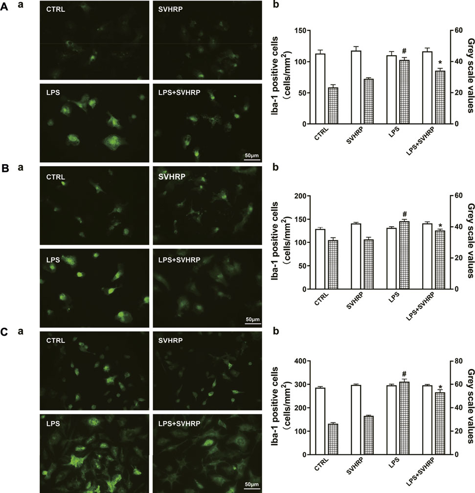
Additionally, there was a mistake in the caption for Figure 2 as published. In Figure 2, the value of the Bar in Figures 2A–C, a is supposed to be 50 instead of 20 μm. The correct Figure 2 appears below.

FIGURE 2. SVHRP attenuates LPS-induced upregulation of Iba-1 in microglia. IF staining for Iba-1 in primary neuron-glia (A), mixed glia (B), and enriched microglia (C) cultures were performed 24 h after LPS treatment. Cells were pretreated with vehicle or SVHRP (20 μg/ml) for 1 h before LPS challenge. (a) Representative images of Iba-1 positive cells (400×, Bar = 50 μm). (b) Cell number and average grey scale for Iba-1 staining were shown. The data were the means ± SEM (n ≥ 3 for each cell preparation). #p < 0.05 compared with CTRL group, *p < 0.05 compared with LPS group.
The authors apologize for this error and state that this does not change the scientific conclusions of the article in any way. The original article has been updated.
All claims expressed in this article are solely those of the authors and do not necessarily represent those of their affiliated organizations, or those of the publisher, the editors and the reviewers. Any product that may be evaluated in this article, or claim that may be made by its manufacturer, is not guaranteed or endorsed by the publisher.
Keywords: SVHRP, anti-inflammation, microglia, NF-κB, MAPKs
Citation: Wu X-F, Li C, Yang G, Wang Y-Z, Peng Y, Zhu D-D, Sui A-R, Wu Q, Li Q-F, Wang B, Li N, Zhang Y, Ge B-Y, Zhao J and Li S (2021) Corrigendum: Scorpion Venom Heat-Resistant Peptide Attenuates Microglia Activation and Neuroinflammation. Front. Pharmacol. 12:791953. doi: 10.3389/fphar.2021.791953
Received: 09 October 2021; Accepted: 05 November 2021;
Published: 26 November 2021.
Edited and reviewed by:
Massimo Grilli, University of Genoa, ItalyCopyright © 2021 Wu, Li, Yang, Wang, Peng, Zhu, Sui, Wu, Li, Wang, Li, Zhang, Ge, Zhao and Li. This is an open-access article distributed under the terms of the Creative Commons Attribution License (CC BY). The use, distribution or reproduction in other forums is permitted, provided the original author(s) and the copyright owner(s) are credited and that the original publication in this journal is cited, in accordance with accepted academic practice. No use, distribution or reproduction is permitted which does not comply with these terms.
*Correspondence: Shao Li, bGlzaGFvODlAZG11LmVkdS5jbg==; Jie Zhao, ZGx6aGFvakAxNjMuY29t
†These authors have contributed equally to this work
Disclaimer: All claims expressed in this article are solely those of the authors and do not necessarily represent those of their affiliated organizations, or those of the publisher, the editors and the reviewers. Any product that may be evaluated in this article or claim that may be made by its manufacturer is not guaranteed or endorsed by the publisher.
Research integrity at Frontiers

Learn more about the work of our research integrity team to safeguard the quality of each article we publish.