 Xueyi Wang
 Yiqi Wang
 Chenghua Lou
94% of researchers rate our articles as excellent or good
Learn more about the work of our research integrity team to safeguard the quality of each article we publish.
Find out more
REVIEW article
Front. Pharmacol., 08 December 2020
Sec. Ethnopharmacology
Volume 11 - 2020 | https://doi.org/10.3389/fphar.2020.577124
Eupatorium lindleyanum DC. (EL) has a long history of traditional use in China to cure coughs, chronic bronchitis, lobar pneumonia, and hypertension. Because of this extensive use of EL in traditional medicine, this present review gives a systematic overview of the conventional applications, phytochemistry, and pharmacological effects of the herb. Literature was systematically searched using the scientific databases ScienceDirect, SciFinder, CNKI, Wiley, Baidu Scholar, SpringerLink, PubMed, Web of Science, and other professional websites. Information was also gathered from books on traditional Chinese herbal medicine, the Chinese Pharmacopoeia and Chinese Materia Medica. To date, many preparations of EL have been widely used clinically to treat various diseases of the respiratory system. More than 100 compounds have been isolated from the herb, including triterpenes, sesquiterpenes, sesquiterpene lactones, flavonoids, acyclic diterpenoids, sterols, and so on. Among them, terpenoids are considered to be the most important bioactive substances in EL. The pharmacological functions of EL, including anti-asthmatic, anti-tussive, anti-inflammatory, anti-hyperlipidemic, anti-hypertensive, anti-virus, and anti-tumor activities, have been widely investigated. However, most of the studies are preclinical research. Further studies are required to examine the underlying mechanisms of action. Traditionally, EL is used for treating many diseases, especially respiratory diseases. Unfortunately, up to now, modern studies have not yet well elucidated the conventional usage of EL. Most importantly, its biological activities and the corresponding constituents are still unclear. Moreover, studies on the pharmacokinetics and toxicity of EL are few, so data on the clinical safety of EL are lacking. Taken together, research work on EL is quite preliminary. More in-depth studies of phytochemistry, pharmacological activities, pharmacokinetics, and toxicity of the herb are needed. This review aims to provide valuable information on EL to guide future investigations and applications.
Eupatorium lindleyanum DC. (EL) (Figure 1), also known as “Yemazhui,” has been traditionally used to treat coughs, chronic bronchitis, and hypertension for thousands of years in China (Editorial Committee of the Administration Bureau of Traditional Chinese Medicine, 1998). The plant is mainly distributed in the Chinese provinces of Jiangsu, Gansu, Shandong, and Hunan. Importantly, Jiangsu Province plant is considered to be an authentic herb. EL has been recorded in the China (Pharmacopoeia, 1977), the Jiangsu Provincial Standard of Local Medicinal Materials 1988, and the China National pharmacopoeia commission, 2015). It possesses the functions of reducing phlegm and relieving cough and asthma (National Pharmacopoeia Commission, 2015).
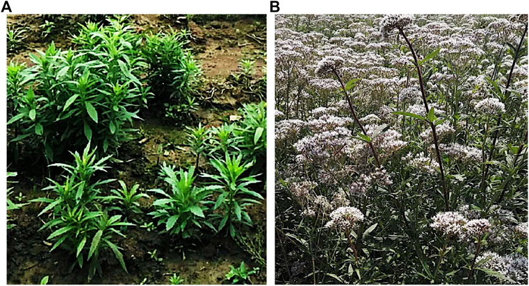
FIGURE 1. Eupatorium lindleyanum DC. (A) Seedlings (B) Plant.
Over the last few decades, researchers have shown interest in the bioactive constituents of EL due to the extensive biological activities of this herb. The components of the plant have been widely investigated and more than 100 compounds have been isolated from the plant. Several types of constituents have been isolated from EL, such as sesquiterpenes, diterpenoids, triterpenoids, volatile oil, flavonoids, and so on (Ito et al., 1979; Yang et al., 2003; Huo et al., 2004; Qian et al., 2004; Yang et al., 2005a; Huo et al., 2006; Ye et al., 2008; Wu et al., 2012a). Also, numerous studies have demonstrated a great number of pharmacological functions of the constituents and fractions from EL, including anti-hyperlipidemic (Zhou et al., 2016), antioxidant (Yan et al., 2011), anti-cancer (Yang et al., 2007), anti-viral (Peng et al., 2008a), and anti-inflammatory effects (Chu et al., 2016). However, review articles about its phytochemistry and pharmacological characteristics are few. Therefore, we have undertaken to make a detailed review of EL by searching a variety of literature from different databases.
The present review was compiled from reported studies on the botany, traditional applications, identified compounds, pharmacological activities, toxicology, and pharmacokinetics of the herb. We also discuss the limitations of the current studies of EL and suggest areas of interest for potential future research. We expect that this review will be useful in providing valuable information for future in-depth investigations and applications.
EL is a perennial herb that grows to a height of approximately 1–2 m. The rhizome is short with fibrous roots, and the stem is erect. The upper part is branched, light brown or purple, and scattered with purple spots. Young plants are covered in dense hairs. Leaves are usually opposite, 3-lobed verticillate, sometimes undivided or deeply divided, and sessile. Lobes are linear lanceolate, margins sparsely serrate, and hairy on both sides with glandular dots below. Leaves have three veins. The capitula are numerous with short stalks, the inflorescence is corymbose; the involucre is bell-shaped with nine lanceolate bracts and each capitulum has five tubular flowers, five-lobed apex, five stamens, and two-lobed stigmas. The tubular flowers are bisexual and purplish. Achenes are black, elliptic, and slightly oblate. Flowering occurs from July to September and fruiting from August to October (Editorial Committee of the Administration Bureau of Traditional Chinese Medicine, 1998; National Pharmacopoeia Commission, 2015).
Because of the effective pharmacological functions of EL, there is a long history of usage of the plant in China. Anecdotally, the history of the herb for treating diseases can be traced back to the “Warring States Period” (475–221 BCE). At that time, it was mainly used to treat horses with respiratory diseases. It has been considered a “folk herb” since ancient times until it was recorded in the China Pharmacopoeia in 1977. According to the China Pharmacopoeia, EL possesses the function of reducing phlegm, clearing away lung heat, and relieving cough and asthma (Editorial Committee of the Administration Bureau of Traditional Chinese Medicine, 1998; National Pharmacopoeia Commission, 2015). Traditionally, it was usually used to treat lung heat, cough, chronic bronchitis, and lobar pneumonia.
The preparations of EL, such as Eupatorium Kechuan powder (野马追克喘散), modified Yemazhui capsule (加味野马追胶囊), sustained-release preparation of Yemazhui (野马追缓释制剂), and Yemazhui syrup (野马追糖浆), are widely used clinically to treat diseases of the respiratory system (Zhu et al., 2012; Su et al., 2016; Zhang, 2017). The applications of EL preparations are summarized in Table 1. In the early 1970s, EL was used to treat patients with chronic bronchitis (People’s Hospital of Xuyi in Jiangsu Province, 1973; Jiangsu Hospital of Jiangsu Province, 1976a). The flavonoids and alkaloids extracted from EL showed significant anti-asthmatic and anti-tussive activities, and markedly improved chronic bronchitis. Su et al. (2016) evaluated the effects of modified Yemazhui capsule in treating patients with acute bronchitis. Sixty patients were involved in the study. Results demonstrated that the modified Yemazhui capsule was effective in treating acute bronchitis. The clinical response rate (CRR) was 96.15%. Also, the symptom scores of the modified Yemazhui capsule-treated group were significantly improved (Su et al., 2016). Consistent with this, a study by Yan (2013) demonstrated that the symptoms of chronic bronchitis were markedly alleviated by treatment with Jiawei Yemazhui capsule. The CRR of Jiawei Yemazhui capsule reached 94.44% (Yan, 2013). Yemazhui syrup is another preparation of EL in common clinical use. Clinical investigations on the effects of Yemazhui syrup on children with chronic bronchitis were evaluated by Zhang (2017). Results demonstrated that the Yemazhui syrup was effective in alleviating chronic bronchitis (96.5%). Importantly, the preparation did not show any significant adverse reactions (Zhang, 2017). It was also reported that Yemazhui syrup was effective in treating patients with lobar pneumonia. Treatment with Yemazhui syrup significantly alleviated lung inflammation and normalized the body temperature and leukocyte counts.
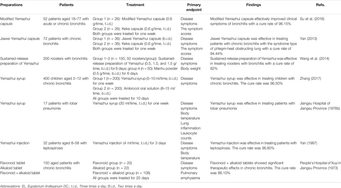
TABLE 1. Clinical trials of EL related preparations.
Results from animal studies also support the above findings. Numerous experiments have documented the therapeutic effects of EL in treating animals with respiratory diseases. It was reported that the sustained-release preparation of Yemazhui showed potential effects in treating roosters with bronchitis (82%) (Wang et al., 2014). Eupatorium Kechuan powder was also demonstrated to be effective in treating diseases of the respiratory system in pigs (93.3%) (Zhu et al., 2012). The effects of EL injection in treating human patients with leptospirosis were reported by (Yan, 1987). According to the report, after treatment for 12.9 h, the body temperature of patients was normalized and the clinical cure rate was 96.8% (Yan, 1987).
The above clinical studies demonstrate the effectiveness of preparations of EL in treating diseases of the respiratory system, especially for bronchitis and lobar pneumonia. However, although the effects of EL in treating leptospirosis were reported, more clinical studies are still required to further verify this pharmacological activity.
Due to the extensive use of EL in traditional Chinese medicine, the bioactive constituents and pharmacological functions of EL have been widely studied. Several categories of phytochemicals have been identified. To date, more than 100 components have been verified in EL, such as triterpenes, sesquiterpenes, sesquiterpene lactones, flavonoids, acyclic diterpenoids, sterols, alkaloids, and so on. Among them, terpenes are considered to be one of the most important constituents in this plant. Table 2 shows all the compounds isolated from EL. The chemical structures are illustrated in Figures 2–7.
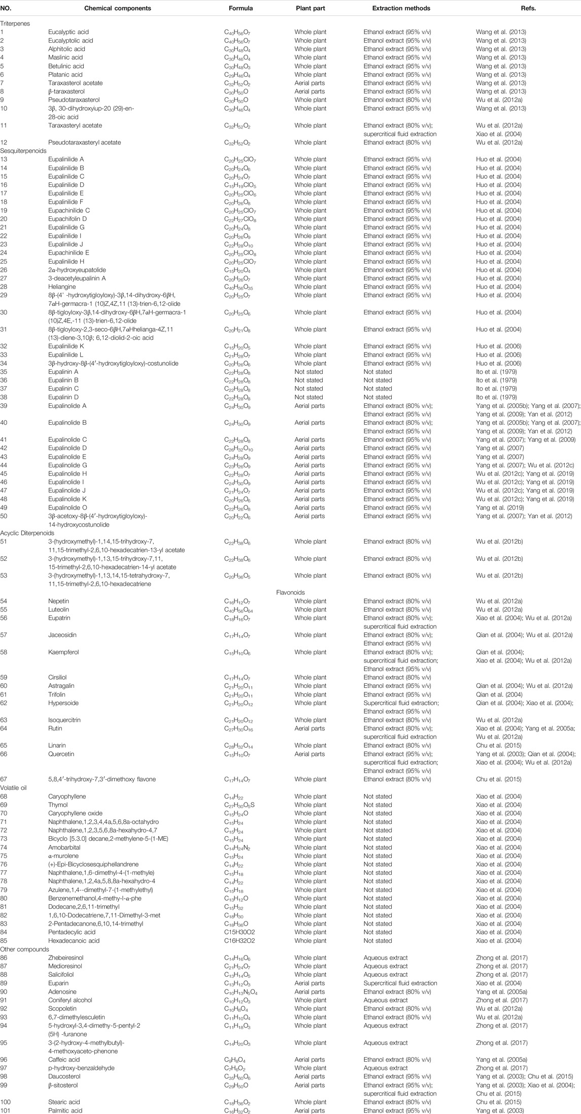
TABLE 2. Chemical components of EL.
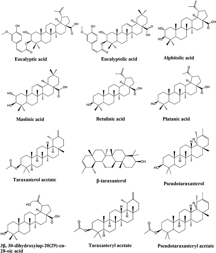
FIGURE 2. Identified triterpenes in EL.
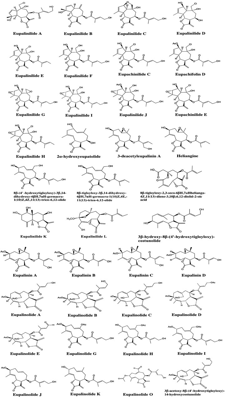
FIGURE 3. Identified sesquiterpenoids in EL.
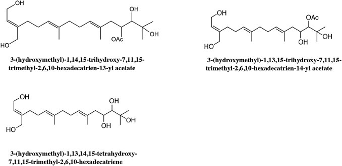
FIGURE 4. Identified acyclic diterpenoids in EL.
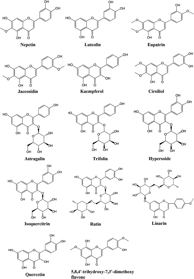
FIGURE 5. Identified flavonoids in EL.
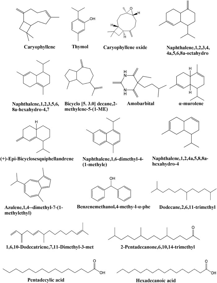
FIGURE 6. Identified compounds in volatile oil of EL.
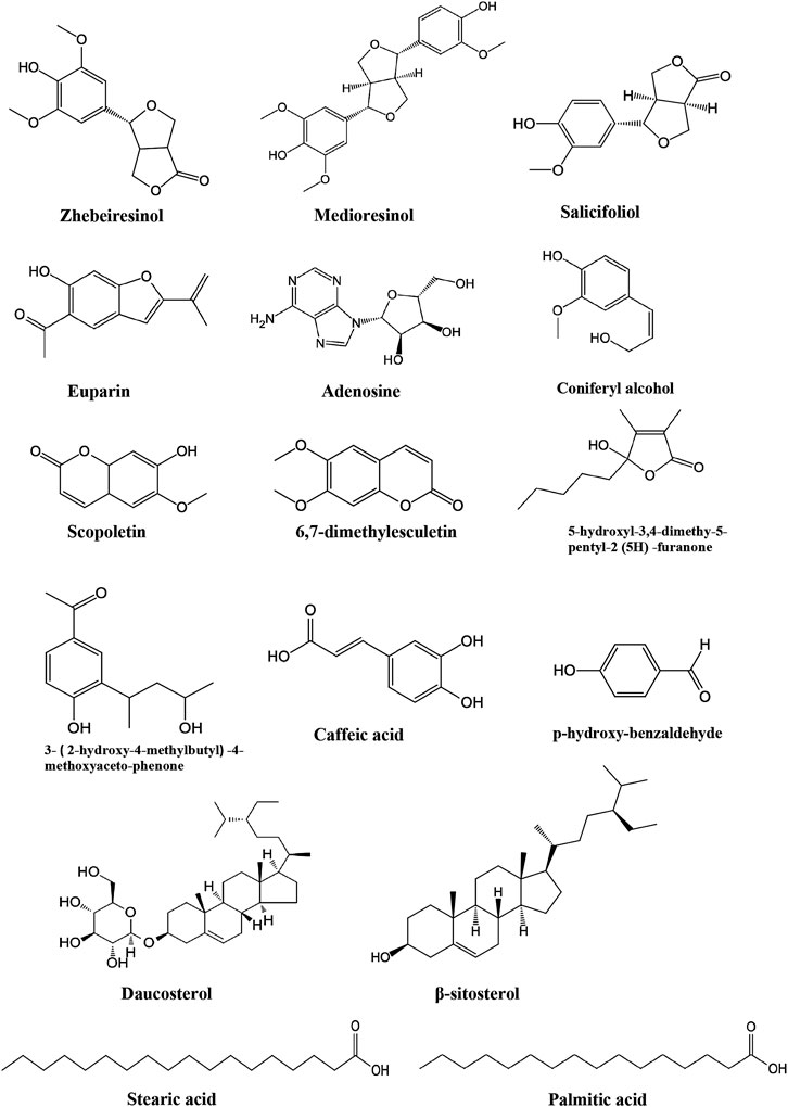
FIGURE 7. Other compounds identified in EL.
The presence of triterpenes in EL has drawn the attention of researchers due to their extensive pharmacological activities in traditional medicine. (Sathya et al., 2014; Sachan et al., 2018; Rios et al., 2020). In the past few years, 12 triterpenes have been identified in EL (Table 2). Their chemical structures are shown in Figure 2. Wang et al. isolated seven triterpene compounds from EL for the first time: alphitolic acid, eucalyptic acid B, eucalyptolic acid, 3β,30-dihydroxyiup-20 (29)-en-28-oic acid, eucalyptic acid, platanic acid, maslinic acid, and betulinic acid (Wang et al., 2013). In addition, taraxasteryl acetate, β-taraxasterol, pseudotaraxasterol, taraxasterol acetate, and pseudotaraxasteryl acetate were identified (Yang et al., 2003; Wu et al., 2012a).
In EL, sesquiterpenoids are commonly considered to be the most important bioactive components. To date, 38 sesquiterpenoids have been identified in EL (Table 2). The structures of these sesquiterpenoids are shown in Figure 3. In 1979, Ito et al. found four new sesquiterpene lactones from EL and named them eupalinins A-D (Ito et al., 1979). Huo et al. isolated 21 sesquiterpene lactones from the plant, including eupalinilides A-J, 2α-hydroxyeupatolide, eupachinilide C, eupachifolin D, 3-deacetyleupalinin A, eupachinilide E, heliangine, eupallnilides K and L, and others (Huo et al., 2004; Huo et al., 2006). Subsequently, 3β-acetoxy-8β-(4′-hydroxytigloyloxy)-14-hydroxycostunolide, 3β-hydroxy-8β-(4′-hydroxytigloyloxy)-costunolide, eupalinolides A-E, G-K and O were identified in the plant (Yang et al., 2005b; Yang et al., 2007; Wu et al., 2012c; Yan et al., 2012; Yang et al., 2019).
To date, only three acyclic diterpenoids isolated from EL have been identified (Table 2): 3-(hydroxymethyl)-1,14,15-trihydroxy-7,11,15-trimethyl-2,6,10-hexadecatrien-13-yl acetate; 3-(hydroxymethyl)-1,13,15-trihydroxy-7,11,15-trimethyl-2,6,10-hexadecatrien-14-yl acetate; and 3-(hydroxymethyl)-1,13,14,15-tetrahydroxy-7,11,15-trimethyl-2,6,10-hexadecatriene (Wu et al., 2012b; Zhong et al., 2017). Figure 4 shows the chemical structures of acyclic diterpenoids in EL.
To date, 15 flavonoids have been isolated from EL (Table 2; Figure 5). Qian et al. identified 6 flavonoids in EL for the first time: jaceosidin, kaempferol, quercetin, astragalin, trifolin, and hypersoide (Qian et al., 2004). Luteolin, isoquerecitrin, rutin, cirsiliol, linarin, quercetin, 5,8,4′-trihydroxy-7,3′-dimethoxy flavone, nepetin and eupatrin were subsequently isolated from the plant (Yang et al., 2003; Xiao et al., 2004; Yang et al., 2005a; Wu et al., 2012a; Chu et al., 2015).
Phytochemical studies on the volatile oil of EL are still preliminary. Chen et al. analyzed the constituents of volatile oil from the flowers of EL. Eighteen compounds were identified, including thymol, caryophyllene, 1,6,10-dodecatriene,7,11-dimethyl-3-met, dodecane,2,6,11-trimethyl, α-murolene, (+)-epi-bicyclosesquiphellandrene, naphthalene,1,6-dimethyl-4-(1-methyle), caryophyllene oxide, benzenemethanol,4-methy-l-α-phe, amobarbital, 2-pentadecanone,6,10,14-trimethyl, pentadecylic acid, hexadecanoic acid, and others (Table 2; Figure 6) (Chen and Yao, 2006).
In addition to the constituents mentioned above, other types of compounds have also been identified from EL, such as fatty acids, sterols, coumarins, and alkaloids (Table 2; Figure 7). The identified compounds include n-hexadecane acid (Yang et al., 2003; Wu et al., 2012a), β-sitosterol (Yang et al., 2003; Chu et al., 2015), daucosterol (Yang et al., 2003; Chu et al., 2015), caffeic acid (Yang et al., 2005), adenosine (Yang et al., 2005), vanillic acid (Chu et al., 2015), palmitic acid (Chu et al., 2015), scopoletin (Wu et al., 2012a), 6,7-dimethylesculetin (Wu et al., 2012a), and butanoic acid (Wu et al., 2012a). In addition, zhebeiresinol, medioresinol, salicifoliol, 3-(2-hydroxy-4-methylbutyl)-4-methoxyaceto-phenone, coniferyl alcohol, and p-hydroxy-benzaldehyde were also identified from EL (Zhong et al., 2017).
As a well-known traditional Chinese medicine, EL has been extensively applied to treat diseases of the respiratory systems. Numerous modern studies have demonstrated that EL and its constituents exhibit potent effects in ameliorating respiratory diseases (Zhu et al., 2012; Su et al., 2016; Zhang, 2017). The pharmacological effects of EL are closely connected with its anti-asthmatic, anti-tussive, and anti-inflammatory functions (Figure 8). Other diverse functions, including anti-hyperlipidemic, anti-hypertensive, anti-virus, anti-tumor, and protective effects on skin damage, were demonstrated in many studies (Figure 8). However, to date, studies of the biological activities of EL are quite preliminary and the related mechanisms of action are not well elucidated. It is unclear which constituents are responsible for the observed biological activities.
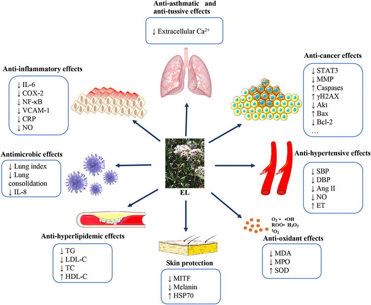
FIGURE 8. Pharmacological effects of EL according to the reported studies.
Respiratory diseases are increasing rapidly and drastically, along with environmental pollution (Verschakelen and Demedts, 1995; Mendes et al., 2020). Existing therapies for respiratory diseases are not highly effective; therefore, it is necessary to develop novel agents. The effects of EL on relieving cough and asthma have been extensively reported (Zhou et al., 2001; Tang et al., 2002; Luo et al., 2008). Pharmacodynamic studies have demonstrated the anti-tussive and anti-asthmatic activities of EL (Luo et al., 2008). It was reported that the water fraction of EL (9.334 g/kg; i.g.) could significantly reduce the cough frequency in a mouse cough model induced by concentrated ammonia. The inhibition rate was around 37.4% (p < 0.05). Significant anti-asthmatic activities of the water fraction (0.467 g/kg; i.g.) and petroleum ether fraction (0.329 g/kg; i.g.) were also reported in the study. The latency of asthma was significantly prolonged by treatment with the water fraction (135.41%) and petroleum ether fraction (135.63%). Experiments also documented that the fractions of petroleum ether (1.258 g/kg; i.g.), chloroform (1.421 g/kg; i.g.) and ethyl acetate (1.267 g/kg; i. g.) possessed significant expectorant activity. Compared with the control group, the phenol red secretions were increased to 146.1% (petroleum ether fraction), 165.0% (chloroform fraction), and 131.7% (ethyl acetate fraction), respectively (Luo et al., 2008). Consistent with this, Zhou et al. (2001) also identified the anti-tussive activity of the water extract of EL in ammonia- and citric acid–induced animal models. Their results demonstrated that cough latency was markedly prolonged (6.59 fold, p < 0.001) and cough frequency was significantly decreased (inhibition rate 80.46%, p < 0.001) on treatment with EL extract (45.1 g [herb]/kg). Anti-asthmatic and expectorant activities of EL (45.1 g [herb]/kg; i.g.) were also demonstrated in in vivo models (Zhou et al., 2001). Tang et al. (2002) investigated the possible anti-asthmatic mechanisms of EL. Studies reported that the EL extract (18.4 g [herb]/L; i.g.) exerted an inhibitory effect on the contraction of tracheal smooth muscle via inhibiting the inflow of extracellular Ca2+, indicating the potent anti-asthmatic mechanism of EL (Tang et al., 2002). The above studies demonstrate that EL shows potential anti-asthmatic and anti-tussive effects in in vivo models. The inhibition of the inflow of extracellular Ca2+ could be one of the main mechanisms for its anti-asthmatic activity.
Inflammation is a critical pathophysiological process in many chronic diseases. The anti-inflammatory activities of numerous natural medicines have been demonstrated (Li et al., 2019; Barragan-Zarate et al., 2020). Studies have documented the anti-inflammatory functions of EL in various models, such as acute lung injury (ALI) and xylene-induced mouse models. The information on anti-inflammatory studies of EL is summarized in Table 3. ALI, induced by diverse predisposing causes, is characterized by acute, progressive respiratory distress and persistent hypoxemia (Guo et al., 2020; Yang et al., 2020). To the best of our knowledge, the inflammatory response is considered as one of the most important factors in the development of ALI (Yu et al., 2015). Therefore, drugs with anti-inflammatory activity are conventionally used clinically for ALI therapy. In the traditional Chinese medicine system, studies have identified the functions of EL on prevention of ALI (Yang et al., 2010; Chu et al., 2015; Li et al., 2019; Chu et al., 2016; Yu et al., 2015; Huang et al., 2019). The mechanisms are mainly based on its anti-inflammatory and antioxidant functions. Experiments demonstrated that EL treatment (22 g/kg; i.g.) significantly reduced the expression levels of inflammatory factors and increased the level of arterial oxygen partial pressure in an ALI model. Also, oleic acid-induced elevation of lung index was significantly inhibited in EL-treated groups (22 g/kg; i.g.) (Yang et al., 2010). In an LPS-induced ALI model, fractions (of chloroform, ethyl acetate, n-butanol, water layer, flavonoids and sesquiterpenes) from EL significantly decreased the lung wet/dry (W/D) ratio and the levels of inflammatory factors, and attenuated pathological changes in the lung (Chu et al., 2015; Chu et al., 2016; Yu et al., 2015; Huang et al., 2019; Li et al., 2019). However, Li et al. (2019) demonstrated that the petroleum ether fraction of EL (30 mg/kg; i.g.) was not effective in LPS-induced ALI models. Jaceosidin, a flavonoid isolated from EL, was also found to be effective on ALI. After administration of jaceosidin (15, 30, and 60 mg/kg; i.g.), suppression of COX-2 and NF-κB was observed (Huang et al., 2019).
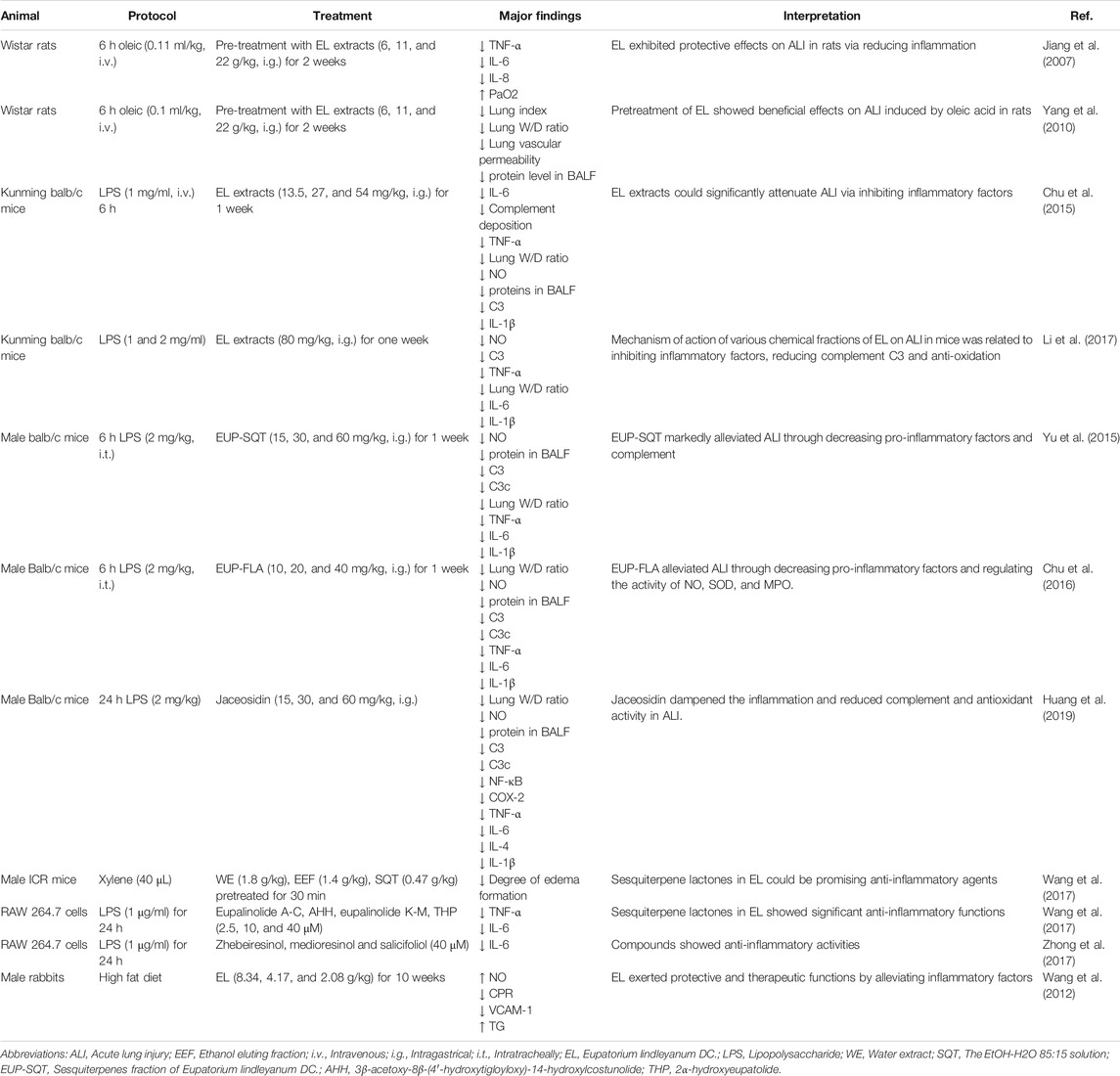
TABLE 3. Anti-inflammatory effects of EL and its potential mechanisms.
Besides the ALI models, the anti-inflammatory functions of EL have also been demonstrated in many other models. In a xylene-induced mouse model, Wang et al. investigated the anti-inflammatory functions of different fractions extracted from EL. Results demonstrated that the sesquiterpene fraction (0.47 g/kg; i.e.) reduced xylene-induced ear edema (21.53%, p < 0.01). In vitro, compounds (2.5, 10, and 40 μM) from EL, such as eupalinolide L, eupalinolide M, and 2α-hydroxyeupatolide, could significantly inhibit inflammatory factors in RAW 264.7 cells (Wang et al., 2017). Experiments also demonstrated the anti-inflammatory activities of zhebeiresinol, medioresinol, and salicifoliol from EL. At 40 μM, treatment with the compounds significantly inhibited IL-6 production. The inhibition rates were 41.6, 74.7, and 35.0%, respectively (Zhong et al., 2017). Importantly, the anti-inflammatory functions of EL were also demonstrated in a rabbit atherosclerosis model. Results indicated that the expression of C-reactive protein (CRP) and mRNA expression of VCAM-1 in EL-treated groups was markedly decreased. In addition, increased NO content was significantly observed in EL-treated groups, which might be a potential anti-inflammatory mechanism of the EL extract in atherosclerosis (Wang et al., 2012).
The above studies demonstrated that the fractions and constituents from EL showed significant anti-inflammatory activities both in vitro and in in vivo model systems. The potential mechanisms were mainly related to the suppression of inflammatory factors, including IL-6, COX-2, and NF-κB.
An imbalance between the production of oxidants and antioxidants often results in oxidative stress (Kemp et al., 2008). Overproduction of oxidants such as malonic aldehyde (MDA) and depletion of SOD and GSH resulted in inflammatory processes and oxidative damage (; Sun et al., 2016; Mo et al., 2020). The antioxidant functions of EL were widely shown in different ALI models (Table 4). In LPS- and oleic acid–induced acute lung injury models, increased levels of SOD and reduced levels of MDA and MPO were significantly detected on treatment with EL compounds and fractions (Zhou et al., 2017). Yan et al. (2011) evaluated the antioxidant effects of various extracts from EL. Their results verified that the water extract (400 μg/ml) and residue water extract (400 μg/ml) showed significant superoxide anion radical scavenging activity (the inhibition rates were 94.55 and 95.77%) (Yan et al., 2011). The antioxidant activity of flavonoids in EL were also assessed with the DPPH (2,2-diphenyl-1-picry-hydrazyl radical) method. Flavonoids showed powerful scavenging activities on DPPH free radical with an IC50 of 10.92 mg/ml (Wang et al., 2010). In addition, Yan et al. developed an ultrasonic-microwave synergistic extraction method to increase the yield rate of antioxidants from EL, which was found to be more effective than other methods (Yan et al., 2013). The above data indicated that EL exhibited significant antioxidant effects in various models through increasing the level of SOD and reducing the levels of MDA and MPO.
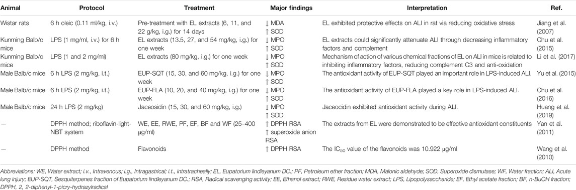
TABLE 4. Antioxidant activities of EL and its potential mechanisms.
Hyperlipidemia, a metabolic disturbance of lipid, is one of the most lethal factors for cardiovascular and cerebrovascular diseases (Pappa et al., 2019; Zeng et al., 2019). It is conventionally characterized by abnormal elevations of LDL-C, TC, and LDL (Zhou et al., 2015). Experiments have demonstrated the anti-hyperlipidemic effects of extracts and compounds from EL. Kaempferol (100 and 300 mg/kg; i.g.) and total flavonoids (50 and 100 mg/kg; i.g.) from EL could markedly lower serum blood lipids, hemorheological parameters, and blood viscosity (He et al., 2014). Most importantly, the contents of TG, LDL-C, and TC were markedly reduced while HDL-C was significantly raised by treatment with EL. These results suggest that EL shows marked preventive and therapeutic effects on hyperlipidemia in rats (Zhou et al., 2017). Consistent with this, Wang et al. (2009) also demonstrated the anti-hyperlipidemia effects of EL (11.28, 22.56, and 45.12 g/kg; i.g.) in an experimental hyperlipemia model. In addition, studies demonstrated that EL (25 g/kg; i.g.) exerted hypolipidemic activity via regulating low density lipoprotein receptor (LDLR) mRNA in hyperlipidemic rats (Chen et al., 2009). The above experiments demonstrate that EL exerts anti-hyperlipidemic effects through regulating LDLR, increasing the content of HDL-C, and decreasing the levels of TG, LDL-C, and TC.
The anti-hypertensive effects of EL were demonstrated in different animal models. Studies demonstrated that the water extract of EL (5 and 10 mg [herb]/mL; i.g.) could significantly suppress the vasoconstriction of the vascular vessels and regulate extracellular calcium influx and intracellular calcium release (Jiang et al., 2007). Moreover, the anti-hypertensive effects of the water decoction of EL were demonstrated in a spontaneously hypertensive rat (SHR) model. SHRs were treated with a water decoction of EL (16.36, 32.73, and 65.45 g/kg; i.g.) for 7 weeks. Rats in the EL-treated groups showed markedly lower systolic blood pressure (SBP) and diastolic blood pressure (DBP) of the caudal artery, indicating the anti-hypertensive effects of the water decoction of EL. Also, decreased levels of NO and angiotensin (Ang Ⅱ) and increased level of endothelin (ET) in serum were observed, suggesting the possible anti-hypertensive mechanisms of EL (Chi, 2016). These experiments demonstrated that EL exerted significant anti-hypertensive activities in vivo. The suppression of Ang Ⅱ and upregulation of ET could be the potential mechanisms of action.
The antimicrobial effects of the EL preparations were reported in many studies (Peng et al., 2008a; Dou et al., 2011). Studies were mainly focused on the compound Yemazhui capsule, in which EL was one of the most important herbs. Peng et al. (2008a) confirmed that the compound Yemazhui capsule (0.1, 0.2, and 0.4 g/ml; i.g.) could effectively inhibit the influenza virus, prolong the lifespan (34.0%), decrease the lung index (16.7%), and inhibit lung consolidation in vivo (Peng et al., 2008a). Furthermore, they also demonstrated the anti-virus effects of compound Yemazhui capsule (0.1, 0.2, and 0.4 g/ml; i.g.) in parainfluenza virus (Peng et al., 2008a). Dou et al. (2011) also demonstrated the anti-virus effects of compound Yemazhui capsule on respiratory syncytial virus (RSV). Results showed that the compound Yemazhui capsule (31.3 mg/L) significantly inhibited the RSV infection. The inhibition rate was around 60.81%. Interestingly, marked suppression of IL-8 in RSV-infected A549 cells was observed. Peng et al. (2008b) also evaluated the anti-bacterial activities of the compound Yemazhui capsule in vivo. Results showed that the compound Yemazhui capsule (8 g [herb]/kg) significantly reduced the mortality of mice after infection with streptococci (Peng et al., 2008b). The mortality was markedly decreased from 70.00% (the control group) to 31.58% (the compound Yemazhui capsule group). These studies demonstrated that the compound Yemazhui capsule showed significant antimicrobial effects in in vitro and in vivo model systems. However, the exact antimicrobial mechanisms of action are still unclear.
Recently, the anti-tumor activities of EL have been widely investigated. Studies have demonstrated that the compounds and fractions from EL showed significant anti-tumor activities (Table 5). The related mechanisms are quite complicated. F1012-2 (5, 10, and 20 μg/ml), an active extract of EL, could significantly inhibit the growth of TNBC cells. Studies demonstrated that F1012-2 suppressed cancer cells through induction of apoptosis and cell cycle arrest (G2/M) (Tian et al., 2018). Significant inhibition of Akt and activation of p38 signaling pathways were observed in the study. Yang et al. (2007) found that the compounds from EL, including eupalinolides A-E and 3β-acetoxy-8β-(40-hydroxytigloyloxy)-14-hydroxycostunolide, could suppress the proliferation of cancer cells. Eupalinolide O (EO), one of the main compounds in EL, was demonstrated to be effective in suppressing the growth of MDA-MB-468 cells. The inhibitory effects of EO (5, 10, and 20 μM) were related to apoptosis induction (Yang et al., 2016). Moreover, some studies also demonstrated that Eupalinolide J (EJ) exhibited significant anticancer activity in TNBC cells. Experiments documented that EJ suppressed the proliferation of cancer cells through induction of apoptosis, disruption of MMP, and suppression of the STAT3 pathway (Lou et al., 2019; Yang et al., 2019). Wu et al. (2020) also demonstrated the anticancer effects of EJ (2.5, 5, 10, and 20 μM) in prostate cancer cells. Their results indicated that EJ suppressed the growth of prostate cancer cells via induction of DNA damage responses. The expression levels of p-Chk1, p-Chk2, and γH2AX were remarkably increased with the treatment of EJ (Wu et al., 2020). The above results indicated that the fractions and compounds isolated from EL showed significant anticancer activities. The possible mechanisms were largely associated with induction of apoptosis, cell cycle arrest, disruption of MMP, induction of DNA damage responses, and suppression of STAT3 and Akt signaling pathways.
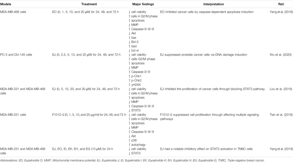
TABLE 5. Anti-tumor activities of EL and its potential mechanisms.
Protective effects of EL on skin damage have been demonstrated. According to the reports, microphthalmia-associated transcription factor (MITF) and heat shock protein 70 (HSP70) play a key role in the production of melanin (Park et al., 2018; Stangl et al., 2018). Experiments demonstrate that EL extract (0.08 mg/ml) could significantly suppress MITF, melanin production, and tyrosinase activity in HSP70-overexpressed cells (Yamashita et al., 2010). Their further study documented that eupalinolide A (EA) and eupalinolide B (EB) were the active HSP-inducers in the EL extracts. After treatment with EA (10 μg/ml) and EB (10 μg/ml), the level of HSP70 in the skin was significantly up-regulated. Importantly, the damage induced by UVB radiation was significantly alleviated, suggesting the effects of both compounds on preventing skin damage and melanin production (Yamashita et al., 2012).
To date, information on toxicological studies of EL is still limited. Li et al. examined the effects of EL in the nervous, cardiovascular, and respiratory systems of rats. Results demonstrated that the extracts of EL did not show any significant effects in rats even at a concentration of 45 g (herb)/kg (Li et al., 2005). Zhou et al. investigated the general pharmacological actions and toxicity of EL. The autonomous movement, cardiovascular and respiratory reactions, and acute and chronic toxicity were tested in rats and mice. Interestingly, EL did not show any significant effects in these tests. The LD50 value was 225.6 g (herb)/kg. No toxic reaction was found in a chronic toxicity test. It was concluded that EL had no obvious influence on normal physiological action and tissues and organs in animals (Zhou et al., 2005). Importantly, clinical findings also supported the above results (Su et al., 2016). The above studies suggested that EL was safe in traditional application. However, further toxicological studies are still needed to test its clinical safety.
To date, there are few reports on the pharmacokinetics of EL. Pharmacokinetic studies should be prioritized to better understand the absorption, distribution, metabolism, and excretion of the active constituents in EL. UHPLC-TOF-MS was used to analyze the mass spectrometric decomposition and metabolic transformation of eupalinolide F in rats. It was demonstrated that eupalinolide F forms a series of 55 related metabolites via undergoing multiple biotransformation pathways (Qin et al., 2019). In addition, Zhang et al. found that Maximum concentrations (Cmax) of HYP, EA, and EB in plasma occurred at 1.80 ± 0.84, 1.00 ± 0.62, and 0.57 ± 0.09 h after treatment with EL extract (625 mg/kg) in rats. Cmax values of EA, EB, and HYP were 144.73 ± 35.90, 371.25 ± 63.91, and 689.60 ± 80.88 ng/ml, respectively. Studies demonstrated that Cmax, AUC0−t, and AUC0−∞ rose in a dose-dependent way with increased dosages of EL fractions (Zhang et al., 2015).
In conclusion, this review emphasizes the significance of the herb in traditional Chinese medicine and summarizes the findings of published studies. The traditional uses, botany, phytochemistry, pharmacological activities, toxicity, and pharmacokinetics of EL have been stated in this paper. EL has been extensively used clinically to treat respiratory diseases. It exhibits anti-inflammatory, antioxidant, anti-tumor, antimicrobial, anti-hyperlipidemic, and anti-hypertensive activities. Terpenes, especially sesquiterpenoids, are considered to be the most important constituents in EL. Moreover, the preparations of EL, including Eupatorium Kechuan powder, modified Yemazhui capsule, Jiawei Yemazhui capsule, sustained-release preparation of Yemazhui, and Yemazhui syrup, have been widely applied in traditional prescriptions to cure many kinds of diseases. Hence, EL has an important role in the traditional Chinese medicine system.
To date, considerable research on EL has been done in various fields, especially in phytochemistry and pharmacological activities. However, challenges still exist. First, although a large number of compounds have been identified in the plant, studies are still necessary to explore all new compounds in EL. Second, various biological effects, such as anti-asthmatic, anti-tussive, anti-inflammatory, anti-tumor, anti-virus, anti-hyperlipidemic, and anti-hypertensive functions, have been widely reported. However, the exact active constituents and potential mechanisms are still unclear. Clinical studies are still necessary to further evaluate these biological functions. Third, toxicity studies, including sub-chronic or acute toxicity, are also very important to determine the safety of EL. Finally, further studies are still needed to investigate in detail the pharmacokinetics and therapeutic doses in humans to better understand the pharmacological activities of EL. This review provides useful background for researchers on the current status of EL investigation and suggests possible directions for future study.
XW, SM, FL, and YW performed the search, screened the papers, edited the tables, and wrote the text. CL designed the study, wrote, and revised the paper.
This work was financially supported by the Zhejiang Chinese Medical University Research Fund Project (2019ZG32).
The authors declare that the research was conducted in the absence of any commercial or financial relationships that could be construed as a potential conflict of interest.
The pictures of the plant were kindly gifted by Gang Yuan in Jiangsu Province of China. We would like to thank Linda Banbury for her kind help on the revision of our manuscript.
EL: Eupatorium lindleyanum DC.
PF Petroleum ether fraction
CRP C-reactive protein
WE Water extract
ALI Acute lung injury
EE Ethanol extract
TC Total cholesterol
RWE Residue water extract
HL Hepatic lipase
EF Ethyl acetate fraction
BF n-BuOH fraction
TG Triglyceride
BALF Bronchoalveolar lavage fluid
DPPH 2, 2-diphenyl-1-picry-hydrazylradical
MDA Malonic aldehyde
SOD Superoxide dismutase
WF Water fraction
HDL-C High density lipoprotein cholesterol
ET Endothelin
RSV Respiratory syncytial virus
LDL-C Low density lipoprotein cholesterol
SHR Spontaneously hypertensive rat
TNBC Triple-negative breast cancer
EO Eupalinolide O
DBP Diastolic blood pressure
MMP Mitochondrial membrane potential
UV Ultraviolet
LDLR Low density lipoprotein receptor
SBP Systolic blood pressure
DBP Diastolic blood pressure
MITF Microphthalmia-associated transcription factor
CRR clinical response rate
EA Eupalinolide A
EB Eupalinolide B
UHPLC-TOF-MS Ultra-high performance liquid chromatography-four-stage rod-time-of-flight mass spectrometry
Cmax The maximum concentrations
HYP Hyperoside
Ang Ⅱ Angiotensin
SBP Systolic blood pressure
LPS Lipopolysaccharide
EEF Ethanol eluting fraction
MPO Myeloperoxidase
LPL Lipoprotein lipase
LCAT Lecithin cholesterol acyltransferase
Barragan-Zarate, G. S., Lagunez-Rivera, L., Solano, R., Pineda-Pena, E. A., Landa-Juarez, A. Y., Chavez-Pina, A. E., et al. (2020). Prosthechea karwinskii, an orchid used as traditional medicine, exerts anti-inflammatory activity and inhibits ROS. J. Ethnopharmacol. 253, 112632. doi:10.1016/j.jep.2020.112632
Chen, J.,, and Yao, C. (2006). Study of chemical components of volatile oil from Chinese herb. J. Anal. Sci. 22 (4), 485–486. doi:10.1016/S1872-2040(07)60022-X
Chen, W., Qin, J., He, H., Li, Q., Wang, K., and Zhou, Y. (2009). Effects of lindley eupatorium herb total flavonoid on blood lipid metabolism in experimental hyperlipidemic rats. J. Third Mil. Med. Univ. 31 (16), 1589–1591. doi:10.3321/j.issn:1000-5404.2009.16.022
Chi, D. (2016). Study on the antihypertensive effects of water decoction of Eupatorium lindleyanum on spontaneously hypertensive rats and corresponding mechanism. China Pharm. 27 (25), 3502–3504. doi:10.6039/j.issn.1001-0408.2016.25.14
Chu, C., Ren, H., Wu, T., and Zhang, J. (2015). Chemical constituents and antipyretic effect of Eupatorium lindleyanum DC. Nat. Prod. Res. Dev. 27, 816–821. doi:10.16333/j.1001-6880.2015.05.012
Chu, C., Ren, H., Xu, N., Xia, L., Chen, D., and Zhang, J. (2016). Eupatorium lindleyanum DC. sesquiterpenes fraction attenuates lipopolysaccharide-induced acute lung injury in mice. J. Ethnopharmacol. 185, 263–271. doi:10.1016/j.intimp.2016.06.032
Dou, J., Peng, Y., Huang, F., Qian, D., and Zhou, C. (2011). Study on Fufang Yemazhui capsule against respiratory syncytial virus. Drug Eval. Res. 34 (2), 85–88. doi:10.7501/j.issn.0253-6376
Editorial Committee of the Administration Bureau of Traditional Chinese Medicine (1998). Chinese materia medica 7. Shanghai, China: Shanghai Science & Technology Press, 839–841.
Guo, R., Li, Y., Han, M., Liu, J., and Sun, Y. (2020). Emodin attenuates acute lung injury in Cecal-ligation and puncture rats. Int. Immunopharm. 85, 106626. doi:10.1016/j.intimp.2020.106626
He, H., Kong, L., Li, X., and Zhou, Y. (2014). Kaempferol vs lindley euqatorium herb total flavonoid for lyperlipemia and hemorheological parameters in rats. J. Third Mil. Med. Univ. 36 (11), 1187–1189. doi:10.16016/j.1000-5404.2014.11.004
Huang, X. L., Wei, X. C., Guo, L. Q., Zhao, L., Chen, X. H., Cui, Y. D., et al. (2019). The therapeutic effects of Jaceosidin on lipopolysaccharide-induced acute lung injury in mice. J. Pharmacol. Sci. 140 (3), 228–235. doi:10.1016/j.jphs.2019.07.004
Huo, J., Yang, S., Ding, J., and Yue, J. (2004). Cytotoxic sesquiterpene lactones from Eupatorium lindleyanum. J. Nat. Prod. 67, 1470–1475. doi:10.1021/np040023h
Huo, J., Yang, S., Ding, J., and Yue, J. (2006). Two new cytotoxic sesquiterpenoids from Eupatorium lindleyanum DC. J. Integr. Plant Biol. 48 (4), 473–477. doi:10.1111/j.1744-7909.2006.00226.x
Ito, K., Sakakibara, Y., Haruna, M., and Lee, K. H. (1979). Four new germacranolides from Eupatorium lindleyanum DC. Chem. Lett. 8, 1469–1472. doi:10.1246/cl.1979.1469
Jiang, T., Tang, C., and Yang, C. (2007). Effects of Eupatorium lindleyanum on vasoconstriction of the vascular vessels. Pharmacol. Clin. Chin. Mater. Med. 23 (5), 124–125. doi:10.3969/j.issn.1001-859X.2007.05.059
Jiangsu Hospital of Jiangsu Province (1976a). A summary of five years’ clinical observation of Yemazhui in treating patients with chronic bronchitis. Jinagsu Med. J. 6, 43. doi:10.19460/j.cnki.0253-3685.1976.06.025
Jiangsu Hospital of Jiangsu Province (1976b). Clinical observation of Yemazhui in treating 17 patients with lobar pneumonia. Jinagsu Med. J. 6, 42. doi:10.19460/j.cnki.0253-3685.1976.06.023
Kemp, M., Go, Y. M., and Jones, D. P. (2008). Nonequilibrium thermodynamics of thiol/disulfide redox systems: a perspective on redox systems biology. Free Radic. Biol. Med. 44 (6), 921–937. doi:10.1016/j.freeradbiomed.2007.11.008
Li, C. L., Tan, L. H., Wang, Y. F., Luo, C. D., Chen, H. B., Lu, Q., et al. (2019). Comparison of anti-inflammatory effects of berberine, and its natural oxidative and reduced derivatives from Rhizoma Coptidis in vitro and in vivo. Phytomedicine 52, 272–283. doi:10.1016/j.phymed.2018.09.228
Li, J., Zhou, Y., and Zeng, M. (2005). Study on general pharmacology of Eupatorium lindleyanum DC. Sichuan J. Physiol. Sci. 27 (1), 42. doi:CNKI:SUN:ZGYA.0.2005-02-005
Li, X. L., Chu, C. J., Wei, X. C., Xia, L., and Zhang, J. (2017). Protective effects of various chemical fractions of YeMaZhui on acute lung injury in mice. West. J. Tradit. Chin. Med. 30 (9), 9–15. doi:10.3969/j.issn.1004-6852.2017.09.003
Lou, C., Chen, Y., Zhang, J., Yang, B., and Zhao, H. (2019). Eupalinolide J suppresses the growth of triple-negative breast cancer cells via targeting STAT3 signaling pathway. Front. Pharmacol. 10, 1071. doi:10.3389/fphar.2019.01071
Luo, Y., Peng, Y., Ye, Q., Shi, L., and Wang, Z. (2008). Pharmacodynamic investigation on the anti-tussive, anti-asthmatic and expectorant fractions in Eupatorium lindleyanum DC. Jiangsu J. Tradit. Chin. Med. 40 (5), 55–57. doi:10.3969/j.issn.1672-397X.2008.08.031
Mendes, L. P. S., Vieira, D. S. R., Gabriel, L. S., Ribeiro-Samora, G. A., Dornelas De Andrade, A., Brandao, D. C., et al. (2020). Influence of posture, sex, and age on breathing pattern and chest wall motion in healthy subjects. Braz. J. Phys. Ther. 24 (3), 240–248. doi:10.1016/j.bjpt.2019.02.00710.1016/j.bjpt.2019.02.007
Mo, M., Li, S., Dong, Z., Li, C., Sun, Y., Li, A., et al. (2020). S-allylmercaptocysteine ameliorates lipopolysaccharide-induced acute lung injury in mice by inhibiting inflammation and oxidative stress via nuclear factor kappa B and Keap1/Nrf2 pathways. Int. Immunopharm. 81, 106273. doi:10.1016/j.intimp.2020.106273
National Pharmacopoeia Commission (1977). Pharmacopoeia of the people’s Republic of China 1. Beijing, China: China Medical Science and Technology Press, 535.
National Pharmacopoeia Commission (2015). Pharmacopoeia of the people’s Republic of China 1. Beijing, China: China Medical Science and Technology Press, 313.
Pappa, E., Rizos, C. V., Filippatos, T. D., and Elisaf, M. S. (2019). Emerging fixed-dose combination treatments for hyperlipidemia. J. Cardiovasc. Pharmacol. Therapeut. 24 (4), 315–322. doi:10.1177/1074248419838506
Park, S. H., Baek, K. H., Shin, I., and Shin, I. (2018). Subcellular Hsp70 inhibitors promote cancer cell death via different mechanisms. Cell Chem. Biol. 25 (10), 1242–1254.e8. doi:10.1016/j.chembiol.2018.06.010
Peng, Y., Dou, J., Huang, F., and Qian, D. (2008a). Antiviral effects of Fufang Yemazhui capsule in vitro and in vivo. Chin. Tradit. Pat. Med. 30 (5), 650–654. doi:10.3969/j.issn.1001-1528.2008.05.009
Peng, Y., Dou, J., Huang, F., and Qian, D. (2008b). Antibacterial and antiviral effect of Fufang Yemazhui capsule in vivo. Lishizhen Med. Mater. Med. Res. 19 (3), 542–544. doi:10.3969/j.issn.1008-0805.2008.03.012
People’s Hospital of Xuyi in Jiangsu Province (1973). Clinical observation of Yemazhui in treating aged patients with chronic bronchitis. J. New Med. 2, 16–18. doi:CNKI:SUN:ZZYZ.0.1973-02-005
Qian, S., Yang, N., Duan, J., Yuan, L., and Tian, L. (2004). Study on the flavonoids of Eupatorium lindleyanum. China J. Chin. Mater. Med. 29 (1), 50–52. doi:10.3321/j.issn:1001-5302.2004.01.015
Qin, W., Liu, X., Wang, Y., Song, X., Yang, Y., and Li, Q. (2019). Analysis of metabolites of eupalinolide F in rats based on UPLC-Q-TOF-MS. Nat. Prod. Res. Dev. 31, 2137–2143. doi:10.16333/j.1001-6880.2019.12.018
Rios, M. Y., Ortega, A., Dominguez, B., Deciga, M., and Rosa, V. (2020). Glaucacetalin E and galphimidin B from Galphimia glauca and their anxiolytic activity. J. Ethnopharmacol. 259, 112939. doi:10.1016/j.jep.2020.112939
Sachan, R., Kundu, A., Jeon, Y., Choi, W. S., Yoon, K., Kim, I. S., et al. (2018). Afrocyclamin A, a triterpene saponin, induces apoptosis and autophagic cell death via the PI3K/Akt/mTOR pathway in human prostate cancer cells. Phytomedicine 51, 139–150. doi:10.1016/j.phymed.2018.10.012
Sathya, S., Sudhagar, S., Sarathkumar, B., and Lakshmi, B. S. (2014). EGFR inhibition by pentacyclic triterpenes exhibit cell cycle and growth arrest in breast cancer cells. Life Sci. 95 (1), 53–62. doi:10.1016/j.lfs.2013.11.019
Stangl, S., Tei, L., De Rose, F., Reder, S., Martinelli, J., Sievert, W., et al. (2018). Preclinical evaluation of the Hsp70 peptide tracer TPP-PEG24-DFO[(89)Zr] for tumor-specific PET/CT imaging. Canc. Res. 78 (21), 6268–6281. doi:10.1158/0008-5472.CAN-18-0707
Su, K., Tan, J., Cai, M., Jiang, Y., Zhu, J., Feng, G., et al. (2016). Clinical observation on the effect of modified Yemazhui Capsule on acute bronchitis & acute exacerbation of chronic bronchitis with the syndrome type of phlegm-heat obstructing lung. J. Emerg. Tradit. Chin. Med. 25 (12), 2229–2231. doi:10.3969/j.issn.1004-745X.2016.12.007
Sun, C. Y., Xu, L. Q., Zhang, Z. B., Chen, C. H., Huang, Y. Z., Su, Z. Q., et al. (2016). Protective effects of pogostone against LPS-induced acute lung injury in mice via regulation of Keap1-Nrf2/NF-kappaB signaling pathways. Int. Immunopharm. 32, 55–61. doi:10.1016/j.intimp.2016.01.007
Tang, C., Jiang, T., and Chen, Z. (2002). Effects of Eupatorium lindleyanum on the contraction of guinea pig tracheal smooth muscle in vitro. Pharmacol. Clin. Chin. Mater. Med. 18 (6), 30–32. doi:10.3969/j.issn.1001-859X.2002.06.018
Tian, S., Chen, Y., Yang, B., Lou, C., Zhu, R., Zhao, Y., et al. (2018). F1012‐2 inhibits the growth of triple negative breast cancer through induction of cell cycle arrest, apoptosis, and autophagy. Phytother. Res. 32 (5), 908–922. doi:10.1002/ptr.6030
Verschakelen, J. A.,, and Demedts, M. G. (1995). Normal thoracoabdominal motions. Influence of sex, age, posture, and breath size. Am. J. Respir. Crit. Care Med. 151, 399–405. doi:10.1164/ajrccm.151.2.7842198
Wang, D., Fang, X., Qi, F., Zhou, Q., Yang, T., and Wang, S. (2014). Clinical investigation on the effects of sustained-release preparation of Yemazhui on rooster bronchitis. Heilongjiang Anim. Sci. Vet. Med. 5, 157–159. doi:CNKI:SUN:HLJX.0.2014-09-055
Wang, F., Li, W., Tao, J., Zhong, Y., and Yang, H. (2013). Triterpenes isolated from traditional Chinese medicine Eupatorium lindleyanum DC. J. Nanchang Univ. 37 (3), 250–254. doi:10.3969/j.issn.1006-0464.2013.03.009
Wang, F., Zhong, H., Fang, S., Zheng, Y., Li, C., Peng, G., et al. (2017). Potential anti-inflammatory sesquiterpene lactones from Eupatorium lindleyanum. Planta Med. 84 (2), 123–128. doi:10.1055/s-0043-117742
Wang, K., Cheng, Y., and Zhou, Y. (2012). Lindley eupatorium herb extract prevents and attenuates vascular inflammation reaction in atherosclerosis rabbits. J. Third Mil. Med. Univ. 34 (18), 1853–1856. doi:10.16016/j.1000-5404.2012.18.005
Wang, K., Qing, J., Cheng, W., Li, Q., and Zhou, Y. (2009). Effect of Lindley eupatorium herb on hemorheological parameters and anti-oxidative enzyme in hyperlipemia rat. Pharmacol. Clin. Chin. Mater. Med. 25 (2), 80–82. doi:CNKI:SUN:ZYYL.0.2009-02-034
Wang, N., Wang, W., Zheng, Y., Li, J., and Li, C. (2010). Study on DPPH free radical scavenging efficiency of flavonoids from Eupatorium lindleyanum DC. China Food Addit 6, 84–87. doi:10.3969/j.issn.1006-2513.2010.06.008
Wu, S., Sun, Q., Chu, C., and Zhang, J. (2012a). Chemical constituents of Eupatorium lindleyanum. China J. Chin. Mater. Med. 37 (7), 937–940. doi:10.4268/cjcmm20120715
Wu, S., Xu, N., Zhang, J., Yao, S., and Chu, C. (2012b). Three new acyclic diterpenoids from Eupatorium lindleyanum DC. J. Asian Nat. Prod. Res. 14 (7), 652–656. doi:10.1080/10286020.2012.684682
Wu, S., Xu, N., Sun, Q., Han, H., and Zhang, J. (2012c). Six new sesquiterpenes from Eupatorium lindleyanum. Helv. Chim. Acta 95, 1637–1644. doi:10.1002/hlca.201200083
Wu, Z., Xu, X., Dai, L., Wang, Y., Yang, B., Zhao, H., et al. (2020). Eupalinolide J induces apoptosis, cell cycle arrest, mitochondrial membrane potential disruption and DNA damage in human prostate cancer cells. J. Toxicol. Sci. 45 (1), 15–23. doi:10.2131/jts.45.15
Xiao, J., Wang, G., Wei, F., and Lin, R. (2004). Chemical constituents of Eupatorium lindleyanum. Chin. Tradit. Herb. Drugs 35 (8), 855–856. doi:10.7501/j.issn.0253-2670.2004.8.392
Yamashita, Y., Hoshino, T., Matsuda, M., Kobayashi, C., Tominaga, A., Nakamura, Y., et al. (2010). HSP70 inducers from Chinese herbs and their effect on melanin production. Exp. Dermatol. 19 (8), e340–e342. doi:10.1111/j.1600-0625.2009.01061.x
Yamashita, Y., Ikeda, T., Matsuda, M., Maji, D., Hoshino, T., and Mizushima, T. (2012). Purification and characterization of HSP-inducers from Eupatorium lindleyanum. Biochem. Pharmacol. 83 (7), 909–922. doi:10.1016/j.bcp.2011.12.040
Yan, G., Ji, L., Luo, Y., and Hu, Y. (2011). Antioxidant activities of extracts and fractions from Eupatorium lindleyanum DC. Molecules 16 (7), 5998–6009. doi:10.3390/molecules16075998
Yan, G., Ji, L., Luo, Y., and Hu, Y. (2012). Preparative isolation and purification of three sesquiterpenoid lactones from Eupatorium lindleyanum DC. by high-speed counter-current chromatography. Molecules 17, 9002–9009. doi:10.3390/molecules17089002
Yan, G., Ji, L., Luo, Y., and Hu, Y. (2013). Ultrasonic-microwave synergistic extraction of antioxidants from Eupatorium lindleyanum DC. J. Nanjing Univ. Technol. 35 (1), 71–75. doi:10.3969/j.issn.1671-7627.2013.01.015
Yan, Q. (1987). Clinical observation of Yemazhui in treating 32 patients with leptospirosis. Jiangsu J. Tradit. Chin. Med. 9, 9.
Yan, X. (2013). Clinical observation on the effect of Jiawei Yemazhui capsule on chronic bronchitis with the syndrome type of phlegm-heat obstructing lung. Heilongjiang J. Tradit. Chin. Med. 6, 27–28. doi:CNKI:SUN:HLZY.0.2013-06-019
Yang, B., Shen, J. W., Zhou, D. H., Zhao, Y. P., Wang, W. Q., Zhu, Y., et al. (2019). Precise discovery of a STAT3 inhibitor from Eupatorium lindleyanum and evaluation of its activity of anti-triple-negative breast cancer. Nat. Prod. Res. 33 (4), 477–485. doi:10.1080/14786419.2017.1396596
Yang, B., Zhao, Y., Luo, C., and Zhao, H. (2016). Eupalinolide O, a novel sesquiterpene lactone from Eupatorium lindleyanum DC., induces cell cycle arrest and apoptosis in human MDA-MB-468 breast cancer cells. Oncol. Rep. 36, 2807–2813. doi:10.3892/or.2016.5115
Yang, H. H., Duan, J. X., Liu, S. K., Xiong, J. B., Guan, X. X., Zhong, W. J., et al. (2020). A COX-2/sEH dual inhibitor PTUPB alleviates lipopolysaccharide-induced acute lung injury in mice by inhibiting NLRP3 inflammasome activation. Theranostics 10 (11), 4749–4761. doi:10.7150/thno.43108
Yang, H., Zhou, Y. D., and He, H. X. (2010). Influence of Eupatorium lindleyanum on pulmonary vascular permeability of oleic acid-induced acute lung injury in rats. China Pharm. 19 (9), 5–6. doi:10.3969/j.issn.1006-4931.2010.09.003
Yang, N., Qian, S., Duan, J., Li, P., and Tian, L. (2007). Cytotoxic sesquiterpene lactones from Eupatorium lindleyanum. J. Asian Nat. Prod. Res. 9 (4), 339–345. doi:10.1080/10286020600727673
Yang, N., Qian, S., Duan, J., and Tian, L. (2003). Studies on the chemical constituents of Eupatorium lindleyanum (I). J. China Pharm. Univ. 34 (3), 220–221.
Yang, N., Tian, L., Qian, S., Duan, J., and Li, P. (2005a). Chemical constituents of Eupatorium lindleyanum (II). Chin. J. Nat. Med. 3 (4), 224–227. doi:CNKI:SUN:ZGTR.0.2005-04-006
Yang, N., Qian, S., Duan, J., Li, P., and Tian, L. (2005b). Two new sesquiterpenes from Eupatorium lindleyanum. Chin. Chem. Lett. 16, 1223–1226.
Yang, N. Y., Duan, J. A., Qian, D. W., and Tian, L. J. (2009). Simultaneous quantification of four sesquiterpene lactones in Eupatorium lindleyanum DC. by RP-LC. Chromatographia 70 (1–2), 205–209. doi:10.1365/s10337-009-1123-y
Ye, G., Huang, X., Li, Z., Fan, M., and Huang, C. (2008). A new cadinane type sesquiterpene from Eupatorium lindleyanum (Compositae). Biochem. Systemat. Ecol. 36, 741–744. doi:10.1016/j.bse.2008.06.003
Yu, S., Shi, M., Liu, C., Liu, Q., Guo, J., Yu, S., et al. (2015). Time course changes of oxidative stress and inflammation in hyperoxia-induced acute lung injury in rats. IranJ. Basic Med. Sci. 18 (1), 98–103. doi:10.22038/IJBMS.2019.13852
Zeng, H., Chen, P., Chang, Q., Zheng, B., and Zhang, Y. (2019). Hypolipidemic effect of polysaccharides from Fortunella margarita (Lour.) Swingle in hyperlipidemic rats. Food Chem. Toxicol. 132, 110663. doi:10.1016/j.fct.2019.110663
Zhang, J., Zhao, F., Yu, X., Lu, X., and Zheng, G. (2015). Pharmacokinetics of eupalinolide A, eupalinolide B and hyperoside from Eupatorium lindleyanum in rats by LC/MS/MS. J. Chromatogr. B Analyt. Technol. Biomed. Life Sci. 995–996, 1–7. doi:10.1016/j.jchromb.2015.04.038
Zhang, Y. (2017). Clinical investigation on the effect of Yemazhui syrup on children chronic bronchitis. J. Imaging Res. Med. Appl. 1 (1), 141–142. doi:10.3969/j.issn.2096-3807.2017.01.091
Zhong, H., Fang, S., Chen, Y., Li, C., Zheng, Y., and Peng, G. (2017). Chemical constituents from Eupatorium lindleyanum and their anti-inflammatory activities. Chin. Tradit. Pat. Med. 39 (2), 329–333. doi:10.3969/j.issn.1001-1528.2017.02.020
Zhou, X., Song, G., Zhang, X., Liang, X., and Li, Q. (2016). Beneficial effects of crude extract of Eupatorium lindleyanum DC. in hyperlipidemia and atherosclerosis. Biotechnol. Biotechnol. Equip. 30, 151–157. doi:10.1080/13102818.2015.1088796
Zhou, X., Zhang, W., Liu, X., Zhang, W., and Li, Y. (2015). Interrelationship between diabetes and periodontitis: role of hyperlipidemia. Arch. Oral Biol. 60 (4), 667–674. doi:10.1016/j.archoralbio.2014.11.008
Zhou, Y., Chen, K., He, H., Jiang, Z., Zeng, M., and Yang, H. (2005). Investigation on general pharmacology of Eupatorium lindleyanum DC. China Pharm. 16 (2), 94–96. doi:10.3969/j.issn.1001-0408.2005.02.006
Zhou, Y., Jiang, Z., He, H., Zeng, M., and Yang, H. (2017). Preventive and therapeutic effect of Lindley Eupatorium herb on experimental hyperpidemia in rats and mice. China Pharm. 18 (3), 178–179. doi:10.3969/j.issn.1001-0408.2007.03.009
Zhou, Y., Wu, Y., Zhu, S., He, H., Qiu, F., and Li, J. (2001). Study on the anti-bacterial, anti-tussive and anti-asthmatic effects of Yemazhui. China Pharm. 12 (12), 716–718. doi:10.3969/j.issn.1001-0408.2001.12.006
Keywords: Eupatorium lindleyanum DC, botany, pharmacology, toxicity, pharmacokinetics, phytochemistry
Citation: Wang X, Ma S, Lai F, Wang Y and Lou C (2020) Traditional Applications, Phytochemistry, and Pharmacological Activities of Eupatorium lindleyanum DC.: A Comprehensive Review. Front. Pharmacol. 8:577124. doi: 10.3389/fphar.2020.577124
Received: 22 September 2020; Accepted: 10 November 2020;
Published: 08 December 2020.
Edited by:
Alexander N. Shikov, Saint-Petersburg State Chemical Pharmaceutical Academy, RussiaReviewed by:
Wei Peng, Chengdu University of Traditional Chinese Medicine, ChinaCopyright © 2020 Wang, Ma, Lai, Wang and Lou. This is an open-access article distributed under the terms of the Creative Commons Attribution License (CC BY). The use, distribution or reproduction in other forums is permitted, provided the original author(s) and the copyright owner(s) are credited and that the original publication in this journal is cited, in accordance with accepted academic practice. No use, distribution or reproduction is permitted which does not comply with these terms.
*Correspondence: Chenghua Lou, bG91LmNoZW5naHVhQGhvdG1haWwuY29t
Disclaimer: All claims expressed in this article are solely those of the authors and do not necessarily represent those of their affiliated organizations, or those of the publisher, the editors and the reviewers. Any product that may be evaluated in this article or claim that may be made by its manufacturer is not guaranteed or endorsed by the publisher.
Research integrity at Frontiers
Learn more about the work of our research integrity team to safeguard the quality of each article we publish.