
95% of researchers rate our articles as excellent or good
Learn more about the work of our research integrity team to safeguard the quality of each article we publish.
Find out more
ORIGINAL RESEARCH article
Front. Pharmacol. , 09 April 2019
Sec. Ethnopharmacology
Volume 10 - 2019 | https://doi.org/10.3389/fphar.2019.00354
This article is part of the Research Topic Assessing the Pharmacological Effects and Therapeutic Efficacy of Traditional Chinese Medicine in Neurological Disease Models View all 16 articles
 Xuan Gong1†
 Hui Xiong1†Sisi Liu1Yutong Liu2Liang Yin1Chuyue Tu1Hua Wang2Zhongqiu Zhao3,4Weiwu Chen5Zhinan Mei1*
The pathogenesis of itchy skin diseases including allergic contact dermatitis (ACD) is complicated and the treatment of chronic itch is a worldwide problem. One traditional Tibetan medicine, Qingpeng ointment (QP), has been used in treatment of ACD in China for years. In this study we used HPLC and LC/MS analysis, combined with a BATMAN-TCM platform, for detailed HPLC fingerprint analysis and network pharmacology of QP, and investigated the anti-inflammatory and antipruritic activities of QP on ACD induced by squaric acid dibutylester (SADBE) in mice. The BATMAN-TCM analysis provided information of effector molecules of the main ingredients of QP, and possible chronic dermatitis-associated molecules and cell signaling pathways by QP. In ACD mice, QP treatment suppressed the scratching behavior induced by SADBE in a dose-dependent manner and inhibited the production of Th1/2 cytokines in serum and spleen. Also, QP treatment reversed the upregulation of mRNAs levels of itch-related genes in the skin (TRPV4, TSLP, GRP, and MrgprA3) and DRGs (TRPV1, TRPA1, GRP, and MrgprA3). Furthermore, QP suppressed the phosphorylation of Erk and p38 in the skin. In all, our work indicated that QP can significantly attenuate the pathological alterations of Th1/2 cytokines and itch-related mediators, and inhibit the phosphorylation of MAPKs to treat the chronic itch.
Chronic itch has been long known not only as a concomitant manifestation of various skin diseases, such as contact dermatitis, eczema, atopic dermatitis, psoriasis, etc., but also occurs in systemic, neurological, or psychiatric diseases. So far, medications for chronic itch still can be simply categorized into topical (emollients and moisturizers, anesthetics, coolants, glucocorticoids, others) and systemic (antihistamines, neuroactive medications, antidepressants, phototherapy, etc.) forms (Yosipovitch and Bernhard, 2013; Pereira and Stander, 2017; Hachisuka et al., 2018; Shevchenko et al., 2018). Due to its heterogeneity, the causes and pathophysiology of chronic itch are complicated. The treatment of chronic itch, especially intractable itch, is a worldwide problem and a great challenge for clinicians.
Complementary and alternative medicine (CAM), including herbal or botanical therapies, traditional Chinese medicine (TCM), dietary supplements, etc., is clinically used to treat intractable skin diseases like psoriasis. A large amount of evidence indicated those CAM managements could effectively relieve the symptoms of diseases or their underlying causes, which cannot be solved by western medicine (Farahnik et al., 2017; Parker et al., 2017; Svendsen et al., 2017; Na Takuathung et al., 2018). For example, it is estimated that there are about 10–62% of patients with psoriasis in the western world seeking the use of CAM (Farahnik et al., 2017; Svendsen et al., 2017). Consistently animal experimental studies have also accumulated a wealth of data demonstrating the effectiveness of the use of CAM in animal models (Na Takuathung et al., 2018).
Allergic contact dermatitis (ACD) is a skin T cell-mediated immune response caused by one or more non-toxic allergens in contact with the skin, and often occurs together with irritant contact dermatitis significantly affecting the quality of life (Kostner et al., 2017). Existing treatments for ACD are still limited to a few traditional methods such as topical steroids and immunosuppressants, etc., etiologically based and efficient treatments are still lacking. Over centuries Chinese medicines have been used in the treatment of ACD, and their main ingredients and possible molecular mechanisms have been gradually recognized (Li and Li, 2013; Li et al., 2013, 2018; Fu et al., 2015). One traditional Tibetan medicine, Qingpeng ointment (QP), has been used in treatment of ACD in China for many years. QP contains nine main ingredients including Oxytropis falcata Bunge, Rheum lhasaense, Aconitum pendulum Busch, Terminalia chebula Retz (stoned), Terminalia billerica (Gaertn.) Roxb, Phyllanthus emblica Linn., Styrax tonkinensis (Pierre) Craib ex Hartw, Tinospora sinensis (Lour.) Merr., and Synthetic musk (Product Manual, Qizheng Tibetan Medicine Co., Ltd., Lanzhou, China). Two previous works reported that QP exerts anti-inflammation and ameliorates ACD in BALB/c mice (Li and Li, 2013; Li et al., 2013, 2018; Fu et al., 2015). However, similar to other TCM experimental studies on ACD they have some common problems. For example, their ACD model was established by topical 4-dinitrofluorobenzene (DNFB), a model that has been used for a long time but still has actual unknown molecule mechanisms (Li and Li, 2013; Li et al., 2013, 2018; Fu et al., 2015). Secondly without exception, they all focus on anti-inflammatory effect and none of them have comprehensively studied the chronic itch associated with ACD.
Squaric acid dibutylester (SADBE), a small molecule hapten, is commonly used in the treatment of alopecia areata and has been used as a popular ACD inducer in mice (Fu et al., 2014; Feng et al., 2017; Luo et al., 2018). Remarkably in recent years neuroimmune mechanisms of SADBE-induced chronic itch were greatly investigated. It was showed that the increased CXCL10/CXCR3 signaling in sensory neurons may contribute to the pathogenesis of the chronic itch in this model (Fu et al., 2014). Most recently we and our collaborators found transient receptor potential (TRP) channels, TRPV1 and TRPA1-expressing sensory neurons or TRPV4 in the TRPV4-expressing epithelial and immune cells in the skin are molecular and cellular basis for SADBE-induced chronic itch (Feng et al., 2017; Luo et al., 2018).
In order to fully understand the pharmacological functions of herbal ingredients or components, we also employed a newly developed bioinformatics analysis tool, BATMAN-TCM (a Bioinformatics Analysis Tool for Molecular mechanism of Traditional Chinese Medicine) established by our collaborator. Unlike the traditional concept of “one drug, one goal,” BATMAN-TCM tries to seek the key molecules targeted by the main components of TCM, and discover the biological networks derived from these molecules (Liu Z. et al., 2016; Zhang et al., 2017). Meanwhile, the data will be compared with the molecular targets and signal pathway networks related to diseases established in the database, and will describe the complicated mechanisms and potential therapeutic values of the drug (Liu Z. et al., 2016; Zhang et al., 2017, 2018). Therefore, this study attempted to introduce BATMANTCM into the ACD study and investigate the anti-inflammatory activities of QP on ACD induced by SADBE in mice, especially whether QP treats the chronic itch, and if it does what the possible mechanism could be.
The QP ointment used in this study was purchased from Tibet Qizheng Tibetan Medicine Co., Ltd. (Lanzhou, China) and the same batch of drug product was sued for all animal experiments, LC/MS analysis and BATMAN-TCM Target Prediction and Analysis. QP ointment contains nine kinds of herbal components, including O. falcata Bunge, R. lhasaense, A. pendulum Busch, T. chebula Retz (stoned), T. Billerica (Gaertn.)Roxb, P. emblica Linn., S. tonkinensis (Pierre) Craib ex Hartw, T. sinensis (Lour.) Merr., and S. musk, which were mixed at a weight ratio of 20:10:15:20:20:20:7:30:5.
For identification of the main constituents, the mass spectral analyses were performed using an Agilent 6540 Q-TOF LC/MS system (Agilent Technologies, Santa Clara, CA, United States). The operating conditions for ESI interface were as following: positive/negative ionization mode; spray voltage, 3.8 kV; gas temperature, 350°C; and the full scan range was from 100 to 1600 m/z. An Agilent 1290 HPLC system equipped with a G1379A HPLC pump, a G1315B DAD detector and a ZORBAX SB-C18 column (250 × 4.6 mm, 5 μm) was used for the HPLC analysis. The mobile was consisted of acetonitrile (solvent A) and 5% methanol aqueous solution (including 0.1% formic acid, solvent B) and the gradient elution program was as below: 0∼10 min, 100% B; 10∼25 min, 0–5% A, 100∼95% B; 25∼60 min, 5∼10% A, 95∼90% B; 60∼100 min, 10∼20% A, 90∼80% B; 100∼150 min, 20∼70% A, 80∼30% B, 150∼180 min, 70% A, 30% B. The flow rate was 1.0 mL/min, and the sample injection volume was 10 μL. The detection wavelength was set at 284 nm.
The target prediction method adopted the BATMAN-TCM platform, which in principle, is a similarity-based method to predict potential drug–target interactions based on the similarity of those known TCMs drug–target interactions. Simply, the combination of QP composition and their corresponding PubChem ID were an input into BATMAN-TCM software for network pharmacological analysis to obtain the results of the “drug-disease” potential target prediction (Liu Z. et al., 2016; Zhang et al., 2017). The analysis was based on multiple source database included TCMID database (providing a giant formula-herb-ingredient association data), DrugBank (downloaded on July 26, 2015), KEGG (version: July 31, 2014), TTD (Therapeutic Target Database) (version: 4.3.02), OMIM (Online Mendelian Inheritance in Man) (downloaded on Mar. 13, 2014), and TTD (version: 4.3.02) (Liu Z. et al., 2016; Zhang et al., 2017).
Male C57BL/6 mice were purchased from Beijing Vital River Laboratory Animal Technology Co., Ltd. (Beijing, China). Animals were single-caged and housed in a SPF-grade environment with water and food ad libitum at temperature (24 ± 2)°C, a 12-h/12-h light/dark cycle lighting off at night (7:00 pm–7:00 am), at least adapted for 1 week before behavior work. All behavioral tests were videotaped above the observation boxes, and assessed by observers blinded to the experimental design. In this study, mice used for behavioral tests were divided into the blank control group (acetone topically), the model group (0.5% SADBE/acetone topically), groups of treatment low-dose, medium-dose and high-dose of QP ointment applied to the shaved nape of the neck (0.625, 1.25, and 2.5 g/kg), or the positive drug (Diphenhydramine Hydrochloride, 10 mg/kg, i.p.). In this study, male mice at 7 weeks old were used for experiments. All animal experimental procedures followed international guidelines for care and use of laboratory animals which were approved by the Animal Ethics Committee of South-central University for Nationalities. SADBE was purchased from J&K Scientific Ltd. (Beijing, China). QP was purchased from Tibet Qizheng Tibetan Medicine Co., Ltd. (Lanzhou, China). Diphenhydramine hydrochloride was purchased from Shanghai Yuanye Bio-Technology Co., Ltd. (Shanghai, China).
The model was mainly established following previous work (Feng et al., 2017; Luo et al., 2018). The mice were sensitized once per day for three consecutive days (from day 0 to day 2) by applying 20 μL of 0.5% SADBE/acetone solution topically to the shaved abdominal skin. 5 days later, 20 μL of 0.5% SADBE/acetone solution was applied topically to shaved mouse neck skin about 3 × 3 cm region in the midline backs once a day for three consecutive days. 3 days later, the paw scratching behavior was recorded by the camera under unmanned conditions for 60 min, and the number of scratches to the shaved part of the neck was counted by people blinded to the experimental design. The treatment group was topically applied to the shaved nape of the neck with low, medium and high dose QP (0.625, 1.25, and 2.5 g/kg) twice a day starting from day 0 to day 12, and once a day on day 13. Diphenhydramine (10 mg/kg i.p.) as the positive drug was injected into the mice on day 13. The following scratching behavior was recorded after 30 min at the last QP and diphenhydramine treatment by observers. The control group was used same volume of acetone other than 0.5% SADBE/acetone. The model group mice were applied to the blank substrate (from Tibet Qizheng Tibetan Medicine Co., Ltd.) at the same time (Feng et al., 2017; Luo et al., 2018).
The skin in midline of the mouse neck was collected and fixed in 4% paraformaldehyde, routinely dehydrated, embedded in paraffin, and sectioned. Hematoxylin & eosin (HE) and toluidine blue staining were performed as previously described (Zhao et al., 2013). After the stained sections were dehydrated, cleared and sealed, they were visualized under a microscope, and images were taken. Image J software was used to measure the relative thickness of the epidermis from HE staining images and count the number of mast cells from toluidine blue staining images.
The total protein from the mouse midline back neck skin and cervical Dorsal Root Ganglion (DRG) (C1–C7) were extracted as according to standard protocols. Simply, a 1% protease inhibitor (Roche, Switzerland) and a 1% phosphatase inhibitor (Roche, Switzerland) were added to the protein lysate. By using this lysate buffer, the mouse tissues were homogenized and sonicated, then placed on ice for 30 min, and centrifuged (12000 g, 4°C, 9 min) to take the supernatants for the next steps. Samples were denatured in a 20% 5× loading buffer and heated in a boiling water bath for 8 min. After cooling, the samples were separated and cryopreservation. Proteins were separated by SDS–PAGE (7 or 10% acrylamide) and blotted onto PVDF membranes. The membrane was incubated with primary antibodies overnight at 4°C, and the primary antibodies involved were anti-P38 (8690S, 1:1000), anti-phospho-P38 (4511S, 1:1000), anti-Erk (4695S, 1:1000), anti-phospho-Erk (4370P, 1:2000), anti-Akt (4691S, 1:1000), anti-phospho-Akt (4060S, 1:2000), anti-Stat3 (12640S, 1:1000), anti-phospho-Stat3 (9145P, 1:2000), anti-IκBα (4812S, 1:1000), anti-phospho-IκBα (2859S, 1:2000) (CST, Danvers, MA, United States) and anti-Tubulin (Proteintech, Wuhan, China, 1:10000). Then the membranes were incubated with the appropriate horseradish peroxidase-conjugated secondary antibodies for 1 h at room temperature. The following immunoreactive polypeptides were visualized using ECL (ZETA, San Francisco, CA, United States) assays, and Tubulin was used as an internal control.
Total RNA was extracted from homogenized DRG neurons or skin samples using the Trizol reagent (TaKaRa, Japan) according to the manufacturer’s instructions. RNA was treated with DNase I and cDNA was synthesized in vitro using a ThermoScript RT-PCR reverse transcription kit. Each reaction was performed in a system containing 10 μL SYBR Mix, 2 μL cDNA, 1 μL primer mix and 7 μL RNase-free water, using a real-time PCR system. Relative mRNA expression levels of different target genes compared to GAPDH or β-Actin were calculated using the 2−ΔΔCt method. The primer sequences (5′ to 3′) involved were showed in Table 1.
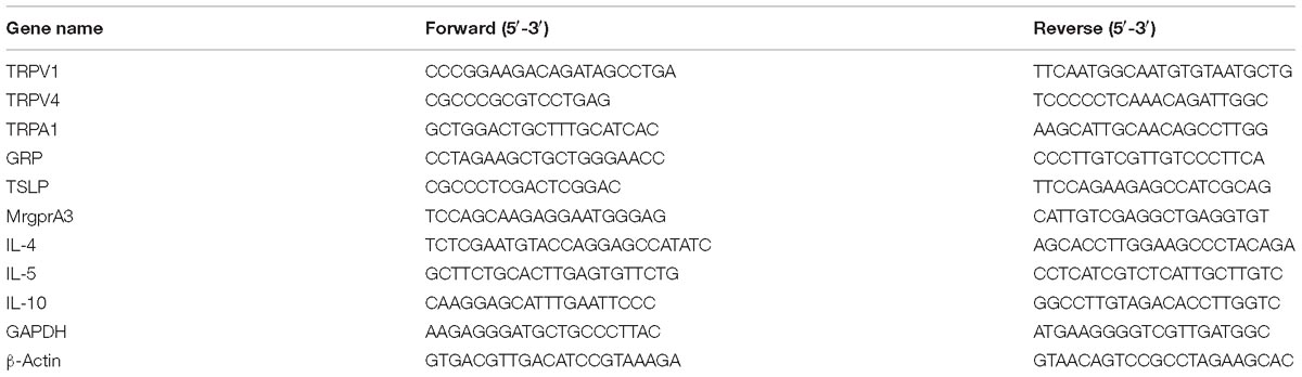
Table 1. Primer sequences.
Detection of IgE, Histamine, TNF-α, IFN-γ, IL-1β, IL-4, IL-5, IL-10, etc., in the spleen and serum of mice using an ELISA kit according to the manufacturer’s instructions. All mouse ELISA kits were purchased from Shanghai Jianglin Biotechnology Co., Ltd.
All data was expressed as the means ± SEM. GraphPad Prism 6 software (San Diego, CA, United States) was used for the data statistical analysis and graphics. Unpaired t-test was used to analyze statistical comparisons between two groups. Multiple comparisons were compared by one-way analysis of variance (ANOVA) followed by Bonferroni’s post hoc tests. p-value < 0.05 was assumed as statistically significant.
Using the current LC/MS method, three main compounds of QP were identified, including gallic acid (1) (Teodoro et al., 2018), corilagin (2) (Salih et al., 2017), and ellagic acid (3) (BenSaad et al., 2017) (their structures showed in Supplementary Figure S1). These compounds are all phenolic acid derivatives and gallic acid as the main unit. In their mass spectrum, fragment ions at m/z 465 [M-169]+ in the 2 indicated the existence of gallic acid unit, and m/z 153, 121 in the 3 indicated the existence of 3,4-dihydroxybenzoic acid and benzoic acid units (Supplementary Figure S1). Five batches purchased from Tibet Qizheng Tibetan Medicine Co., Ltd. were also examined based on the results of LC/MS. These batch numbers were as follows: 161130 (S1), 170308 (S2), 170812 (S3), 171134 (S4), and 180513 (S5). The retention times of three compounds were 12.03, 57.82, and 81.41 min, respectively (Figures 1A,B).
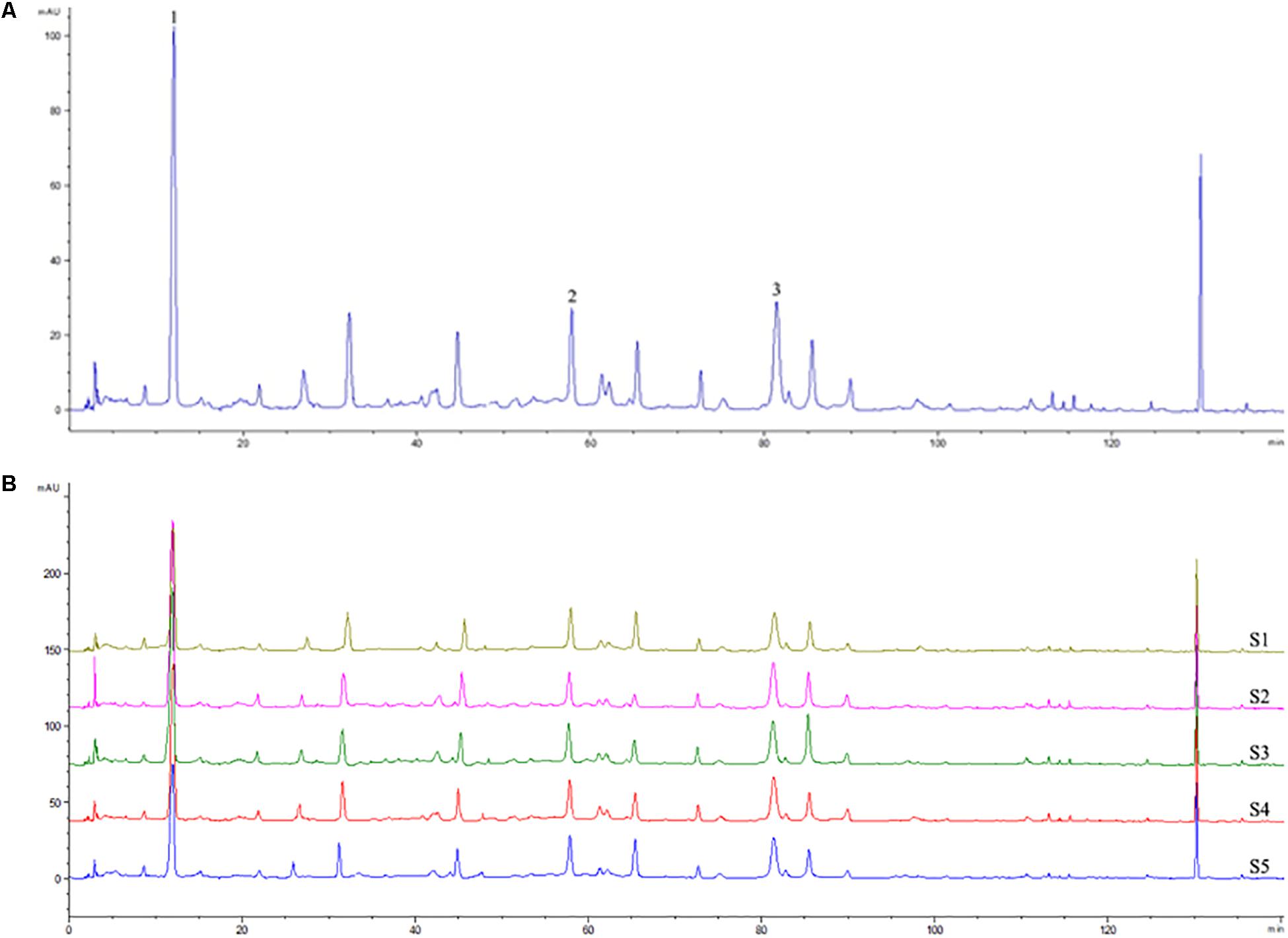
Figure 1. HPLC fingerprint analysis of QP ointment. The HPLC chromatographic of QP at 284 nm wavelength was recorded (A). There are at least 3 phenotypic peaks, which are derived from gallic acid (1), corilagin (2), and ellagic acid (3), respectively. HPLC fingerprint of 5 batches of QP was also presented (B).
To establish the related target prediction and network/pathway analysis for QP to treat chronic dermatitis, the current work used the platform of BATMAN-TCM and obtained the interactive network diagram of QP-chronic dermatitis (Figure 2). In the view of the association network, based on QP’s drug targets and biological pathways, targets of chronic dermatitis diseases and predicted pathways, the prediction results are divided into three types of nodes including composition-targets association, disease-targets association and their overlapping association. A complete gene names of targets and biological pathways for QP’s drug and/or chronic dermatitis, see MS Excel worksheets 1 and 2 in Supplementary Table S1.
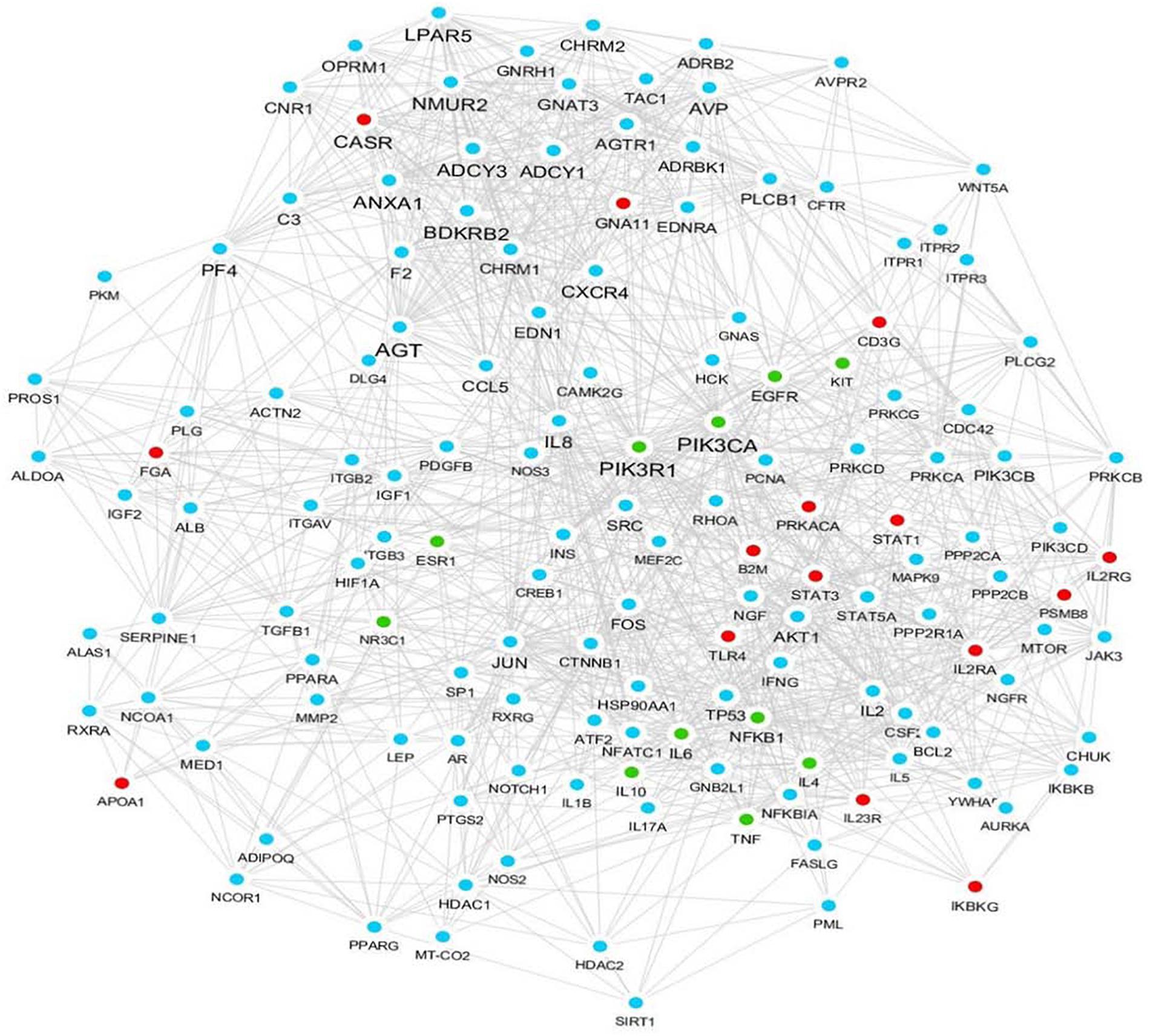
Figure 2. QP ingredient-target-pathway/chronic dermatitis association network. Red nodes correspond to the predicted key targets of chronic dermatitis, such as TLR4, STAT1, STAT3, PRKACA, PSMB8, IL2RG, CD3G, etc. Blue nodes correspond to the key targets of QP ointment, such as IL-8, CXCR4, JUN, IL-1β, IL-17A, etc., Green nodes represent the key targets associate to both QP and chronic dermatitis, such as IL-4, IL-6, IL-10, TNF, NFκB1, PIK3R3, PIK3CA, EGFR, KIT, ESR1, NR3C1, etc.
Skin allergies and chronic dermatitis have been caused by topical application of SADBE, a popular ACD inducer in mice (Fu et al., 2014; Feng et al., 2017; Luo et al., 2018). In this study, we followed the schematic experimental protocol (Fu et al., 2014; Feng et al., 2017; Luo et al., 2018) and pretreated mice with SADBE in the abdomen for 3 days (sensitization) and re-stimulated after 3 days topically on the neck (challenge), which induced robust spontaneous scratching behavior in mice (Figure 3A). Previous analyses indicated that no corticosteroid existed in QP (Li and Li, 2013; Li et al., 2013) and antihistamines have been used traditionally in the attenuation of experimental and clinical pruritus (Yosipovitch and Bernhard, 2013; Mahapatra et al., 2014; Pereira and Stander, 2017; Hachisuka et al., 2018; Shevchenko et al., 2018), therefore in this study we used a classic antihistamine, diphenhydramine, as the positive control medicine. Similar to previous reports (Fu et al., 2014; Feng et al., 2017; Luo et al., 2018), compared with the blank control mice, SADBE indeed induced robust scratching behaviors (p < 0.0001) (Figure 3B). All three QP treatments (0.625, 1.25, and 2.5 g/kg) and the positive drug (diphenhydramine, 10 mg/kg) significantly decreased the scratching behavior than that in the model group (p < 0.05). It is worth noting that QP can work in a dose-dependent manner to suppress the pruritus response in the ACD model (Figure 3B).
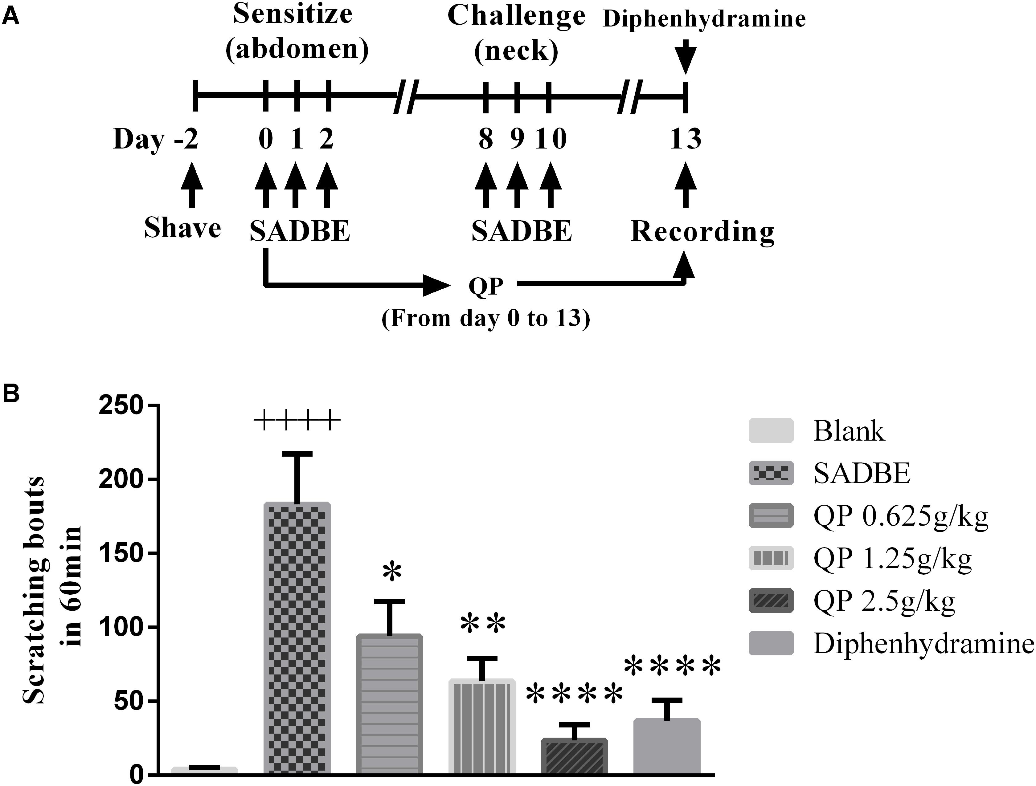
Figure 3. QP reduced the itching caused by allergic contact dermatitis induced by SADBE. (A) A Schematic diagram shows the experimental protocol. 0.5% SADBE/acetone was topically treated daily 3 times to the mouse abdomen, and re-stimulated topically daily 3 times to the neck back skin after a 5 days’ break. Spontaneous scratching was videotaped and analyzed afterward. (B) The behavior results of SADBE mice in different controls or after three QP (0.625, 1.25, and 2.5 g/kg), and the positive drug (diphenhydramine, 10 mg/kg). Data was expressed as mean ± SEM. ∗p < 0.05, ∗∗p < 0.01, and ∗∗∗∗p < 0.0001, compared with the model group. ++++p < 0.0001, compared with the blank control group. One-way ANOVA followed by post hoc test (n = 5 each group). The method and criteria to present and statistically analyze the data in (B) were followed the same in all other work in this paper unless mentioned specifically.
We next examined the SADBE induced skin lesions and whether the QP treatments were beneficial to them. From the skin general appearance observed, compared with the model group, mice after treated with different doses of QP showed some signs of reduced skin lesions (Figure 4A). However, from the results of HE (Figures 4B,C) and toluidine blue (Figures 4D,E) staining, there was no significant differences after any QP or diphenhydramine treatment as compared with the model group, and by the measurement of skin thickness and the number of mast cells, which have been frequently used as indicators for the skin inflammation and immune cell infiltration. The data indicated, in mice SADBE initiated thickening of skin and increased infiltration of mast cells, which could not be significantly inhibited by QP.
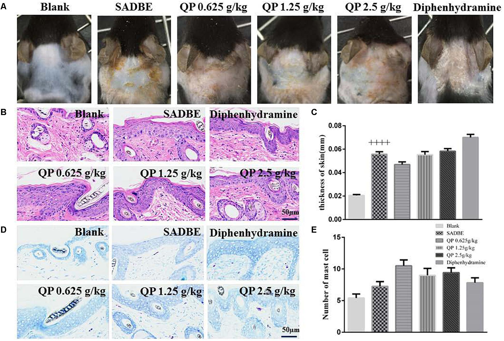
Figure 4. Effect of QP on skin histological changes in SADBE mice. (A) The midline dorsal neck skin images of each group of mice were showed on day 13. The neck skin lesions in QP treatment groups were fewer than those in the model group. (B,C) showed representative HE staining images and measured skin thickness of each group as labeled, respectively. (D,E) showed representative toluidine blue images and cell counts of each group as labeled, respectively. Data was expressed as mean ± SEM. ++++p < 0.0001, compared to the blank group. One-way ANOVA (n = 5 each group). Scale bar: 50 μm.
In this study we tried to take advantage of the information from our network pharmacological study (Figure 2 and Supplementary Table S1) for molecular analysis. Upon the results of potential effector molecules they provided, we analyzed the mRNA expression levels of IL-5, IL-10, TRPV4, TSLP, GRP, and MrgprA3 in the skin from different groups of mice. The experimental results showed that the mRNA levels of IL-5, IL-10, TRPV4, TSLP, GRP, and MrgprA3 in the skin of the model group were significantly higher than those in the blank control group. Compared with the model group, QP treatments (especially at the high dose group) can significantly reduce the enhanced IL-5, IL-10, TRPV4, TSLP, GRP, and MrgprA3 mRNA levels in the skin of SADBE mice (Figure 5). The positive control drug diphenhydramine, was able to significantly decrease that of IL-10, GRP, and MrgprA3.
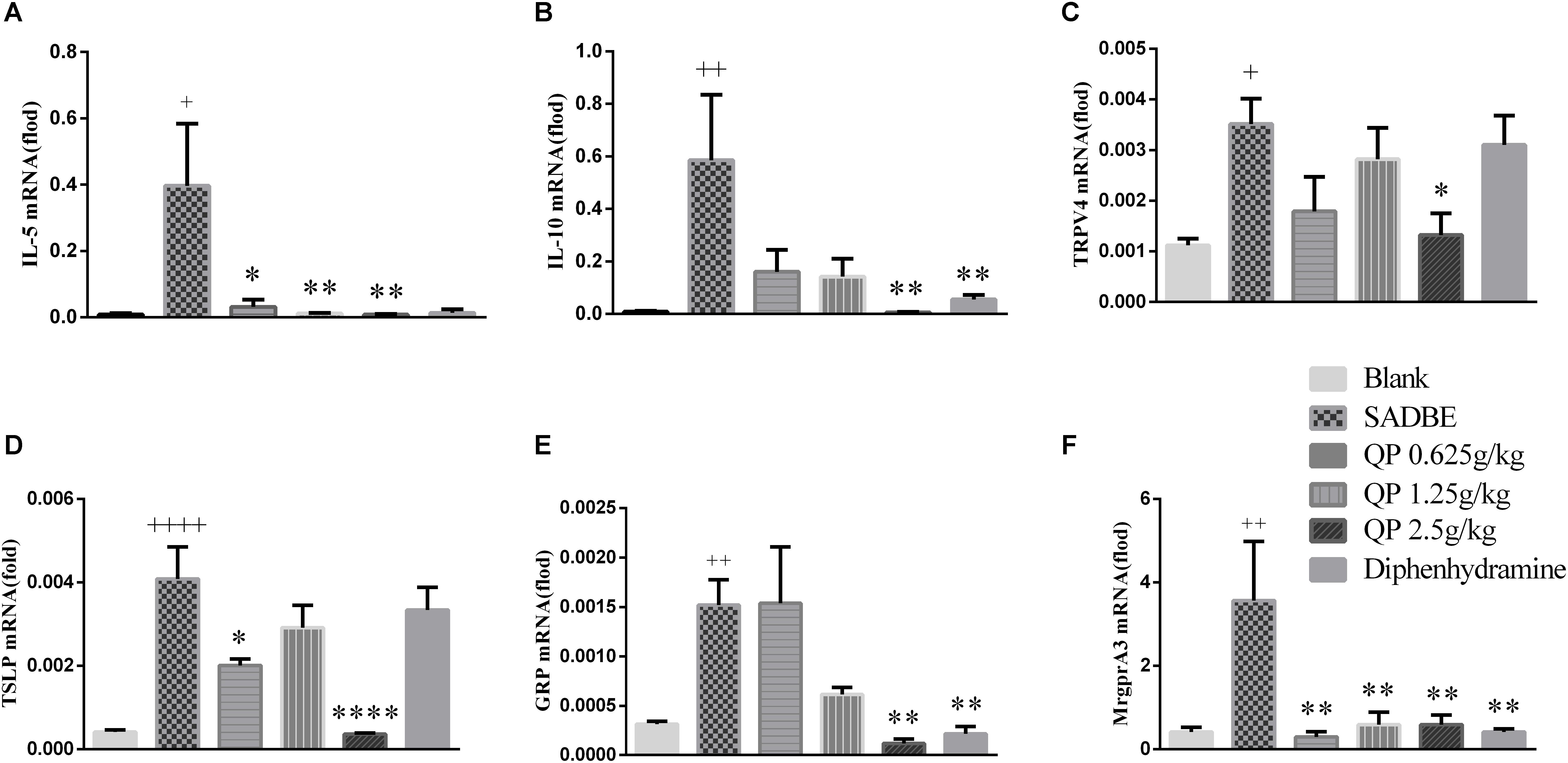
Figure 5. The mRNA of IL-5 (A), IL-10 (B), TRPV4 (C), TSLP (D), GRP (E), and MrgprA3 (F) were detected in mouse skin. After mice were treated with different doses of QP ointment and diphenhydramine, the targeted molecular mRNA levels were measured as well as that in the model group or the blank control group. Data was expressed as mean ± SEM. ∗p < 0.05, ∗∗p < 0.01, and ∗∗∗∗p < 0.0001, compared with the model group. +p < 0.05, ++p < 0.01, and ++++p < 0.0001, compared with the blank control group. One-way ANOVA (n = 5 each group).
It is well known a number of intracellular kinase cascades would be activated by inflammatory mediators and participate in the generation and maintenance of immune responses in dermatitis (Ayush et al., 2013; Bechara et al., 2017; Oetjen et al., 2017; Li et al., 2018; Xiao and Xiao, 2018). Considering the information from our network pharmacological study (Figure 2 and Supplementary Table S1), expression of several important protein kinases was detected in the skin of mice (Figures 6A–F). Compared with the blank control group, the level of phosphorylated P38 (Figure 6C) in the skin of the model group increased significantly. Compared with the model group, the high-dose QP significantly decreased the expression of p-P38 and p-Erk (Figures 6B,C) in the skin. Detection of the p-Stat3, p-Akt and p-IκBα expression in the skin of mice revealed that there was no significant difference in their expression levels among the groups (Figures 6A,D–F).
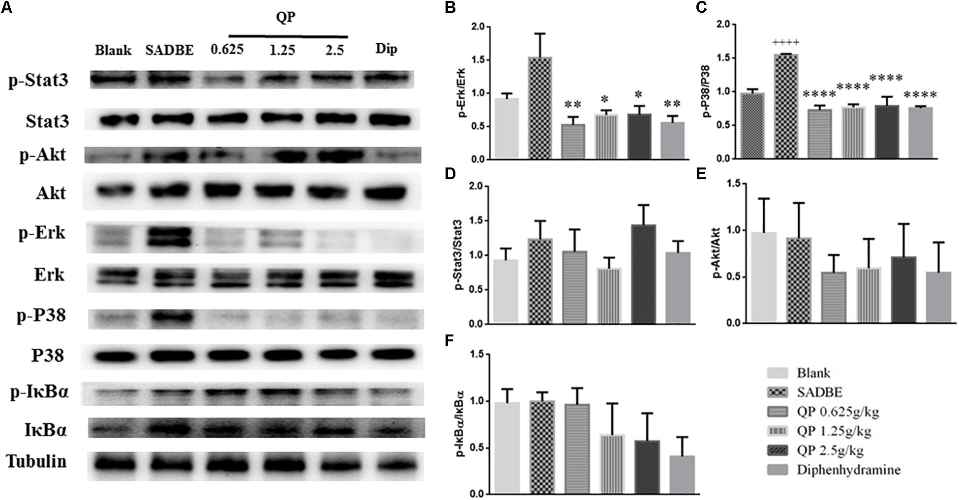
Figure 6. QP suppressed the phosphorylation of MAPKs in the skin of SADBE mice. Representative western blots images of p-Stats, Stats, p-Akt, Akt, p-Erk, Erk, p-P38, p38, p-IκBα, IκBα, and Tubulin, respectively (A). Expression of p-Erk (B) and p-P38 (C) but not p-Stat3 (D), p-Akt (E), and p-IκBα (F) in the skin of SADBE mice was significantly reduced QP compared to that in the model group. Data was expressed as mean ± SEM. ∗p < 0.05, ∗∗p < 0.01, and ∗∗∗∗p < 0.0001, compared to the model group. ++++p < 0.0001, compared to the blank control group. ANOVA (n = 3 each group)1. The related original western blot images were showed in Supplementary Figures S3, S4.
The DRG is the site where the primary neuronal cells are clustered and also a critical site for the development itch sensory information in the periphery (Han et al., 2013; Liu Z. et al., 2016; Kittaka and Tominaga, 2017; Zhang et al., 2017). Real-time PCR was used to measure the mRNA levels of TRPV1 (Figure 7A), TRPA1 (Figure 7B), GRP (Figure 7C), and MrgprA3 (Figure 7D) in the DRG of mice. As shown in Figure 7, the levels of TRPV1, GRP, and MrgprA3 mRNA in the DRG of model group were significantly higher than those in the blank control group. The positive drug diphenhydramine was effective in reducing TRPA1 and TRPV1 mRNA levels. Even though the low-dose QP had no significant effect on the four itch-related genes, however, compared with that of the model group, TRPV1 and GRP mRNA levels were significantly reduced by the medium-dose of QP, and TRPV1, TRPA1, GRP, and MrgprA3 mRNA levels were markedly reduced by the high-dose of QP.
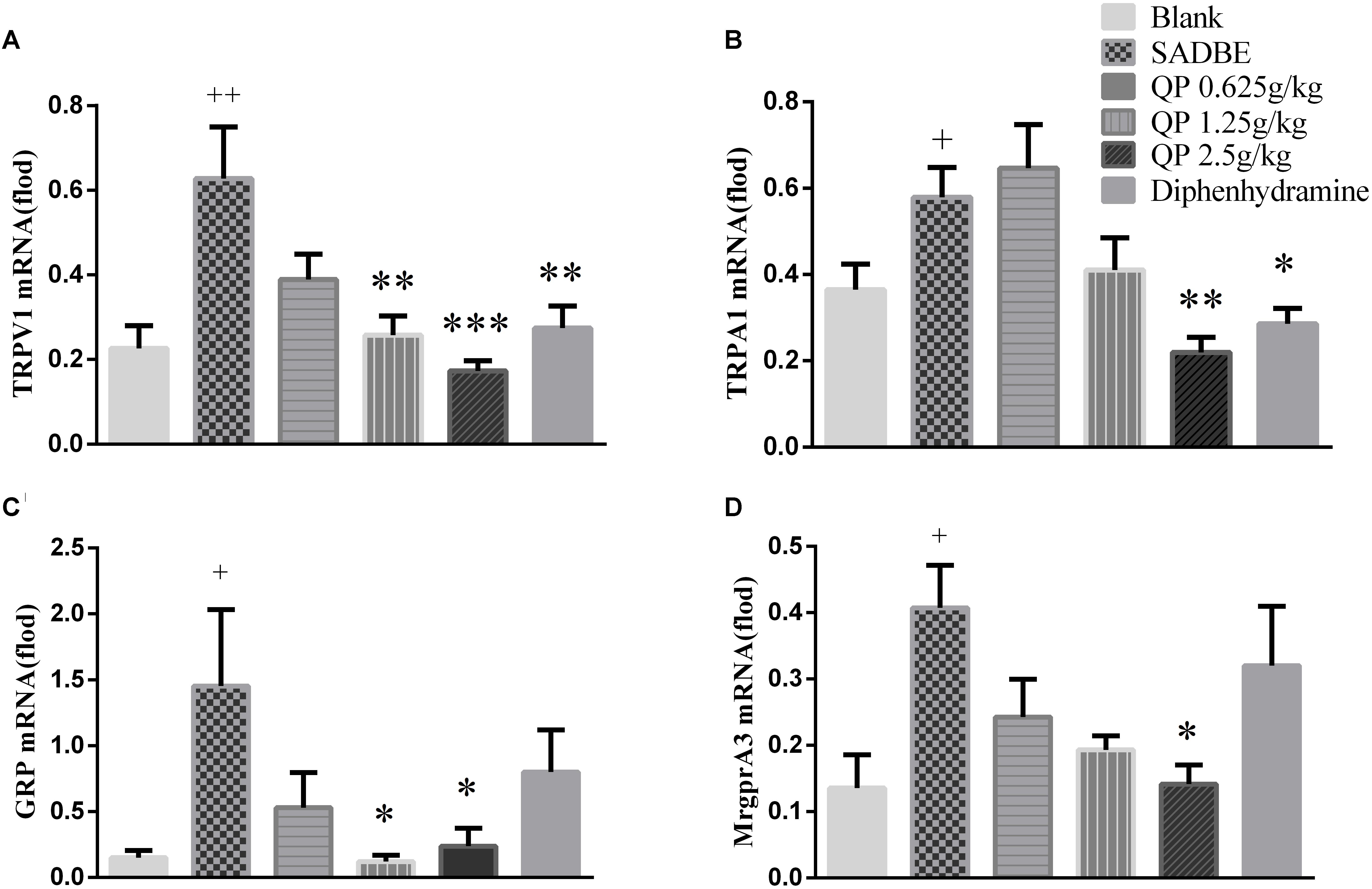
Figure 7. The mRNA of TRPV1 (A), TRPA1 (B), GRP (C) and MrgprA3 (D) were detected in mice DRG. After mice were treated with different doses of QP ointment and diphenhydramine, the targeted molecular mRNA levels were measured as well as that in the model group or the blank control group. Data was expressed as mean ± SEM. ∗p < 0.05, ∗∗p < 0.01, and ∗∗∗p < 0.001, compared to the model group. +p < 0.05 and ++p < 0.01, compared to the blank control group. ANOVA (n = 5 each group).
By using the IgE, IL-4, IL-5, IL-10, and TNF-α Elisa kits, the levels of IgE (Figure 8A), IL-4 (Figure 8B), IL-5 (Figure 8C), IL-10 (Figure 8D), TNF-α (Figure 8E) in the serum and TNF-α (Figure 8F) in the spleen were detected. Compared with that in the blank control mice, the production of IgE, IL-5, IL-10 and TNF-α in the model group mice were significantly increased. Positive control drug could significantly reduce IL-4, IL-5, and IL-10 and TNF-α content in serum, but QP showed good inhibitory effect in reducing the content of all these cytokines in the serum or spleen. Among them, the high-dose of QP showed the most striking effect which significantly to all markers detected.
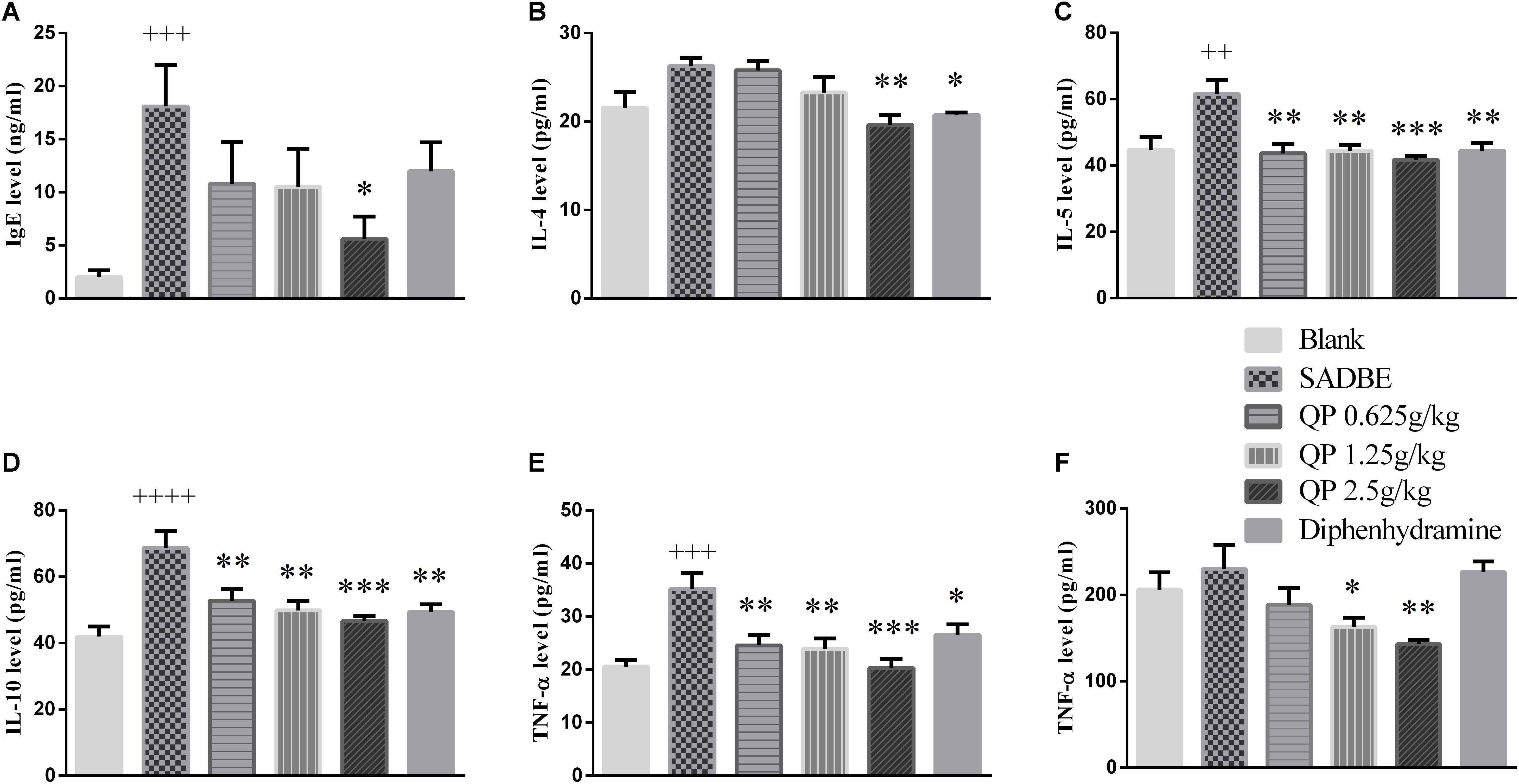
Figure 8. Effect of QP on the levels of different cytokines or IgE in the serum or spleen of SADBE mice. Among them, (A–E) showed the levels of IgE, IL-4, IL-5, IL-10, and TNF-α in the serum of each group, and (F) showed of TNF-α in the spleen of each group. ∗p < 0.05, ∗∗p < 0.01, and ∗∗∗p < 0.001, compared to the model group. ++p < 0.01, +++p < 0.001, and ++++p < 0.0001, compared to the blank control group. ANOVA (n = 5 each group).
Recent advances in experimental and computational studies have shown that, unlike some single drugs of western medicine, natural medicines or TCM mostly contain multiple components and exert their therapeutic effects by acting on multiple targets (Liu Z. et al., 2016; Zhang et al., 2017, 2018). Therefore, it is necessary to introduce modern technologies and systematic methods, such as HPLC fingerprint analysis and network pharmacology, to reveal the pharmacological functions of these drugs and predict their action mechanisms (Liu Z. et al., 2016; Zhang et al., 2017, 2018). In this study the HPLC analysis was used to characterize the phytochemical features of QP ointment and 3 common peaks were proposed as the fingerprints of its multiple chemical constituents. According to the fragment ion at m/z 153 in the mass spectrum indicated that QP contained major phenolic constituents with the central structural unit of gallic acid. Previously this type compounds had been showed treatment effects on ACD or contact dermatitis (Mo et al., 2014; Fu et al., 2015). However, even though gallic acid, corilagin, and ellagic acid were identified as major components of QP, there may be other effective or novel structural compounds in QP since there are several peaks of HPLC fingerprint analysis. Further investigation is needed to investigate what they are and whether they are effective ingredients for the treatment of ACD.
On the other hand, BATMAN-TCM is a bioinformatics analysis tool developed by our collaborators (Liu Z. et al., 2016; Zhang et al., 2017). In this study by using BATMAN-TCM, we analyzed the potential targets of each component of QP, and generated a current ACD’s disease-related gene database. Our results suggested that molecules such as IL-8, CXCR4, JUN, IL-1β, IL-17A, etc., are potential effector molecules or downstream molecules of QP, and molecules such as TLR4, STAT1, STAT3, PRKACA, etc., are chronic dermatitis-associated targets, while IL-4, IL-6, IL-10, TNF, NFκB1, etc., may cover by both. As far as we know, this study is the first use of modern methods of network pharmacology to explore the therapeutic effect of Tibetan medicine on allergic contact dermatitis. Certainly, this method still has some limitations. For example, it mainly relies on the continuous updating and improvement of the relevant online database (Liu Z. et al., 2016; Zhang et al., 2017). The results given by BATMAN-TCM were suggested to be combined with the existing literature and research progress, and the results of animal and cytological experiments were to make the most appropriate judgment. The mRNA-seq technology can provide a method for rapidly distinguishing altered mRNA after drug treatment (Duan et al., 2019), if possible, the mRNA-seq work for studying transcriptome expression in a QP-treated ACD mouse model will help to understand and explain the mechanism of action of QP.
Our experimental results highlighted the key role of several Th1/2 cytokines for QP to treat the chronic itch. T helper (Th) cells have been defined into different subsets by different cytokine profiles secreted. For example, Th1 cells are characterized by the secretion of IL-2, IFN-γ and TNF-α, etc., while Th2 cells secrete IL-4, IL-5, and IL-13, etc (Raphael et al., 2015). Th1 cytokines, like TNF-α/TNFR1 signaling, were showed in animal behavior as required for both acute and chronic itch (Miao et al., 2018). More strikingly Th2 cytokines have been reported as highly involving in the inflammation response directly mediating chronic itch. For example, recent data suggest IL-4, IL-31, and IL-33 all have their own receptors expressing in both mouse and human sensory neurons, and these cytokines enhance neuronal responsiveness to multiple pruritogens (Cevikbas et al., 2014; Kaesler et al., 2014; Liu B. et al., 2016; Oetjen et al., 2017). Our data indicate that high level of interleukin IL-5 was detected in ACD mouse skin, and significantly increased IgE, IL-4, IL-5, IL-10 levels in serum, and as wells as elevated TNF-α in both serum and spleen. On the other hand, QP can significantly decrease the upregulation of these molecules in the serum, spleen and skin. In all, these data would not only support some recent chronic itch progress related to our studies (Zhao et al., 2013; Cevikbas et al., 2014; Kaesler et al., 2014; Liu B. et al., 2016; Oetjen et al., 2017), but also indicated QP can play anti-inflammatory and antipruritic actions through these molecules in multiple places (serum, spleen, skin, and DRG).
Over the past 10 years, effects of various itch mediators expressed by primary sensory neurons and skin cells (like keratinocytes) have been extensively studied. For example, keratinocytes released thymic stromal lymphopoietin (TSLP) could activate sensory neurons and immune cells for itch (Wilson et al., 2013). Several itch specific neuropeptides, like GRP (Sun and Chen, 2007), somatostatin (Huang et al., 2018), and specific neurons like MrgprA3+ (Han et al., 2013) and GRPR+ (Sun et al., 2009) cells have been identified for the itch signal transmission. TRP channels, especially thermosensitive TRP channels including TRPV1, A1,V4 etc., receive multimodal stimuli and required for itch sensations (Liu Z. et al., 2016; Kittaka and Tominaga, 2017; Zhang et al., 2017). Research studies including our work proved both TRPA1 and TRPV1 channels are required for generating spontaneous scratching induced by SADBE and TRPV4-expressing epithelial and immune cells in the skin dynamically mediate chronic itch (Liu Z. et al., 2016; Zhang et al., 2017). In this study, our work demonstrated the mRNA of TRPV1, TRPA1, GRP, and MrgprA3 were elevated in the SADBE mice DRG and /or skin, which were significantly decreased by QP to a different extent.
Signal transduction originating from membrane-bound Th1/2 cytokine receptors is a complex network that leads to intracellular gene expression and ultimately regulates cellular activities. Among them, the mitogen activated protein kinase (MAPK) (Ayush et al., 2013; Li et al., 2018; Xiao and Xiao, 2018), the nuclear factor kappa-light-chain-enhancer of activated B cells (NF-κB) (Xiao and Xiao, 2018), the Janus kinase (JAK)/signal transducer and activator of transcription (STAT) (Bechara et al., 2017; Oetjen et al., 2017) have been intensively studied and strongly implicated with the pathogenesis of skin diseases and inflammation-controlling interventions. Our work supported, probably QP works through p-P38 and p-Erk cell signaling pathways in skin (not DRG, see Supplementary Figure S2, S5) to treat the chronic itch in ACD mice.
The pathogenesis of chronic skin diseases including ACD is complicated, including at least skin barrier dysfunctions, allergy/immunity, and pruritus (Liu Z. et al., 2016; Kittaka and Tominaga, 2017; Kostner et al., 2017; Zhang et al., 2017; Hachisuka et al., 2018). Cytokines can be produced by a wide array of immune cells, including neutrophils, eosinophils, mast cells, macrophages, and T cells (Liu Z. et al., 2016; Kittaka and Tominaga, 2017; Kostner et al., 2017; Zhang et al., 2017; Hachisuka et al., 2018). Therefore, even though we have some understanding about the possible cytokines, itch-related molecules, and related intracellular signaling pathways for QP to work with, the specific subsets of the cell types, what exact ingredients, and how they function through these cells and molecular network systems, etc., need to be further clarified. Future research on experiments in vitro, genetic engineering animals, etc., may help address these issues.
Via a HPLC system and a BATMAN-TCM platform, for the first time we performed detailed HPLC fingerprint analysis and network pharmacology studies of QP ointment. By in-depth animal and molecular analysis, we identified a number of chronic dermatitis-associated Th1/2 cytokines, itch mediators, and cellular signaling pathways in the skin, DRG and serum for QP to treat the inflammation and chronic itch associated with ACD. Our data clearly suggested, upon stimulation, cytokines and itch-related mediators are released from the skin and neuronal tissues, and QP could effectively work on both the skin and sensory neurons to treat the chronic itch.
All animal experimental procedures followed international guidelines for care and use of laboratory animals which were approved by the Animal Ethics Committee of South-central University for Nationalities.
ZM designed and supervised the study. XG, HX, ZZ, and ZM analyzed the data and wrote the manuscript. XG and HX mainly performed the experiments. LY, SL, CT, HW, YL, and WC helped the experiments.
This research was supported by grant from the National Major Scientific and Technological Special Project for “Significant New Drugs Development” of China (Grant No. 2017ZX09301060) and the Hubei Provincial Technical Innovation Program (Grant No. 2017AHB067).
WC was employed by the company Tibet Cheezheng Tibetan Medicine Co., Ltd.
The remaining authors declare that the research was conducted in the absence of any commercial or financial relationships that could be construed as a potential conflict of interest.
The Supplementary Material for this article can be found online at: https://www.frontiersin.org/articles/10.3389/fphar.2019.00354/full#supplementary-material
DATA SHEET S1 | Three main compounds of QP (Figure S1). QP suppressed the phosphorylation of MAPKs in the DRG of SADBE mice (Supplementary Figure S2). The data of the original western blot images (Supplementary Figures S3–S5).
TABLE S1 | The key targets of chronic dermatitis or QP (Sheet 1), and targets corresponding to key signaling pathways (Sheet 2) based on the platform of BATMAN-TCM.
Ayush, O., Lee, C. H., Kim, H. K., Im, S. Y., Cho, B. H., and Lee, H. K. (2013). Glutamine suppresses DNFB-induced contact dermatitis by deactivating p38 mitogen-activated protein kinase via induction of MAPK phosphatase-1. J. Invest. Dermatol. 133, 723–731. doi: 10.1038/jid.2012.373
Bechara, R., Antonios, D., Azouri, H., and Pallardy, M. (2017). Nickel sulfate promotes IL-17A producing CD4+ T cells by an IL-23-dependent mechanism regulated by TLR4 and Jak-STAT pathways. J. Invest. Dermatol. 137, 2140–2148. doi: 10.1016/j.jid.2017.05.025
BenSaad, L. A., Kim, K. H., Quah, C. C., Kim, W. R., and Shahimi, M. (2017). Anti-inflammatory potential of ellagic acid, gallic acid and punicalagin A&B isolated from Punica granatum. BMC Complement. Altern. Med. 17:47. doi: 10.1186/s12906-017-1555-0
Cevikbas, F., Wang, X., Akiyama, T., Kempkes, C., Savinko, T., Antal, A., et al. (2014). A sensory neuron-expressed IL-31 receptor mediates T helper cell-dependent itch: involvement of TRPV1 and TRPA1. J. Allergy Clin. Immunol. 133, 448–460. doi: 10.1016/j.jaci.2013.10.048
Duan, X., Han, L., Peng, D., Chen, W., Peng, C., Xiao, L., et al. (2019). High throughput mRNA sequencing reveals potential therapeutic targets of tao-hong-Si-Wu decoction in experimental middle cerebral artery occlusion. Front. Pharmacol. 9:1570. doi: 10.3389/fphar.2018.01570
Farahnik, B., Sharma, D., Alban, J., and Sivamani, R. K. (2017). Topical botanical agents for the treatment of psoriasis: a systematic review. Am. J. Clin. Dermatol. 18, 451–468. doi: 10.1007/s40257-017-0266-0
Feng, J., Yang, P., Mack, M. R., Dryn, D., Luo, J., Gong, X., et al. (2017). Sensory TRP channels contribute differentially to skin inflammation and persistent itch. Nat. Commun. 8:980. doi: 10.1038/s41467-017-01056-8
Fu, K., Qu, L., Shimada, S. G., Nie, H., and LaMotte, R. H. (2014). Enhanced scratching elicited by a pruritogen and an algogen in a mouse model of contact hypersensitivity. Neurosci. Lett. 579, 190–194. doi: 10.1016/j.neulet.2014.03.062
Fu, R., Zhang, Y., Peng, T., Guo, Y., and Chen, F. (2015). Phenolic composition and effects on allergic contact dermatitis of phenolic extracts Sapium sebiferum (L.) Roxb. leaves. J. Ethnopharmacol. 162, 176–180. doi: 10.1016/j.jep.2014.12.072
Hachisuka, J., Chiang, M. C., and Ross, S. E. (2018). Itch and neuropathic itch. Pain 159, 603–609. doi: 10.1097/j.pain.0000000000001141
Han, L., Ma, C., Liu, Q., Weng, H. J., Cui, Y., Tang, Z., et al. (2013). A subpopulation of nociceptors specifically linked to itch. Nat. Neurosci. 16, 174–182. doi: 10.1038/nn.3289
Huang, J., Polgar, E., Solinski, H. J., Mishra, S. K., Tseng, P. Y., Iwagaki, N., et al. (2018). Circuit dissection of the role of somatostatin in itch and pain. Nat. Neurosci. 21, 707–716. doi: 10.1038/s41593-018-0119-z
Kaesler, S., Volz, T., Skabytska, Y., Koberle, M., Hein, U., Chen, K. M., et al. (2014). Toll-like receptor 2 ligands promote chronic atopic dermatitis through IL-4-mediated suppression of IL-10. J. Allergy Clin. Immunol. 134, 92–99. doi: 10.1016/j.jaci.2014.02.017
Kittaka, H., and Tominaga, M. (2017). The molecular and cellular mechanisms of itch and the involvement of TRP channels in the peripheral sensory nervous system and skin. Allergol. Int. 66, 22–30. doi: 10.1016/j.alit.2016.10.003
Kostner, L., Anzengruber, F., Guillod, C., Recher, M., Schmid-Grendelmeier, P., and Navarini, A. A. (2017). Allergic contact dermatitis. Immunol. Allergy Clin. North Am. 37, 141–152. doi: 10.1016/j.iac.2016.08.014
Li, W., Liu, F., Wang, J., Long, M., and Wang, Z. (2018). microRNA-21-mediated inhibition of mast cell degranulation involved in the protective effect of berberine on 2,4-dinitrofluorobenzene-induced allergic contact dermatitis in rats via p38 pathway. Inflammation 41, 689–699. doi: 10.1007/s10753-017-0723-1
Li, Y., and Li, L. F. (2013). Topical application of a Chinese medicine, Qingpeng ointment, ameliorates 2, 4-dinitrofluorobenzene-induced allergic contact dermatitis in BALB/c mice. Eur. J. Dermatol. 23, 803–806. doi: 10.1684/ejd.2013.2185
Li, Y. Z., Lu, X. Y., Jiang, W., and Li, L. F. (2013). Anti-inflammatory effect of qingpeng ointment in atopic dermatitis-like murine model. Evid. Based Complement. Alternat. Med. 2013:907016. doi: 10.1155/2013/907016
Liu, B., Tai, Y., Achanta, S., Kaelberer, M. M., Caceres, A. I., Shao, X., et al. (2016). IL-33/ST2 signaling excites sensory neurons and mediates itch response in a mouse model of poison ivy contact allergy. Proc. Natl. Acad. Sci. U.S.A. 113, E7572–E7579. doi: 10.1073/pnas.1606608113
Liu, Z., Guo, F., Wang, Y., Li, C., Zhang, X., Li, H., et al. (2016). BATMAN-TCM: a bioinformatics analysis tool for molecular mechANism of traditional Chinese medicine. Sci. Rep. 6:21146. doi: 10.1038/srep21146
Luo, J., Feng, J., Yu, G., Yang, P., Mack, M. R., Du, J., et al. (2018). Transient receptor potential vanilloid 4-expressing macrophages and keratinocytes contribute differentially to allergic and nonallergic chronic itch. J. Allergy Clin. Immunol. 141, 608–619.e7. doi: 10.1016/j.jaci.2017.05.051
Mahapatra, S., Albrecht, M., Behrens, B., Jirmo, A., Behrens, G., Hartwig, C., et al. (2014). Delineating the role of histamine-1- and -4-receptors in a mouse model of Th2-dependent antigen-specific skin inflammation. PLoS One 9:e87296. doi: 10.1371/journal.pone.0087296
Miao, X., Huang, Y., Liu, T. T., Guo, R., Wang, B., Wang, X. L., et al. (2018). TNF-alpha/TNFR1 signaling is required for the full expression of acute and chronic itch in mice via peripheral and central mechanisms. Neurosci. Bull. 34, 42–53. doi: 10.1007/s12264-017-0124-3
Mo, J., Panichayupakaranant, P., Kaewnopparat, N., Songkro, S., and Reanmongkol, W. (2014). Topical anti-inflammatory potential of standardized pomegranate rind extract and ellagic acid in contact dermatitis. Phytother. Res. 28, 629–632. doi: 10.1002/ptr.5039
Na Takuathung, M., Wongnoppavich, A., Panthong, A., Khonsung, P., and Chiranthanut, N. (2018). Antipsoriatic effects of wannachawee recipe on imiquimod-induced psoriasis-like dermatitis in BALB/c Mice. Evid. Based Complement. Alternat. Med. 2018:7931031. doi: 10.1155/2018/7931031
Oetjen, L. K., Mack, M. R., Feng, J., Whelan, T. M., Niu, H., Guo, C. J., et al. (2017). Sensory neurons co-opt classical immune signaling pathways to mediate chronic itch. Cell 171, 217–228.e13. doi: 10.1016/j.cell.2017.08.006
Parker, S., Zhang, C. S., Yu, J. J., Lu, C., Zhang, A. L., and Xue, C. C. (2017). Oral Chinese herbal medicine versus placebo for psoriasis vulgaris: a systematic review. J. Dermatolog. Treat. 28, 21–31. doi: 10.1080/09546634.2016.1178377
Pereira, M. P., and Stander, S. (2017). Chronic pruritus: current and emerging treatment options. Drugs 77, 999–1007.
Raphael, I., Nalawade, S., Eagar, T. N., and Forsthuber, T. G. (2015). T cell subsets and their signature cytokines in autoimmune and inflammatory diseases. Cytokine 74, 5–17. doi: 10.1016/j.cyto.2014.09.011
Salih, E. Y. A., Fyhrquist, P., Abdalla, A. M. A., Abdelgadir, A. Y., Kanninen, M., Sipi, M., et al. (2017). LC-MS/MS tandem mass spectrometry for analysis of phenolic compounds and pentacyclic triterpenes in antifungal extracts of Terminalia brownii (Fresen). Antibiotics 6:E37. doi: 10.3390/antibiotics6040037
Shevchenko, A., Valdes-Rodriguez, R., and Yosipovitch, G. (2018). Causes, pathophysiology, and treatment of pruritus in the mature patient. Clin. Dermatol. 36, 140–151. doi: 10.1016/j.clindermatol.2017.10.005
Sun, Y. G., and Chen, Z. F. (2007). A gastrin-releasing peptide receptor mediates the itch sensation in the spinal cord. Nature 448, 700–703. doi: 10.1038/nature06029
Sun, Y. G., Zhao, Z. Q., Meng, X. L., Yin, J., Liu, X. Y., and Chen, Z. F. (2009). Cellular basis of itch sensation. Science 325, 1531–1534. doi: 10.1126/science.1174868
Svendsen, M. T., Jeyabalan, J., Andersen, K. E., Andersen, F., and Johannessen, H. (2017). Worldwide utilization of topical remedies in treatment of psoriasis: a systematic review. J. Dermatolog. Treat. 28, 374–383. doi: 10.1080/09546634.2016.1254331
Teodoro, G. R., Gontijo, A. V. L., Salvador, M. J., Tanaka, M. H., Brighenti, F. L., Delbem, A. C. B., et al. (2018). Effects of acetone fraction from Buchenavia tomentosa aqueous extract and gallic acid on Candida albicans biofilms and virulence factors. Front. Microbiol. 9:647. doi: 10.3389/fmicb.2018.00647
Wilson, S. R., The, L., Batia, L. M., Beattie, K., Katibah, G. E., McClain, S. P., et al. (2013). The epithelial cell-derived atopic dermatitis cytokine TSLP activates neurons to induce itch. Cell 155, 285–295. doi: 10.1016/j.cell.2013.08.057
Xiao, Z., and Xiao, S. (2018). The anti-inflammatory effect of fructus kochiae on allergic contact dermatitis rats via pERK1/2/TLR4/NF-kappaB pathway activation. Evid. Based Complement. Alternat. Med. 2018:1096920. doi: 10.1155/2018/1096920
Yosipovitch, G., and Bernhard, J. D. (2013). Clinical practice. Chronic pruritus. N. Engl. J. Med. 368, 1625–1634. doi: 10.1056/NEJMcp1208814
Zhang, B., Wang, X., Li, Y., Wu, M., Wang, S. Y., and Li, S. (2018). Matrine is identified as a novel macropinocytosis inducer by a network target approach. Front. Pharmacol. 9:10. doi: 10.3389/fphar.2018.00010
Zhang, M., Wu, H., Guo, F., Yu, Y., Wei, J., Geng, Y., et al. (2017). Identification of active components in Yixinshu Capsule with protective effects against myocardial dysfunction on human induced pluripotent stem cell-derived cardiomyocytes by an integrative approach. Mol. Biosyst. 13, 1469–1480. doi: 10.1039/c6mb00813e
Keywords: Tibetan medicine, chronic itch, allergic contact dermatitis, MAPK, mice
Citation: Gong X, Xiong H, Liu S, Liu Y, Yin L, Tu C, Wang H, Zhao Z, Chen W and Mei Z (2019) Qingpeng Ointment Ameliorates Inflammatory Responses and Dysregulation of Itch-Related Molecules for Its Antipruritic Effects in Experimental Allergic Contact Dermatitis. Front. Pharmacol. 10:354. doi: 10.3389/fphar.2019.00354
Received: 29 September 2018; Accepted: 21 March 2019;
Published: 09 April 2019.
Edited by:
Jiahong Lu, University of Macau, ChinaReviewed by:
Jianli Tao, Boston Children’s Hospital, Harvard Medical School, United StatesCopyright © 2019 Gong, Xiong, Liu, Liu, Yin, Tu, Wang, Zhao, Chen and Mei. This is an open-access article distributed under the terms of the Creative Commons Attribution License (CC BY). The use, distribution or reproduction in other forums is permitted, provided the original author(s) and the copyright owner(s) are credited and that the original publication in this journal is cited, in accordance with accepted academic practice. No use, distribution or reproduction is permitted which does not comply with these terms.
*Correspondence: Zhinan Mei, bWVpemhpbmFuQDE2My5jb20=
†These authors have contributed equally to this work
Disclaimer: All claims expressed in this article are solely those of the authors and do not necessarily represent those of their affiliated organizations, or those of the publisher, the editors and the reviewers. Any product that may be evaluated in this article or claim that may be made by its manufacturer is not guaranteed or endorsed by the publisher.
Research integrity at Frontiers
Learn more about the work of our research integrity team to safeguard the quality of each article we publish.