95% of researchers rate our articles as excellent or good
Learn more about the work of our research integrity team to safeguard the quality of each article we publish.
Find out more
REVIEW article
Front. Pharmacol. , 17 June 2014
Sec. Pharmacology of Ion Channels and Channelopathies
Volume 5 - 2014 | https://doi.org/10.3389/fphar.2014.00144
This article is part of the Research Topic CaMKII in Cardiac Health and Disease View all 19 articles
The cardiac voltage gated Ca2+ current (ICa) is critical to the electrophysiological properties, excitation-contraction coupling, mitochondrial energetics, and transcriptional regulation in heart. Thus, it is not surprising that cardiac ICa is regulated by numerous pathways. This review will focus on changes in ICa that occur during the cardiac action potential (AP), with particular attention to Ca2+-dependent inactivation (CDI), Ca2+-dependent facilitation (CDF) and how calmodulin (CaM) and Ca2+-CaM dependent protein kinase (CaMKII) participate in the regulation of Ca2+ current during the cardiac AP. CDI depends on CaM pre-bound to the C-terminal of the L-type Ca2+ channel, such that Ca2+ influx and Ca2+ released from the sarcoplasmic reticulum bind to that CaM and cause CDI. In cardiac myocytes CDI normally pre-dominates over voltage-dependent inactivation. The decrease in ICa via CDI provides direct negative feedback on the overall Ca2+ influx during a single beat, when myocyte Ca2+ loading is high. CDF builds up over several beats, depends on CaMKII-dependent Ca2+ channel phosphorylation, and results in a staircase of increasing ICa peak, with progressively slower inactivation. CDF and CDI co-exist and in combination may fine-tune the ICa waveform during the cardiac AP. CDF may partially compensate for the tendency for Ca2+ channel availability to decrease at higher heart rates because of accumulating inactivation. CDF may also allow some reactivation of ICa during long duration cardiac APs, and contribute to early afterdepolarizations, a form of triggered arrhythmias.
The cardiac L-type Ca2+ channel (LTCC) current (ICa) is an important contributor to overall cardiac electrophysiology and arrhythmias, excitation-contraction coupling (ECC; it causes further intracellular Ca2+ release and activation of the myofilaments), mitochondrial energy regulation, cell death and transcriptional regulation (Bers, 2008). ICa is mainly via the Cav1.2 α1 LTCC isoform, although the Cav1.3 isoform is expressed in some atrial cells (especially pacemaker cells). That pore-forming α1 subunit also carries the intrinsic voltage-dependent gating properties (Perez-Reyes et al., 1989) and many key regulatory sites. However, the mature LTCC in heart is a complex containing also a β as well as an α2-δ subunit that influence LTCC trafficking and gating (Shirokov et al., 1998; Bichet et al., 2000; Wei et al., 2000; Dzhura and Neely, 2003). Cav1.2 has four major domains (I-IV), each of which contains six transmembrane segments (S1-S6), where positive charges in the S4 segments participate as voltage sensors and the S5-S6 loop is the locus of the ion-conducting pore (Bers, 2001).
The rapid upstroke or phase 0 of the cardiac action potential (AP) is driven by Na+ current (INa) in most cardiac myocytes, and causes voltage-dependent activation of ICa. In pacemaker cells in the sino-atrial and atrio-ventricular node, it is ICa activation that is responsible for the rapid upstroke of the AP. ICa activation is a bit slower than INa activation, but starts early during the cardiac AP. The early repolarization phase of the AP (phase 1) can enhance ICa because of an increase in electrochemical driving force, i.e., membrane potential (Em) is further from the Ca2+ equilibrium potential (ECa; Sah et al., 2002). However, both depolarization and the rise in local intracellular [Ca2+] ([Ca2+]i) begin the processes of voltage- and Ca2+-dependent inactivation (VDI and CDI), which continues during the plateau phase of the AP (phase 2) causing a progressive decrease in ICa. As rapid and terminal AP repolarization ensue (phase 3) the LTCC undergoes de-activation, but then recovery from inactivation is both time and Em-dependent. Thus, for LTCC to recover full availability between beats, some time must elapse and that recovery time depends on Em (e.g., at −80 and −50 mV the time constant is about 100 and 400 ms, respectively).
ICa amplitude and gating properties are influenced by myriad regulatory pathways, but here we will focus on the Ca2+-dependent mechanisms that shape the ICa occurring during the AP in ventricular myocytes. Hence, this review will describe how the Ca2+ sensing protein calmodulin (CaM) mediates CDI, and is involved in the activation of CaMKII, a serine/threonine-specific protein kinase which is a key mediator of ECC. Note that, although CaMKII activation can also be Ca2+-independent (see accompanying article by Erickson, 2014), here we will focus on the main activation mechanism, which is Ca2+/CaM dependent. Moreover, the particular structure of this kinase (well described in this series by Pellicena and Schulman, 2014) confers to CaMKII the ability to integrate oscillatory Ca2+ signals, because CaMKII activity depends on both frequency and duration of previous Ca2+/CaM pulses (De Koninck and Schulman, 1998; Saucerman and Bers, 2008). We will show how the CaMKII-dependent LTCC phosphorylation mediates the Ca2+-dependent facilitation (CDF) of ICa, and how this process can eventually lead to Em or Ca2+ instabilities in ventricular myocytes.
Inactivation of ICa is driven by VDI and CDI (Kass and Sanguinetti, 1984; Lee et al., 1985; Hadley and Hume, 1987). Several studies have shown that the Ca2+-sensing protein CaM mediates CDI by interacting with the carboxyl tail of the LTCC α1 subunit (Zuhlke and Reuter, 1998; Peterson et al., 1999; Qin et al., 1999; Zuhlke et al., 1999; Pate et al., 2000), a cytoplasmic region that contains an EF-hand region and an IQ motif. At rest, CaM is pre-bound to the LTCC at or near the IQ motif (Erickson et al., 2001; Pitt et al., 2001). Upon ICa activation and consequent Ca2+ release from the sarcoplasmic reticulum (SR), local [Ca2+]i rises, causing Ca2+ to bind to CaM and induce inactivation. The details of the CDI process are not totally resolved, and may involve multiple regions of the channel, including the I-II loop that is thought to be key for VDI (Kim et al., 2004; Cens et al., 2006). An intriguing new hypothesis has emerged from detailed studies from the Yue lab (Ben Johny et al., 2013). During diastole, the C-lobe of apoCaM (CaM without any Ca2+ bound) would be associated with the IQ domain, and its N-lobe associated with the pre-IQ domain (between the IQ locus and the upstream EF-hand domain). Ca2+ binding to the N-lobe of CaM (the faster, low-affinity site) would cause the N-lobe to shift and bind to part of the LTCC N-terminal domain (which they call the NSCaTE module), and thereby trigger N-lobe CDI. Then when Ca2+ also binds to the C-lobe of CaM (the higher affinity, slower binding lobe) the C-lobe shifts its binding from the IQ domain to a position just upstream of the Pre-IQ region where the N-lobe had been bound. If Ca2+ binds only to the C-lobe (e.g., if the N-lobe is unavailable) then the C-lobe does a similar sort of shift on its own, and mediates C-lobe CDI. For cardiac Cav1.2 channels, overall CDI and C-lobe-CDI are relatively similar, while N-lobe CDI alone was not apparent (Peterson et al., 1999). That differs from some neuronal P/Q, N or R type Ca2+ channels, where N-lobe CDI seems to be dominant (Liang et al., 2003).
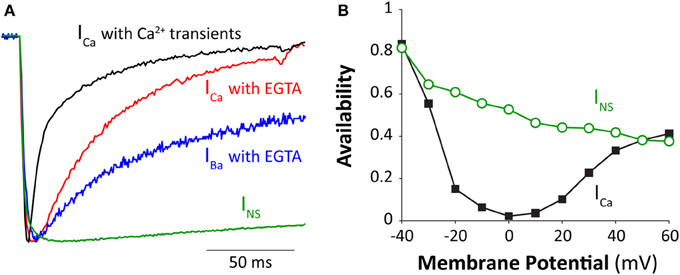
Figure 1A shows ICa inactivation kinetics in a rabbit ventricular myocyte under different Ca2+ conditions. The time to half inactivation (t1/2) increases from 17 to 37 ms when normal Ca2+ transients are abolished (e.g., by buffering the intracellular Ca2+ with 10 mM EGTA). Note that EGTA is a relatively slow buffer and cannot abolish very local [Ca2+] elevation around the mouth of the channel (although in this case SR Ca2+ release is prevented). In absence of extracellular Ca2+, LTCC are permeable to Ba2+, and this current (IBa) has been often studied to differentiate VDI and CDI (Lee et al., 1985; Peterson et al., 2000; Cens et al., 2006), despite a modest ability of Ba2+ to induce inactivation (Ferreira et al., 1997). When Ba2+ is the charge carrier (and intracellular Ca2+ is buffered), IBa inactivation is further slowed (t1/2 = 161 ms).

Figure 1. Inactivation of cardiac Ca2+ channel. (A) Normalized ICa, IBa, and INS elicited by a square voltage pulse at room temperature to 0 mV (except INS at −30 mV to obtain comparable activation state). ICa was recorded under both perforated patch (where normal SR Ca2+ release and Ca2+ transients occur) and ruptured patch conditions with cells dialyzed with 10 mM EGTA (to prevent global Ca2+ transients). IBa was also recorded with ruptured patch (with 10 mM EGTA in the pipette). Extracellular [Ca2+] and [Ba2+] were both 2 mM and INS was measured in divalent-free conditions (10 mM EDTA inside and out) with extracellular [Na+] at 20 mM and intracellular [Na+] at 10 mM. Peak currents were 1370, 808, 780, and 5200 pA and were attained at 5, 7, 10, and 14 ms for ICa (perforated), ICa (ruptured), IBa and INS respectively, with t1/2 of current decline of 17, 37, 161, and > 500 ms respectively. (B) Amplitude of INS and ICa through LTCC (at −10 mV) after 500 ms pulses to the indicated Em in guinea-pig ventricular myocytes (modified from Bers, 2001 with permission, data from Hadley and Hume, 1987).
In the absence of divalent ionic species, LTCC is permeable to monovalent cations and is referred to as non-specific monovalent current (INS, mostly carried by Na+ and Cs+). INS inactivates only very slowly at this voltage at room temperature (t1/2 > 500 ms; Figure 1A), but exhibits VDI, which becomes faster at more positive voltages (Hadley and Hume, 1987; Grandi et al., 2010). INS inactivation is incomplete (after 500 ms) even at more positive Em (Figure 1B). The additional ICa inactivation at intermediate Em has an U-shaped Em-dependence (as does inward ICa amplitude, maximal at about 0 mV), reflecting the contribution of CDI. Note that at +50–60 mV little Ca2+ enters during ICa, and the extent of ICa and INS inactivation is similar. It is tempting to speculate that INS inactivation properties might provide pure VDI characteristics that are relevant for ICa. However, INS can actually inactivate faster than IBa at positive voltages, so we think that using INS to assess VDI characteristics for ICa is likely to be invalid (Grandi et al., 2010). However, IBa inactivation is also not purely VDI, because inactivation is IBa-amplitude dependent (Brunet et al., 2009) and Ba2+ can partially substitute for Ca2+ in CDI (Ferreira et al., 1997). To resolve this we have attempted to carefully account for the weak Ba2+-dependent inactivation and refine the characteristics of VDI vs. CDI in cardiac myocytes in a computational analysis (Morotti et al., 2012). That is, most prior work using IBa to characterize VDI had slightly overestimated VDI. This is certainly not meant to discourage the use of IBa vs. ICa as a means to study CDI, just that this IBa is not entirely devoid of divalent-dependent inactivation.
Given the role of ICa in sustaining the AP plateau, CDI and VDI are important determinant for AP duration (APD) regulation. Inhibition of ICa inactivation induces AP prolongation, and has pro-arrhythmic consequences (see section “Arrhythmogenic consequences of CaMKII-dependent ICa effects”). For example, impaired VDI has been observed in Timothy syndrome (Splawski et al., 2004, 2005; Brunet et al., 2009), an inherited disease characterized by severe ventricular arrhythmias and sudden cardiac death. The expression of mutant Ca2+-insensitive CaM (via adenovirus) in adult guinea-pig cardiomyocytes also prevents CDI and causes dramatic AP prolongation (Alseikhan et al., 2002). Moreover, some human patients with arrhythmias resembling long QT syndrome have linked mutations in the Ca2+ binding domains in one of the three CaM genes (which otherwise encode the identical CaM protein; Crotti et al., 2013). A loss of CDI also characterizes the more common pathologic condition of heart failure (HF), where marked AP prolongation and associated defective Ca2+ cycling have been reported (Beuckelmann et al., 1992). It is interesting to note that, at first, the down-regulation of repolarizing K+ currents (Ito and IK1) was thought to be responsible for the increased APD seen in HF. Only in the late 1990s the pivotal role of CDI became clear, when it was first proposed in a theoretical study in dog (Winslow et al., 1999), and then experimentally observed in a guinea pig model of HF (Ahmmed et al., 2000). So clearly defective ICa CDI can be arrhythmogenic in people.
APD regulation is fundamental to control the Ca2+ level in myocytes, which is functionally important with respect to the Ca2+ requirements for myofilament activation, and thus contractility. Indeed, CDI is a physiological negative feedback mechanism that limits excessive Ca2+ entry in myocytes. When the myocyte has relatively high Ca2+ load, a large Ca2+ transient enhances ICa inactivation (limiting further Ca2+ influx). Conversely, when myocyte Ca2+ is low and SR Ca2+release is small, there is less CDI and enhanced Ca2+ entry that increases intracellular Ca2+ content (Puglisi et al., 1999; Eisner et al., 2000; Bers and Grandi, 2009). Notably, Na+/Ca2+ exchange also participates in this negative feedback (i.e., higher Ca2+ transients limit Ca2+ entry and increase Ca2+ extrusion from the myocyte via Na+/Ca2+ exchange).
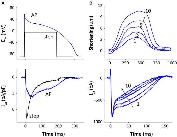
The time course of ICa during the AP is significantly different compared to that seen during a square voltage pulse [Figure 2A, rabbit ventricular myocyte, 25°C, with 10 mM EGTA to prevent Ca2+ transients (Yuan et al., 1996)]. Peak ICa during the AP is lower and occurs later than during a square pulse, with larger ICa late in the AP. The later ICa peak is because at the AP peak (+50 mV) Ca2+ channels activate rapidly, but the driving force for Ca2+ (Em–ECa) is initially low, because Em is close to the reversal potential for ICa (ECa ~ +60 mV). As Em repolarizes, the driving force increases faster than channel inactivation, producing a larger current at later times during the AP (Sah et al., 2002). Sipido et al. (1995) first investigated how Ca2+ released from the SR modulates ICa performing “classic” voltage-clamp experiments, and observed that CDI increases as SR Ca2+ release gets larger. Our group confirmed this observation in a more “physiological” condition, as shown in Figure 2B, where repeated AP-clamps are performed as the SR Ca2+ stores are reloaded, such that contractions get progressively larger (beat 1–10; Puglisi et al., 1999). One can see the contribution of SR Ca2+ release to CDI as the Ca2+ transients and contractions get larger. Integration of the Ca2+ influx via ICa during these ten pulses (which approach the steady state) shows that the ICa-dependent influx decreases from 12 to 6 μmol/L cytosol, indicating that ICa inactivation due to SR Ca2+ release decreases net Ca2+ influx by about 50%. These experiments were done at both 25 and 35°C. At 35°C peak ICa occurs earlier and is higher, but also inactivates faster and the AP duration is also shorter. The net result is that there is very little difference between these temperatures for the integral of Ca2+ influx during the AP (with SR Ca2+ release fully functional).

Figure 2. ICa inactivation during the AP. (A) Rabbit ventricular myocytes (at 25°C) were voltage-clamped with either a square voltage step or an AP waveform (measured from 5 other cells under physiological conditions). All other currents were blocked, e.g., by replacement of K+ with Cs+ and Na+ with TEA (inside and out) and cells were dialyzed with 10 mM EGTA to prevent Ca2+ transients (data from Yuan et al., 1996, modified from Bers, 2001 with permission). (B) After SR Ca2+ was depleted by a brief caffeine-application (with Na+), a series of AP-clamps were given, and contraction and ICa recovered to steady state over 10 sequential pulses at 25°C in rabbit ventricular myocyte (modified from Bers, 2001 with permission, data from Puglisi et al., 1999).
Using a combination of AP and square voltage-clamp protocols, Linz and Meyer (1998) assessed the time-course of ICa inactivation during the AP in different Ca2+ homeostasis conditions. Their analysis pointed out that, in physiological condition, CDI is the overwhelmingly dominant inactivation on the time scale of an AP, as recapitulated in the theoretical study by Greenstein and Winslow (2002). Moreover, Linz and Meyer (1998) showed that CDI is mostly controlled by Ca2+ released from the SR during the initial part of the AP, then by Ca2+ entered through the LTCCs. These results are well described by our recent computational study that updated the balance of VDI and CDI in the context of a detailed Ca2+ cycling electrophysiological myocyte model (Morotti et al., 2012).
At increased heart rates, there is typically an increase in Ca2+ transient amplitude (known sometimes as the positive force-frequency relationship) in normal hearts in species other than rat and mouse (Bers, 2001). The higher Ca2+ transients also typically decline faster at high heart rates (known a frequency-dependent acceleration of relaxation; Bers, 2001). Thus, ICa inactivation is expected to be faster, based on the above discussion. The higher heart rate could also shorten the diastolic interval and increase diastolic [Ca2+]i, which might reduce ICa availability. Indeed, while ICa recovery from inactivation is classically time and Em-dependent (Hadley and Hume, 1987), we showed that elevations of [Ca2+]i could slow recovery from inactivation, especially under conditions where SR Ca2+ uptake is depressed and diastolic Em is slightly depolarized (Altamirano and Bers, 2007), as can be the case in human HF (Sipido et al., 1998). This sort of diastolic [Ca2+]i effect on LTCC availability is probably of only minor relevance under normal physiological conditions and heart rates in healthy hearts, but may be more of a factor under pathophysiological conditions. That is, in HF there is an increased likelihood that peak ICa will decrease at high heart rates, and that might contribute to limiting the more negative force-frequency relationship observed in HF (Sipido et al., 1998).
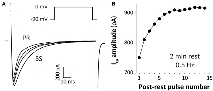
Several early studies reported progressive increases in ICa amplitude and prominent slowing of inactivation that was observed during increased frequency of voltage-clamp pulses from physiological holding potentials (~ −80 mV), as shown in the example in Figure 3 (Lee, 1987; Boyett and Fedida, 1988; Tseng, 1988; Hryshko and Bers, 1990). This phenomenon is not reproduced if holding Em is more depolarized (e.g., −40 mV) where a negative staircase is observed, or in the absence of Ca2+ (e.g., when Ba2+ is the charge carrier). This ICa staircase was also stronger when local Ca2+ influx was amplified by SR Ca2+ release. Thus, this phenomenon is termed Ca2+-dependent facilitation of ICa.

Figure 3. Ca2+-dependent facilitation of ICa. ICa at 0.5 Hz (from −90 to 0 mV) after a 2 min rest period in a ferret ventricular myocyte. The first post-rest pulse (PR) and the second, third and steady state (SS) pulses are shown in (A) and the whole post-rest ICa “staircase” in (B) (modified from Bers, 2001 with permission, data from Hryshko and Bers, 1990).
CDF and CDI co-exist under physiological conditions, and this may be why ICa facilitation was masked by holding Em near −40 mV. That is, recovery from inactivation at that Em is slow, so the records were dominated by a negative ICa staircase that was attributable to CDI and incomplete ICa recovery from inactivation. It has been proposed that the facilitatory mechanism may partly offset reduced Ca2+ channel availability at high heart rates (caused by direct CDI), contributing to improving cardiac performance during exercise (Ross et al., 1995). While CDI responds rapidly (in response to local [Ca2+]i during the same beat), CDF occurs more slowly (over several beats). Indeed, biphasic effects of [Ca2+]i on unitary ICa have been reported (Hirano and Hiraoka, 1994). Some studies even claimed that progressive decrease in SR Ca2+ release (negative staircase in rat) and CDI are responsible for the observed CDF (Guo and Duff, 2003, 2006). However, because CDF is quite similar in species that exhibit positive Ca2+ transients staircases and even when SR Ca2+ release is blocked this seems unlikely to be the case (Hryshko and Bers, 1990).
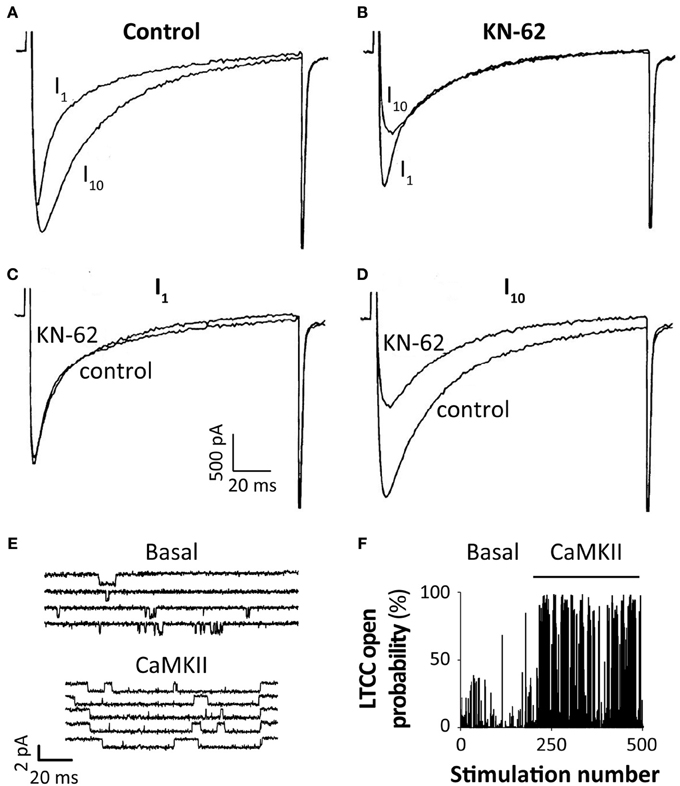
About 20 years ago three groups independently demonstrated that Ca2+-dependent ICa facilitation is mediated by CaMKII-dependent phosphorylation of LTCC (Anderson et al., 1994; Xiao et al., 1994; Yuan and Bers, 1994). Xiao et al. (1994) also observed that sarcolemmal CaMKII activation correlates qualitatively with the changes in ICa. All three studies reported that pharmacological inhibition of CaMKII abolishes CDF in mammalian cardiomyocytes (Figures 4A–D). Anderson's group extended this work by characterizing the CaMKII-dependent effect on single channel ICa recorded in excised inside-out patches (Dzhura et al., 2000). They showed that addition of activated CaMKII to the cytoplasmic side of the sarcolemma results in phosphorylation of the LTCC complex, inducing high-activity (mode 2) gating that is characterized by long frequent openings (Figures 4E,F), consistently with the macroscopic effect of CDF.

Figure 4. CaMKII-dependent regulation of ICa. Superimposed ICa traces from the first (I1) and tenth (I10) voltage-clamp pulse from −90 to 0 mV at 2 Hz in a single rabbit ventricular myocyte obtained in control condition (A) or after 10 min equilibration with the CaMKII inhibitor KN-62 (1 μM) (B); I1 and I10 obtained in the two conditions are respectively shown (superimposed) in panels (C,D) (modified from Yuan and Bers, 1994 with permission). (E) A single LTCC current (channel openings are seen as downward deflections from baseline) is elicited by repetitive depolarizing voltage-clamp steps (from −70 to 0 mV) and reveals infrequent, brief openings under basal conditions (upper panel). CaMKII (bottom) causes frequent and prolonged LTCC openings compared with baseline. Panel (F) shows that the probability of LTCC opening during a depolarizing voltage-clamp step is dramatically increased upon addition of CaMKII, compared with basal conditions (modified from Anderson, 2004 with permission, data from Dzhura et al., 2002).
Since CDF is observed when cells are dialyzed with 10 mM EGTA (but is abrogated by 20 mM BAPTA), the active CaMKII must be highly localized near the channels (Hryshko and Bers, 1990). Although the CaMKII-dependent phosphorylation of LTCC has been studied for a long time, the molecular bases of this phenomenon are not still completely understood. In particular, it is debated which LTCC subunit is involved, since multiple candidate phosphorylation sites have been identified in both the pore-forming α1C subunit and the auxiliary β2 subunit (Sun and Pitt, 2011).
Some early studies suggested that the IQ motif on the α1C subunit is involved in CDF (Wu et al., 2001). Wu et al. showed that in rabbit ventricular myocytes ICa facilitation could be nearly abolished by the CaMKII inhibitory peptide AC3-I, but could then be rescued by cell dialysis with a peptide resembling the Ca2+ channel IQ domain, called “IQ-mimetic peptide.” This may also relate to early studies of CDI with wild-type and mutant α1C in Xenopus oocytes, where it was found that isoleucine point mutations in the IQ domain could either enhance (Ile to Ala) or abolish (Ile to Glu) CDF (Zuhlke et al., 1999).
More recent studies in heterologous cells indicate that CaMKII may directly bind and phosphorylate the α1C subunit. In oocytes CaMKII could phosphorylate the α1C subunit (Hudmon et al., 2005). Hudmon et al. (2005) also showed that tethering of CaMKII to the Cav1.2 C-terminus is an essential molecular feature of CDF, because mutations to a putative C-terminus binding site prevent CDF. Other recent studies support the idea of CaMKII-dependent phosphorylation of the pore-forming α1C subunit, and propose possible phosphorylation sites. Erxleben et al. (2006) studied the increase in mode 2 activity of rabbit Cav1.2 channels seen in neurons in two pathologic conditions of cyclosporin neurotoxicity and Timothy syndrome. They found that mode 2 activity increases through a CaMKII-dependent mechanism involving respectively Ser-1517 (at the end of the S6 helix in domain IV), and Ser-439 (at the end of the S6 helix in domain I). Wang et al. (2009) expressed guinea pig Cav1.2 channel in Chinese hamster ovary, and found that CaMKII phosphorylates Thr-1603 residue (Thr-1604 in rabbit) within the pre-IQ region in the C-terminal tail of the Cav1.2 channel. In HEK cells ICa facilitation was decreased by the single mutations (to Ala) in Ser-1512 and Ser-1570 (two serines that flank the C-terminal EF-hand motif), and abolished by the double mutation S1512A/S1570A (Lee et al., 2006). Furthermore, Blaich et al. (2010) observed impaired ICa facilitation in mice with knockin mutations at the Ser-1512 and Ser-1570 (to Ala) phosphorylation sites, and confirmed that Cav1.2 channel is modulated by CaMKII-dependent phosphorylation in the murine heart.
In contrast to that data implicating sites on the pore-forming α1 subunit, other results point to CaMKII-dependent phosphorylation of regulatory β subunits. In particular, it was reported that CDF is mediated by phosphorylation of the β2a subunit, at Thr-498 in isolated adult rat (Grueter et al., 2006) and rabbit (Koval et al., 2010) ventricular myocytes. Grueter et al. (2006) first investigated whether, and in which conditions, CaMKII can directly bind to a β2a subunit (expressed as a glutathione S-transferase, GST, fusion protein). They found such high affinity binding when CaMKII was in the active (i.e., autophosphorylated) state. By screening a library of GST-fusion proteins, they identified the β2a region that bound to CaMKII, and verified that CaMKII would phosphorylate this region. Among the different possible phosphorylation sites present in this region, only the mutation of Thr-498 to Ala (T498A) impaired CaMKII-phosphorylation. Expressing T498A β2a with Cav1.2 in tsA201 cells resulted in impaired CaMKII-dependent increase in channel open probability, and ablation of CaMKII-mediated whole cell ICa facilitation has been observed in rat cardiomyocytes (Grueter et al., 2006). It was also shown that Leu-493 present in the β2a and β1a (but not present in β3 and β4) subunits was important for high affinity CaMKII binding, and that mutation of Leu-493 to Ala (L493A) substantially reduced CaMKII binding, but did not interfere with β2a phosphorylation at Thr-498 (Grueter et al., 2008; Abiria and Colbran, 2010). Other studies have shown that overexpression of β2a, which can dramatically increase ICa, causes cellular Ca2+ overload, and facilitates arrhythmogenesis, apoptosis and hypertrophic signaling (Chen et al., 2005; Koval et al., 2010; Chen et al., 2011). Koval et al. (2010) showed that prevention of intracellular Ca2+ release by ryanodine, by inhibition of CaMKII activity, or expression of β2a T498A or L493A mutants could reduce Ca2+ entry and improved cell survival.
Despite much effort aimed at the detailed molecular mechanism for CaMKII-dependent ICa facilitation, more work will be required to develop a fully satisfying explanation. It may be that sites on both the α and β subunit are important, that the α−β subunit interaction is critical, and there may also be more than one CaMKII binding domain and phosphorylation target. The CaM involved in activating the CaMKII that is associated with the LTCC seems unlikely to be the same CaM that is involved in CDI, since that CaM appears dedicated and bound strongly even at low [Ca2+]i not to fully dissociate from the CDI regulatory sites.
CaMKII-dependent modulation of ICa is characterized by both increased current amplitude and slowed inactivation, and can result in an overall increase in Ca2+ entry, which can be pro-arrhythmic. Intracellular Ca2+ overload is associated with increased propensity of spontaneous SR Ca2+ release, which can lead to delayed afterdepolarizations (DADs) because of the transient inward current carried by the Na+/Ca2+ exchanger (in the Ca2+ extrusion mode). In a theoretical study (Morotti et al., 2012), we also showed that, when CDI is dramatically impaired, the same mechanism can be responsible for the development of early afterdepolarizations (EADs) during the prolonged AP plateau. It has also been shown that the CaMKII-dependent shift of LTCC into mode 2 gating can explain the global ICa facilitation typically measured (Hashambhoy et al., 2009). That group also showed that higher mode 2 activity can favor the development of EADs because of ICa reactivation during the AP plateau (Tanskanen et al., 2005; Hashambhoy et al., 2010). For a further detailed review about mathematical modeling of CaMKII-mediated regulation of LTCC see the accompanying article in this series by Greenstein et al. (2014).
Studying different conditions in which the AP is forcibly prolonged, Anderson's group obtained the first experimental evidence for the role of CaMKII in the development of afterdepolarizations in rabbit ventricular myocytes. They showed that the development of EADs (due to ICa reactivation during the prolonged plateau) is prevented by CaMKII inhibition (with KN-93 or AC3-I) (Anderson et al., 1998; Wu et al., 1999a), and that AC3-I also prevents the development of DADs caused by the increased Na+/Ca2+ exchanger current (Wu et al., 1999b). They observed the development of EADs due to CaMKII-dependent enhancement of LTCC open probability in a transgenic mouse model of cardiac hypertrophy as well (Wu et al., 2002). This model, together with increased CaMKII, showed an increased propensity for ventricular arrhythmias, which can be prevented by CaMKII-inhibition. Increased CaMKII levels have been observed also in a murine model of pressure overload HF (Wang et al., 2008). In this model, CaMKII-dependent activation of ICa is already maximal and CDF cannot be induced, suggesting an important role of CaMKII in remodeling in failing myocytes.
It is now well known that CaMKII is hyperactive in several forms of cardiac diseases (Anderson et al., 2011; Swaminathan et al., 2012; Vincent et al., 2014), and interesting insights about ICa modulation have been provided by studies on animal models in which CaMKII is overexpressed or inhibited. Both chronic CaMKII overexpression in transgenic mouse myocytes and acute overexpression in rabbit myocytes cause increase in ICa amplitude and slowing in inactivation (consistent with CDF), and ICa could be reduced back to control levels by blocking CaMKII with KN-93 or AIP (Maier et al., 2003; Kohlhaas et al., 2006). Conversely, two different mouse models with CaMKII inhibition (Zhang et al., 2005; Picht et al., 2007) are characterized by complete inhibition of ICa facilitation. Notably, Picht et al. used a CaMKII inhibitory peptide (AIP) genetically targeted to the SR, consistent with the notion that CaMKII involved in ICa facilitation being localized at junctions between the SR and sarcolemma. Interestingly, Xu et al. (2010) showed that ICa facilitation was significantly reduced in a CaMKII-knockout mouse model. They also found an increase in Cav1.2 expression, which may be due to a compensatory mechanism for the reduced CaMKII-dependent facilitation over the long-term CaMKII inhibition.
In fact, other studies suggest that CaMKII activity can influence LTCC expression (Meffert et al., 2003; Shi et al., 2005; Ishiguro et al., 2006), based on the evidence that CaMKII phosphorylates the nuclear factor-kappaB (NFkB) component p65, causing its nuclear translocation, and consequent release of NFkB-dependent inhibition of Cav1.2 channel expression. Xu et al. (2010) found a significant reduction of p65 nuclear translocation in their transgenic myocytes.
Beyond LTCC, CaMKII influences many other targets within the cell (Bers and Grandi, 2009), many of which play important roles in modulating the cardiac ECC. An accurate analysis of the arrhythmogenic consequences of CaMKII-dependent LTCC phosphorylation cannot neglect, among the various targets, the effects on phospholamban (PLB) and ryanodine receptors (RyRs). CaMKII phosphorylation of PLB releases its inhibition on Ca2+-sensitivity of SR Ca2+ pump (Simmerman and Jones, 1998), thus causing an increase in the pump affinity for Ca2+. When RyRs are phosphorylated, their sensitivity for cytosolic Ca2+ (Li et al., 1997; Wehrens et al., 2004) and passive leak (Ai et al., 2005; Guo et al., 2006) are enhanced. Thus, consequences of CaMKII-dependent phosphorylation of RyRs and PLB are increased SR Ca2+ uptake and release, resulting in an increase in Ca2+ transient amplitude, which further activates CaMKII, and this can have arrhythmogenic consequences. Integrated mathematical models have been helpful in quantitatively understanding the complex interactions among these players. Soltis and Saucerman (2010) demonstrated the key role of RyR phosphorylation in the prominent positive feedback that associates the CaMKII-dependent increase in Ca2+ signal to a further increase in CaMKII activity. They also showed that the CaMKII-Ca2+-CaMKII feedback is enhanced by β-adrenergic stimulation (which further enhances Ca2+ signal). We recently extended their work, by studying the synergy of Na+ handling with Ca2+ and CaMKII signaling, since CaMKII hyperactivity in HF has also been associated with late INa and intracellular [Na+] ([Na+]i) overload (Wagner et al., 2006; Grandi and Herren, 2014). We found that a significant gain in [Na+]i (~ 3–4 mM), which is what happens in HF (Despa et al., 2002), induces an increase in Ca2+ and consequent Ca2+-dependent CaMKII activation, which in turn enhances Na+ and Ca2+ signals, leading to a pro-arrhythmic condition. We also showed that, in condition of CaMKII overexpression, the CaMKII-Na+-Ca2+-CaMKII feedback is predominant, and leads to a hyper-phosphorylation of RyRs responsible for spontaneous SR Ca2+ release and DADs development (Morotti et al., 2014).
CaMKII has numerous targets in cardiac myocytes, and we must assume that under normal physiological conditions this orchestrates a response that is acutely adaptive. However, when CaMKII becomes chronically activated in disease, by autophosphorylation and oxidation (Anderson et al., 2011; Swaminathan et al., 2012), O-GlcNAcylation (Erickson et al., 2013) or possibly nitrosylation (Gutierrez et al., 2013), these regulatory systems may become maladaptive. The key CaMKII-dependent regulation of LTCC is ICa facilitation, a moderate increase in ICa amplitude and slowing of ICa inactivation in response to changes in heart rate. It seems likely that ICa facilitation is a normal adaptation to increased heart rate, to ensure Ca2+ channel availability and the integrity of ECC (which might otherwise be depressed by CDI or encroachment into recovery from inactivation). However, when this system is chronically on in pathological states it may contribute to inappropriate Ca2+ loading of the myocytes, and contribute to worsening pathology via poor diastolic function or arrhythmias triggered by EADs or DADs, altered ICa restitution or cardiac alternans. The detailed molecular mechanisms remain to be fully resolved, but work over the past 10–20 years has paved the way for further clarification in the near future.
Donald M. Bers received a research grant from Gilead Sciences in May 2013. Gilead Sciences was in no way involved in the design, funding, execution, or interpretation of this study. Stefano Morotti has nothing to disclose.
This study was supported by NIH grants R37-HL30077, R01-HL105242, and P01-HL80101 and the Fondation Leducq Transatlantic CaMKII Alliance (to Donald M. Bers), and by a postdoctoral fellowship from the American Heart Association (to Stefano Morotti). We are grateful to Dr. Eleonora Grandi for her critical reading of the manuscript.
Abiria, S. A., and Colbran, R. J. (2010). CaMKII associates with CaV1.2 L-type calcium channels via selected beta subunits to enhance regulatory phosphorylation. J. Neurochem. 112, 150–161. doi: 10.1111/j.1471-4159.2009.06436.x
Ahmmed, G. U., Dong, P. H., Song, G., Ball, N. A., Xu, Y., Walsh, R. A., et al. (2000). Changes in Ca(2+) cycling proteins underlie cardiac action potential prolongation in a pressure-overloaded guinea pig model with cardiac hypertrophy and failure. Circ. Res. 86, 558–570. doi: 10.1161/01.RES.86.5.558
Ai, X., Curran, J. W., Shannon, T. R., Bers, D. M., and Pogwizd, S. M. (2005). Ca2+/calmodulin-dependent protein kinase modulates cardiac ryanodine receptor phosphorylation and sarcoplasmic reticulum Ca2+ leak in heart failure. Circ. Res. 97, 1314–1322. doi: 10.1161/01.RES.0000194329.41863.89
Alseikhan, B. A., DeMaria, C. D., Colecraft, H. M., and Yue, D. T. (2002). Engineered calmodulins reveal the unexpected eminence of Ca2+ channel inactivation in controlling heart excitation. Proc. Natl. Acad. Sci. U.S.A. 99, 17185–17190. doi: 10.1073/pnas.262372999
Altamirano, J., and Bers, D. M. (2007). Effect of intracellular Ca2+ and action potential duration on L-type Ca2+ channel inactivation and recovery from inactivation in rabbit cardiac myocytes. Am. J. Physiol. Heart Circ. Physiol. 293, H563–H573. doi: 10.1152/ajpheart.00469.2006
Anderson, M. E. (2004). Calmodulin kinase and L-type calcium channels; a recipe for arrhythmias? Trends Cardiovasc. Med. 14, 152–161. doi: 10.1016/j.tcm.2004.02.005
Anderson, M. E., Braun, A. P., Schulman, H., and Premack, B. A. (1994). Multifunctional Ca2+/calmodulin-dependent protein kinase mediates Ca(2+)-induced enhancement of the L-type Ca2+ current in rabbit ventricular myocytes. Circ. Res. 75, 854–861. doi: 10.1161/01.RES.75.5.854
Anderson, M. E., Braun, A. P., Wu, Y., Lu, T., Wu, Y., Schulman, H., et al. (1998). KN-93, an inhibitor of multifunctional Ca++/calmodulin-dependent protein kinase, decreases early afterdepolarizations in rabbit heart. J. Pharmacol. Exp. Ther. 287, 996–1006.
Anderson, M. E., Brown, J. H., and Bers, D. M. (2011). CaMKII in myocardial hypertrophy and heart failure. J. Mol. Cell. Cardiol. 51, 468–473. doi: 10.1016/j.yjmcc.2011.01.012
Ben Johny, M., Yang, P. S., Bazzazi, H., and Yue, D. T. (2013). Dynamic switching of calmodulin interactions underlies Ca2+ regulation of CaV1.3 channels. Nat. Commun. 4, 1717. doi: 10.1038/ncomms2727
Bers, D. M. (2001). Excitation-Contraction Coupling and Cardiac Contractile Force. Dordrecht; Boston: Kluwer Academic Publishers. doi: 10.1007/978-94-010-0658-3
Bers, D. M. (2008). Calcium cycling and signaling in cardiac myocytes. Annu. Rev. Physiol. 70, 23–49. doi: 10.1146/annurev.physiol.70.113006.100455
Bers, D. M., and Grandi, E. (2009). Calcium/calmodulin-dependent kinase II regulation of cardiac ion channels. J. Cardiovasc. Pharmacol. 54, 180–187. doi: 10.1097/FJC.0b013e3181a25078
Beuckelmann, D. J., Nabauer, M., and Erdmann, E. (1992). Intracellular calcium handling in isolated ventricular myocytes from patients with terminal heart failure. Circulation 85, 1046–1055. doi: 10.1161/01.CIR.85.3.1046
Bichet, D., Cornet, V., Geib, S., Carlier, E., Volsen, S., Hoshi, T., et al. (2000). The I-II loop of the Ca2+ channel alpha1 subunit contains an endoplasmic reticulum retention signal antagonized by the beta subunit. Neuron 25, 177–190. doi: 10.1016/S0896-6273(00)80881-8
Blaich, A., Welling, A., Fischer, S., Wegener, J. W., Kostner, K., Hofmann, F., et al. (2010). Facilitation of murine cardiac L-type Ca(v)1.2 channel is modulated by calmodulin kinase II-dependent phosphorylation of S1512 and S1570. Proc. Natl. Acad. Sci. U.S.A. 107, 10285–10289. doi: 10.1073/pnas.0914287107
Boyett, M. R., and Fedida, D. (1988). The effect of heart rate on the membrane currents of isolated sheep Purkinje fibres. J. Physiol. 399, 467–491.
Brunet, S., Scheuer, T., and Catterall, W. A. (2009). Cooperative regulation of Ca(v)1.2 channels by intracellular Mg(2+), the proximal C-terminal EF-hand, and the distal C-terminal domain. J. Gen. Physiol. 134, 81–94. doi: 10.1085/jgp.200910209
Cens, T., Rousset, M., Leyris, J. P., Fesquet, P., and Charnet, P. (2006). Voltage- and calcium-dependent inactivation in high voltage-gated Ca(2+) channels. Prog. Biophys. Mol. Biol. 90, 104–117. doi: 10.1016/j.pbiomolbio.2005.05.013
Chen, X., Nakayama, H., Zhang, X., Ai, X., Harris, D. M., Tang, M., et al. (2011). Calcium influx through Cav1.2 is a proximal signal for pathological cardiomyocyte hypertrophy. J. Mol. Cell. Cardiol. 50, 460–470. doi: 10.1016/j.yjmcc.2010.11.012
Chen, X., Zhang, X., Kubo, H., Harris, D. M., Mills, G. D., Moyer, J., et al. (2005). Ca2+ influx-induced sarcoplasmic reticulum Ca2+ overload causes mitochondrial-dependent apoptosis in ventricular myocytes. Circ. Res. 97, 1009–1017. doi: 10.1161/01.RES.0000189270.72915.D1
Crotti, L., Johnson, C. N., Graf, E., De Ferrari, G. M., Cuneo, B. F., Ovadia, M., et al. (2013). Calmodulin mutations associated with recurrent cardiac arrest in infants. Circulation 127, 1009–1017. doi: 10.1161/CIRCULATIONAHA.112.001216
De Koninck, P., and Schulman, H. (1998). Sensitivity of CaM kinase II to the frequency of Ca2+ oscillations. Science 279, 227–230. doi: 10.1126/science.279.5348.227
Despa, S., Islam, M. A., Weber, C. R., Pogwizd, S. M., and Bers, D. M. (2002). Intracellular Na(+) concentration is elevated in heart failure but Na/K pump function is unchanged. Circulation 105, 2543–2548. doi: 10.1161/01.CIR.0000016701.85760.97
Dzhura, I., and Neely, A. (2003). Differential modulation of cardiac Ca2+ channel gating by beta-subunits. Biophys. J. 85, 274–289. doi: 10.1016/S0006-3495(03)74473-7
Dzhura, I., Wu, Y., Colbran, R. J., Balser, J. R., and Anderson, M. E. (2000). Calmodulin kinase determines calcium-dependent facilitation of L-type calcium channels. Nat. Cell Biol. 2, 173–177. doi: 10.1038/35004052
Dzhura, I., Wu, Y., Colbran, R. J., Corbin, J. D., Balser, J. R., and Anderson, M. E. (2002). Cytoskeletal disrupting agents prevent calmodulin kinase, IQ domain and voltage-dependent facilitation of L-type Ca2+ channels. J. Physiol. 545, 399–406. doi: 10.1113/jphysiol.2002.021881
Eisner, D. A., Choi, H. S., Diaz, M. E., O'Neill, S. C., and Trafford, A. W. (2000). Integrative analysis of calcium cycling in cardiac muscle. Circ. Res. 87, 1087–1094. doi: 10.1161/01.RES.87.12.1087
Erickson, J. R. (2014). Mechanisms of CaMKII activation. Front. Pharmacol. 5:59. doi: 10.3389/fphar.2014.00059
Erickson, J. R., Pereira, L., Wang, L., Han, G., Ferguson, A., Dao, K., et al. (2013). Diabetic hyperglycaemia activates CaMKII and arrhythmias by O-linked glycosylation. Nature 502, 372–376. doi: 10.1038/nature12537
Erickson, M. G., Alseikhan, B. A., Peterson, B. Z., and Yue, D. T. (2001). Preassociation of calmodulin with voltage-gated Ca(2+) channels revealed by FRET in single living cells. Neuron 31, 973–985. doi: 10.1016/S0896-6273(01)00438-X
Erxleben, C., Liao, Y., Gentile, S., Chin, D., Gomez-Alegria, C., Mori, Y., et al. (2006). Cyclosporin and Timothy syndrome increase mode 2 gating of CaV1.2 calcium channels through aberrant phosphorylation of S6 helices. Proc. Natl. Acad. Sci. U.S.A. 103, 3932–3937. doi: 10.1073/pnas.0511322103
Ferreira, G., Yi, J., Rios, E., and Shirokov, R. (1997). Ion-dependent inactivation of barium current through L-type calcium channels. J. Gen. Physiol. 109, 449–461. doi: 10.1085/jgp.109.4.449
Grandi, E., and Herren, A. W. (2014). CaMKII-dependent regulation of cardiac Na+ homeostasis. Front. Pharmacol. 5:41. doi: 10.3389/fphar.2014.00041
Grandi, E., Morotti, S., Ginsburg, K. S., Severi, S., and Bers, D. M. (2010). Interplay of voltage and Ca-dependent inactivation of L-type Ca current. Prog. Biophys. Mol. Biol. 103, 44–50. doi: 10.1016/j.pbiomolbio.2010.02.001
Greenstein, J. L., Foteinou, P. T., Hashambhoy-Ramsay, Y. L., and Winslow, R. L. (2014). Modeling CaMKII-mediated regulation of L-type Ca2+ channels and ryanodine receptors in the heart. Front. Pharmacol. 5:60. doi: 10.3389/fphar.2014.00060
Greenstein, J. L., and Winslow, R. L. (2002). An integrative model of the cardiac ventricular myocyte incorporating local control of Ca2+ release. Biophys. J. 83, 2918–2945. doi: 10.1016/S0006-3495(02)75301-0
Grueter, C. E., Abiria, S. A., Dzhura, I., Wu, Y., Ham, A. J., Mohler, P. J., et al. (2006). L-type Ca2+ channel facilitation mediated by phosphorylation of the beta subunit by CaMKII. Mol. Cell 23, 641–650. doi: 10.1016/j.molcel.2006.07.006
Grueter, C. E., Abiria, S. A., Wu, Y., Anderson, M. E., and Colbran, R. J. (2008). Differential regulated interactions of calcium/calmodulin-dependent protein kinase II with isoforms of voltage-gated calcium channel beta subunits. Biochemistry 47, 1760–1767. doi: 10.1021/bi701755q
Guo, J., and Duff, H. J. (2003). Inactivation of ICa-L is the major determinant of use-dependent facilitation in rat cardiomyocytes. J. Physiol. 547, 797–805. doi: 10.1113/jphysiol.2002.033340
Guo, J., and Duff, H. J. (2006). Calmodulin kinase II accelerates L-type Ca2+ current recovery from inactivation and compensates for the direct inhibitory effect of [Ca2+]i in rat ventricular myocytes. J. Physiol. 574, 509–518. doi: 10.1113/jphysiol.2006.109199
Guo, T., Zhang, T., Mestril, R., and Bers, D. M. (2006). Ca2+/Calmodulin-dependent protein kinase II phosphorylation of ryanodine receptor does affect calcium sparks in mouse ventricular myocytes. Circ. Res. 99, 398–406. doi: 10.1161/01.RES.0000236756.06252.13
Gutierrez, D. A., Fernandez-Tenorio, M., Ogrodnik, J., and Niggli, E. (2013). NO-dependent CaMKII activation during beta-adrenergic stimulation of cardiac muscle. Cardiovasc. Res. 100, 392–401. doi: 10.1093/cvr/cvt201
Hadley, R. W., and Hume, J. R. (1987). An intrinsic potential-dependent inactivation mechanism associated with calcium channels in guinea-pig myocytes. J. Physiol. 389, 205–222.
Hashambhoy, Y. L., Greenstein, J. L., and Winslow, R. L. (2010). Role of CaMKII in RyR leak, EC coupling and action potential duration: a computational model. J. Mol. Cell. Cardiol. 49, 617–624. doi: 10.1016/j.yjmcc.2010.07.011
Hashambhoy, Y. L., Winslow, R. L., and Greenstein, J. L. (2009). CaMKII-induced shift in modal gating explains L-type Ca(2+) current facilitation: a modeling study. Biophys. J. 96, 1770–1785. doi: 10.1016/j.bpj.2008.11.055
Hirano, Y., and Hiraoka, M. (1994). Dual modulation of unitary L-type Ca2+ channel currents by [Ca2+]i in fura-2-loaded guinea-pig ventricular myocytes. J. Physiol. 480(Pt 3), 449–463.
Hryshko, L. V., and Bers, D. M. (1990). Ca current facilitation during postrest recovery depends on Ca entry. Am. J. Physiol. 259, H951–H961.
Hudmon, A., Schulman, H., Kim, J., Maltez, J. M., Tsien, R. W., and Pitt, G. S. (2005). CaMKII tethers to L-type Ca2+ channels, establishing a local and dedicated integrator of Ca2+ signals for facilitation. J. Cell. Biol. 171, 537–547. doi: 10.1083/jcb.200505155
Ishiguro, K., Green, T., Rapley, J., Wachtel, H., Giallourakis, C., Landry, A., et al. (2006). Ca2+/calmodulin-dependent protein kinase II is a modulator of CARMA1-mediated NF-kappaB activation. Mol. Cell. Biol. 26, 5497–5508. doi: 10.1128/MCB.02469-05
Kass, R. S., and Sanguinetti, M. C. (1984). Inactivation of calcium channel current in the calf cardiac Purkinje fiber. Evidence for voltage- and calcium-mediated mechanisms. J. Gen. Physiol. 84, 705–726. doi: 10.1085/jgp.84.5.705
Kim, J., Ghosh, S., Nunziato, D. A., and Pitt, G. S. (2004). Identification of the components controlling inactivation of voltage-gated Ca2+ channels. Neuron 41, 745–754. doi: 10.1016/S0896-6273(04)00081-9
Kohlhaas, M., Zhang, T., Seidler, T., Zibrova, D., Dybkova, N., Steen, A., et al. (2006). Increased sarcoplasmic reticulum calcium leak but unaltered contractility by acute CaMKII overexpression in isolated rabbit cardiac myocytes. Circ. Res. 98, 235–244. doi: 10.1161/01.RES.0000200739.90811.9f
Koval, O. M., Guan, X., Wu, Y., Joiner, M. L., Gao, Z., Chen, B., et al. (2010). CaV1.2 beta-subunit coordinates CaMKII-triggered cardiomyocyte death and afterdepolarizations. Proc. Natl. Acad. Sci. U.S.A. 107, 4996–5000. doi: 10.1073/pnas.0913760107
Lee, K. S. (1987). Potentiation of the calcium-channel currents of internally perfused mammalian heart cells by repetitive depolarization. Proc. Natl. Acad. Sci. U.S.A. 84, 3941–3945. doi: 10.1073/pnas.84.11.3941
Lee, K. S., Marban, E., and Tsien, R. W. (1985). Inactivation of calcium channels in mammalian heart cells: joint dependence on membrane potential and intracellular calcium. J. Physiol. 364, 395–411.
Lee, T. S., Karl, R., Moosmang, S., Lenhardt, P., Klugbauer, N., Hofmann, F., et al. (2006). Calmodulin kinase II is involved in voltage-dependent facilitation of the L-type Cav1.2 calcium channel: identification of the phosphorylation sites. J. Biol. Chem. 281, 25560–25567. doi: 10.1074/jbc.M508661200
Li, L., Satoh, H., Ginsburg, K. S., and Bers, D. M. (1997). The effect of Ca(2+)-calmodulin-dependent protein kinase II on cardiac excitation-contraction coupling in ferret ventricular myocytes. J. Physiol. 501(Pt 1), 17–31. doi: 10.1111/j.1469-7793.1997.017bo.x
Liang, H., DeMaria, C. D., Erickson, M. G., Mori, M. X., Alseikhan, B. A., and Yue, D. T. (2003). Unified mechanisms of Ca2+ regulation across the Ca2+ channel family. Neuron 39, 951–960. doi: 10.1016/S0896-6273(03)00560-9
Linz, K. W., and Meyer, R. (1998). Control of L-type calcium current during the action potential of guinea-pig ventricular myocytes. J. Physiol. 513(Pt 2), 425–442. doi: 10.1111/j.1469-7793.1998.425bb.x
Maier, L. S., Zhang, T., Chen, L., DeSantiago, J., Brown, J. H., and Bers, D. M. (2003). Transgenic CaMKIIdeltaC overexpression uniquely alters cardiac myocyte Ca2+ handling: reduced SR Ca2+ load and activated SR Ca2+ release. Circ. Res. 92, 904–911. doi: 10.1161/01.RES.0000069685.20258.F1
Meffert, M. K., Chang, J. M., Wiltgen, B. J., Fanselow, M. S., and Baltimore, D. (2003). NF-kappa B functions in synaptic signaling and behavior. Nat. Neurosci. 6, 1072–1078. doi: 10.1038/nn1110
Morotti, S., Edwards, A. G., McCulloch, A. D., Bers, D. M., and Grandi, E. (2014). A novel computational model of mouse myocyte electrophysiology to assess the synergy between Na+ loading and CaMKII. J. Physiol. 592, 1181–1197. doi: 10.1113/jphysiol.2013.266676
Morotti, S., Grandi, E., Summa, A., Ginsburg, K. S., and Bers, D. M. (2012). Theoretical study of L-type Ca(2+) current inactivation kinetics during action potential repolarization and early afterdepolarizations. J. Physiol. 590, 4465–4481. doi: 10.1113/jphysiol.2012.231886
Pate, P., Mochca-Morales, J., Wu, Y., Zhang, J. Z., Rodney, G. G., Serysheva, I. I., et al. (2000). Determinants for calmodulin binding on voltage-dependent Ca2+ channels. J. Biol. Chem. 275, 39786–39792. doi: 10.1074/jbc.M007158200
Pellicena, P., and Schulman, H. (2014). CaMKII inhibitors: from research tools to therapeutic agents. Front. Pharmacol. 5:21. doi: 10.3389/fphar.2014.00021
Perez-Reyes, E., Kim, H. S., Lacerda, A. E., Horne, W., Wei, X. Y., Rampe, D., et al. (1989). Induction of calcium currents by the expression of the alpha 1-subunit of the dihydropyridine receptor from skeletal muscle. Nature 340, 233–236. doi: 10.1038/340233a0
Peterson, B. Z., DeMaria, C. D., Adelman, J. P., and Yue, D. T. (1999). Calmodulin is the Ca2+ sensor for Ca2+-dependent inactivation of L-type calcium channels. Neuron 22, 549–558. doi: 10.1016/S0896-6273(00)80709-6
Peterson, B. Z., Lee, J. S., Mulle, J. G., Wang, Y., de Leon, M., and Yue, D. T. (2000). Critical determinants of Ca(2+)-dependent inactivation within an EF-hand motif of L-type Ca(2+) channels. Biophys. J. 78, 1906–1920. doi: 10.1016/S0006-3495(00)76739-7
Picht, E., DeSantiago, J., Huke, S., Kaetzel, M. A., Dedman, J. R., and Bers, D. M. (2007). CaMKII inhibition targeted to the sarcoplasmic reticulum inhibits frequency-dependent acceleration of relaxation and Ca2+ current facilitation. J. Mol. Cell. Cardiol. 42, 196–205. doi: 10.1016/j.yjmcc.2006.09.007
Pitt, G. S., Zuhlke, R. D., Hudmon, A., Schulman, H., Reuter, H., and Tsien, R. W. (2001). Molecular basis of calmodulin tethering and Ca2+-dependent inactivation of L-type Ca2+ channels. J. Biol. Chem. 276, 30794–30802. doi: 10.1074/jbc.M104959200
Puglisi, J. L., Yuan, W., Bassani, J. W., and Bers, D. M. (1999). Ca(2+) influx through Ca(2+) channels in rabbit ventricular myocytes during action potential clamp: influence of temperature. Circ. Res. 85, e7–e16. doi: 10.1161/01.RES.85.6.e7
Qin, N., Olcese, R., Bransby, M., Lin, T., and Birnbaumer, L. (1999). Ca2+-induced inhibition of the cardiac Ca2+ channel depends on calmodulin. Proc. Natl. Acad. Sci. U.S.A. 96, 2435–2438. doi: 10.1073/pnas.96.5.2435
Ross, J. Jr. Miura, T., Kambayashi, M., Eising, G. P., and Ryu, K. H. (1995). Adrenergic control of the force-frequency relation. Circulation 92, 2327–2332. doi: 10.1161/01.CIR.92.8.2327
Sah, R., Ramirez, R. J., and Backx, P. H. (2002). Modulation of Ca(2+) release in cardiac myocytes by changes in repolarization rate: role of phase-1 action potential repolarization in excitation-contraction coupling. Circ. Res. 90, 165–173. doi: 10.1161/hh0202.103315
Saucerman, J. J., and Bers, D. M. (2008). Calmodulin mediates differential sensitivity of CaMKII and calcineurin to local Ca2+ in cardiac myocytes. Biophys. J. 95, 4597–4612. doi: 10.1529/biophysj.108.128728
Shi, X. Z., Pazdrak, K., Saada, N., Dai, B., Palade, P., and Sarna, S. K. (2005). Negative transcriptional regulation of human colonic smooth muscle Cav1.2 channels by p50 and p65 subunits of nuclear factor-kappaB. Gastroenterology 129, 1518–1532. doi: 10.1053/j.gastro.2005.07.058
Shirokov, R., Ferreira, G., Yi, J., and Rios, E. (1998). Inactivation of gating currents of L-type calcium channels. Specific role of the alpha 2 delta subunit. J. Gen. Physiol. 111, 807–823. doi: 10.1085/jgp.111.6.807
Simmerman, H. K., and Jones, L. R. (1998). Phospholamban: protein structure, mechanism of action, and role in cardiac function. Physiol. Rev. 78, 921–947.
Sipido, K. R., Callewaert, G., and Carmeliet, E. (1995). Inhibition and rapid recovery of Ca2+ current during Ca2+ release from sarcoplasmic reticulum in guinea pig ventricular myocytes. Circ. Res. 76, 102–109. doi: 10.1161/01.RES.76.1.102
Sipido, K. R., Stankovicova, T., Flameng, W., Vanhaecke, J., and Verdonck, F. (1998). Frequency dependence of Ca2+ release from the sarcoplasmic reticulum in human ventricular myocytes from end-stage heart failure. Cardiovasc. Res. 37, 478–488. doi: 10.1016/S0008-6363(97)00280-0
Soltis, A. R., and Saucerman, J. J. (2010). Synergy between CaMKII substrates and beta-adrenergic signaling in regulation of cardiac myocyte Ca(2+) handling. Biophys. J. 99, 2038–2047. doi: 10.1016/j.bpj.2010.08.016
Splawski, I., Timothy, K. W., Decher, N., Kumar, P., Sachse, F. B., Beggs, A. H., et al. (2005). Severe arrhythmia disorder caused by cardiac L-type calcium channel mutations. Proc. Natl. Acad. Sci. U.S.A. 102, 8089–8096; discussion 8086–8088. doi: 10.1073/pnas.0502506102
Splawski, I., Timothy, K. W., Sharpe, L. M., Decher, N., Kumar, P., Bloise, R., et al. (2004). Ca(V)1.2 calcium channel dysfunction causes a multisystem disorder including arrhythmia and autism. Cell 119, 19–31. doi: 10.1016/j.cell.2004.09.011
Sun, A. Y., and Pitt, G. S. (2011). Pinning down the CaMKII targets in the L-type Ca(2+) channel: an essential step in defining CaMKII regulation. Heart Rhythm 8, 631–633. doi: 10.1016/j.hrthm.2010.10.001
Swaminathan, P. D., Purohit, A., Hund, T. J., and Anderson, M. E. (2012). Calmodulin-dependent protein kinase II: linking heart failure and arrhythmias. Circ. Res. 110, 1661–1677. doi: 10.1161/CIRCRESAHA.111.243956
Tanskanen, A. J., Greenstein, J. L., O'Rourke, B., and Winslow, R. L. (2005). The role of stochastic and modal gating of cardiac L-type Ca2+ channels on early after-depolarizations. Biophys. J. 88, 85–95. doi: 10.1529/biophysj.104.051508
Tseng, G. N. (1988). Calcium current restitution in mammalian ventricular myocytes is modulated by intracellular calcium. Circ. Res. 63, 468–482. doi: 10.1161/01.RES.63.2.468
Vincent, K. P., McCulloch, A. D., and Edwards, A. G. (2014). Towards a hierarchy of mechanisms in CaMKII-mediated arrhythmia. Front. Pharmacol. 5:110. doi: 10.3389/fphar.2014.00110
Wagner, S., Dybkova, N., Rasenack, E. C., Jacobshagen, C., Fabritz, L., Kirchhof, P., et al. (2006). Ca2+/calmodulin-dependent protein kinase II regulates cardiac Na+ channels. J. Clin. Invest. 116, 3127–3138. doi: 10.1172/JCI26620
Wang, W. Y., Hao, L. Y., Minobe, E., Saud, Z. A., Han, D. Y., and Kameyama, M. (2009). CaMKII phosphorylates a threonine residue in the C-terminal tail of Cav1.2 Ca(2+) channel and modulates the interaction of the channel with calmodulin. J. Physiol. Sci. 59, 283–290. doi: 10.1007/s12576-009-0033-y
Wang, Y., Tandan, S., Cheng, J., Yang, C., Nguyen, L., Sugianto, J., et al. (2008). Ca2+/calmodulin-dependent protein kinase II-dependent remodeling of Ca2+ current in pressure overload heart failure. J. Biol. Chem. 283, 25524–25532. doi: 10.1074/jbc.M803043200
Wehrens, X. H., Lehnart, S. E., Reiken, S. R., and Marks, A. R. (2004). Ca2+/calmodulin-dependent protein kinase II phosphorylation regulates the cardiac ryanodine receptor. Circ. Res. 94, e61–e70. doi: 10.1161/01.RES.0000125626.33738.E2
Wei, S. K., Colecraft, H. M., DeMaria, C. D., Peterson, B. Z., Zhang, R., Kohout, T. A., et al. (2000). Ca(2+) channel modulation by recombinant auxiliary beta subunits expressed in young adult heart cells. Circ. Res. 86, 175–184. doi: 10.1161/01.RES.86.2.175
Winslow, R. L., Rice, J., Jafri, S., Marban, E., and O'Rourke, B. (1999). Mechanisms of altered excitation-contraction coupling in canine tachycardia-induced heart failure, II: model studies. Circ. Res. 84, 571–586. doi: 10.1161/01.RES.84.5.571
Wu, Y., Dzhura, I., Colbran, R. J., and Anderson, M. E. (2001). Calmodulin kinase and a calmodulin-binding “IQ” domain facilitate L-type Ca2+ current in rabbit ventricular myocytes by a common mechanism. J. Physiol. 535, 679–687. doi: 10.1111/j.1469-7793.2001.t01-1-00679.x
Wu, Y., MacMillan, L. B., McNeill, R. B., Colbran, R. J., and Anderson, M. E. (1999a). CaM kinase augments cardiac L-type Ca2+ current: a cellular mechanism for long Q-T arrhythmias. Am. J. Physiol. 276, H2168–H2178.
Wu, Y., Roden, D. M., and Anderson, M. E. (1999b). Calmodulin kinase inhibition prevents development of the arrhythmogenic transient inward current. Circ. Res. 84, 906–912. doi: 10.1161/01.RES.84.8.906
Wu, Y., Temple, J., Zhang, R., Dzhura, I., Zhang, W., Trimble, R., et al. (2002). Calmodulin kinase II and arrhythmias in a mouse model of cardiac hypertrophy. Circulation 106, 1288–1293. doi: 10.1161/01.CIR.0000027583.73268.E7
Xiao, R. P., Cheng, H., Lederer, W. J., Suzuki, T., and Lakatta, E. G. (1994). Dual regulation of Ca2+/calmodulin-dependent kinase II activity by membrane voltage and by calcium influx. Proc. Natl. Acad. Sci. U.S.A. 91, 9659–9663. doi: 10.1073/pnas.91.20.9659
Xu, L., Lai, D., Cheng, J., Lim, H. J., Keskanokwong, T., Backs, J., et al. (2010). Alterations of L-type calcium current and cardiac function in CaMKII{delta} knockout mice. Circ. Res. 107, 398–407. doi: 10.1161/CIRCRESAHA.110.222562
Yuan, W., and Bers, D. M. (1994). Ca-dependent facilitation of cardiac Ca current is due to Ca-calmodulin-dependent protein kinase. Am. J. Physiol. 267, H982–H993.
Yuan, W., Ginsburg, K. S., and Bers, D. M. (1996). Comparison of sarcolemmal calcium channel current in rabbit and rat ventricular myocytes. J. Physiol. 493(Pt 3), 733–746.
Zhang, R., Khoo, M. S., Wu, Y., Yang, Y., Grueter, C. E., Ni, G., et al. (2005). Calmodulin kinase II inhibition protects against structural heart disease. Nat. Med. 11, 409–417. doi: 10.1038/nm1215
Zuhlke, R. D., Pitt, G. S., Deisseroth, K., Tsien, R. W., and Reuter, H. (1999). Calmodulin supports both inactivation and facilitation of L-type calcium channels. Nature 399, 159–162. doi: 10.1038/20200
Keywords: CaMKII, calcium channel, calcium current inactivation, calcium current facilitation, calcium current staircase
Citation: Bers DM and Morotti S (2014) Ca2+ current facilitation is CaMKII-dependent and has arrhythmogenic consequences. Front. Pharmacol. 5:144. doi: 10.3389/fphar.2014.00144
Received: 01 May 2014; Paper pending published: 26 May 2014;
Accepted: 02 June 2014; Published online: 17 June 2014.
Edited by:
Andrew G. Edwards, Oslo University Hospital and Simula Research Laboratory, NorwayReviewed by:
Joseph L. Greenstein, The Johns Hopkins University, USACopyright © 2014 Bers and Morotti. This is an open-access article distributed under the terms of the Creative Commons Attribution License (CC BY). The use, distribution or reproduction in other forums is permitted, provided the original author(s) or licensor are credited and that the original publication in this journal is cited, in accordance with accepted academic practice. No use, distribution or reproduction is permitted which does not comply with these terms.
*Correspondence: Donald M. Bers, Department of Pharmacology, University of California Davis, 451 E. Health Sciences Drive, GBSF room 3513, Davis, CA, 95616, USA e-mail:ZG1iZXJzQHVjZGF2aXMuZWR1
Disclaimer: All claims expressed in this article are solely those of the authors and do not necessarily represent those of their affiliated organizations, or those of the publisher, the editors and the reviewers. Any product that may be evaluated in this article or claim that may be made by its manufacturer is not guaranteed or endorsed by the publisher.
Research integrity at Frontiers

Learn more about the work of our research integrity team to safeguard the quality of each article we publish.