 Li Wang
 Shan Li2
94% of researchers rate our articles as excellent or good
Learn more about the work of our research integrity team to safeguard the quality of each article we publish.
Find out more
CASE REPORT article
Front. Pediatr., 03 February 2025
Sec. Pediatric Otolaryngology
Volume 13 - 2025 | https://doi.org/10.3389/fped.2025.1451316
Rhabdomyoma is an exceedingly rare benign soft tissue tumor of skeletal muscle origin, classified into cardiac and extracardiac types based on location. Extracardiac rhabdomyoma further includes adult, genital, and fetal types depending on the degree of differentiation. Most patients are between 40 and 70 years old, with a mean age of 60 years and a male predominance. This case report presents a 9-year-old boy diagnosed with an oropharyngeal tumor, initially presenting with a 6-month history of a foreign body sensation in the throat, presenting as night-time snoring. Postoperative histological examination revealed adult rhabdomyoma, characterized by specific immunohistochemical and histologic traits including cytoplasmic positivity for muscle-specific Actin (MSA), Desmin, Myogenin, and MYOD1, large polygonal skeletal muscle cells and frequent extensive vacuolization. This report highlights the unusual age of presentation for this variant and underscores the need for heightened clinical awareness to ensure accurate diagnosis and effective management of such rare occurrences.
Rhabdomyomas are exceptionally rare benign soft tissue tumors originating from skeletal muscle cells (1), benign rhabdomyomas arise frequently in the head and neck (2), and constituting merely 2% of all skeletal muscle tumors (3). They are classified into cardiac and extracardiac types according to their location (4). Extracardiac rhabdomyomas are particularly rare (5, 6) and are further divided into fetal, juvenile, and adult subtypes based on histological features rather than the age of occurrence, although fetal rhabdomyoma typically presents in newborns and early childhood (1).
Adult rhabdomyomas predominantly affecting individuals between 40 and 70 years old, with a mean age of 60 years, and showing a male predominance (7). To date, the occurrence of adult-type rhabdomyoma in children's pharyngeal region has not been documented globally.
A 9-year-old boy was referred with a 6-month history of a foreign body sensation in the throat, presenting as night-time snoring. Fiberlaryngoscopic examination revealed a round mass with a pedicle in the pharynx, approximately 2 cm in diameter. The surface of the mass was smooth, with the pedicle rooted at the upper pole of the tonsil (Figure 1). The boy underwent a transoral excision of the oropharyngeal tumor.
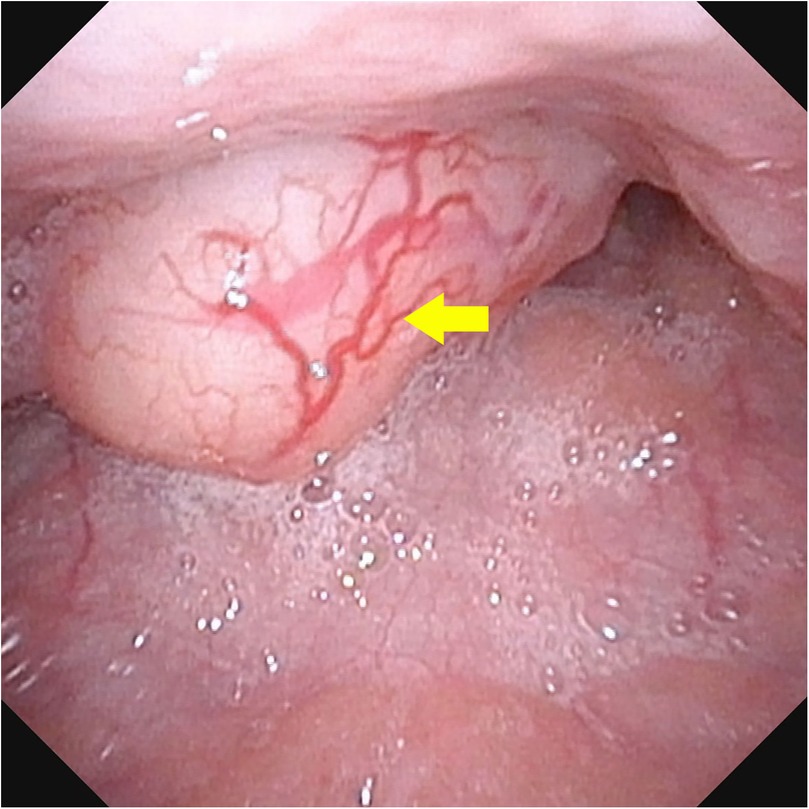
Figure 1. Fiberlaryngoscopic examination revealed a round mass with a pedicle in the pharynx, approximately 2 cm in diameter. The surface of the mass was smooth, with the pedicle rooted at the upper pole of the tonsil.
Macroscopically, the mass measured 2.2 × 2.0 × 1.3 cm, was well-circumscribed, and on cut surface, appeared deep gray with a tough, braided texture. Microscopically, the cells exhibited abundant eosinophilic, granular cytoplasm with well-defined borders, large polygonal skeletal muscle cells and frequent extensive vacuolization. Immunohistochemical staining was positive for MSA, Desmin, MYOD1 (Figure 2) and Myogenin, leading to a final pathological diagnosis of adult rhabdomyoma.
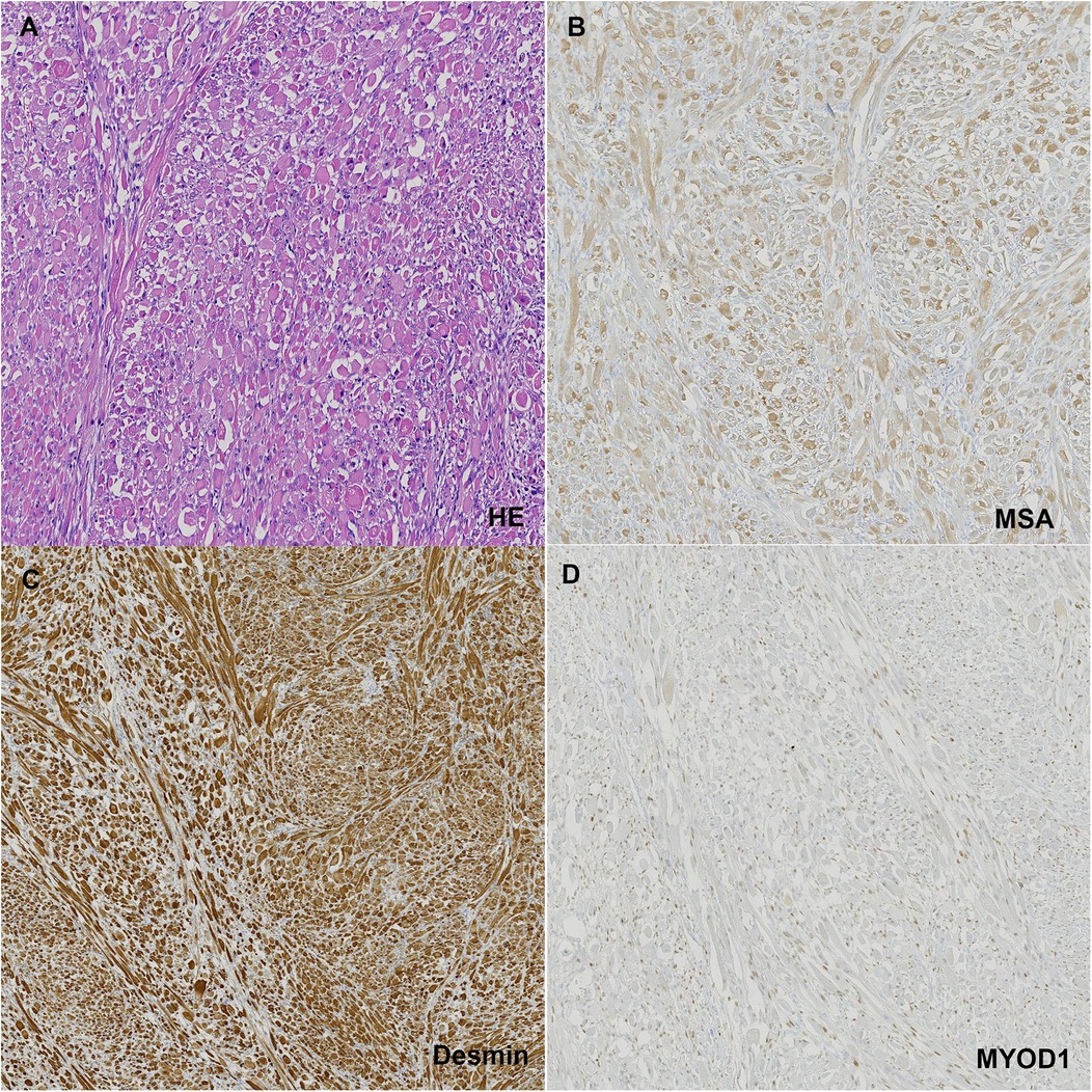
Figure 2. (A) HE staining of the tumor tissue; (B) positive staining with MSA; (C) positive staining with desmin; (D) positive staining with MYOD1.
The postoperative course was uneventful, and the boy was discharged five days after the intervention, with complete resolution of symptoms. He has been followed up for 18 months without any recurrence.
Extracardiac rhabdomyomas are exceedingly rare tumors, constituting less than 2% of neoplasms of striated muscle origin. Adult-type rhabdomyomas are rare benign tumors with a significantly lower incidence compared to their malignant counterpart, rhabdomyosarcoma (8). Adult rhabdomyomas predominantly affect the head and neck region, originating from the musculature of the third and fourth branchial arches. Most cases are solitary (70%) and typically occur in males over 50 years old (9). However, the occurrence of adult rhabdomyoma originating from a child's pharyngeal region is exceptionally rare. To our knowledge, this is the first reported case of an adult rhabdomyoma in the pharyngeal region of child.
The signs and symptoms at the time of presentation depend on the tumor's location. In our case, the boy experienced a sensation of a foreign body in the throat and presented with night-time snoring for six months. Reported symptoms in the literature include hearing loss, a mass in the submandibular triangle, swelling in the nasopharynx, hemoptysis, and obstructive sleep apnea, though pure dysphagia is rare (7, 10, 11).
Current diagnostic imaging modalities, including ultrasound, CT, and MRI, have not proven to be highly specific for the diagnosis of adult rhabdomyoma (8). In our case, there was an objective image of a mass with a pedicle in the pharynx, prompting us to perform direct excision. It is importance to combine imaging with histopathological confirmation, especially in pediatric patients where rare tumors like rhabdomyomas are less frequently considered. While imaging is useful for localization and surgical planning, histopathology remains the definitive diagnostic method. The gold standard treatment is surgery. Despite adult rhabdomyomas being benign, up to 42% may recur due to incomplete resection (12). Moreover, residual tumors may have malignant potential (13).
The microscopic appearance of adult rhabdomyomas is relatively consistent, characterized by polygonal, closely packed cells with prominent nuclei located centrally or peripherally. The cytoplasm is eosinophilic, granular, and shows extensive vacuolization in some cells. In our case, these features were observed. Immunohistochemical diagnosis features of rhabdomyoma include cytoplasmic positivity for MSA, Desmin, myoglobin, and MYOD1 (14).
Adult rhabdomyoma in children is an exceptionally rare occurrence and presents a diagnostic challenge due to its atypical manifestation in pediatric head, oral cavity, and neck tumors. Surgical resection is the treatment of choice, and careful long-term follow-up is essential. This case highlights the importance of considering adult rhabdomyoma in the differential diagnosis of pediatric pharyngeal masses, despite its rarity in this age group. Monitoring for recurrence is crucial in our clinical center to ensure optimal management and outcomes for this patient.
The original contributions presented in the study are included in the article/Supplementary Material, further inquiries can be directed to the corresponding author.
Written informed consent was obtained from the individual(s) for the publication of any potentially identifiable images or data included in this article.
LW: Investigation, Methodology, Writing – original draft. SL: Investigation, Resources, Writing – original draft. LS: Resources, Supervision, Writing – review & editing.
The author(s) declare that no financial support was received for the research, authorship, and/or publication of this article.
The authors declare that the research was conducted in the absence of any commercial or financial relationships that could be construed as a potential conflict of interest.
All claims expressed in this article are solely those of the authors and do not necessarily represent those of their affiliated organizations, or those of the publisher, the editors and the reviewers. Any product that may be evaluated in this article, or claim that may be made by its manufacturer, is not guaranteed or endorsed by the publisher.
1. Müller S. Update from the 4th edition of the world health organization of head and neck tumours: tumours of the oral cavity and Mobile tongue. Head Neck Pathol. (2017) 11(1):33–40. doi: 10.1007/s12105-017-0792-3
2. Jo VY, Demicco EG. Update from the 5th edition of the world health organization classification of head and neck tumors: soft tissue tumors. Head Neck Pathol. (2022) 16(1):87–100. doi: 10.1007/s12105-022-01425-w
3. Agamanolis DP, Dasu S, Krill CE Jr. Tumors of skeletal muscle. Hum Pathol. (1986) 17(8):778–95. doi: 10.1016/S0046-8177(86)80198-8
4. Smythe JF, Dyck JD, Smallhorn JF, Freedom RM. Natural history of cardiac rhabdomyoma in infancy and childhood. Am J Cardiol. (1990) 66(17):1247–9. doi: 10.1016/0002-9149(90)91109-J
5. Shemen L, Spiro R, Tuazon R. Multifocal adult rhabdomyomas of the head and neck. Head Neck. (1992) 14(5):395–400. doi: 10.1002/hed.2880140511
6. Rosenman D, Gertner R, Fradis M, Podoshin L, Misslevitsch A, Boss JH. Rhabdomyoma of the larynx. J Laryngol Otol. (1986) 100(5):607–10. doi: 10.1017/S0022215100099746
7. Di Sant'Agnese PA, Knowles DM 2nd. Extracardiac rhabdomyoma: a clinicopathologic study and review of the literature. Cancer. (1980) 46(4):780–9.<780::AID-CNCR2820460423>3.0.CO;2-K
8. Khalaf MG, Haddad R, Akiki M, Khazen J, Melkane AE. Multifocal adult rhabdomyoma of the head and neck: case report and systematic review of the literature. Int J Oral Maxillofac Surg. (2021) 50(3):327–34. doi: 10.1016/j.ijom.2020.07.018
9. da Silva Leonel ACL, Dos Santos ST, de Amorim Carvalho EJ, de Castro JFL. de almeida OP, da cruz perez DE. Oral adult rhabdomyoma. Head Neck Pathol. (2021) 15(4):1253–60. doi: 10.1007/s12105-021-01371-z
10. Kapadia SB, Meis JM, Frisman DM, Ellis GL, Heffner DK, Hyams VJ. Adult rhabdomyoma of the head and neck: a clinicopathologic and immunophenotypic study. Hum Pathol. (1993) 24(6):608–17. doi: 10.1016/0046-8177(93)90240-H
11. Andersen CB, Elling F. Adult rhabdomyoma of the oropharynx recurring three times within thirty-five years. Acta Pathol Microbiol Immunol Scand. (1986) 94(4):281–4. doi: 10.1111/j.1699-0463.1986.tb02995.x
12. Limbach AL, Goyal A. Adult rhabdomyoma: a challenging diagnosis on cytology. CytoJournal. (2012) 9:20. doi: 10.4103/1742-6413.100124
13. Andrade NN, Gandhewar T, Aggarwal N, Mathai P. Adult rhabdomyoma of the tongue in a child: report of a case and a literature appraisal. Contemp Clin Dent. (2018) 9(1):2–4. doi: 10.4103/ccd.ccd_835_17
Keywords: adult-type rhabdomyoma, extracardiac rhabdomyomas, pharyngeal, child, case report
Citation: Wang L, Li S and Sun L (2025) Case Report: Unveiling the unexpected: a rare case of adult-type rhabdomyoma in a 9-year-old boy. Front. Pediatr. 13:1451316. doi: 10.3389/fped.2025.1451316
Received: 19 June 2024; Accepted: 15 January 2025;
Published: 3 February 2025.
Edited by:
Shuai Ren, Affiliated Hospital of Nanjing University of Chinese Medicine, ChinaReviewed by:
Miguel A. Guzman, Saint Louis University, United StatesCopyright: © 2025 Wang, Li and Sun. This is an open-access article distributed under the terms of the Creative Commons Attribution License (CC BY). The use, distribution or reproduction in other forums is permitted, provided the original author(s) and the copyright owner(s) are credited and that the original publication in this journal is cited, in accordance with accepted academic practice. No use, distribution or reproduction is permitted which does not comply with these terms.
*Correspondence: Lingling Sun, Nzg5Njc2ODBAcXEuY29t
Disclaimer: All claims expressed in this article are solely those of the authors and do not necessarily represent those of their affiliated organizations, or those of the publisher, the editors and the reviewers. Any product that may be evaluated in this article or claim that may be made by its manufacturer is not guaranteed or endorsed by the publisher.
Research integrity at Frontiers
Learn more about the work of our research integrity team to safeguard the quality of each article we publish.